Artificial Intelligence for Pediatric Ophthalmology
←
→
Page content transcription
If your browser does not render page correctly, please read the page content below
Artificial Intelligence for Pediatric Ophthalmology
                                                                                 Julia E. Reid, MD m,† and Eric Eaton, PhD ‡

                                                          Purpose of review
                                                          Despite the impressive results of recent artificial intelligence (AI) applications to general ophthalmol-
arXiv:1904.08796v1 [physics.med-ph] 6 Apr 2019

                                                          ogy, comparatively less progress has been made toward solving problems in pediatric ophthalmology
                                                          using similar techniques. This article discusses the unique needs of pediatric ophthalmology patients
                                                          and how AI techniques can address these challenges, surveys recent applications of AI to pediatric
                                                          ophthalmology, and discusses future directions in the field.

                                                          Recent findings
                                                          The most significant advances involve the automated detection of retinopathy of prematurity (ROP),
                                                          yielding results that rival experts. Machine learning (ML) has also been successfully applied to the clas-
                                                          sification of pediatric cataracts, prediction of post-operative complications following cataract surgery,
                                                          detection of strabismus and refractive error, prediction of future high myopia, and diagnosis of read-
                                                          ing disability via eye tracking. In addition, ML techniques have been used for the study of visual
                                                          development, vessel segmentation in pediatric fundus images, and ophthalmic image synthesis.

                                                          Summary
                                                          AI applications could significantly benefit clinical care for pediatric ophthalmology patients by opti-
                                                          mizing disease detection and grading, broadening access to care, furthering scientific discovery, and
                                                          improving clinical efficiency. These methods need to match or surpass physician performance in clinical
                                                          trials before deployment with patients. Due to widespread use of closed-access data sets and software
                                                          implementations, it is difficult to directly compare the performance of these approaches, and repro-
                                                          ducibility is poor. Open-access data sets and software implementations could alleviate these issues,
                                                          and encourage further AI applications to pediatric ophthalmology.

                                                          Keywords
                                                          pediatric ophthalmology, machine learning, artificial intelligence, deep learning

                                                 INTRODUCTION                                              atric ophthalmology, despite the pressing need. In
                                                                                                           the United States, there is a shortage of pediatric
                                                 The increased availability of ophthalmic data, cou- ophthalmologists [12] and fellowship positions con-
                                                 pled with advances in artificial intelligence (AI) and tinue to go unfilled [13]. Globally, this shortage is
                                                 machine learning (ML), offer the potential to pos- even more pronounced and devastating—for exam-
                                                 itively transform clinical practice. Recent applica- ple, retinopathy of prematurity (ROP), now in its
                                                 tions of ML techniques to general ophthalmology third epidemic, has resulted in irreversible blindness
                                                 have demonstrated the potential for automated dis- in over 50,000 premature infants due to worldwide
                                                 ease diagnosis [1], automated prescreening of primary shortages of trained specialists and other barriers to
                                                 care patients for specialist referral [2], and scientific adequate care [14, 15].
                                                 discovery [3], among others. Acting as a complement
                                                 to ophthalmologists, these and future applications
                                                 have the potential to optimize patient care, reduce mNemours / Alfred I. duPont Hospital for Children, Division of
                                                 costs and barriers to access, limit unnecessary refer- Pediatric Ophthalmology, Wilmington, DE; † Thomas Jefferson
                                                 rals, permit objective monitoring, and enable early University,        Departments of Pediatrics and Ophthalmology,
                                                                                                           Philadelphia, PA; and ‡ University of Pennsylvania, Department
                                                 disease detection.                                        of Computer and Information Science, Philadelphia, PA
                                                     To date, most AI applications have focused on
                                                 adult ophthalmic diseases, as discussed by several re- Correspondence to Julia E. Reid, MD, Division of Pediatric
                                                                                                           Ophthalmology, 1600 Rockland Road, Wilmington, DE 19803,
                                                 views [4–11]. Comparatively little progress has been USA. email: julia.e.reid@nemours.org
                                                 made in applying AI and ML techniques to pedi-

                                                                                                              1
Artificial Intelligence for Pediatric Ophthalmology Julia E. Reid & Eric Eaton
be fully cyclopleged. Ancillary testing that requires
KEY POINTS patient cooperation may not be possible in an awake
child, and eye exams under anesthesia are not un-
• Pediatric ophthalmology has unique aspects that common. Similarly, children are typically placed un-
must be considered when designing AI applications,
including disease prevalence, cause, presentation, di- der general anesthesia for eye procedures, whereas
agnosis, and treatment, which differ from adults. adults may require only topical or local anesthesia.
Techniques for more accurate diagnosis and disease
• Most recent AI applications focus on ROP or congen- prediction could help reduce the high cost and risk of
ital cataracts, although many other areas of pediatric
ophthalmology could benefit from AI. repeated exams and surgeries under anesthesia.
Other distinguishing factors pertain to the pedi-
• Reproducibility and comparability between current atric patient’s growth and development. In most chil-
AI approaches is poor, and would be improved with dren, visual development occurs from birth until age
open-access data sets and software implementations.
7 or 8; eye diseases affecting children during this pe-
• Evaluation on experimental data sets should be aug- riod can cause permanent vision loss due to ambly-
mented with clinical validation prior to deployment opia or reduced visual abilities. Additionally, during
with patients. development, significant ocular growth occurs, caus-
ing changes in refractive error that complicate surgi-
cal planning for congenital cataract patients.
Retinal imaging, too, differs for pediatric and
UNIQUE CONSIDERATIONS FOR adult patients. Factors such as children’s lack of fix-
PEDIATRIC OPHTHALMOLOGY ation and small pupils can create blur, partial occlu-
sion, and illumination defects, all of which degrade
Ophthalmic disease prevalence, cause, presentation, image quality. For infants being screened for ROP,
diagnosis, and treatment all differ between adult and their fundus images are more variable and have more
pediatric patients—dissimilarities that are important visible choroidal vessels, making classification com-
to consider when developing AI applications. paratively difficult [16].
Common diseases in children include amblyopia,
strabismus, nasolacrimal duct obstruction (NLDO),
CLINICAL APPLICATIONS OF AI
retinopathy of prematurity (ROP), and congenital
eye diseases. The adult population, by contrast, is This section surveys recent AI applications to pedi-
affected by cataracts, dry eye, macular degeneration, atric ophthalmology, organized by disease (see Ta-
diabetic retinopathy, and glaucoma. For diseases that ble 1). The approaches discussed in this survey
occur in both children and adults, the presentation, would more precisely be called applications of ML—
cause, and treatment often differ. Glaucoma is a good the largest subfield of AI concerned with learning
example, as the cause and presentation in congeni- models from data. We have provided a brief overview
tal glaucoma patients are both unlike those in adult- of AI and ML and their relationship in supplemental
onset glaucoma patients. Optimal management of material, but the interested reader is encouraged to
glaucoma, including surgery, also differs for these two consult a more extensive tutorial on these topics [e.g.
populations. 5]. To limit its scope, this review focuses on appli-
Infants and children have distinct characteristics cations with a goal of having the AI aspects directly
from adults that affect their ophthalmology visits. impact clinical practice; we omit studies where ML
Given their developmental capabilities, there is gen- was used primarily for statistical analysis.
erally less information gleaned from a single eye exam
of a child, so several visits may be required to accu-
rately diagnose or characterize that child’s disease. Retinopathy of Prematurity (ROP)
There is also a stronger reliance on the objective The most significant AI advances in pediatric oph-
exam because of the infant’s or child’s inability to thalmology apply to ROP, a leading cause of child-
effectively communicate. Children’s short attention hood blindness worldwide [14, 15, 40]. In addition
spans and unpredictable behavior often necessitate to the shortage of trained providers [14, 15, 41], ROP
a quick exam that allows the physician to gain the exams are difficult, clinical impressions are subjective
child’s trust while keeping him or her at ease. De- and vary among examiners [23, 42, 43], and disease
spite this, there are portions of the clinic visit that management is time-intensive, requiring several serial
take longer, such as restraining a child to adminis- exams. AI applications have focused on detecting the
ter dilating drops and then waiting for that child to presence and grading of ROP or plus disease from
2Artificial Intelligence for Pediatric Ophthalmology Julia E. Reid & Eric Eaton
Table 1. Summary of ML-based techniques for pediatric ophthalmic disease detection and diagnosis
Approach Predicted category Sensitivity Specificity AUROC Accuracy Method summary
(Approx. devel. year) (%) (%) (%)
Retinopathy of prematurity (ROP)
DeepROP [17 ]
Experimental data set Cloud-based platform. Set of
(2018) Presence of ROP 96.64 99.33 0.995 97.99 fundus images → two CNNs
Severe (vs Mild) ROP 88.46 92.31 0.951 90.38 (modified Inception-BN nets
Clinical test pretrained on ImageNet): one
Presence of ROP 84.91 96.90 – 95.55 predicts presence, and the
Severe (vs Mild) ROP 93.33 73.63 – 76.42 other severity
i-ROP-DL [18 ]
Clinically significant ROP – – 0.914 – Applies a linear formula to
(2018) Type 1 ROP 94 79 0.960 – the probabilities output by
Type 2 ROP – – 0.867 – i-ROP-DL (see below) to yield
Pre-plus disease – – 0.910 – a severity score on a 1–9 scale
MiGraph [19] Presence of ROP 99.4 95.0 0.98 97.5 SIFT features from image
(2016) patches → multiple instance
learning graph-kernel SVM
VesselMap [20] Severe ROP Semiautomated tool that uses
(2007) From mean arteriole diameter – – 0.93 – classic image analysis to mea-
From mean venule diameter – – 0.87 – sure vessel diameter
ROP: Plus or pre-plus disease
i-ROP-DL [21 ]
Plus disease [18 ]
– – 0.989 – CNN-output (U-net) ves-
(2018) Pre-plus disease [18 ]
– – 0.910 – sel segmentations → CNN
Plus disease [21 ]
93 94 0.98 91.0 (InceptionV1 pretrained on
Pre-plus or worse disease [21 ] 100
94 0.94 – ImageNet) to classify as
normal/pre-plus/plus
CNN + Bayes [16] Plus disease (per image) 82.5 98.3 – 91.8 CNN (InceptionV1 pretrained
(2016) (per exam) 95.4 94.7 – 93.6 on ImageNet) adapted to out-
put the Bayesian posterior
i-ROP [22] Plus disease 93 – – 95 SVM with a kernel derived
(2015) Pre-plus or worse disease 97 – – – from a GMM of tortuosity and
dilation features from manually
segmented images
Naı̈ve Bayes [23] Plus/pre-plus/none (SVM-RFE) – – – 79.41 Naı̈ve Bayes with SVM-RFE or
(2015) Plus disease (ReliefF) – – – 88.24 ReliefF vessel feature selection
CAIAR [24] Plus (from venule width) – – 0.909 – Generative vessel model fit to
(2008) Plus (from arteriole tortuosity) – – 0.920 – a multi-scale representation of
the retinal image
ROPtool [26] Plus tortuosity (eye) 95 78 – 87.50 User-guided tool that traces
(2007) (quadrant) 85 77 0.885 80.63 centerlines of retinal vessels to
Pre-plus tortuosity (quadrant) 89 82 0.875 – measure tortuosity
RISA [27] Plus disease (from arteriole 93.8 93.8 0.967 – Logistic regression on geomet-
(2005) and venule curvature and ric features computed for each
tortuosity, venule diameter) segment of the vascular tree
IVAN [24] Plus (from venule width) – – 0.909 – Measures vessel width via clas-
(2002) sic image analysis
Abbreviations: AUROC – area under the receiver operating characteristic curve; GMM – Gaussian mixture model
digital fundus photos. Beyond the benefits of auto- and width via classic image analysis, including Vessel
mated ROP screening and objective assessment, dig- Finder [47], VesselMap [20], ROPtool [26], RISA [27,
ital retinal imaging may cause less pain and stress for 48, 49], CAIAR [24, 25], and IVAN [24, 50], all of
infants undergoing ROP screening compared to indi- which require at least one manual step from the user.
rect ophthalmoscopy [44] and enable neonatology-led Recent work suggests other potential vessel measure-
screening programs [45]. ments correlated with plus disease, such as a decrease
Early computational approaches to detecting plus in the openness of the major temporal arcade an-
disease from fundus images focused on vessel tor- gle [51]. Once extracted, retinal vessel measurements
tuosity. One early attempt to objectively quantify have been used as features for various predictive mod-
tortuosity used the spatial frequency of manual ves- els of plus disease, including linear models such as lo-
sel tracings [46]. Since then, there have been sev- gistic regression [27] and naı̈ve Bayes [23], as well as
eral tools developed to determine vessel tortuosity non-linear models trained by support vector machines
3Artificial Intelligence for Pediatric Ophthalmology Julia E. Reid & Eric Eaton
Table 1. (Continued)
Approach Predicted category Sensitivity Specificity AUROC Accuracy Method summary
(Approx. devel. year) (%) (%) (%)
Pediatric cataracts
Post-operative CLR and/or High IOP (RF) 62.5 76.9 0.722 70.0 Demographic and cataract
complication (NB) 73.1 66.7 0.719 70.0 severity evaluation data →
prediction [28] Central lens regrowth (RF) 66.7 72.2 0.743 72.0 class-balancing using SMOTE
(2019) (NB) 61.1 68.8 0.735 66.0 → random forest (RF) and
High IOP (RF) 63.6 71.8 0.735 70.0 naı̈ve Bayes (NB) classifiers
(NB) 54.5 69.2 0.719 66.0
CS-ResCNN [29] Severe posterior Slit-lamp images → automat-
(2017) capsular opacification 89.66 93.19 0.9711 92.24 ically crop to lens → CNN
(ResNet pretrained on Ima-
geNet) with cost-sensitive loss
CC-Cruiser [30] Multi-center trial Cloud-based platform. Slit-
(2016) Cataract presence [31 ]
89.7 86.4 – 87.4 lamp images → automatically
Opacity area grading [31 ]
91.3 88.9 – 90.6 crop to lens → three CNNs
Density grading [31 ]
85.3 67.9 – 80.2 (AlexNets) to predict: cataract
Location grading [31 ]
84.2 50.0 – 77.1 presence, severity (area, den-
Treatment [31 ]
86.7 44.4 – 70.8 sity, location), and treatment
Experimental data set (surgery or follow-up)
Cataract presence [32 ]
96.83 97.28 0.9686 97.07
Area grading [32 ]
90.75 86.63 0.9892 89.02
Density grading [32 ]
93.94 91.05 0.9743 92.68
Location grading [32 ]
93.08 82.70 0.9591 89.28
Strabismus
RF-CNN [33 ]
Strabismus presence 93.30 96.17 0.9865 93.89 Two-stage CNN: eye regions
(2018) segmented from face images
via R-FCN → 11-layer CNN
SVM + VGG-S [34] Strabismus presence 94.1 96.0 – 95.2 Eye-tracking gaze maps →
(2017) CNN (VGG-S pretrained on
ImageNet) features → SVM
Pediatric Vision Central vs. paracentral fixation Signals from retinal birefrin-
Screener [35] Experimental evaluation 100.0 100.0 – – gence scanning → two-layer
(2017) Clinical evaluation 98.51 100.0 – – feed-forward neural net
Vision screening
AVVDA [36] Strabismus and/or RE – – – 76.9 Features from Brückner red re-
(2008) Strabismus 82 – – – flex imaging and eccentric fixa-
High refractive error (RE) 90 – – – tion video → C4.5 decision tree
Reading disability (RD)
SVM-RFE [37] High risk for RD, ages 8–9 95.5 95.7 – 95.6 SVM with feature selection
(2016) trained on eye-tracking data
Polynomial SVM [38] RD in adults, children ages 11+ – – – 80.18 SVM trained on eye-tracking
(2015) and demographic features
Approach Predicted category AUROC AUROC AUROC Method summary
(Approx. devel. year) (at 3 years) (at 5 years) (at 8 years)
Refractive error (RE)
Random forest [39 ]
Internal evaluation Age, spherical equivalent (SE),
(2018) High myopia onset 0.903-0.986 0.875-0.901 0.852-0.888 and progression rate of SE be-
Clinical test tween two visits was used by a
High myopia onset 0.874-0.976 0.847-0.921 0.802-0.886 random forest for prediction
High myopia at age 18 0.940-0.985 0.856-0.901 0.801-0.837
(SVMs) [22]. For predicting ROP, Rani et al. [19] also tems, which include Worrall et al. [16], i-ROP-DL
employ an SVM, but instead use SIFT [52] features [18, 21], and DeepROP [17], demonstrate agree-
extracted from retinal image patches and frame the ment with expert opinion [16, 18] and better disease
problem in a multiple instance learning [53] setting. detection than some experts [17, 21].
Recent approaches to ROP and plus disease de- Like many ML methods, these systems can pro-
tection are mostly based on convolutional neural net- vide a confidence score in their predictions. i-ROP-
works (CNN), which take fundus images as input DL exploits this notion directly by combining the pre-
and do not require manual annotation. These sys- diction probabilities via a linear formula to compute
4Artificial Intelligence for Pediatric Ophthalmology Julia E. Reid & Eric Eaton
Table 2. Pediatric ROP data sets used in deep learning CC-Cruiser [30–32] is a cloud-based platform
that can automatically detect cataracts from slit-
Approach Data set Patients Images Labels lamp images, grade them, and recommend treatment.
DeepROP Chengdu 1,273 20,795 normal, mild ROP,
[17 ]
severe ROP
After automatically cropping the slit-lamp image to
i-ROP-DL i-ROP 898 5,511 normal, plus, the lens region, it uses three separate CNNs (modified
[21 ]
pre-plus AlexNets [61]) to predict three aspects: cataract pres-
CNN + Bayes Canada 35 1,459 normal, plus ence, grading (opacity area, density, location), and
[16] London – 106 normal, plus treatment recommendation (surgery or non-surgical
follow-up). CC-Cruiser was evaluated in a multi-
center randomized controlled trial within five oph-
an ROP severity score, which can serve as an ob- thalmology clinics, demonstrating significantly lower
jective quantification of disease; a similar idea could performance in diagnosing cataracts (87.4%) and rec-
provide finer grading of plus disease [21]. ommending treatment (70.8%) than experts (99.1%
For their core predictive networks, all these CNN- and 96.7%, respectively), but achieving high patient
based systems use versions of the Inception architec- satisfaction for its rapid evaluation [31].
ture [54, 55] with transfer learning [56, 57] by pre- Children who require surgery face potential com-
training on ImageNet, giving them similar founda- plications that differ from those that adults face [62].
tions. However, these approaches differ in prepro- Zhang et al. applied random forests and naı̈ve Bayes
cessing (e.g., i-ROP-DL [21] uses a U-net [58] to classifiers to predict two common post-operative com-
perform automatic vessel segmentation) and postpro- plications, central lens regrowth and high intraocular
cessing (e.g., i-ROP-DL [18] outputs the ROP sever- pressure (IOP), from a patient’s demographic infor-
ity score; Worrall et al. [16] outputs the Bayesian mation and cataract severity evaluation [28]. Another
posterior). DeepROP processes a set of fundus im- approach [29] uses a CNN to detect severe posterior
ages per case, taking a multiple instance learning [53] capsular opacification warranting surgery, employing
approach, while the other two deep learning meth- a ResNet [63] pretrained on ImageNet with a cost-
ods classify single images. The other key difference is sensitive loss to handle data set imbalance.
that these systems are trained on different non-public
ROP data sets of varying sizes and labelings (Ta-
ble 2). The use of non-public data sets and closed im- Strabismus
plementations (only DeepROP is open source) com- Strabismus affects 1 in 50 children and can cause am-
plicates comparison and reproducibility [59]. blyopia, interfere with binocularity, and have lasting
Current methods for ROP detection are capable psychosocial effects [64–68]. A CNN was used to de-
of coarse-grained classification, such as discriminat- tect strabismus based on visual manifestation in the
ing severe from mild ROP; they do not specifically eye regions of facial photos [33], which would be
assess disease stage or zone (e.g., [17]). In fact, all especially useful for telemedical evaluation. For in-
systems except DeepROP [17] and MiGraph [19] ex- office evaluation, which in contrast permits the use
amine only the posterior pole view, either ignoring of specialized screening instruments, strabismus can
other views or explicitly cropping them out. While be detected using a CNN based on fixation devia-
the literature suggests that severe disease rarely de- tions from eye-tracking data [34], or with very high
velops without changes in posterior pole vasculature sensitivity and specificity from retinal birefringence
[60], providing additional outputs of the zone and scanning [35].
stage could improve the interpretability of the sys-
tem’s assessment and improve performance.
Vision Screening
Pediatric Cataracts Like strabismus, refractive error can cause ambly-
opia, but is difficult for pediatricians to detect.
Pediatric cataracts are more variable than adult Instrument-based vision screening is recommended
cataracts, and surgical removal depends upon [69] and most devices have adjustable thresholds for
cataract severity and deprivational amblyopia risk. signaling a screening failure. Using video frames from
Slit lamp exams enable cataract visualization but can one such instrument that combines Brückner pupil
be challenging and subjective, and slit lamp image red reflex imaging and eccentric photorefraction, Van
quality can vary (e.g., based on the child’s coopera- Eenwyk et al. trained a variety of ML classifiers to de-
tiveness, image amplification, and interference from tect amblyogenic risk factors in young children, with
eyelashes and other eye disease or structures) [32]. the most successful being a C4.5 decision tree [70].
5Artificial Intelligence for Pediatric Ophthalmology Julia E. Reid & Eric Eaton
Reading Disability many visual proficiencies, such as facial recognition,
are facilitated by the gradual increase in visual acu-
Reading disability affects approximately 10% of chil-
ity during normal visual development. When tested
dren [38], but objective and efficient testing for it is
in CNNs via initial training with blurred images,
lacking [37]. Abnormal eye tracking is non-causally
gradual acuity development increased generalization
associated with reading disability [37, 38]. Two stud-
performance and encouraged the development of re-
ies used SVMs to identify reading disability from eye
ceptive fields with a broader spatial extent [103].
movements during reading, either predicting reading
These results provide a possible explanation for the
disability risk in children ages 8–9 [37], or detect-
decreased visual proficiencies of congenital cataract
ing reading disability in adults and children ages 11+
patients, and suggest the potential for temporary re-
[38]. The children in both of these studies are older
fractive undercorrection to help restore visual devel-
than the optimal age for diagnosis, so validation in a
opment [103].
younger cohort could be useful.
Pediatric Retinal Vessel Segmentation
Refractive Error
Although many programs have been developed for
High myopia is associated with numerous vision-
vessel segmentation in adults or premature infants,
threatening complications [71]. Children at risk for
fundus images in older children have unique traits,
high myopia can take low-dose atropine to halt or
including light artifacts, that complicate segmenta-
slow myopic progression [72, 73]1 , but it can be dif-
tion [104]. Fraz et al. [104] developed an ensemble
ficult to determine for which children to recommend
of bagged decision trees that use multi-scale analysis
this treatment [39]. Lin et al. [39] predicted high
with multiple filter types to do vessel segmentation in
myopia in children from clinical measures using a ran-
pediatric fundus images. Another tool, CAIAR [25],
dom forest, showing good predictive performance for
has been validated in school-aged children [105]. CA-
up to 8 years into the future. Further work has the
IAR was first applied to infants with ROP and uses a
potential to guide prophylactic treatment.
generative model of the vessels fit via maximum like-
lihood to a multi-scale representation of the retinal
Non-Pediatric Applications image [25].
AI has been applied to various adult ophthalmic dis-
eases, including diabetic retinopathy [1, 74–77], AMD Ophthalmic Image Synthesis
[78–83], sight-threatening retinal disease [2, 84–89],
Through their multi-layered representation, deep
glaucoma [90–92], intraocular lens calculation [93],
learning methods such as generative adversarial net-
and keratoconus [94]. It has also been used for robot-
works [106] are able to synthesize novel realistic
assisted repair of epiretinal membranes [95], retinal
images, including retinal fundus images [107, 108].
vessel segmentation [96–99], and systemic disease pre-
Such synthesized images can compensate for data
diction from fundus images [100]. For a detailed re-
scarcity, preserve patient privacy, and depict vari-
view, see [4–11].
ations on or combinations of diseases for resident
education [109, 110].
OTHER OPHTHALMIC APPLICATIONS One recent technique to synthesize high-
resolution images, progressive growing of GANs
This section reviews applications of ML to pediatric (PGGANs), was used to synthesize realistic fundus
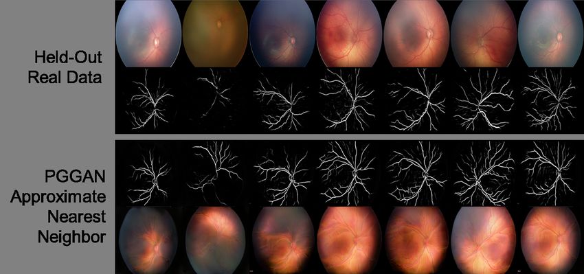
ophthalmology that are not tied to specific diagnoses. images of ROP (see examples in Figure 1) [111].
The PGGAN was trained on ROP fundus images
Visual Development in combination with vessel segmentation maps
obtained from a pre-trained U-net CNN [58]. GANs
ML has the potential to provide scientific insight have also been used to synthesize retinal images of
into visual development. For example, adults who diabetic retinopathy, including the ability to control
had cataract surgery and aphakic correction in in- high-level aspects of the presentation [77, 112].
fancy have exhibited diminished facial processing ca- While many of the GAN-synthesized images display
pabilities [101, 102]. This impairment was originally believable pathologic features, some do contain
blamed on early visual deprivation [101, 102], but “checkerboard” and other generation artifacts.
more recently, it was conjectured to be caused by
the aphakic correction and high initial acuity experi- 1 Note: this usage of atropine is not approved by the FDA.
enced by these infants [103]. The hypothesis is that
6Artificial Intelligence for Pediatric Ophthalmology Julia E. Reid & Eric Eaton
Figure 1. Real (top row) and synthetic (bottom row) fundus images of ROP with their corresponding vessel
segmentations [111]. The top row shows real images that were not included in the training set, and the bottom
row shows the most similar synthesized images. (Image from [111], reused with permission.)
CURRENT LIMITATIONS AND and multi-task learning [117, 118] techniques may of-
FUTURE DIRECTIONS fer a solution to this problem, providing mechanisms
to adapt adult models to pediatric patients given a
Current applications to pediatric ophthalmology have small amount of pediatric ophthalmic data. These
several limitations that offer avenues for future work. methods could also reuse knowledge across models
Disagreement on reference standards An ML of different diseases or populations—for example, in-
classifier’s performance is fundamentally limited by tegrating knowledge across multiple smaller pediatric
the quality of the training data, which are manually data sets of different ophthalmic diseases to help com-
labeled by clinicians. However, there is often signifi- pensate for the lack of data on any one disease. No-
cant variation of the diagnosis and treatment among tice that, by pretraining on ImageNet, many of the
physicians, given the same case information [23, 42, CNN-based methods surveyed here already employ
43, 113], which complicates determination of the cor- transfer learning of basic image features to compen-
rect labels. When ML was used to identify factors sate for using small data sets; transferring from adult
influencing ROP experts’ decisions for plus disease ophthalmic data sets may provide further advantages.
diagnosis, the most important features were venous Poor reproducibility and comparability Al-
tortuosity and vascular branching [23, 43], neither of most all the ML studies discussed here, even those
which are part of the standard “plus disease” defi- that focus on the same disease, are trained and eval-
nition of arteriolar tortuosity and venular dilatation uated on different data sets. In many cases, the data
[114, 115]. Most approaches use the majority label sets and software source code are not available pub-
from multiple experts as the label for each training licly, complicating reproducibility and scientific com-
instance, or combine the majority label given to im- parison across algorithms [59].
agery with the clinical diagnosis [116]. An alternative Most ML research relies on publicly accessible
approach puts cases with any amount of disagreement data sets and software implementations for evalua-
up for adjudication among the experts, resulting in a tion and comparison. One simple way to encourage
consensus label and reducing errors, as demonstrated further applications of AI to pediatric ophthalmol-
for diabetic retinopathy [76]. ogy is through the public release of data sets in strict
Need for pediatric-specific models It would be compliance with HIPAA regulations, and with special
advantageous for pediatric ophthalmology to benefit regard to the additional HIPAA restrictions for mi-
from the large amount of work in AI for adult oph- nors. Even small pediatric ophthalmic data sets could
thalmology. However, due to the unique aspects of be of use when used in combination with adult data
pediatric disease manifestation, ML models trained through transfer learning techniques, as mentioned
on adult patients may make errors when directly ap- above. For the largest impact, these open data sets
plied to pediatric patients. Transfer learning [56, 57] should be hosted in a widely used ML repository.
7Artificial Intelligence for Pediatric Ophthalmology Julia E. Reid & Eric Eaton
Lack of temporal information Most of these Financial support and sponsorship
systems detect disease based upon one snapshot in
E.E.’s work was partially supported by the Lifelong
time, without consideration of longitudinal imaging
Learning Machines program from DARPA/MTO un-
of the case [16]. In some diseases, such as ROP, rapid
der grant #FA8750-18-2-0117. The funders had no
change is associated with poorer outcomes [47, 119],
role in the research presented in this article, nor in its
suggesting that temporal information may have a role
preparation, review, or approval. The views and con-
in predicting severe disease.
clusions contained herein are those of the authors and
Uninterpretable “black-box” models Despite should not be interpreted as necessarily representing
their predictive power, the “black-box” nature of the official policies or endorsements, either expressed
most state-of-the-art ML methods, such as deep or implied, of DARPA or the U.S. Government.
neural networks, complicates their application in
medicine. It is often challenging to quantitatively
Conflicts of interest
interpret the inference process of such models, under-
standing how they arrived at their predictions [120, There are no conflicts of interest.
121]. Since they focus on correlations between the
input and desired output, in some cases ML models REFERENCES
may fixate on confounding factors instead of patho-
logical information [122]. Interpretable ML methods Papers of particular interest, published within the annual
provide a potential solution to benefit clinicians, al- period of review, have been highlighted as:
lowing, for example, examination of intermediate de- of special interest
cision steps within a deep network, natural language of outstanding interest
justifications for a decision, or visualization of image
features that contribute to a decision [121]. While 1. Gulshan V, Peng L, Coram M, et al. De-
these methods seek to improve the interpretability of velopment and validation of a deep learning
black-box models, other approaches seek to improve algorithm for detection of diabetic retinopa-
the predictive power of models that are already inter- thy in retinal fundus photographs. JAMA
pretable, such as the MediBoost algorithm for grow- 2016;316:2402–2410.
ing decision trees via gradient boosting [123]. 2. De Fauw J, Ledsam JR, Romera-Paredes B,
et al. Clinically applicable deep learning for
CONCLUSION diagnosis and referral in retinal disease. Na-
ture Medicine 2018;24:1342–1350.
There is a large potential for current and future AI 3. Varadarajan AV, Poplin R, Blumer K, et
applications to pediatric ophthalmology, and there al. Deep learning for predicting refractive
are some diseases, such as NLDO, congenital glau- error from retinal fundus images. Inves-
coma, and congenital ptosis, without any published tigative Ophthalmology and Visual Science
applications of AI to our knowledge. Automated dis- 2018;59:2861–2868.
ease detection, the most common use case, could aug-
4. Roach L. Artificial intelligence. Eyenet Mag-
ment telemedical efforts to broaden access to care,
azine 2017:77–83.
improve efficiency, and result in earlier diagnoses.
However, other less-utilized capabilities of this tech- 5. Consejo A, Melcer T, and Rozema JJ. In-
nology, including disease grading and outcome predic- troduction to machine learning for oph-
tion, have the potential to enhance clinical care. All thalmologists. Seminars in Ophthalmology
AI methods deployed in clinical care must ultimately 2019;34:19–41.
match or surpass physician performance while meet- 6. Ting DSW, Pasquale LR, Peng L, et al. Ar-
ing the unique requirements of both clinicians and pe- tificial intelligence and deep learning in oph-
diatric patients, suggesting the need to augment eval- thalmology. British Journal of Ophthalmol-
uations on experimental data sets with clinical trials. ogy 2018:2018–313173.
7. Lee A, Taylor P, Kalpathy-Cramer J, and Tu-
Acknowledgements fail A. Machine learning has arrived! Oph-
We would like to thank Jing Jin, MD, José Marcio thalmology 2017;124:1726–1728.
Luna, PhD, and Jorge Mendez for their helpful feed- 8. Rahimy E. Deep learning applications in oph-
back on this article. thalmology. Current Opinion in Ophthalmol-
ogy 2018;29:254–260.
8Artificial Intelligence for Pediatric Ophthalmology Julia E. Reid & Eric Eaton
9. Caixinha M and Nunes S. Machine learning The i-ROP-DL deep learning system is the
techniques in clinical vision sciences. Current first to detect specific ROP classifications, in-
Eye Research 2017;42:1–15. cluding clinically significant, type 1, and type
10. American Academy of Ophthalmology. The 2 ROP. This model could potentially be a
future of artificial intelligence in ophthalmol- useful telemedical tool for identifying referral-
ogy. AAO Mid-Year Forum 2018. warranted ROP.
11. Du XL, Li WB, and Hu BJ. Applica- 19. Rani P, Elagiri Ramalingam R, Rajamani
tion of artificial intelligence in ophthalmol- KT, et al. Multiple instance learning: Robust
ogy. International Journal of Ophthalmology validation on retinopathy of prematurity. In-
2018;11:1555–1561. ternational Journal of Control Theory and
Applications 2016;9:451–459.
12. Estes R, Estes D, West C, et al. The Amer-
ican Association for Pediatric Ophthalmol- 20. Rabinowitz MP, Grunwald JE, Karp KA,
ogy and Strabismus workforce distribution et al. Progression to severe retinopathy pre-
project. Journal of American Association dicted by retinal vessel diameter between 31
for Pediatric Ophthalmology and Strabismus and 34 weeks of postconception age. Archives
2007;11:325–329. of Ophthalmology 2007;125:1495–1500.
13. Dotan G, Karr DJ, and Levin AV. Pedi- 21. Brown JM, Campbell JP, Beers A, et al. Au-
atric ophthalmology and strabismus fellow- tomated diagnosis of plus disease in retinopa-
ship match outcomes, 2000-2015. Journal of thy of prematurity using deep convolu-
American Association for Pediatric Ophthal- tional neural networks. JAMA Ophthalmol-
mology and Strabismus 2017;21:1–181. ogy 2018;136:803–810.
14. Gilbert C. Retinopathy of prematurity: A The i-ROP-DL system detects plus disease in
global perspective of the epidemics, popula- infants with ROP more accurately than the
tion of babies at risk and implications for con- majority of experts in this study. This article
trol. Early Human Development 2008;84:77– highlights a deep learning method with the
82. ability to surpass physician performance.
15. Quinn G. Retinopathy of prematurity blind- 22. Ataer-Cansizoglu E, Bolon-Canedo V, Camp-
ness worldwide: phenotypes in the third epi- bell JP, et al. Computer-based image analy-
demic. Eye and Brain 2016;8:31–36. sis for plus disease diagnosis in retinopathy
of prematurity: Performance of the “i-ROP”
16. Worrall DE, Wilson CM, and Brostow GJ.
system and image features associated with ex-
Automated retinopathy of prematurity case
pert diagnosis. Translational Vision Science
detection with convolutional neural net-
& Technology 2015;4:5.
works. In: Workshop on Deep Learning and
Data Labeling for Medical Applications (LA- 23. Bolón-Canedoa V, Ataer-Cansizoglub E, Er-
BELS/DLMIA). 2016:68–76. dogmusb D, et al. Dealing with inter-
expert variability in retinopathy of prema-
17. Wang J, Ju R, Chen Y, et al. Auto-
turity: A machine learning approach. Com-
mated retinopathy of prematurity screening
puter Methods and Programs in Biomedicine
using deep neural networks. EBioMedicine
2015;122:1–15.
2018;35:361–368.
24. Shah DN, Wilson CM, Ying Gs, et al. Semi-
The DeepROP system for ROP detection is automated digital image analysis of posterior
trained on the largest data set to date, and pole vessels in retinopathy of prematurity.
is the first to detect severe ROP using fun- Journal of American Association for Pediatric
dus images that include the peripheral retina. Ophthalmology and Strabismus 2009;13:504–
This deep learning approach demonstrates 506.
the potential benefits of fine-grained ROP
classification. 25. Wilson CM, Cocker KD, Moseley MJ, et al.
Computerized analysis of retinal vessel width
18. Redd TK, Campbell JP, Brown JM, et al. and tortuosity in premature infants. Inves-
Evaluation of a deep learning image assess- tigative Ophthalmology and Visual Science
ment system for detecting severe retinopathy 2008;49:3577–3585.
of prematurity. British Journal of Ophthal-
mology 2018:2018–313156.
9Artificial Intelligence for Pediatric Ophthalmology Julia E. Reid & Eric Eaton
26. Wallace DK, Zhao Z, and Freedman SF. A This system is the first to detect strabis-
pilot study using “ROPtool” to quantify plus mus remotely from digital facial images. As a
disease in retinopathy of prematurity. Journal telemedical application, this could help deter-
of American Association for Pediatric Oph- mine which children require an ophthalmol-
thalmology and Strabismus 2007;11:381–387. ogy referral for strabismus.
27. Gelman R, Jiang L, Du YE, et al. Plus disease 34. Chen Z, Fu H, Lo WL, and Chi Z. Stra-
in retinopathy of prematurity: Pilot study of bismus recognition using eye-tracking data
computer-based and expert diagnosis. Jour- and convolutional neural networks. Journal of
nal of American Association for Pediatric Healthcare Engineering 2018:7692198.
Ophthalmology and Strabismus 2007;11:532– 35. Gramatikov BI. Detecting central fixation by
540. means of artificial neural networks in a pe-
28. Zhang K, Liu X, Jiang J, et al. Prediction diatric vision screener using retinal birefrin-
of postoperative complications of pediatric gence scanning. BioMedical Engineering On-
cataract patients using data mining. Journal line 2017;16:52.
of Translational Medicine 2019;17:2. 36. Van Eenwyk J, Agah A, Giangiacomo J, and
29. Jiang J, Liu X, Zhang K, et al. Automatic di- Cibis G. Artificial intelligence techniques for
agnosis of imbalanced ophthalmic images us- automatic screening of amblyogenic factors.
ing a cost-sensitive deep convolutional neu- Transactions of the American Ophthalmolog-
ral network. BioMedical Engineering OnLine ical Society 2008;106:64–73.
2017;16:132. 37. Nilsson Benfatto M, Öqvist Seimyr G,
30. Long E, Lin H, Liu Z, et al. An artificial intel- Ygge J, et al. Screening for dyslexia us-
ligence platform for the multihospital collab- ing eye tracking during reading. PLOS ONE
orative management of congenital cataracts. 2016;11:e0165508.
Nature Biomedical Engineering 2017;1:0024. 38. Rello L and Ballesteros M. Detecting readers
31. Lin H, Li R, Liu Z, et al. Diagnostic effi- with dyslexia using machine learning with eye
cacy and therapeutic decision-making capac- tracking measures. In: Proceedings of the 12th
ity of an artificial intelligence platform for Web for All Conference (W4A). ACM Press,
childhood cataracts in eye clinics: A mul- 2015:16.
ticentre randomized controlled trial. EClini- 39. Lin H, Long E, Ding X, et al. Prediction of
calMedicine 2019. myopia development among Chinese school-
This study describes a multi-center random- aged children using refraction data from
ized controlled trial evaluating the perfor- electronic medical records: A retrospective,
mance of the CC-Cruiser system for cataract multicentre machine learning study. PLOS
diagnosis and treatment—an important step Medicine 2018;15:e1002674.
toward a real-world clinical application of AI This study predicts the development of high
to pediatric ophthalmology. myopia in children up to 8 years in advance.
32. Liu X, Jiang J, Zhang K, et al. Localiza- Such prediction could potentially be used to
tion and diagnosis framework for pediatric guide atropine prophylaxis.
cataracts based on slit-lamp images using 40. Steinkuller PG, Du L, Gilbert C, et al.
deep features of a convolutional neural net- Childhood blindness. Journal of AAPOS
work. PLOS ONE 2017;12:e0168606. 1999;3:26–32.
This study describes a cloud-based ML plat- 41. American Academy of Ophthalmology. Oph-
form, CC-Cruiser, that accurately detects thalmologists warn of shortage in specialists
cataract presence, area, density, and location. who treat premature babies with blinding
Such an approach could detect cataracts in eye condition. AAO Press Release 2006-07-13
the primary care setting or serve as a comple- 2006.
ment to the pediatric ophthalmologist’s eval-
42. Wallace DK, Quinn GE, Freedman SF, and
uation.
Chiang MF. Agreement among pediatric oph-
33. Lu J, Fan Z, Zheng C, et al. Automated thalmologists in diagnosing plus and pre-plus
strabismus detection for telemedicine appli- disease in retinopathy of prematurity. Journal
cations. arXiv 1809.02940 2018. of AAPOS 2008;12:352–356.
10Artificial Intelligence for Pediatric Ophthalmology Julia E. Reid & Eric Eaton
43. Ataer-Cansizoglu E, Kalpathy-Cramer J, You 53. Dietterich TG, Lathrop RH, and Lozano-
S, et al. Analysis of underlying causes of inter- Pérez T. Solving the multiple instance prob-
expert disagreement in retinopathy of prema- lem with axis-parallel rectangles. Artificial
turity diagnosis. Methods of Information in Intelligence 2002;89:31–71.
Medicine 2015;54:93–102. 54. Szegedy C, Wei Liu, Yangqing Jia, et al.
44. Moral-Pumarega MT, Caserı́o-Carbonero S, Going deeper with convolutions. In: IEEE
De-La-Cruz-Bértolo J, et al. Pain and stress Conference on Computer Vision and Pattern
assessment after retinopathy of prematu- Recognition (CVPR). IEEE, 2015.
rity screening examination: Indirect ophthal- 55. Ioffe S and Szegedy C. Batch normalization:
moscopy versus digital retinal imaging. BMC Accelerating deep network training by reduc-
Pediatrics 2012;12:132. ing internal covariate shift. Proceedings of the
45. Gilbert C, Wormald R, Fielder A, et al. Po- International Conference on Machine Learn-
tential for a paradigm change in the detec- ing 2015.
tion of retinopathy of prematurity requiring 56. Pan SJ and Yang Q. A survey on transfer
treatment. Archives of Disease in Childhood - learning. IEEE Transactions on Knowledge
Fetal and Neonatal Edition 2016;101:F6–F9. and Data Engineering 2010;22:1345–1359.
46. Capowski J, Kylstra J, and Freedman S. A 57. Weiss K, Khoshgoftaar TM, and Wang D.
numeric index based on spatial frequency for A survey of transfer learning. Journal of Big
the tortuosity of retinal vessels and its appli- Data 2016;3:9.
cation to plus disease in retinopathy of pre-
maturity. Retina 1995;15:490–500. 58. Ronneberger O, Fischer P, and Brox T. U-
net: Convolutional networks for biomedical
47. Heneghan C, Flynn J, O’Keefe M, and Cahill image segmentation. Medical Image Com-
M. Characterization of changes in blood ves- puting and Computer-Assisted Intervention
sel width and tortuosity in retinopathy of pre- (MICCAI) 2015:234–241.
maturity using image analysis. Medical Image
Analysis 2002;6:407–429. 59. Celi LA, Citi L, Ghassemi M, and Pol-
lard TJ. The PLOS ONE collection on ma-
48. Swanson C, Cocker KD, Parker KH, et al. chine learning in health and biomedicine: To-
Semiautomated computer analysis of ves- wards open code and open data. PLOS ONE
sel growth in preterm infants without and 2019;14:e0210232.
with ROP. British Journal of Ophthalmology
2003;87:1474–1477. 60. Early Treatment For Retinopathy Of Pre-
maturity Cooperative Group. Revised indica-
49. Gelman R, Martinez-Perez ME, Vanderveen tions for the treatment of retinopathy of pre-
DK, et al. Diagnosis of plus disease in maturity: Results of the early treatment for
retinopathy of prematurity using retinal im- retinopathy of prematurity randomized trial.
age multiscale analysis. Investigative Opthal- Arch Ophthalmol 2003;121:1684–1694.
mology & Visual Science 2005;46:4734–4738.
61. Krizhevsky A, Sutskever I, and Hinton GE.
50. Sherry LM, Jin Wang J, Rochtchina E, et al. ImageNet classification with deep convolu-
Reliability of computer-assisted retinal vessel tional neural networks. Advances in Neural
measurement in a population. Clinical and Information Processing Systems 2012:1097–
Experimental Ophthalmology 2002;30:179– 1105.
182.
62. Whitman MC and Vanderveen DK. Compli-
51. Oloumi F, Rangayyan RM, and Ells AL. cations of pediatric cataract surgery. Semi-
Quantification of the changes in the openness nars in Ophthalmology 2014;29:414–420.
of the major temporal arcade in retinal fun-
dus images of preterm infants with plus dis- 63. He K, Zhang X, Ren S, and Sun J. Deep resid-
ease. Investigative Ophthalmology & Visual ual learning for image recognition. In: IEEE
Science 2014;55:6728–6735. Conference on Computer Vision and Pattern
Recognition (CVPR). IEEE, 2016:770–778.
52. Lowe DG. Distinctive image features from
scale-invariant keypoints. International Jour- 64. Elston J. Concomitant strabismus. In: Pae-
nal of Computer Vision 2004;60:91–110. diatric Ophthalmology. Ed. by Taylor D. Ox-
ford: Blackwell Science, 1997.
11Artificial Intelligence for Pediatric Ophthalmology Julia E. Reid & Eric Eaton
65. Adams GGW and Sloper JJ. Update on 78. Lee CS, Baughman DM, and Lee AY. Deep
squint and amblyopia. Journal of the Royal learning is effective for classifying normal ver-
Society of Medicine 2003;96:3–6. sus age-related macular degeneration OCT
66. Mojon-Azzi SM and Mojon DS. Strabis- images. Ophthalmology Retina 2017;1:322–
mus and employment: The opinion of head- 327.
hunters. Acta Ophthalmologica 2009;87:784– 79. Rohm M, Tresp V, Müller M, et al. Pre-
788. dicting visual acuity by using machine learn-
67. Mojon-Azzi SM, Kunz A, and Mojon DS. ing in patients treated for neovascular age-
The perception of strabismus by children related macular degeneration. Ophthalmol-
and adults. Graefe’s Archive for Clinical and ogy 2018;125:1028–1036.
Experimental Ophthalmology 2011;249:753– 80. Klimscha S, Waldstein SM, Schlegl T, et al.
757. Spatial correspondence between intraretinal
fluid, subretinal fluid, and pigment epithelial
68. Mohney BG, McKenzie JA, Capo JA, et al.
detachment in neovascular age-related macu-
Mental illness in young adults who had stra-
lar degeneration. Investigative Opthalmology
bismus as children. Pediatrics 2008;122:1033–
& Visual Science 2017;58:4039.
1038.
81. Bogunovic H, Montuoro A, Baratsits M, et
69. American Academy of Pediatrics. Visual sys-
al. Machine learning of the progression of in-
tem assessment in infants, children, and
termediate age-related macular degeneration
young adults by pediatricians. Pediatrics
based on OCT imaging. Investigative Opthal-
2016;137:e20153596.
mology & Visual Science 2017;58:BIO141.
70. Quinlan J. C4.5: Programs for Machine 82. Grassmann F, Mengelkamp J, Brandl C, et
Learning. Morgan Kaufmann Publishers, al. A deep learning algorithm for predic-
1993. tion of age-related eye disease study sever-
71. Ikuno Y. Overview of the complications of ity scale for age-related macular degeneration
high myopia. Retina 2017;37:2347–2351. from color fundus photography. Ophthalmol-
72. Clark TY and Clark RA. Atropine 0.01% eye- ogy 2018;125:1410–1420.
drops significantly reduce the progression of 83. Schlanitz FG, Baumann B, Kundi M, et al.
childhood myopia. Journal of Ocular Phar- Drusen volume development over time and its
macology and Therapeutics 2015;31:541–545. relevance to the course of age-related macular
73. Chia A, Lu QS, and Tan D. Five-year clinical degeneration. British Journal of Ophthalmol-
trial on atropine for the treatment of myopia ogy 2017.
2: Myopia control with atropine 0.01% eye- 84. Ohsugi H, Tabuchi H, Enno H, and Ishitobi
drops. Ophthalmology 2016;123:391–399. N. Accuracy of deep learning, a machine-
learning technology, using ultra-wide-field
74. Gargeya R and Leng T. Automated identi-
fundus ophthalmoscopy for detecting rheg-
fication of diabetic retinopathy using deep
matogenous retinal detachment. Scientific
learning. Ophthalmology 2017;124:962–969.
Reports 2017;7:9425.
75. Soto-Pedre E, Navea A, Millan S, et al. Eval-
85. Zhen Y, Chen H, Zhang X, et al. Assessment
uation of automated image analysis software
of central serous chorioretinopathy (CSC) de-
for the detection of diabetic retinopathy to
picted on color fundus photographs using
reduce the ophthalmologists’ workload. Acta
deep learning. arXiv 1901.04540 2019.
Ophthalmologica 2014.
86. Schlegl T, Waldstein SM, Bogunovic H, et al.
76. Krause J, Gulshan V, Rahimy E, et al.
Fully automated detection and quantification
Grader variability and the importance of
of macular fluid in OCT using deep learning.
reference standards for evaluating machine
Ophthalmology 2018;125:549–558.
learning models for diabetic retinopathy.
Ophthalmology 2018;125:1264–1272. 87. Prahs P, Radeck V, Mayer C, et al. OCT-
based deep learning algorithm for the eval-
77. Pujitha AK and Sivaswamy J. Retinal im- uation of treatment indication with anti-
age synthesis for CAD development. Proceed- vascular endothelial growth factor medica-
ings of the International Conference on Image tions. Graefe’s Archive for Clinical and Ex-
Analysis and Recognition 2018:613–621. perimental Ophthalmology 2017;256:91–98.
12Artificial Intelligence for Pediatric Ophthalmology Julia E. Reid & Eric Eaton
88. Bagheri A, Persano Adorno D, Rizzo P, et 98. Knudtson MD, Lee KE, Hubbard LD, et
al. Empirical mode decomposition and neural al. Revised formulas for summarizing reti-
network for the classification of electroretino- nal vessel diameters. Current Eye Research
graphic data. Medical & Biological Engineer- 2003;27:143–149.
ing & Computing 2014;52:619–628. 99. Ng J, Clay ST, Barman SA, et al. Maxi-
89. Kermany DS, Goldbaum M, Cai W, et al. mum likelihood estimation of vessel parame-
Identifying medical diagnoses and treatable ters from scale space analysis. Image and Vi-
diseases by image-based deep learning. Cell sion Computing 2010;28:55–63.
2018;172:1122–1131. 100. Poplin R, Varadarajan AV, Blumer K, et al.
90. Omodaka K, An G, Tsuda S, et al. Classifi- Prediction of cardiovascular risk factors from
cation of optic disc shape in glaucoma using retinal fundus photographs via deep learning.
machine learning based on quantified ocular Nature Biomedical Engineering 2018;2:158–
parameters. PLOS ONE 2017;12:e0190012. 164.
91. Li Z, He Y, Keel S, et al. Efficacy of a 101. Lewis TL, Mondloch CJ, Maurer D, et al. The
deep learning system for detecting glaucoma- effect of early visual deprivation on the de-
tous optic neuropathy based on color fundus velopment of face detection. Developmental
photographs. Ophthalmology 2018;125:1199– Science 2013;16:728–742.
1206. 102. Grady CL, Mondloch CJ, Lewis TL, and
92. Martin KR, Mansouri K, Weinreb RN, et Maurer D. Early visual deprivation from con-
al. Use of machine learning on contact lens genital cataracts disrupts activity and func-
sensor-derived parameters for the diagnosis tional connectivity in the face network. Neu-
of primary open-angle glaucoma. American ropsychologia 2014;57:122–139.
Journal of Ophthalmology 2018;194:46–53. 103. Vogelsang L, Gilad-Gutnicka S, Ehrenberga
93. Clarke GP and Burmeister J. Comparison of E, et al. Potential downside of high initial
intraocular lens computations using a neu- visual acuity. Proceedings of the National
ral network versus the Holladay formula. Academy of Sciences 2018;115:11333–11338.
Journal of Cataract & Refractive Surgery
This article proposes that high initial acuity
1997;23:1585–1589. can disrupt visual development, and suggests
94. Hwang ES, Perez-Straziota CE, Kim SW, et it as an explanation of why adults with a his-
al. Distinguishing highly asymmetric kerato- tory of congenital cataract surgery in infancy
conus eyes using combined Scheimpflug and may exhibit deficient facial recognition. Their
spectral-domain OCT analysis. Ophthalmol- hypothesis is supported by experimental re-
ogy 2018;125:1862–1871. sults that use convolutional neural networks
95. Edwards TL, Xue K, Meenink HC, et al. to model visual development, and could be
First-in-human study of the safety and via- used to improve neural network training.
bility of intraocular robotic surgery. Nature 104. Fraz MM, Rudnicka AR, Owen CG, and Bar-
Biomedical Engineering 2018;2:649–656. man SA. Delineation of blood vessels in pedi-
96. Lahiri A, Roy AG, Sheet D, and Biswas PK. atric retinal images using decision trees-based
Deep neural ensemble for retinal vessel seg- ensemble classification. International Journal
mentation in fundus images towards achiev- of Computer Assisted Radiology and Surgery
ing label-free angiography. International Con- 2014;9:795–811.
ference of the IEEE Engineering in Medicine 105. Owen CG, Rudnicka AR, Mullen R, et al.
and Biology Society (EMBC) 2016:1340– Measuring retinal vessel tortuosity in 10-year-
1343. old children: Validation of the Computer-
97. Maji D, Santara A, Ghosh S, et al. Deep neu- Assisted Image Analysis of the Retina (CA-
ral network and random forest hybrid archi- IAR) program. Investigative Opthalmology &
tecture for learning to detect retinal vessels Visual Science 2009;50:2004–2010.
in fundus images. International Conference of 106. Goodfellow I, Pouget-Abadie J, Mirza M,
the IEEE Engineering in Medicine and Biol- et al. Generative adversarial nets. Advances
ogy Society (EMBC) 2015:3029–3032. in Neural Information Processing Systems
2014;27:2672–2680.
13Artificial Intelligence for Pediatric Ophthalmology Julia E. Reid & Eric Eaton
107. Zhao H, Li H, and Cheng L. Synthesizing fila- 118. Zhang Y and Yang Q. An overview of
mentary structured images with GANs. arXiv multi-task learning. National Science Review
1706.02185 2017. 2018;5:30–43.
108. Costa P, Galdran A, Meyer MI, et al. 119. Wallace DK, Kylstra JA, and Chesnutt DA.
End-to-end adversarial retinal image synthe- Prognostic significance of vascular dilation
sis. IEEE Transactions on Medical Imaging and tortuosity insufficient for plus disease in
2018;37:781–791. retinopathy of prematurity. Journal of AA-
109. Yi X, Walia E, and Babyn P. Generative ad- POS 2000;4:224–229.
versarial network in medical imaging: A re- 120. Doshi-Velez F and Kim B. Towards a rigor-
view. arXiv 1809.07294 2019. ous science of interpretable machine learning.
110. Finlayson SG, Kohane IS, and Oakden- arXiv:1702.08608 2017.
Rayner L. Towards generative adversarial 121. Gilpin LH, Bau D, Yuan BZ, et al. Explaining
networks as a new paradigm for radiology ed- explanations: An overview of interpretability
ucation. arXiv:1812.01547 2018. of machine learning. Proceedings of the 5th
111. Beers A, Brown J, Chang K, et al. High- IEEE International Conference on Data Sci-
resolution medical image synthesis using pro- ence and Advanced Analytics (DSAA) 2018.
gressively grown generative adversarial net- 122. Zech JR, Badgeley MA, Liu M, et al. Vari-
works. arXiv 1805.03144 2018. able generalization performance of a deep
This is the first example of realistic synthe- learning model to detect pneumonia in chest
sized ROP fundoscopic images. Synthesized radiographs: A cross-sectional study. PLOS
Medicine 2018;15:e1002683.
images would be an effective way to aug-
ment data sets and resident education with- 123. Valdes G, Luna JM, Eaton E, et al. Med-
out compromising patient privacy. iBoost: A patient stratification tool for
112. Niu Y, Gu L, Lu F, et al. Pathological evi- interpretable decision making in the era
dence exploration in deep retinal image diag- of precision medicine. Scientific Reports
nosis. Proceedings of the AAAI Conference 2016;6:37854.
on Artificial Intelligence 2019.
113. Chiang MF, Jiang L, Gelman R, et al. Inter-
expert agreement of plus disease diagnosis in
retinopathy of prematurity. Archives of Oph-
thalmology 2007;125:875–880.
114. Committee for the Classification of Retinopa-
thy of Prematurity. An international clas-
sification of retinopathy of prematurity.
Archives of Ophthalmology 1984;102:1130–
1134.
115. International Committee for the Classifica-
tion of Retinopathy of Prematurity. The In-
ternational Classification of Retinopathy of
Prematurity revisited. Archives of Ophthal-
mology 2005;123:991–999.
116. Ryan MC, Ostmo S, Jonas K, et al. De-
velopment and evaluation of reference stan-
dards for image-based telemedicine diagnosis
and clinical research studies in ophthalmol-
ogy. AMIA Annual Symposium Proceedings
2014:1902–1910.
117. Ruder S. An overview of multi-task learning
in deep neural networks. arXiv 1706.05098
2017.
14You can also read