Induction of a local muscular dystrophy using electroporation in vivo: an easy tool for screening therapeutics - Nature
←
→
Page content transcription
If your browser does not render page correctly, please read the page content below
www.nature.com/scientificreports
OPEN Induction of a local muscular
dystrophy using electroporation
in vivo: an easy tool for screening
therapeutics
Aline Derenne1,2,3, Alexandra Tassin2,3, Thuy Hang Nguyen2, Estelle De Roeck2,
Vincianne Jenart2, Eugénie Ansseau1, Alexandra Belayew2, Frédérique Coppée1,
Anne‑Emilie Declèves1 & Alexandre Legrand2*
Intramuscular injection and electroporation of naked plasmid DNA (IMEP) has emerged as a potential
alternative to viral vector injection for transgene expression into skeletal muscles. In this study, IMEP
was used to express the DUX4 gene into mouse tibialis anterior muscle. DUX4 is normally expressed
in germ cells and early embryo, and silenced in adult muscle cells where its pathological reactivation
leads to Facioscapulohumeral muscular dystrophy. DUX4 encodes a potent transcription factor
causing a large deregulation cascade. Its high toxicity but sporadic expression constitutes major issues
for testing emerging therapeutics. The IMEP method appeared as a convenient technique to locally
express DUX4 in mouse muscles. Histological analyses revealed well delineated muscle lesions 1-week
after DUX4 IMEP. We have therefore developed a convenient outcome measure by quantification
of the damaged muscle area using color thresholding. This method was used to characterize
lesion distribution and to assess plasmid recirculation and dose–response. DUX4 expression and
activity were confirmed at the mRNA and protein levels and through a quantification of target
gene expression. Finally, this study gives a proof of concept of IMEP model usefulness for the rapid
screening of therapeutic strategies, as demonstrated using antisense oligonucleotides against DUX4
mRNA.
Electroporation (EP), also named electro-transfection, is a non-viral method allowing an enhanced cellular
uptake of various type of exogenous molecules such as DNA, RNA, proteins or chemicals. The procedure is
based on the application of short electric pulses that transiently permeabilize cell membrane permitting cellular
and nuclear entrance of large particles1–4. Neumann et al. were the first to report in 1982 that pulsed electric
fields could efficiently introduce linear or circular DNA into mouse lyoma cells in c ulture5. The mechanisms by
which EP facilitates DNA transport across cell membrane, cytoplasm and nuclear membranes are still debated.
Electric pulses induce electrophoretic forces that facilitate migration of charge-carrying molecules, such as
naked DNA plasmids (pDNA), maximizing their interaction with cell m embranes6. Cells exposed to an electrical
field present a change in transmembrane potential. At a critical threshold value, a re-orientation of membrane
phospholipids occurs, leading to the formation of small hydrophilic openings called electropores. These
breakdowns are reversible and allow water, ions and membrane-impermeable molecule flow2,3,7,8. By this way,
pDNA can enter into cells and its encoded transgene can be expressed. However, recent studies demonstrated the
importance of endocytosis pathways (both clathrin- and caveolin-mediated endocytosis) for DNA internalization
following EP3,4,9,10, notably in mouse muscles11. Since its discovery, numerous advances were made in the field
and EP was applied with success both in vitro and in vivo in various cell and tissue types. Today, EP has many
biomedical applications. Most studies are related to anticancer drug delivery, also called electrochemotherapy,
using intratumoral injection coupled with EP to enhance cellular uptake of a therapeutic agent presenting
1
Department of Metabolic and Molecular Biochemistry, Research Institute for Health Sciences and Technology,
University of Mons, Mons, Belgium. 2Department of Respiratory Physiology, Pathophysiology and Rehabilitation,
Research Institute for Health Sciences and Technology, University of Mons, Mons, Belgique. 3These authors
contributed equally: Aline Derenne and Alexandra Tassin. *email: alexandre.legrand@umons.ac.be
Scientific Reports | (2020) 10:11301 | https://doi.org/10.1038/s41598-020-68135-7 1
Vol.:(0123456789)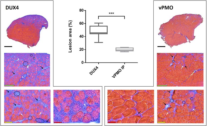
www.nature.com/scientificreports/
high intrinsic toxicity but low plasma membrane permeability12–14. Besides its direct clinical application in
the field of cancer, EP is broadly used as a gene delivery tool15 that can be applied either for vaccination16–20,
immunotherapy (especially in cancer a pplications21,22), gene therapy23–26, genome e diting27,28 and generation of
induced pluripotent stem cells29,30. Number of phase I and II clinical trials are underway or have been completed,
demonstrating the safety and efficacy of this procedure3. EP has several advantages compared to other tools for
gene delivery. Indeed, pDNA are easy to modify and prepare, inexpensive to produce in large scale, and may
be administered multiple times without significant inflammation or immune response. pDNA are injected as
“naked” molecules meaning that no additional chemicals are associated, limiting risks and undesirable side
effects1,2. Unlike viral vectors, there is no safety c onsiderations1,31. EP procedure is relatively simple and easy to
set up and does not need expensive instrumentation. This procedure allows increased levels of gene transfer and
expression, nearing those of viral vector, and is applicable from cells transfection to drug and therapeutic gene
delivery into living tissues from rodents to humans2,3.
Skeletal muscles constitute attractive targets for gene therapy, due to their accessibility and high
vascularization. Notably, skeletal muscles were used for systemic delivery of therapeutic protein such as
erythropoietin32, coagulation factors33 or anti-inflammatory cytokines34. Muscle EP protocols, first described
in35, have been optimized over time to improve transfection efficacy and transgene expression level1,36–40. In the
context of muscle dystrophies, EP methodology was explored as a potential route for treatment. EP was notably
reported as efficient to transduce constructs encoding dystrophin in the mdx mouse and dog models of Duchenne
Muscular Dystrophy (DMD)41,42. Contrary to DMD, resulting from the loss of dystrophin, Facioscapulohumeral
muscular dystrophy (FSHD)43 is a gain of function disease caused by the inappropriate expression in skeletal
muscle of DUX4, a gene normally only expressed in germline and early e mbryogenesis44–55. The DUX4 gene
encodes a transcription factor that deregulates a large molecular n etwork53–60. However, the precise mechanisms
by which DUX4 leads to clinical symptoms still must be clarified. Even though various therapeutic strategies
are emerging, there is currently no curative treatment for FSHD. Several drug-based therapies aiming either for
muscle improvement (anti-inflammatory a pproach61, β2-adrenergic a gonists62–65, antioxidants66) or inhibition
of DUX4 expression such as mitogen-activated protein kinase inhibitors67 (Losmapimod, Fulcrum therapeutics,
NCT04003974 ) are investigated in clinical t rials68,69. In parallel, gene therapy has been explored to reduce or
avoid DUX4 protein expression and/or activity by controlling D4Z4 locus methylation70,71, or to silence DUX4
mRNA. Among those, antisense oligonucleotides (AOs) and siRNA targeting the DUX4 mRNA and preventing
its translation have been developed and successfully tested in vitro and in vivo54,72–75. Development of in vivo
proof-of-concept studies for emerging therapies are now required as a next step towards clinical trials. Several
hurdles such as DUX4 toxicity and its stochastic low expression have made the generation of an animal model
recapitulating all the pathophysiological aspects of FSHD very challenging. FSHD-like mouse models have now
been described, each of them possessing their own advantages and limits59,76–82. Some of these mouse models
allow inducible conditional DUX4 expression, bypassing DUX4 high toxicity during embryonic development
and enabling mice to grow up and develop muscular dystrophy78–80,83. These models open new ways to investigate
molecular mechanisms leading to FSHD symptoms. However, the use of inducible transgenic models is often time
consuming and costly. It is especially an issue in the current context of FSHD where high throughput molecule
screenings are required to identify new potential therapeutics. In the present study, we describe a convenient
in vivo model of DUX4 local muscle expression using an EP procedure. This model is simple, unexpansive,
reproducible and associated with an easy read out that facilitates quantitative analysis. Therefore, this model can
be useful at the forefront for high throughput therapeutic screening.
Results
Hyaluronidase pre‑treatment improves gene expression following naked DNA injection and
electroporation in vivo. Mouse TA muscles were injected with the pCMV-lacZ reporter plasmid and then
electroporated (IMEP procedure). In order to determine whether muscle pre-treatment with hyaluronidase
(which digests hyaluronic acid, a major constituent of the extracellular matrix) could modify gene electroporation
efficacy, naked DNA injection was preceded (hIMEP group) or not (IMEP group) by an intramuscular injection
(IM) of hyaluronidase. TA muscles were harvested 7 days after injection. The reporter expression level was
evaluated by X-gal staining of β-galactosidase activity on cryosections from proximal, medial and distal
muscle regions and averaged for each group. As observed in Fig. 1, hyaluronidase pre-treatment significantly
improved gene electroporation efficiency as shown by the increased β-galactosidase-positive (β-gal+) muscle
area (Fig. 1A–C). Quantification of β-gal+ muscle surface showed a three-fold increase in mouse TA pre-treated
with hyaluronidase, with median value of 36.4% (p < 0.001, hIMEP vs IMEP, Fig. 1C). The hIMEP procedure was
therefore applied in the next steps of the study.
TA electroporation with DUX4‑expression plasmid induces readily quantifiable muscle
lesions. Because of the known DUX4 toxicity in human FSHD muscles, we first checked whether a DUX4
local expression induced by the hIMEP procedure could impact muscle structure in mice. To this aim, the TA
muscles were injected and electroporated with pCIneo-DUX4 expression plasmid using the hIMEP procedure.
The empty pCIneo plasmid was used as negative control. In both conditions, a simultaneous injection of the
pCMV-lacZ reporter vector was performed to facilitate the location of muscle areas having incorporated the
transgenes. Mice were sacrificed 1 week later and TA muscles were harvested, quickly frozen, cryosectioned
and stained with X-gal to detect reporter gene activity, or with Hematoxylin–Eosin–Heidenhain blue (HEB) for
histological evidence of muscle damage.
One-week post-injection, TA muscles electroporated with the control plasmid presented a normal histological
structure with peripheral nuclei in geometric fibres surrounded by a thin layer of endomysial extracellular matrix,
Scientific Reports | (2020) 10:11301 | https://doi.org/10.1038/s41598-020-68135-7 2
Vol:.(1234567890)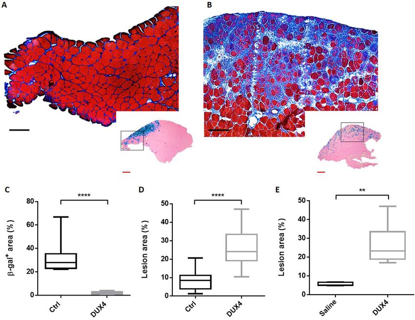
www.nature.com/scientificreports/

                            Figure 1.  Hyaluronidase pre-treatment improves β-galactosidase expression in mouse Tibialis Anterior muscle
                            (TA). (A,B) Representative sections of TA electroporated (A) without hyaluronidase pre-treatment (IMEP)
                            or (B) with hyaluronidase pre-treatment 2 h before the electroporation procedure (hIMEP). TA muscles were
                            injected by IMEP or hIMEP with 40 µg of pCMV-lacZ reporter plasmid. TA were harvested 1-week post-
                            injection and cryosections stained with X-gal (blue) and counterstained with Eosin (pink). Scale 500 µm. (C)
                            Percentage of surface area expressing β-galactosidase (β-gal+) quantified by color thresholding using ImageJ.
                            Data are represented as boxplots, ****p < 0.0001 Mann–Whitney Rank Sum Test; n = 4 for each group. The graph
                            was generated using GraphPad Prism 6.01.

                            as shown by HEB staining (Fig. 2A). A restricted area exhibiting some smaller fibres, central nuclei and a slight
                            focal inflammatory infiltrate around the site of injection (β-gal+ area) was sometimes observed (data not shown).
                            X-gal staining in control TA confirms electroporation efficiency as shown by the presence of grouped β-gal+
                            fibres (Fig. 2A, Box). In contrast, TA muscles electroporated with the DUX4-expression plasmid showed a large
                            damaged region containing atrophic fibres with high size variability, some regenerating fibres characterized by
                            central nuclei, a marked inflammatory infiltrate and an accumulation of conjunctive tissue (fibrosis) (Fig. 2B;
                            SI Fig. S1 online). Interestingly, X-gal staining in adjacent sections showed a lower number of β-gal+ myofibres
                            with a different spatial distribution as compared to TA muscles injected with the control plasmid (Fig. 2B, Box).
                            Indeed, a median value of 2.1% of the total muscle surface was positive for β-gal in the DUX4 expression group
                            against 27.97% in the control group (p < 0.001, Fig. 2C). As expected, lesion area quantification on HEB-colored
                            total cryosections indicated that the percentage of damaged surface area was significantly higher in TA muscles
                            injected with the DUX4-expression plasmid as compared to the control group (p < 0.001) with a median value
                            of 25.1 and 8.5%, respectively (Fig. 2D).
                                We also investigated the plasmid recirculation potential following hIMEP. Indeed, we were wondering whether
                            the recirculation of the DUX4 expression plasmid would be sufficient to cause transfection of the contralateral
                            TA resulting in quantifiable lesions. To this aim, both TA muscles from the same mouse were electroporated,
                            one using pCIneo-DUX4, the other one using saline solution. One week after hIMEP, quantification of the
                            damaged area on HEB-stained cryosections showed no detectable lesion in the saline-injected TA, in contrast
                            to the contralateral TA injected with the DUX4-expression plasmid which exhibited a significant lesion area
                            (Fig. 2E). This suggested that no plasmid recirculation occurred, allowing an independent treatment of both TA
                            from the same animal.

                            Dose–response analysis. A dose–response analysis was performed by electroporating mouse TA muscles
                            with increasing doses of the DUX4-expression plasmid or control plasmid, concomitantly with the pCMV-lacZ
                            reporter vector. The quantification of muscle damages (HEB staining) was (1) reported either to total muscle
                            section or (2) to the injected area, corresponding to the β-gal+ region. Regardless of the quantification method,
                            no statistical difference was observed following the injection of different doses of the control plasmid (1–20–
                            40 µg pCIneo; one-way ANOVA on the ranks followed by Dunn’s post hoc test, p = 0.959 and 0.153 in total and
                            injected area, respectively, data not shown). The data from these three control groups were therefore pooled in
                            a single control group (Fig. 3). Regarding the muscle damage quantification reported to total section (Fig. 3A),
                            a significant increase of damaged surface between the control group and all DUX4 groups was demonstrated
                            (p < 0.01). Similar results were obtained when the injected area was the only area considered for the quantification
                            (Fig. 3B). However, there was no significant difference in the percentage of damaged area obtained after injection
                            of increasing doses of pCIneo-DUX4 using either quantification system (p = 0.894 and 0.957, reported to total
                            section or to injected area, respectively). Data from the four DUX4 expression groups have thus been grouped
                            (in Fig. 3C) to compare results obtained from both quantification methods. As expected, the percentage of
                            damaged muscle surface area was higher when quantified within the injected region, as compared to the
                            percentage calculated on total muscle section. This difference is highly statistically relevant in the DUX4 group
                            (p < 0.0001) and significant in the control groups (p = 0.01) where the altered muscle area was limited to 7.4% of
                            the total muscle section and to 7.3% when calculated in the injected region (Fig. 3C).

Scientific Reports |   (2020) 10:11301 |               https://doi.org/10.1038/s41598-020-68135-7                                             3

                                                                                                                                         Vol.:(0123456789)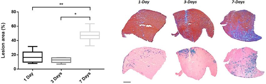
www.nature.com/scientificreports/

                                      Figure 2.  hIMEP of DUX4-expression plasmid induces muscle lesions. (A,B) Representative sections of TA
                                      electroporated with 10 µg of pCMV-lacZ and 40 µg of (A) pCIneo or (B) pCIneo-DUX4 plasmids. TA muscles
                                      were harvested 1-week post injection and cryosections from distal, medial and proximal regions stained with
                                      X-gal to assess β-gal+ areas (small pictures). Adjacent sections were stained with HEB coloration for muscle
                                      damages evaluation (large pictures). Scales 50 µm (red) and 100 µm (black). (C,D) Percentage of surface area
                                      (C) expressing β-galactosidase (β-gal+) and (D) damaged in mouse TA 1-week post hIMEP using 10 µg of
                                      pCMV-lacZ concomitant with 40 µg of pCIneo (Ctrl) or pCIneo-DUX4 (DUX4) plasmids. β-gal+ and lesion
                                      area percentage were evaluated on total section stained with X-gal or HEB respectively, and quantified by color
                                      thresholding using ImageJ. (E) Recirculation test. Both TA from same mouse were electroporated, one using
                                      40 µg of pCIneo-DUX4, the other saline solution. One week after hIMEP, lesion area was evaluated on HEB
                                      stained TA cryosections by color thresholding using ImageJ. All results are presented as boxplots, **p < 0.01 and
                                      ****p < 0.0001 Mann–Whitney Rank Sum Test; n = 5 for each group except in (E) n = 2. Graphs were generated
                                      using GraphPad Prism 6.01.

                                      Distribution of the lesions through Tibialis Anterior muscles. In order to investigate DUX4
                                      expression distribution through TA following hIMEP with pCIneo-DUX4, muscle damages were quantified
                                      in 3 different TA regions (proximal, medial and distal region). Since no statistically significant difference was
                                      observed between tested doses considering each region individually (One way Anova, p = 0.871, 0.821 and
                                      0.395 in the proximal, medial and distal region, respectively, data not shown), data corresponding to the 1-, 5-,
                                      20-, 40 µg DUX4 plasmid groups were respectively pooled for each muscle region. As illustrated in Fig. 4, at
                                      7 days post-injection, the percentage of damaged muscle area in the proximal and medial part of the TA were
                                      not statistically different, with respective medians of 47.11% and 46.69% (p: NS, Friedman Repeated Measures
                                      Analysis of Variance on Ranks). Regarding the distal muscle part, the percentage of lesion was significantly
                                      lower than in the other muscle regions (p < 0.05) with a median of 25.6% (Fig. 4). Considering these results, the
                                      subsequent analyses were based on the proximal and medial TA parts to minimize variability.

                                      Time‑course analysis. The development of the TA lesions was evaluated 1-, 3- and 7-days after the hIMEP
                                      procedure by using 1 µg of the DUX4-expression plasmid and a concomitant injection of pCMV-lacZ reporter.

          Scientific Reports |   (2020) 10:11301 |              https://doi.org/10.1038/s41598-020-68135-7                                            4

Vol:.(1234567890)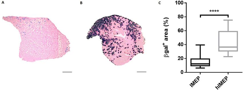
www.nature.com/scientificreports/
Figure 3. Dose–response of muscle lesion area in mouse TA 1-week post hIMEP procedure. (A,B) Lesion
area percentage was evaluated (A) on total cryosection or (B) in the injected area (defined by β-gal+ region)
from distal, medial and proximal part of TA electroporated with different doses of control or DUX4-expression
plasmid and 10 µg of pCMV-lacZ. Sections were stained with HEB and lesions were quantified by color
thresholding using ImageJ. Control groups (Ctrl) include 1-, 20-, 40-µg pCIneo-injected groups as there was
no statistical difference among them (one-way ANOVA followed by Dunn’s post hoc test, p = 0.959 and 0.153
for total and injected region quantifications respectively, NS). (C) Comparison of injured area percentages
quantified on total section (black) or in injected area (defined by the β-gal+ region) (grey). DUX4 groups include
1-, 5-, 20-, 40-µg pCIneo-DUX4-injected groups as there was no statistical difference among them (one-way
ANOVA followed by Dunn’s post hoc test, p = 0.894 and 0.957 for total and injected regions respectively, NS). All
results are presented as boxplots, ****p < 0.0001, **p < 0.01, (vs ctrl (A,B) or as indicated (C)). (A,B) One-way
ANOVA on the ranks followed by Dunn’s post hoc test; n = 3, except 5 µg DUX4 (n = 4), 40 µg DUX4 (n = 5) and
Ctrl (n = 11). (C) Wilcoxon signed rank test; n = 11 (ctrl) or 15 (DUX4). Graphs were generated using GraphPad
Prism 6.01.
Figure 4. Lesion distribution through TA regions. Mouse TAs were electroporated with different doses of
pCIneo-DUX4 (1, 5,10, 20 and 40 µg) and 10 µg of pCMV-lacZ. Muscle damaged area 1 week following TA
hIMEP was evaluated in the injected area (β-gal+) of HEB stained cryosections from distal, medial and proximal
regions of TA (color thresholding, ImageJ). Since no statistical difference was observed between tested doses
considering each region individually, data were respectively pooled to form single proximal, medial and distal
groups (data not shown, One-way Anova, p = 0.871, 0.821 and 0.395 in proximal, medial and distal region
respectively). All data are presented as boxplots, *p < 0.05 and **p < 0.01. Friedman Repeated Measures Analysis
of Variance on Ranks; n = 15 for each group. Graphs were generated using GraphPad Prism 6.01.
Muscle damages were then evaluated in TA proximal and medial parts by focusing on the injected area (β-gal+
region). At 1- and 3-days after injection, a slight thickening of the extracellular matrix and a straight mark with
some mononuclear cells and regenerating fibres (probably corresponding to the injection site) were observed.
This led to a global ‘lesion’ percentage defined by HEB staining of 16.3% and 12.9% at 1- and 3-days post-
injection, respectively. At 7 days after electroporation, extensive injuries, a marked inflammatory infiltrate and
an extracellular matrix expansion were detected. At this time point, damaged surface area represented 47.2% of
the injected area (Fig. 5, median value, p < 0.05 7-days vs 1- and 3-days).
Expression of DUX4 and its target genes in hIMEP mice. DUX4 expression at the mRNA level was
investigated by 3′RACE in hIMEP treated mice. In addition, mRNA levels of two DUX4 target genes (Wfdc3,
Zscan4c), commonly used as biomarkers of DUX4 transcriptional activity78–80, were quantified along with
the mRNA level of Myog a myogenic marker known to be downregulated in FSHD, contrary to Wfdc3 and
Scientific Reports | (2020) 10:11301 | https://doi.org/10.1038/s41598-020-68135-7 5
Vol.:(0123456789)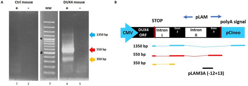
www.nature.com/scientificreports/
Figure 5. Time-course analysis. TA muscles were electroporated with 1 µg of pCIneo-DUX4 and 10 µg of
pCMV-LacZ. Lesion area was evaluated 1-, 3- or 7-days after hIMEP in the injected area (β-gal+) on HEB stained
cryosections of medial and proximal TA regions (color thresholding, ImageJ). Results are presented as boxplots,
*p < 0.05 and **p < 0.01. One-way ANOVA on the ranks followed by Dunn’s post hoc test; n = 3 for each group.
Scale 1 mm. The graph was generated using GraphPad Prism 6.01.
Figure 6. Confirmation of DUX4 mRNA expression by 3′RACE. (A) Nested PCR was used to amplify the 3′
end of DUX4 transcripts from total RNA extracted from TA muscles electroporated with 1 µg of control (left
panel) or DUX4-expression pDNA (right panel). Representative cropped gels. Lanes 1 and 2 come from one
gel; lanes 3, 4 and 5 come from a second gel ran in parallel. Corresponding full-length gels are available in
supplementary information (SI Fig. S2 online). 3′RACE were performed 1-, 3- and 7-days post hIMEP. Wells
(−) show negative controls without retro-transcription. Several fragments were detected (~ 1,350 bp, ~ 550 bp
and ~ 350 bp; colored arrows) in each time point (not shown), cloned, sequenced and analyzed to confirm
DUX4 specificity. (B) Schematic representation (not to scale) of 3′RACE products analysis. In silico alignment
confirmed conservation of vPMO [pLAM3A (− 12 + 13)] target sequence (black line).
Zscan4c84–86. To this aim, total mRNAs were extracted from mouse TAs treated by hIMEP (1-, 3- and 7-days)
using 1 µg of pCIneo-DUX4 or control plasmid pCIneo concomitant with reporter vector pCMV-lacZ. The 3′RACE
products were separated by electrophoresis on agarose gel and 3 distinct product lengths (~ 1,350 bp, ~ 550 bp
and ~ 350 bp) were observed. These fragments were only detected in the DUX4 expression group and at all
investigated time points. Interestingly, the 550-bp fragment presented with the highest intensity and frequency
(8/12 tested samples), suggesting a higher expression level of the corresponding mRNA as compared to the
others. No specific fragment was detected in control mice (Fig. 6; SI Fig. S2 online). All PCR products were
cloned, sequenced and analyzed to ensure DUX4 specificity. We also confirmed, by in silico analysis, presence
of the target sequence for antisense oligonucleotide pLAM3A (− 12 + 13) (described in54,72) in the 550-bp
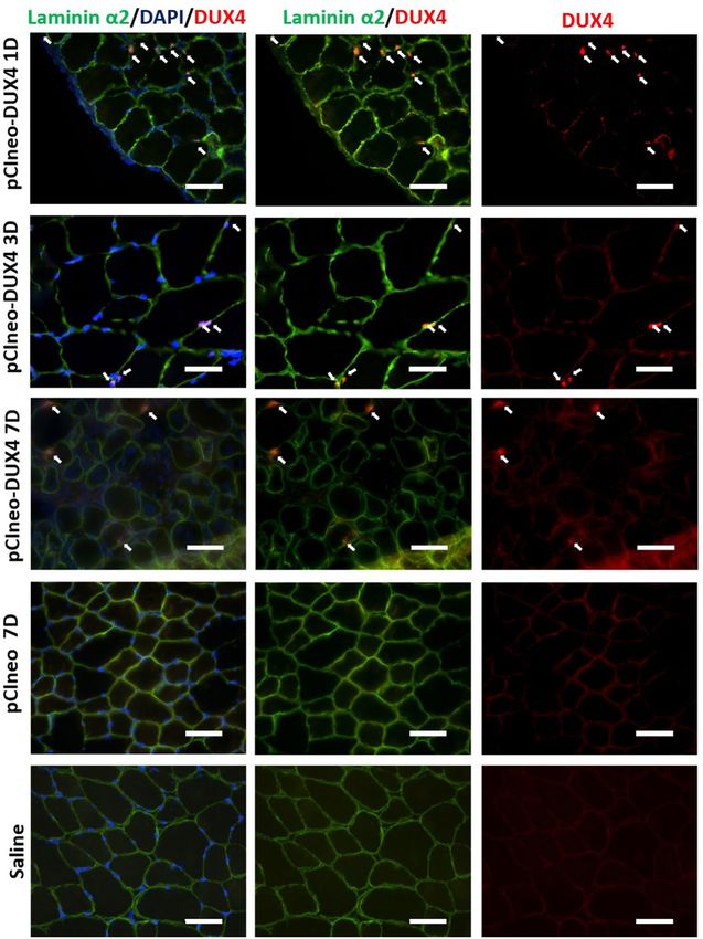
sequences. DUX4 expression was also evaluated at the protein level by immunofluorescence (Fig. 7) confirming
the presence of DUX4-positive nuclei, only in TA muscles injected with pCIneo-DUX4 at all investigated time-
points. RT-qPCR analysis of DUX4 target gene mRNAs highlighted a statistical increase of Wfdc3 and Zscan4c
expression at each time point in the DUX4 group compared to the control group (p < 0.001). However, there was
no statistical difference between time points for these genes. Myog mRNA levels were stable in control groups
regardless of the time point but significantly decreased at day 1 post hIMEP in the DUX4 expression group. Myog
Scientific Reports | (2020) 10:11301 | https://doi.org/10.1038/s41598-020-68135-7 6
Vol:.(1234567890)www.nature.com/scientificreports/
Figure 7. Confirmation of DUX4 protein expression by Immunofluorescence. Mouse TAs were injected by
hIMEP with 10 µg pCMV-lacZ plasmid concomitantly with 40 µg either pCIneo-DUX4 or pCIneo, or with a
saline solution (negative control), as indicated. Cryosections were analyzed 1, 3 and 7 days post-injection by
immunofluorescence with antibodies directed against either DUX4 (E5-5) (red) or laminin α2 (green) to stain
the myofibre basal lamina (for details, see “Methods”). DAPI was used to visualize nuclei (blue). Pictures were
taken with a Nikon Eclipse 80i microscope and merged using NIS-Elements software. Scale bar 50 µm.
Scientific Reports | (2020) 10:11301 | https://doi.org/10.1038/s41598-020-68135-7 7
Vol.:(0123456789)www.nature.com/scientificreports/
Figure 8. RTqPCR analyses of DUX4 target genes (Wfdc3, Zscan4c) and Myog expression level in TA muscle.
TAs were injected by hIMEP with either 1 µg of control pCIneo DNA, 1 µg of pCIneo-DUX4- or with 50 µl of
saline solution and harvested 1-, 3- or 7-days post injection. Total RNA was extracted with Trizol and qPCRs
were performed in duplicates using SYBR Green FastStart Essential DNA Green Master. Analyzes were
performed with LightCycler 96 software. At each time, results obtained from control plasmid and saline solution
injected groups were pooled to form a single control group, as no statistical difference could be highlighted
between them for all tested genes (one-way ANOVA on the ranks followed by Dunn’s post hoc test). Results
are presented as fold difference to Rplp0 for Wfdc3 and Zscan4c and as relative to control for Myog. All data are
presented as boxplots, *p < 0.05, **p < 0.01, ***p < 0.001 and #p < 0.05 vs DUX4 7 days, One-way ANOVA on
the ranks followed by Dunn’s post hoc test n = 14 (Ctrl) or 7 (DUX4) (Wfdc3 and Zscan4c) and n = 8 (ctrl) or 4
(DUX4) (Myog). Graphs were generated using GraphPad Prism 6.01.
expression then increased over time. However, we could not detect any significant difference between DUX4 and
control groups at 3- and 7-days post hIMEP (Fig. 8).
Preliminary evaluation of antisense oligonucleotide injected into DUX4 hIMEP mouse. Given
that the DUX4 hIMEP mouse model was developed in order to easily test the efficiency of therapeutics
in vivo, we investigated antisense oligonucleotide pLAM3A (− 12 + 13) (described in54,72) with the vPMO
(octa-guanidine conjugated Phosphorodiamidate Morpholino Oligomer) chemistry as a proof-of-concept
study. This vPMO silences DUX4 mRNA translation by interfering with intron II splicing. Its efficiency was
previously demonstrated in vitro54 and in vivo72 in another mouse model87. To this aim, mouse TA muscles were
electroporated using 20 µg of pCIneo-DUX4 plasmid by hIMEP and treated 6 h later with a single intraperitoneal
injection of 250 µg of vPMO pLAM3A − 12 + 13. Seven days after injection, a highly significant 2.5-fold decrease
of histological lesion area was observed in TA muscles of mice treated with the vPMO, as compared to untreated
hIMEP DUX4 mice (p < 0.001) (Fig. 9).
Discussion
Non-viral methods for transgene delivery have received increased attentions thanks to their relative safety,
simplicity and cost-effectiveness compared to the administration of viral vectors which were found associated
with cytotoxicity, inflammation and immune r esponse1,2. Initially, the main drawback of naked pDNA gene
transfer was its low transfection efficiency limiting its use in fundamental research or therapeutic applications.
Indeed, as there are no specific DNA transporters at the surface of mammalian cells, naked plasmids can only
be internalized by non-specific endocytosis. As this is a slow process, most of the injected DNA is retained into
extracellular space and degraded by endogenous DNAses88. Nevertheless, electroporation (EP) was shown to
improve transgene uptake and expression both in vitro and in vivo2,3,7. Previous studies have demonstrated the
importance of various factors influencing efficacy and safety of pDNA EP in several tissues, inter alia, skeletal
muscles. These factors include electrical parameters (intensity, duration, pulse number and frequency), electrode
type and geometry, plasmid features including its length and GC content and finally, additional elements such as
the host age or tissue permeability (e.g. normal vs DMD muscle41). In the present study, we applied optimized EP
parameters according to Mir et al.89 who have demonstrated the influence of voltage-to-distance (between both
electrodes) ratio, pulse length, number and frequency for muscle EP in vivo. Their observations suggested that 8
pulses of 200 V/cm, 20 ms and 2 Hz was an optimal combination for high transgene expression in mouse skeletal
muscle. Those parameters have been largely and successfully used in other studies1,3,38,40,41,90,91. Different types
of electrode can be used, such as plate electrodes, penetrating needle electrodes or multi-electrode a rrays2,3,38.
Here, we chose to use plate electrodes which have the advantage to apply a large and uniform electrical field92,93,
to be less invasive and less damaging than the needle one38,40. Furthermore, the efficiency of gene transfer
essentially depends on the effective DNA electrophoresis and distribution in the area where the electrical field
is applied. However, the extracellular matrix (ECM) surrounding the myofibre surface limits a homogenous
distribution of pDNA and thereby reduces transfection efficiency. Different enzymes modifying connective
tissue permeability such as hyaluronidase or collagenase were tested to improve the distribution of naked
pDNA33,39,40,94–96. Hyaluronidase is a mucolytic enzyme that depolymerizes N-acetyl-hexosamine glycosidic
bonds in hyaluronic acid, interfering with its barrier function in the ECM96. Intact hyaluronic acid binds
water molecules and forms a gelatin-like matrix that provides a structural support for the surrounding tissue.
Scientific Reports | (2020) 10:11301 | https://doi.org/10.1038/s41598-020-68135-7 8
Vol:.(1234567890)www.nature.com/scientificreports/
Figure 9. vPMO treatment of mice electroporated with DUX4-expression plasmid decreases TA muscle lesions.
TA muscles were injected by hIMEP with 20 µg of pCineo-DUX4 plasmid. Six hours later, mice received either
an intraperitoneal injection of 250 µg of vPMO pLAM3A (− 12 + 13) targeting DUX4 mRNA (vPMO group) or
no supplemental treatment (DUX4 group). Percentage of lesion area was evaluated 7 days post IMEP by color
thresholding using ImageJ on HEB colored cryosections of TA proximal and medial parts. Results are presented
as Box plots, ***p < 0,001 by Mann–Whitney Rank Sum Test; n = 4 for each group. The graph was generated
using GraphPad Prism 6.01. Black arrows show fibrosis, white arrows atrophic fibres, black circles inflammatory
infiltrate. Scales 1 mm (black) 250 µm (green), 100 µm (yellow) and 50 µm (red).
Hyaluronic acid fragments resulting from hyaluronidase digestion are still able to bind water molecules but fail
to form a functional viscous gel. Consequently, the density of glycosaminoglycans is reduced, allowing a better
DNA electrophoresis, improving its contact with the plasma membrane, thus increasing the transfection r ate95,97.
Consistent with previous studies, we observed an increased reporter gene (pCMV-lacZ) expression level when
hyaluronidase was injected 2 h before the IMEP procedure. Histological analysis did not reveal any apparent
muscle alterations linked to hyaluronidase pre-treatment, consistent with other o bservations36,39,94–96. Moreover,
96
hyaluronidase is currently used in clinical a pplications supporting that it is not by itself harmful for tissue.
In this study, the EP procedure was used in order to develop a simple mouse model of DUX4 local expression
in skeletal muscle, with the end purpose of rapid therapeutic testing for FSHD. The development of a mouse
model recapitulating the full FSHD phenotype and molecular signature is very challenging due to the need for
DUX4 precise expression timeframe in the early embryo98,99. Attempts at the generation of transgenic mouse
models revealed that a tight regulation of DUX4 expression was essential to obtain viable mice able to reach
adulthood and to develop a dystrophic phenotype76,77. The most recently developed murine models allow a
controlled DUX4 expression: two of these are the iDUX4 mouse [2.7]77 and its improved version iDUX4pA78,
both doxycycline-regulated. In addition the F LExDUX479,83 and the TIC-DUX480 mice allow conditional
expression of human DUX4-fl following Cre recombination. When crossed with the appropriate cre-driver line
(tamoxifen-inducible cre line, cre expressed from a muscle-specific promoter), those models allow the appearance
of a progressive myopathy, useful for investigations on FSHD pathophysiological mechanisms. However, the
use of inducible transgenic models is often time consuming and costly. Therefore, cheap convenient in vivo
models, with a local well delineated muscle lesion, although not representing the whole disease complexity, may
constitute excellent tools in the frame of initial high-throughput therapeutic screening. Moreover, the IMEP
model is customizable. For instance, a construct containing a weaker p romoter100 could be used in order to mimic
a more “physiological” DUX4 expression level. This type of model would be useful to better understand FSHD
pathophysiological mechanisms. In addition, the contribution of modifier genes could be similarly investigated.
In the present study, we have demonstrated that a single injection of pCIneo-DUX4 plasmid into mouse TA by
hIMEP leads to the development of muscle damage after 7 days. These damages are directly linked to DUX4
expression, as evidenced by the absence of comparable lesions in muscles electroporated in the same conditions
with control pDNA. Lesions in muscles injected with DUX4-expression plasmid are characterized by the presence
of degenerating fibres with a heterogeneity in size, including numerous small fibres. Large mononuclear cell
Scientific Reports | (2020) 10:11301 | https://doi.org/10.1038/s41598-020-68135-7 9
Vol.:(0123456789)www.nature.com/scientificreports/
infiltration, extracellular matrix thickening, fibrosis and central nucleation are also observed as described in
other DUX4 s tudies59,78–80. Conversely, control TAs display a normal histology without dystrophic pattern but
some of them showed a thin and straight lesion characterized by fibres with central nuclei, sign of regeneration
events most probably due to the needle injury caused by the intra-muscular injection. Since our results suggest
an absence of significant systemic dissemination of pDNA following hIMEP, both hindlimbs could be used
as independent systems, fitting therefore with the animal ethics principle of reduction. The local character
of this myopathy model also presents two other advantages. First, only the injected muscle is impaired, so
mice do not show any phenotype59,78–80. Second, the localized lesion observed 7 days after administration of
DUX4 pDNA by hIMEP facilitates therapeutic evaluation. To this aim, we developed an easy and reproducible
quantification method to evaluate the percentage of damaged muscle surface area. To ensure the distinction
between healthy and injured area, we improved the basic hematoxylin–eosin histological coloration by adding
a step of Heidenhain’s blue counterstaining that results in a high blue-pink contrast, easily distinguishable by
automatic color thresholding. As expected, the percentage of damaged surface area is significantly higher in the
injected area (β-gal+) than in the total TA section in DUX4 pDNA-injected mice. Interestingly, in the DUX4
group, we noticed a decrease of the β-gal+ surface area and a different spatial distribution of β-gal+ fibres which
appear more scattered, as compared to the control group. This could be explained by the DUX4 toxicity. Indeed,
since both plasmids are co-injected, it is reasonable to hypothesize that most fibres expressing the LacZ plasmid
also express DUX4. Due to its high toxicity, the DUX4 protein likely causes fibre degeneration, sarcolemmal
damages and a consecutive loss of β-gal signal.
We were not able to highlight a reduction of muscle damage by decreasing DUX4 plasmid doses. If the absence
of dose–response is surprising, different hypotheses may be suggested. First, those results are in agreement
with the high myotoxic potential of DUX4, even when initially expressed in a limited number of myonuclei, as
described previously either in patients52,55,101,102 or in FSHD animal models78–80. As described by Tassin et al.55 and
confirmed by Ferreboeuf et al.102, the model of “DUX4 nuclear spreading” and the subsequent amplification of
the DUX4-induced cascade could partly explain this phenomenon. Second, direct DUX4-induced deregulation
pathways are most probably followed by feedback loop processes generating a self-sustaining system involving
e.g. oxidative stress and/or inflammation66,103. By this way, structural muscle damages observed in our study
certainly result from pathophysiological processes that have become independent from the initial DUX4 boost.
Regarding spatial distribution of lesions within the TA muscle, we did not find any significant difference in
lesion extent between medial and proximal sections, but a lower percentage of damaged surface area was observed
in the distal TA part. This result was unexpected given that the injected transgene could in theory diffuse along
the entire myofibres. Moreover, as DUX4 is a transcription factor and harbors nuclear localization s ignals104, it
can be transported into several neighboring nuclei in a myotube, as described in previous s tudies55,102. Parameters
such as electrode shape and positioning may be involved in inter-regional variation of hIMEP efficiency. Since TA
is not cylinder- but cone-shaped, the distances between each muscle region and the electrodes are not identical
along the whole muscle. As the distal TA part is located close to the tendon, the contact with electrode and the
resulting electrical field was probably lower in this region, despite the use of a conductive gel. Nevertheless, pDNA
may theoretically diffuse through the entire myofibre. However, since pDNA is administrated through a single
injection, a dilution phenomenon could explain a lower expression in the regions remote from the injection
site55. In addition, a limited diffusion of nuclear proteins in muscle fibres has already been m entioned105,106. The
same observations were reported in porcine muscle following EP: in that study transgene expression was limited
to the muscle area delineated by the electrical field but no mechanistic hypothesis was p roposed107. The DUX4
hIMEP procedure is therefore associated to the rapid development of localized muscle lesions. Although this
pattern allows to provide an easy read-out for therapeutic screening, the study of pathophysiological mechanisms
underlying consequences of DUX4 muscle expression requires a more scattered distribution pattern of the
transgene, closer to the one observed in FSHD muscles. This pattern may be obtained by using another route of
delivery of naked pDNA. Indeed, the hydrodynamic injection of pDNA via the saphenous vein, first described
by Hagstrom et al.108,109 allowed a more diffuse expression of the transgene in a panel of hindlimb muscles.
Regarding time-course investigations, no damages were observed 1 and 3 days post-hIMEP with DUX4
expression plasmid, while DUX4 mRNA and protein were already detected. Seven days after the hIMEP
procedure, a severe muscle lesion had developed. Although the presence of early damages between the 3rd and
the 7th day post-injection cannot be excluded, the period of 7 days seems appropriate to obtain quantifiable and
well-delineated muscle lesions consecutive to DUX4 expression. It seems also reasonable to hypothesize that a
delay is necessary for the establishment of pathophysiological processes which will ultimately overcome skeletal
muscle compensatory mechanisms. Since the expression of DUX4 target genes (“DUX4 footprint genes”) is
a good marker of DUX4 protein expression and transcriptional a ctivity78–80, DUX4 target gene signature was
investigated at the mRNA level to confirm the production of a functional DUX4 protein. As expected, at all-time
points, Wfdc3 and Zscan4c expressions were induced in DUX4 pDNA-injected TAs as compared to controls.
Myog, a marker of muscle regeneration, presented a decreased expression 1-day post-injection. This is similar
to results published by other groups showing that Myog expression was decreased by DUX4 expression84–86. The
progressive increase over time is consistent with histological regeneration features (central nuclei) observed
7-days after IMEP. DUX4 protein level was also investigated and as expected given our expression data on DUX4
mRNA and its target genes, DUX4 protein could be detected at all-time points.
Finally, given the relevance of our mouse model and quantification system in the framework of antisense
therapy, a preclinical proof-of-concept study was performed with an antisense oligonucleotide (AO) directed
against the DUX4 mRNA54,74. To this aim, we used pLAM3A (− 12 + 13) AO targeting a splice site in the DUX4
mRNA 3′UTR region. This AO was previously synthesized with a 2′OMe chemistry (phosphorothioate backbone)
and successfully tested in vitro either on cells overexpressing DUX4 or on FSHD primary m yotubes54. The same
AO synthetized as a vPMO (octa-guanidine dendrimer-conjugated vivo morpholino) was used in a preliminary
Scientific Reports | (2020) 10:11301 | https://doi.org/10.1038/s41598-020-68135-7 10
Vol:.(1234567890)www.nature.com/scientificreports/
study in vivo showing its ability to decrease DUX4 mRNA expressed from an AAV vector injected in mouse
TA74. In that study, the vPMO efficiency was monitored through DUX4 mRNA detection and semi-quantification
using 3′RACE. In the present study, we first confirmed that the target sequence of this AO was conserved in
the DUX4 mRNA detected in mouse TA electroporated with the DUX4-expression plasmid. We then tested
this vPMO on the DUX4 hIMEP model and demonstrated it could prevent the development of DUX4-induced
histological muscle damages.
In conclusion, we have developed a rapid and easy-to-use mouse model of DUX4 local over-expression. Even
if the mouse in which TAs are injected with pCIneo-DUX4 by hIMEP does not recapitulate all the complexity of
FSHD pathophysiology, this model has an added value in the forefront of pre-clinical evaluations, particularly
in a context in which high throughput therapeutic screening is still necessary. This model could also be applied
for the injection of other expression vectors to model various gain-of-function muscular diseases in order to
test potential therapeutics.
Methods
Ethics statement. All animal experiments met the Belgian national standard requirements regarding
animal care and were conducted in accordance with the Ethics and Welfare Committee of the University of
Mons. Protocols were approved by the Ethics and Welfare Committee of the University of Mons (reference
number LE016/03).
Animals. Female C57BL/6 mice, aged between 8 and 12 weeks, were purchase from Charles River laboratories
(France). Mice were housed in a conventional animal colony and maintained at 35–40% relative humidity with
a constant room temperature (21 °C) and natural day/night light cycle (12–12 h). Food and water were provided
ad libitum and animals were subjected to an adaptation period of 7 days before starting experiments.
Plasmids. The commercial reporter plasmid pCMV-lacZ encoding the Escherichia coli β-galactosidase
protein was used to assess gene electrotransfer efficiency. The pCIneo-DUX4 plasmid encoding the full length
DUX4 protein is detailed in110. This plasmid contains the complete DUX4 ORF followed by the pLAM region
isolated from the 4q35 fragment of a patient with FSHD. The pCIneo backbone vector from Promega (USA)
was used as negative control. Expression plasmids used in this study are driven by cytomegalovirus (CMV)
promoter. All DNA plasmids were produced by PlasmidFactory (Bielefeld, Germany) in a research grade quality
for pre-clinical and veterinary studies.
Intra‑muscular injection and electroporation (hIMEP). Adult female C57BL/6 mice were
anesthetized by inhalation of 4% isoflurane in an induction chamber then maintained with 2% isoflurane using a
mouse anesthesia mask. Hind limbs were shaved and 40 µg of hyaluronidase from bovine testes (Sigma-Aldrich,
USA) diluted in 20 µl sterile saline buffer (0.9% NaCl, Physiodose, Gilbert Laboratories, France) were injected
through the skin into the tibialis anterior (TA) muscle. Mice were then allowed to recover from anesthesia in
their cages. After two hours, mice were re-anesthetized by an intra-peritoneal injection of Ketamine 100 mg/
kg (Anestketin, Eurovet animal health) and Xylasine 10 mg/kg (Sigma-Aldrich, USA). Each TA was injected,
with a Hamilton syringe, through the skin with one dose of naked pDNA (1, 5, 10, 20 or 40 µg) diluted in an
equal volume of sterile saline buffer (50 µl). Flat parallel electrodes were placed on each side of the TA and good
contact between electrodes and the overlying skin was ensured by use of a conductive gel (Rodisonic, Pannoc,
Belgium). A train of eight square-wave pulses at a voltage of 95 V (200 V/cm; voltage to distance ratio calculated
on the hind limb thickness mean) and a duration of 20 ms at 500 ms interval (2 Hz) was generated using an
EMKA stimulator. After electroporation, animals were transferred back into their cages and were monitored
until complete recovery from anesthesia. Mice were daily checked and then sacrificed by an intraperitoneal
injection of Nembutal (120 mg/kg, CEVO) 1-, 3- or 7-days post electroporation. For vPMO preliminary test,
both mouse TAs were electroporated with 20 µg of pCIneo-DUX4 plasmid. Six hours after the procedure, mice
received either an intra-peritoneal injection of 250 µg of vPMO pLAM3A − 12 + 13 (Gene tools, described i n54,72)
or no supplemental treatment. TA were harvested 7 days after hIMEP.
Tissue preparation and histology. At the indicated euthanasia time points, right and left TAs were
removed, embedded in OCT compound (VWR) and frozen in liquid nitrogen-cooled isopentane. Eight µm
thick cryostat sections from proximal, medial and distal part of TA were cut using a Leica cryotome and serial
sections were colored respectively with X-gal staining (β-gal staining kit, Invitrogen) to assess percentage and
localization of muscle area expressing the electrotransfered genes, and with Hematoxylin–Eosin–Heidenhain
blue (HEB) to evaluate the percentage of damaged muscle area. HEB coloration consists in a basic Hematoxylin–
Eosin coloration followed by a 45-s incubation in Heidenhain’s Blue staining (mix of orange G and Aniline
Blue, Sigma-Aldrich, USA), allowing an intense blue labeling of fibrotic fibresand collagenous tissues which
improves contrast with healthy myofibres to facilitate muscle lesion quantifications. Slides were then scanned
using the NanoZoomer-SQ Digital slide scanner (Hamamatsu Photonics). Images were processed by color
thresholding using ImageJ 1.52a software, Rasband, W.S., ImageJ, U. S. National Institutes of Health, Bethesda,
MA, USA, https://imagej.nih.gov/ij/, 1997–2018. The blue surface area was measured (threshold parameters:
Hue = 35–255/50–255, saturation = 53–255/53–255 for X-gal/HEB staining respectively) and reported to the
complete surface section. Measurements were performed on total section and on the injected area defined by
X-gal staining (SI Fig. S3 online).
Scientific Reports | (2020) 10:11301 | https://doi.org/10.1038/s41598-020-68135-7 11
Vol.:(0123456789)www.nature.com/scientificreports/
Immunofluorescence. Tissue cryosections were fixed with 4% paraformaldehyde/PBS on ice for 20 min,
permeabilized with 0.25% TritonX-100/PBS for 10 min, then incubated with blocking solution (5% normal goat
serum (Dako), 2% BSA, 0.01% TritonX-100/PBS) for 30 min. Sections were then incubated with anti-DUX4
(rabbit monoclonal E5-5; 1:200, Abcam) and anti-laminin α (rat monoclonal; 1:100; Sigma-Aldrich) primary
antibodies at 4 °C overnight. They were subsequently incubated with secondary antibodies Alexa 555 Goat
anti-rabbit IgG (1:500, Biowest) and Alexa 488 Goat anti-rat Ig (1:500, Invitrogen) at room temperature for 1 h.
Stained sections were mounted with EverBrite Mounting Medium with DAPI (Biotium) for nuclear staining.
Pictures were taken with a Nikon Eclipse 80i microscope and merged using NIS-Elements software.
RNA isolation, 3′RACE and real time quantitative PCR. RNA extraction. TA muscles were trimmed
between proximal-medial and medial-distal parts using Leica cryotome (30 sections of 50 µm in both regions).
These slices were ground into liquid nitrogen, homogenized in 1 ml of TRIzol reagent (Invitrogen) and RNA was
isolated according to the manufacturer’s directions. Total RNA was then treated with DNAse I kit (amplification
grade, ThermoFisher).
3′RACE. cDNAs were synthetized using SuperScript III Reverse transcriptase kit (Invitrogen) and the 3′RACE
adaptor of the RLM-RACE kit (5′-GCGAGCACAGAATTAATACGACTCACTATAGGTTTTTTTTTTTTN-
3′, Ambion). The resulting cDNAs were amplified by nested PCR using PrimeSTAR Max DNA polymerase
(Takara-bioJapan). The specific outer primer for DUX4 cDNA amplification was: 5′-AGGCGCAACCTCTCC
TAG AAAC-3′ and the inner primer was: 5′-TGGAAGCACCCCTCAGCGAGGAA-3′. Cycling conditions for
outer PCR were as follows: initial denaturation step at 98 °C for 3 min, followed by amplification step with 25
cycles of 10 s at 98 °C, 10 s at 58 °C and 5 s at 72 °C, ended by 5 min at 72 °C. Same cycling conditions were used
for inner PCR except the last step of the amplification which was 10 s at 72 °C instead of 5 s. The 3′RACE PCR
products were analyzed by electrophoresis on 1%-agarose gel and staining with ethidium bromide (gel pictures
provided Fig. 6 were made in compliance with the digital image and integrity policies of Scientific Reports).
The products were then extracted from agarose band using PCR clean up gel extraction kit (Macherey–Nagel),
cloned into pJET1.2/blunt (ThermoFisher), amplified in E. coli and sequenced (Genewiz Inc.) to confirm specific
DUX4 mRNA amplification and vPMO targeted sequence conservation.
RT‑qPCR. cDNAs were synthetized using Maxima First Strand cDNA Synthesis kit (ThermoFisher). All
qPCRs were performed in duplicates using SYBR Green FastStart Essential DNA Green Master (Roche) and
following primers (10 µM): Wfdc3 (5′-CTTCCATGTCAGGAGCTGTG-3′, 5′-ACCAGGATTCTGGGACAT
TG-3′) and Zscan4c (5′-GATTATTGGCCACAGGACAAG-3′, 5′-TCAGGGTGCTGTTCTTTCTG-3′) from76,
Myog (5′-GAGACATCCCCCTATTTCTACCA-3′, 5′-GCTCAGTCCGCTCATAGCC-3′) and Rplp0 (5′-TCA
TCCAGCAG GTGTTCG-3′, 5′-AGCAAGTGGGAAGGTGTAA-3′) custom designed (Eurogentec). Cycling
conditions were as follows: initial denaturation step at 95 °C for 10 min, followed by 40 cycles of 15 s at 95 °C and
60 s at 60 °C. qPCR results were analysis with LightCycler 96 software (Roche).
Statistical analyses. Statistical analyses were done using GraphPad Prism version 6.01 for Windows,
GraphPad Prism software, La Jolla, CA, USA, www.graphpad.com. Data were tested for normality of distribution,
using the Kolmogorov–Smirnov test, and all considered as non-parametric. Differences between experimental
groups were statistically evaluated by a Mann Whitney rank sum test (hyaluronidase pre-injection, DUX4
expression and vPMO evaluation), one-way analysis of variance (ANOVA) on the ranks followed by a Dunn’s
post hoc test for multiple comparison (dose–response, time–response and qPCR analysis), Friedman repeated
ANOVA on the ranks (muscle distribution) or Wilcox on signed rank test (quantification on total vs injected
area). Differences were considered statistically significant at a p value < 0.05. All data are represented as boxplot
for non-parametric statistical tests (with minimum to maximum whiskers).
Received: 1 February 2020; Accepted: 9 June 2020
References
1. Sokołowska, E. & Błachnio-Zabielska, A. U. A critical review of electroporation as a plasmid delivery system in mouse skeletal
muscle. Int. J. Mol. Sci. 20, 2776. https://doi.org/10.3390/ijms20112776 (2019).
2. Cervia, L. D. & Yuan, F. Current progress in electrotransfection as a nonviral method for gene delivery. Mol. Pharm. 15,
3617–3624 (2018).
3. Young, J. L. & Dean, D. A. Electroporation-mediated gene delivery. Adv. Genet. 89, 49–88 (2015).
4. Rosazza, C., Meglic, S. H., Zumbusch, A., Rols, M.-P. & Miklavcic, D. Gene electrotransfer: A mechanistic perspective. Curr.
Gene Ther. 16, 98–129 (2016).
5. Neumann, E., Schaefer-Ridder, M., Wang, Y. & Hofschneider, P. H. Gene transfer into mouse lyoma cells by electroporation in
high electric fields. EMBO J. 1, 841–845 (1982).
6. Pucihar, G., Kotnik, T., Miklavčič, D. & Teissié, J. Kinetics of transmembrane transport of small molecules into
electropermeabilized cells. Biophys. J. 95, 2837–2848 (2008).
7. Shi, J. et al. A review on electroporation-based intracellular delivery. Molecules 23, 3044. https://doi.org/10.3390/molecules2
3113044 (2018).
8. Jakutavičiūtė, M., Ruzgys, P., Tamošiūnas, M., Maciulevičius, M. & Šatkauskas, S. Physical methods for drug and gene delivery
through the cell plasma membrane. Adv. Anat. Embryol. Cell Biol. 227, 73–92 (2017).
9. Antov, Y., Barbul, A., Mantsur, H. & Korenstein, R. Electroendocytosis: Exposure of cells to pulsed low electric fields enhances
adsorption and uptake of macromolecules. Biophys. J. 88, 2206–2223 (2005).
Scientific Reports | (2020) 10:11301 | https://doi.org/10.1038/s41598-020-68135-7 12
Vol:.(1234567890)You can also read