Management of Morbidity and Mortality in a New Zealand White Rabbit Model of Steroid-Induced Osteonecrosis of the Femoral Head - Ingenta Connect
←
→
Page content transcription
If your browser does not render page correctly, please read the page content below

Comparative Medicine                                                                                                                         Vol 71, No 1
Copyright 2021                                                                                                                             February 2021
by the American Association for Laboratory Animal Science                                                                                    Pages 86–98

                                                                       Case Study

       Management of Morbidity and Mortality in a
       New Zealand White Rabbit Model of Steroid-
        Induced Osteonecrosis of the Femoral Head

              Kerriann M Casey,1* Felicity Gore,2 José G Vilches-Moure,1 Masahiro Maruyama,3 Stuart B Goodman,2,3
                                            Yunzhi Peter Yang,2,3,4 and Samuel W Baker1

   Steroid-induced osteonecrosis of the femoral head (SONFH) is a condition documented in humans and animals exposed
to chronic steroid administration. The rabbit has become a preferred animal model for investigating the pathogenesis and
treatment of SONFH due to its shared femoral vascular anatomy with human patients, relative size of the femoral head,
and general fecundity. However, morbidity and mortality are frequent during the steroid induction period, prior to surgical
manipulation. These problems are poorly reported and inadequately described in the literature. In this study, we report the
clinical, gross, and histopathologic findings of New Zealand white (NZW) rabbits undergoing the steroid induction phase
of the SONFH model. Severe weight loss (>30%), lipemia, hypercholesterolemia, hyperglycemia, and elevations in ALT and
AST were consistent findings across all rabbits, although these changes did not differentiate asymptomatic rabbits from
those that became clinically symptomatic or died. Euthanized and spontaneously deceased rabbits exhibited hepatomegaly,
hepatic lipidosis/glycogenosis, and hepatocellular necrosis, in addition to a lipid-rich and proteinaceous thoracic effusion.
A subset of rabbits developed opportunistic pulmonary infections with Bordetella bronchiseptica and Escherichia coli and
small intestine infections with Lawsonia intracellularis superimposed on hepatic and thoracic disease. Together, these find-
ings allowed us to establish a clinical decision-making flowchart that reduced morbidities and mortalities in a subsequent
cohort of SONFH rabbits. Recognition of these model-associated morbidities is critical for providing optimal clinical care
during the disease induction phase of SONFH.

Abbreviations: MPS, methylprednisolone; NZW, New Zealand white; SONFH, steroid-induced osteonecrosis of the femoral head

DOI: 10.30802/AALAS-CM-20-000071

   Osteonecrosis of the femoral head (ONFH), or avascular                             literature review categorized proposed pathogenic mecha-
necrosis of the femoral head (ANFH), is a condition in both                           nisms as follows: 1) disorders of lipid metabolism, 2) de-
humans4,12,21 and animals5 that is associated with inadequate                         creased osteogenic capacity of bone marrow mesenchymal
vascularization and subsequent death of trabecular bone and                           stem cells, 3) insufficient vascular supply, 4) inflammation
bone marrow of the femoral head. Briefly, diminished or altered                       and apoptosis, and 5) genetic polymorphisms and noncoding
vascular supply to the femoral head leads to trabecular bone                          RNA.29 The complexity of SONFH and variability of patient
weakening, with subsequent femoral head collapse and result-                          demographics suggest the underlying pathogenesis is likely
ing coxofemoral arthritis.12                                                          to be multifactorial.16
   Underlying pathogeneses of ONFH can be broadly catego-                                Several animal models have been developed to study the
rized into traumatic (that is physical trauma) and nontraumatic                       pathogenesis and potential therapeutic strategies for SONFH.33,34
etiologies.21 Nontraumatic etiologies include chronic steroid                         Numerous animal species have been explored as candidate
administration, alcohol consumption, and blood dyscrasias,                            models for SONFH including mice, rats, rabbits, chickens,
among others.4 Among nontraumatic etiologies, prolonged ste-                          emus, and to a lesser extent, dogs, pigs, and sheep.34 While each
roid administration for systemic diseases such as rheumatoid                          species has various advantages and disadvantages, the rabbit is
arthritis, systemic lupus erythematosus, and organ transplan-                         frequently chosen due to its femoral vascular anatomy, which
tation is the most common underlying cause of steroid in-                             is similar to human patients, relative size of the femoral head,
duced ONFH (SONFH) in human patients.33 Although various                              and general fecundity.34 In the rabbit model, SONFH can be es-
mechanisms have been proposed to explain the pathogenesis                             tablished via 3 main induction protocols: 1) intramuscular (IM)
of SONFH, the underlying cause(s) remain elusive. A recent                            injection of methylprednisolone (MPS) alone, 2) IM injection
                                                                                      of MPS along with intravenous (IV) lipopolysaccharide (LPS),
                                                                                      or 3) IM injection of MPS along with IV allogeneic serum (for
Received: 13 Aug 2020. Revision requested: 11 Oct 2020. Accepted: 30 Oct 2020.        example horse serum).34 The two latter induction protocols aim
1
  Department of Comparative Medicine, 2Department of Bioengineering, 3Department of
Orthopedic Surgery, and 4Department of Material Science and Engineering, Stanford     to create the underlying proinflammatory conditions associated
University School of Medicine, Stanford, California                                   with SONFH and are thus used to emulate underlying nontrau-
    *
     Corresponding author. Email: kmcasey@stanford.edu                                matic causes of SONFH in humans.
86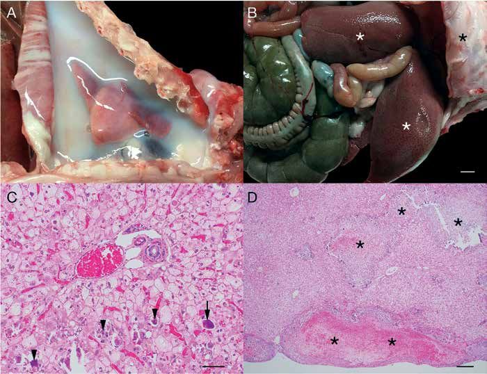
Morbidity and mortality in NZW rabbits undergoing SONFH

   From October 2016 to January 2017, a total of 4 male New                SONFH Induction and Surgery. At the start of the study, rab-
Zealand white (NZW) rabbits was submitted to necropsy for               bits were weighed and given a single IM injection of MPS (20
unexpected death during the induction phase of a SONFH                  mg/kg) in the quadricep muscle group to induce SONFH. Af-
model. These initial deaths prompted a systematic analysis              ter 4 wk, rabbits were randomly assigned to an experimental
of a subsequent cohort of rabbits undergoing SONFH induc-               treatment group involving the placement of experimental grafts
tion.                                                                   into the femoral head after core decompression surgery. Core
   Thus, during the period of August 2018 to May 2019, a second         decompression surgery involves the removal of a core of bone
cohort of SONFH was established in rabbits. Briefly, the SONFH          from the femoral head and neck, the standard surgical treat-
model was induced in male and female NZW rabbits via a sin-             ment for femoral head necrosis in humans. Further discussion
gle IM injection of MPS (20 mg/kg). Interventional surgical pro-        of the experimental treatments is beyond the scope of this study.
cedures were scheduled to occur 4 wk after SONFH induction.             Briefly, rabbits were sedated with ketamine (30 mg/kg SQ) and
During the 4-wk induction period, significant comorbidities and         xylazine (3 mg/kg SQ), intubated, and administered 1% to 4%
deaths occurred in varying subsets of rabbits. Clinical monitor-        isoflurane as needed for the 30-min procedure. Preoperative
ing and intervention were initiated to treat symptomatic rabbits        buprenorphine (0.03 mg/kg IV) was provided for preemptive
and to identify critical points of interventional therapy. A full       analgesia, and cefazolin (25 mg/kg IM) was provided for anti-
complement of diagnostics including bloodwork, radiographs,             biotic coverage and continued twice a day at the same dose and
necropsy, histopathology, microbiologic culture, and PCR test-          route for 2 d after surgery. A 2 cm incision was made to expose
ing was implemented to further characterize the nature of any           the femur immediately distal to the greater trochanter. Core de-
underlying clinical disease(s).                                         compression of the femoral head and neck was performed using
   Herein we report the clinical presentations, therapeutic in-         a 3 mm diameter drill under fluoroscopic guidance (Mini C-arm
terventions, and postmortem findings from rabbits developing            Fluoroscan Imaging System, model 1000-0005, Orthoscan, AZ).
comorbidities related to the induction period of SONFH. A lit-          The incision was closed using 2 buried layers of 3-0 polyglycolic
erature review over the past 30 y sheds light on the prevalence         acid suture (Ethicon, CA). A lidocaine (2 mg/kg) splash block
of SONFH-related complications and/or mortalities reported              was performed prior to closing the skin. After the surgical pro-
in primary research articles using this model. Our goal is to           cedures, the xylazine was reversed with atipamezole (0.3 mg/
help clinicians and pathologists working with rabbit models of          kg IM) and rabbits were allowed to recover. Once fully awake,
SONFH to better understand model-associated comorbidities               they were given buprenorphine SR (0.15 mg/kg SQ, Zoopharm)
and help determine points of clinical intervention to minimize          for postoperative analgesia. The use of nonsteroidal antiinflam-
model-associated mortalities.                                           matory drugs was contraindicated due to the experimental de-
                                                                        sign. During the 4-wk induction period, rabbits underwent no
                 Materials and Methods                                  additional experimental manipulation. The end of the experi-
   Literature Review. To examine the frequency of SONFH-                ment was set at 12-wk after MPS administration.
related deaths in research articles, we identified publications            Clinical Case Management. Throughout induction and the
in peer-reviewed literature by searching the PubMed database            peri-surgical period, rabbits were monitored daily by both in-
(1990 - present). Boolean search terms included: (osteonecrosis         vestigators and the Veterinary Service Center trained animal
AND “femoral head” AND rabbit AND steroid) and (“avascu-                health technicians for general overall health. If clinical signs
lar necrosis” AND “femoral head” AND rabbit AND steroid).               were observed (for example: loss of appetite, weight loss greater
   The literature search yielded a total of 99 unique articles from     than 20% from baseline, reduced or abnormal feces, respira-
1991 to 2020. After abstract review, 23 articles were excluded          tory impairment), clinical intervention was directed and im-
because they fell under one of 3 categories: 1) non-English, 2)         plemented by the veterinary staff and included daily weight
review article, 3) in-vitro work only. Thus, a total of 76 articles     monitoring and ancillary diagnostics. Depending on the clinical
were reviewed for mention of SONFH-related rabbit mortality.            presentation, additional diagnostic tests were performed in-
   Animals. All experimental procedures were approved by the            cluding complete blood counts (CBC), serum biochemistry, and
Stanford Institutional Animal Care and Use Committee (IA-               radiographs. When appropriate, therapeutics were initiated.
CUC). Research adhered to the principles stated in the 2011 edi-        Therapeutics included any combination of subcutaneous (SQ)
tions of the National Research Council’s Guide for the Care and         fluids (10 to 20 mL/kg 0.9% NaCl), Critical Care dietary support
Use of Laboratory Animals.8 The facility in which this research         (Oxbow, NE), and enrofloxacin (5 to 10 mg/kg PO). Rabbits that
was conducted is PHS assured, USDA registered, and fully ac-            failed to respond to clinical management were humanely eutha-
credited by AAALAC, International.                                      nized via an IV injection of pentobarbital containing euthanasia
   A total of 39 (n = 23 male; n = 16 female) NZW rabbits (West         solution (100 mg/kg) administered under deep sedation.
Oregon Rabbit Company, Philomath, OR) ranging in weight                    Necropsy and Histopathology. Rabbits that were found de-
from 3.5 to 4.0 kg were used to establish SONFH. Rabbits were           ceased or were euthanized due to clinical signs (n = 10 total
used in a staggered fashion from the periods of October 2016            rabbits; n = 4 index rabbits and n = 6 study rabbits) were
to January 2017 (index mortalities) and September 2018 to May           submitted for necropsy and histopathology. A cohort of clini-
2019. Rabbits were negative for the following infectious agents:        cally healthy rabbits (n = 19) that reached the experimental
Pasteurella multocida, Salmonella spp, Clostridium pilliforme, cilia-   endpoint (12 wk after MPS administration) also underwent
associated respiratory bacillus, Treponema cuniculi, Encephalitozoon    necropsy examination for comparison. Routine tissue sam-
cuniculi, Eimeria stiedae. Rabbits were acclimated for 1 wk prior       ples were collected and immersion-fixed in 10% neutral buff-
to SONFH induction in a conventional temperature-controlled             ered formalin for at least 72 h. Formalin-fixed tissues were
facility with a 12-h light/dark cycle. Rabbits were housed indi-        processed routinely, embedded in paraffin, sectioned at 5 µm,
vidually in standard, commercially available cages (Allentown,          and stained with hematoxylin and eosin (H and E). Select sec-
NJ) and were allowed ad libitum access to rabbit chow (Teklad           tions were stained with Gram stain (to identify bacteria) and
rabbit diet 2030, WI), a daily rotation of edible enrichment items,     Periodic acid-Schiff (PAS; to highlight glycogen accumulation
and reverse-osmosis water.                                              and spirochete organisms).

                                                                                                                                      87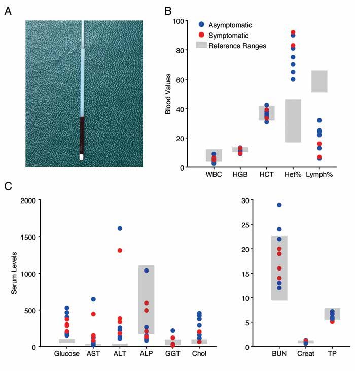
Vol 71, No 1
Comparative Medicine
February 2021

   Ancillary Diagnostics. Based on the clinical presentation and/                  rabbits due to an extensive postmortem interval. All livers were
or gross necropsy findings, ancillary diagnostics were per-                        markedly enlarged, friable, and exhibited an enhanced reticular
formed at the time of tissue collection. Additional diagnostics                    pattern (Figure 1 B).
included any combination of pleural fluid cytology, microbio-                         Histologically, livers exhibited varying degrees of diffuse he-
logic culture (aerobic and/or anaerobic) of the nasal turbinates,                  patic lipidosis and hepatic glycogenosis (Figure 1 C). Centri-
thoracic cavity, abdominal cavity, and/or pericardial space, and                   lobular to midzonal hepatocellular degeneration and necrosis
PCR for Lawsonia intracellularis. Pleural fluids were evaluated                    was moderate to severe and was often accompanied by single
inhouse and a select case was reviewed by a board-certified vet-                   cell necrosis and scattered mineralization. Occasionally, coalesc-
erinary clinical pathologist at the University of California - Da-                 ing regions of hepatocellular necrosis were randomly scattered
vis, Veterinary Medical Teaching Hospital, Clinical Diagnostic                     throughout the subcapsular parenchyma (Figure 1 D). A single
Laboratory Service. Aerobic and anaerobic microbial cultures                       rabbit exhibited small foci of hepatic abscessation with intral-
were evaluated inhouse using the Biolog system. Results were                       esional bacterial cocci (note: Staphylococcus aureus cultured from
compared with identification using MALDI-TOF at the Uni-                           this rabbit’s thoracic effusion). The fourth rabbit did not un-
versity of California, Davis, Veterinary Medical Teaching Hos-                     dergo histologic evaluation due to severe postmortem tissue
pital, Clinical Diagnostic Laboratory Service. Formalin-fixed                      autolysis.
paraffin-embedded tissues scrolls (3, 25-µm-thick) of lesioned                        Clinical Case Management. Based on the knowledge gained
small intestine were submitted to Charles River Laboratories for                   from the index mortalities, robust clinical management of a
TaqMan PCR. Based on histologic findings, the aforementioned                       second SONFH cohort was implemented. Between September
samples were evaluated for Lawsonia intracellularis via PCR.                       2018 and early May 2019, 35 rabbits (16 female and 19 male)
   Statistical Analysis. Statistical analyses were performed us-                   underwent the SONFH induction protocol. The average weights
ing StatPlus:mac (AnalystSoft, Walnut CA). Summary data are                        at the time of MPS administration were 4.6 ± 0.1 kg (females)
presented as mean with SEM. Differences were considered sig-                       and 4.3 ± 0.1 kg (males). Over the course of the study, all rabbits
nificant at P ≤ 0.05.                                                              experienced initial weight gain after MPS injection, followed by
                                                                                   a precipitous drop in weight and loss of muscle mass (Figure 2
                                  Results                                          A). Twenty-nine rabbits survived to the experimental endpoint.
   Literature Review. To determine if SONFH-related mortali-                       For these rabbits, the time from MPS injection to body weight
ties had been previously reported in the research literature, we                   nadir was 35.5 ± 2.6 d. On average, rabbits lost a total of 36 ± 1%
reviewed a total of 76 articles meeting predetermined inclusion                    of body weight. By the end of the study, surviving rabbits had
criteria (see Materials and Methods). Of these, 23 out of 76 ar-                   regained weight to 95 ± 2% of their starting weight. A compari-
ticles (30%) reported mortalities during the SONFH induction                       son of male and female rabbits that survived to the study end-
period (Table 1).3,6,9-11,13-15,17-20,22,23,25-27,30-32,35-37 The average length   point (Figures 2 B through D) showed no significant differences
of induction was 6.7 wk. The mean percentage of mortalities                        in maximal percentage weight loss (males 27 ± 2%, females 24
during the SONFH induction period was 12% (range: 6% to                            ± 2%), the number of days taken to reach body weight nadir
20%). This mortality rate excluded deaths related to invasive                      (males 34.6 ± 2.5 d, female 36.8 ± 5.3 d), or the rate of weight
experimental procedures (that is surgical manipulation outside                     loss (male 0.8 ± 0.1%/day, female 0.7 ± 0.1%/day). The rate of
of the SONFH induction period). Despite reports of mortalities,                    weight loss was not statistically different between rabbits that
only 6 out of 23 articles (26%) proposed causes for mortality                      survived to the study endpoint and those that did not (survi-
events, with minimal to absent description of necropsy findings,                   vors 0.8 ± 0.1%/day, euthanized 1.0 ± 0.1%/day, unpaired t test
and no mention of potential etiologic agents responsible for any                   P = 0.06). (Figure 2 E). All animals that were euthanized or died
reported “infections.” A total of 5 out of 23 articles (22%) refer-                were within the maximal weight loss bounds of both male and
enced antibiotic usage as a component of the induction protocol.                   female surviving rabbits (Figure 2 A).
   Eight out of 76 articles (10%) specifically stated that deaths                     Other than weight loss, the most common presenting morbid-
did not occur throughout the study period. The average length                      ity was abnormal feces. Five out of 35 rabbits presented with a
of induction in this group of studies was 4.5 wk. Qualita-                         combination of diarrhea and small, irregular fecal pellets. On
tively, the literature review provided no information regarding                    presentation these rabbits were bright, alert and responsive,
whether breed, sex, weight, or induction protocol had contrib-                     but had reduced food intake and abnormal feces. Abdominal
uted to the presence or absence of deaths. The remaining 45 out                    auscultation typically revealed reduced bowel sounds, although
of 76 articles (59%) did not specify whether deaths had occurred                   borborygmi were also reported. These rabbits were treated by
during the study.                                                                  providing SQ fluids, enrofloxacin, and additional high-fiber
   Index Mortalities. Between October 2016 and January 2017, 4                     food enrichment items. All rabbits presenting in this manner
out of 18 male NZW rabbits were submitted for necropsy due                         were successfully treated with this regimen and were asymp-
to unanticipated deaths during the SONFH induction period.                         tomatic for the rest of the study.
Three out of 4 rabbits were found dead with few premonitory                           Three out of 35 rabbits presented with nonspecific clinical
clinical signs. The fourth rabbit was euthanized due to lethargy,                  signs associated with sick rabbits. These included quiet attitude
tachypnea, and tachycardia. Duration from MPS administration                       (3/3), reduced activity (3/3), reduced/no food intake (3/3), re-
to death varied from 8 d to 4 wk.                                                  duced/scant fecal production (3/3), and elevated respiratory
   At necropsy, all rabbits exhibited mild to severe, white,                       rate (1/3), in addition to the profound weight loss common to
opaque, gelatinous thoracic effusion that compressed lung pa-                      all rabbits after SONFH induction. These animals did not re-
renchyma (Figure 1 A). Cytologically, the effusion consisted of                    spond to therapy and were euthanized for humane reasons.
proteinaceous fluid, lipid, and scattered lymphocytes. Micro-                         Complete Blood Counts and Serum Biochemistry. Blood was
biologic culture (aerobic and anaerobic) of the thoracic effu-                     collected 4 wk after MPS administration from a subset of 7 as-
sion was performed on 2 rabbits; one yielded small numbers                         ymptomatic rabbits. Other than weight loss associated with
of Staphylococcus aureus, while the other was culture negative.                    SONFH induction, these rabbits were deemed clinically healthy.
Microbiologic culture was not performed on the remaining 2                         Bloodwork from these 7 rabbits was compared with bloodwork

88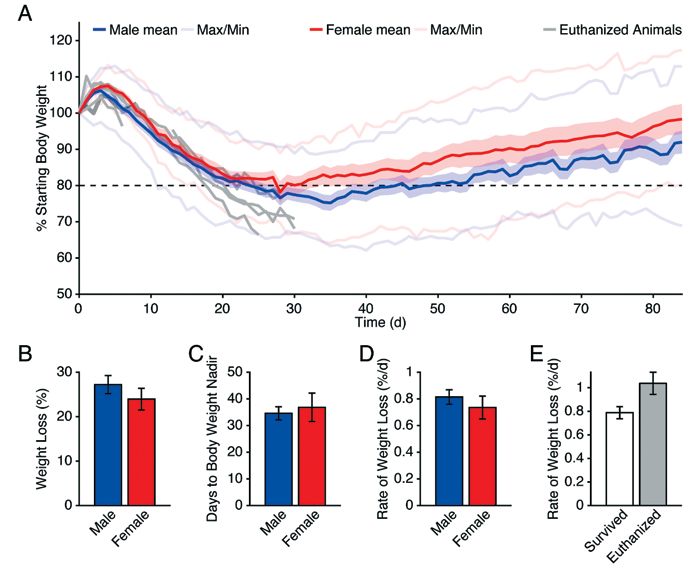
Morbidity and mortality in NZW rabbits undergoing SONFH

Table 1. Reported mortalities in SONFH literature. All reported drug concentrations, routes of administration, and dosing regimens are noted herein.
Quotationed descriptions are as reported in the cited manuscript(s). The mortality denominator reflects the number of animals receiving the induc-
tion protocol (that is sham rabbits excluded). NR = not reported; NA = not applicable; M = male; F = female; IM = intramuscular; IV = intravascular;
IP = intraperitoneal
                                        Weight                                       Induction      Length of    Mortalities
Author (Year)          Breed      Sex    (kg)           Induction protocol           antibiotic     induction      (%)            Cause of death
Pan and           “clean-grade NR         NR     1. dexamethasone sodium            gentamicin        8 wk        2/10 (20)             NR
colleagues (2020)   rabbits”                     phosphate (20 mg/kg, IP, every
                                                 3d for 8 wk)
Peng and              New         NR      NR     1. Escherichia coli endotoxin         none           6 wk         “dead                NR
colleagues (2019)    Zealand                     (10 μg/kg, IV, 2 doses q24h)                                     animals”
                      white                      2. methylprednisolone                                              (NA)
                                                 (40 mg/kg, IM, 3 doses q24h)
Ren and              Japanese     M     2.3–2.7 1. prednisolone acetate                none          3–6 wk       7/50 (14)             NR
colleagues (2018)      white
Maruyama and New Zealand M              3.5–4.0 1. methylprednisolone acetate          none           4 wk        2/24 (8.3)            NR
colleagues (2018) white                         (20 mg/kg, IM, single dose)
Song and          New Zealand F         2.8–3.5 1. lipopolysaccharide                  none           6 wk        9/45 (20)             NR
colleagues (2017)    white                      (10 μg/kg, IV, single dose)
                                                 2. methylprednisolone acetate
                                                 (20 mg/kg, IM, 3 doses q24h)
Karakaplan and        New         M     2.0–2.5 1. methylprednisolone acetate          none           6 wk       4/30 (13.3)            NR
colleagues (2017)    Zealand                    (40 mg/kg, IM, single dose)
                      white
Zhai and              New         NR      2–3    1. lipopolysaccharide                 none         2–12 wk       6/60 (10)             NR
colleagues (2016)    Zealand                     (10 μg/kg, IV, single dose)
                      white                      2. methylprednisolone acetate
                                                 (20 mg/kg, IM, 3 doses q24h)
Zhang and             New        M/F 1.5–2.5 1. lipopolysaccharide                   penicillin       6 wk        5/50 (10)      “acute diarrhea”
colleagues (2015)    Zealand                 (10 μg/kg, IV, single dose)            (100,000 U,                                       (n = 3)
                      white                      2. methylprednisolone acetate       IP, single
                                                 (20 mg/kg, IM, 3 doses q24h)          dose)
Li and colleagues     New         M       NR     1. lipopolysaccharide                 none           6 wk        5/65 (7.7)        “infection”
(2015)               Zealand                     (10 μg/kg, IV, single dose)
                      white                      2. methylprednisolone acetate
                                                 (20 mg/kg, IM, 3 doses q24h)
Kang and             Japanese     M     2.8–3.4 1. methylprednisolone acetate          none           2 wk        4/68 (5.9)      “pneumonia”
colleagues (2015)      white                    (20 mg/kg, IM, single dose)
Fan and               New         M      2.5–3   1. lipopolysaccharide               penicillin       6 wk        4/48 (8.3)        “infection”
colleagues (2014)    Zealand                     (10 μg/kg, IV, single dose)        (200,000 U,
                      white                      2. methylprednisolone acetate       IM, single
                                                 (20 mg/kg, IM, 3 doses q24h)          dose)
Wu and                New         NR      NR     1. lipopolysaccharide                 none           6 wk        4/40 (10)             NR
colleagues (2013)    Zealand                     (10 μg/kg, IV, single dose)
                      white                      2. methylprednisolone acetate
                                                 (20 mg/kg, IM, 3 doses q24h)
Wang and              New        M/F 2.6–3.2 1. horse serum                          penicillin       2 wk       5/40 (12.5)            NR
colleagues (2012)    Zealand                 (10 mL/kg, IV, single dose)            (10,000,000
                      white                      2. horse serum                     U, IP, daily,
                                                 (6 mL/kg, IV, 3 wk post 1.)            7 d)
                                                 3. methylprednisolone
                                                 (45 mg/kg, IP, 3 doses q24h)
Sun and               New         M      2–2.5   1. lipopolysaccharide                 none          10 wk        6/40 (15)             NR
colleagues (2011)    Zealand                     (10 μg/kg, IV, single dose)
                      white                      2. methylprednisolone acetate
                                                 (20 mg/kg, IM, 3 doses q24h)
Kuribayashi and      Japanese     M     3.3–3.9 1. methylprednisolone acetate          none           4 wk        9/50 (18)             NR
colleagues (2010)      white                    (20 mg/kg, IM, single dose)
Sun and               New         M     3.5–4.5 1. lipopolysaccharide                  none          10 wk       9/65 (13.8)            NR
colleagues (2009)    Zealand                    (10 μg/kg, IV, single dose)
                      white                      2. methylprednisolone acetate
                                                 (20 mg/kg, IM, 3 doses q24h)

                                                                                                                                                   89
Vol 71, No 1
Comparative Medicine
February 2021
Table 1. Continued
Weight Induction Length of Mortalities
Author (Year) Breed Sex (kg) Induction protocol antibiotic induction (%) Cause of death
Sheng and New M 3.5–4 1. lipopolysaccharide none 0–2 wk 2/25 (8) NR
colleagues (2009) Zealand (10 μg/kg, IV, single dose)
white 2. methylprednisolone acetate
(20 mg/kg, IM, 3 doses q24h)
Pan and New M 2.5 ± 0.2 1. prednisolone acetate penicillin 9 wk 9/52 (17.3) GI hemorrhage/
colleagues (2009) Zealand (12.25 mg/kg, IM, twice (4 mg/ shock (n = 4);
white weekly for 8 wk) kg), IM, pulmonary infec-
weekly, 9 tion; heart failure
wk) (n = 3); liver/kid-
ney failure (n = 2)
Wu and New M 3.5–4.5 1. lipopolysaccharide none 6 wk 4/64 (6.3) NR
colleagues (2008) Zealand (10 μg/kg, IV, 2 doses)
white 2. methylprednisolone
(20 mg/kg, IM, 3 doses q24h)
Pengde and Japanese M 2.8–3.4 1. methylprednisolone acetate none 2–12 wk 3/54 (5.6) “pneumonia”
colleagues (2008) white (20 mg/kg, IM, single dose)
Chen and NR NR 2.0–2.5 1. dexamethasone none 4–16 wk 1/15 (6.7) NR
colleagues (2008) (7.5 mg/kg, IM, 2 doses
at 1-wk interval)
Miyanishi and Japanese M 3.3–3.9 1. methylprednisolone acetate none 4 wk 5/90 (5.6) NR
colleagues (2006) white (20 mg/kg, IM, single dose)
Yamamoto and New M 3.0–4.5 1. lipopolysaccharide none 4 wk 2/10 (20) NR
colleagues (1995) Zealand (100 μg/kg, IV, 2 doses q24h)
white 2. methylprednisolone
(20 mg/kg, IM, 3 doses q24h
from 4 symptomatic rabbits. For hematology, summarized in surgery. Of these, 29/31 rabbits survived to the experimental
Figure 3 B, the only finding of clinical significance in either as- endpoint. One rabbit was euthanized prior to recovery from
ymptomatic and symptomatic rabbits was a change in the ratio anesthesia due to poor oxygen saturation and arterial blood
of heterophils and lymphocytes, with an increase in the per- gas readings, combined with severe pleural effusion on radio-
centage of heterophils (72 ± 4%, reference range: 17% to 46%), graphs. The second rabbit was euthanized 2 d after surgery due
and a reduction in the percentage of lymphocytes (21 ± 3%, to urine retention that did not resolve despite fluid support, re-
reference range: 51% to 66%), relative to reference ranges. He- peated passing of a transurethral catheter, and medical therapy.
matology results were otherwise unremarkable for all rabbits. The cause of the urinary retention was unknown, but was not
Serum was visibly lipemic (Figure 3 A), and serum chemistry thought to be associated with the model. Of rabbits that sur-
showed an elevation in cholesterol (Figure 3 C; 289 ± 51 mg/dL, vived, 3/29 were unable to maintain peripheral blood oxygen
reference range: 39 to 99 mg/dL). Glucose was also profoundly saturation greater than 92% after extubation; this resolved af-
elevated (298 ± 61 mg/dL, reference range: 50 to 102 mg/dL), ter 4 h in an oxygen chamber (Intensive Care Unit Model 2000,
suggesting a state of insulin resistance. Liver cellular enzymes, Snyder MFG).
aspartate aminotransferase (AST) and alanine aminotransferase Necropsy and Histopathology. Of the 35 rabbits undergoing
(ALT), were also elevated above the reference range. Hepato- SONFH induction, 6 were submitted for necropsy and histopa-
biliary enzymes alkaline phosphatase (ALP) and γ-glutamyl thology evaluation due to spontaneous death or a severe clinical
transferase (GGT) were not elevated. All other serum chemistry condition. Table 2 summarizes the signalment, clinical signs,
parameters including total protein (TP), blood urea nitrogen gross findings, histopathologic findings, and ancillary diagnos-
(BUN), and creatinine (Creat) were within normal range. No tic results for these 6 rabbits. Briefly, 4 were euthanized, and
changes were seen in electrolytes. No significant correlations 2 died spontaneously. Duration from MPS administration to
were found between maximum weight loss and levels of glu- death varied from 9 d to 31 d with a mean of 26 ± 3 d.
cose, AST, ALT, or cholesterol (Pearson correlation, P > 0.05). Generally, 2 distinct patterns emerged when evaluating these
No differences in hematology (Figure 3 B) or serum chemistry 6 rabbits together with the initial 4 index rabbits. The first pat-
values (Figure 3 C) were detected between symptomatic and tern of disease was characterized by a sterile, opaque, lipid-rich,
asymptomatic rabbits. proteinaceous thoracic effusion with pulmonary collapse and/
Radiographs. Radiographic imaging was performed for a or hepatomegaly, with varying degrees of lipidosis, glycogeno-
subset of 5 rabbits at 4-wk after MPS administration. Other sis, and single-cell to coalescing hepatocellular necrosis. Both
than MPS-associated weight loss, these rabbits were clinically pleural effusion and hepatic lesions were identified in 4/4 in-
healthy. Figures 4 A and 4 B show characteristic radiographic dex cases and 2/6 cases from the study cohort. All rabbits (4/4
findings including mild pleural effusion and hepatomegaly as index cases and 6/6 study cohort rabbits) exhibited moderate
defined by extension of the liver margin beyond the caudal bor- to marked hepatomegaly, with varying levels of the aforemen-
der of the ribs. tioned histologic findings.
Surgery. Of the animals that received SONFH induction, 31/35 The second pattern of disease was opportunistic infec-
underwent subsequent anesthesia and core decompression tion. When present, opportunistic infections were typically
90Morbidity and mortality in NZW rabbits undergoing SONFH
Figure 1. Thoracic and hepatic lesions in index mortalities following MPS administration. A. Lipid-rich proteinaceous thoracic effusion result-
ing in atelectasis of the lung parenchyma. B. Marked hepatomegaly (white asterisks) with an enhanced reticular pattern. Black asterisk denotes
caudal aspect of right lateral thoracic wall. Scale bar = 1 cm. C. Diffuse hepatic glycogenosis and lipidosis expanding periportal hepatocytes.
Single cell necrosis (arrow) and mineralization (arrowheads) were frequent. Hematoxylin and eosin, scale bar = 50 μm. D. Broad regions of
subcapsular to intraparenchymal (asterisks) hepatic necrosis, hemorrhage, and mineralization were occasionally observed. Hematoxylin and
eosin, scale bar = 200 μm.
superimposed on the gross and histologic findings described No gross abnormalities were present in any of the remaining
above. In 2/6 study rabbits, opportunistic infection with Esch- 18 rabbits.
erichia coli and/or Bordetella bronchiseptica was identified via mi-
crobiologic culture. These infections typically resulted in severe Discussion
fibrinous heterophilic bronchopneumonia (Figures 4 C and 4 D) Despite being a commonly used model of SONFH, rabbits
and rhinitis ± myocarditis and/or septicemia. Gram negative subjected to this induction protocol are susceptible to steroid-
rods were identified in affected tissues via Gram stain (Figure related morbidities and mortalities. Within our cohorts, the most
4 D, inset). In 2/6 study rabbits, mild to severe granulomatous common clinical signs were severe weight and muscle loss, ab-
and proliferative enteritis due to Lawsonia intracellularis infec- normal appetite and fecal output, elevated respiratory rate, and
tion (Figures 4 E and 4 F) was identified. Lamina propria mac- sudden death. Although bloodwork revealed lipemia, hyper-
rophages and multinucleated giant cells contained abundant cholesterolemia, hyperglycemia, and elevated liver enzymes
PAS-positive material, a classic histologic finding in Lawsonia (AST and ALT), no single parameter (including weight loss)
intracellularis-infected animals (Figure 4 F, inset). PAS-positive could distinguish rabbits that survived to the experimental end-
organisms were also present within the apical cytoplasm of in- point from those that did not. Rabbits that failed to respond to
fected enterocytes (not shown). clinical management or died spontaneously typically displayed
End of Study Rabbits. A total of 19 asymptomatic rabbits un- 2 distinct yet overlapping patterns of disease. The first pattern
derwent gross necropsy at their experimental endpoints (12 wk was typified by a lipid-rich proteinaceous thoracic effusion
after MPS administration) to evaluate for disease. Of these, only in combination with hepatomegaly, hepatic lipidosis, hepatic
one rabbit exhibited chronic pulmonary granulomas; these were glycogenosis, and/or hepatic necrosis. The second pattern in-
culture-positive for Escherichia coli and Bordetella bronchiseptica. volved opportunistic infection(s) superimposed on the first dis-
ease pattern. The most common opportunistic pathogens were
91Vol 71, No 1 Comparative Medicine February 2021 Figure 2. Change in rabbit body weight following MPS administration. A. Percent change in body weight following MPS administration (day zero) for male (blue, mean weight, n = 17) and female (red, mean weight, n = 12) rabbits that survived to their experimental endpoint (‘survi- vors’) and euthanized (gray, individual animals, n = 6) rabbits. Light lines represent minimum and maximum values for male (blue) and female (red) survivors. B. Mean of maximum percent weight loss following MPS administration for male (n = 17) and female (n = 12) survivors (un- paired t test P = 0.27). C. Mean number of days from MPS administration to body weight nadir for male (n = 17) and female (n = 12) survivors (unpaired t test P = 0.68). D. Rate of weight loss from MPS administration to body weight nadir for male (n = 17) and female (n = 12) survivors (unpaired t test P = 0.42). E. Rate of weight loss for survivors (n = 29) or euthanized rabbits (n = 5) over the first 25 d following MPS administra- tion (unpaired t test P = 0.06). All data for figures are presented as mean ± SE unless otherwise noted. Bordetella bronchiseptica and Escherichia coli respiratory infections absence of mortalities during the induction period, suggesting and Lawsonia intracellularis small intestinal infections. Together, that the true overall incidence of mortalities among SONFH these findings allowed us to establish a SONFH decision-mak- studies may be higher than 30%. ing flowchart, as discussed below. Recognition of these model- Despite the significant weight loss seen during the induc- related morbidities is essential for managing rabbits throughout tion period, weight loss itself did not differentiate rabbits that the SONFH induction period. survived to the experimental endpoint from those that did not. Over the past 30 y, 30% of primary research articles using Establishing appropriate humane endpoints for animals un- the rabbit SONFH model have reported mortalities within the dergoing invasive biomedical research is essential for ensuring induction period. In these studies, the average mortality rate animal welfare. Common humane endpoints include criteria re- during the induction period alone was 12% (range: 6% to 20%). lated to weight loss (typically greater than 20% below baseline) Unfortunately, none of the available study parameters (that is or to body condition score (typically a body condition score of breed, sex, weight, induction protocol, prophylactic antibiotic less than 2/5), although body condition scores remain poorly administration, and length of induction) seemed to predict mor- validated in rabbits.28 If either criterion is met, rapid interven- tality rates across these studies. Furthermore, no trends in these tion or removal of the animal(s) from the study is generally parameters were seen when evaluating studies that specifically recommended. These metrics provide clear and objective data noted an absence of mortalities (10% of studies). Moreover, 59% points that can be obtained by research personnel and animal of the experimental studies did not reference the presence or care professionals alike, with little-to-no training on collection 92
Morbidity and mortality in NZW rabbits undergoing SONFH

Figure 3. Changes in blood work following MPS administration. A. Capillary tube containing centrifuged blood from an asymptomatic animal
exhibiting severe lipemia. B. Selected hematology parameters from asymptomatic (blue, n = 7) and symptomatic (red, n = 2) rabbits. Gray boxes
indicate inhouse reference ranges. C. Selected serum chemistry parameters from asymptomatic (blue, n = 7) and symptomatic (red, n = 4) rabbits.
Gray boxes indicate inhouse reference ranges. Abbreviations: white blood cells (WBC); hemoglobin (HGB); hematocrit (HCT); heterophils (Het);
lymphocytes (Lymph); aspartate aminotransferase (AST); alanine aminotransferase (ALT); alkaline phosphatase (ALP); γ-glutamyl transferase
(GGT); cholesterol (Chol); blood urea nitrogen (BUN); creatinine (Creat); total protein (TP).

or interpretation. However, rabbits subjected to the SONFH in-              Abnormalities in hematology and serum chemistry values
duction protocol initially lost significantly more than 20% body         did not aid significantly in the differentiation of rabbits that
weight, and the severity of weight loss did not significantly            would remain asymptomatic from those that would display
correlate with mortality, indicating that using weight loss as           clinical signs or die spontaneously. For example, inversion of the
the sole criterion for euthanasia may be inappropriate for this          heterophil to lymphocyte ratio has been associated with bacte-
model. In addition, rabbits lost both muscle mass and adipose            rial infection (as was seen in a subset of necropsied rabbits), but
tissue, making body condition scoring challenging and compli-            also occurs due to stressful events and the release of cortisol.2
cating its use as an effective or reasonable endpoint.                   Therefore, the profoundly inverted heterophil:lymphocyte ratio
                                                                                                                                            93
Vol 71, No 1
Comparative Medicine
February 2021
Table 2. Mortality characteristics of study rabbits. F = female; M = male; SD = spontaneous death; E = euthanasia; + = mild, ++ = moderate,
+++ = severe; NP = not performed; E. coli = Escherichia coli; B. bronchi = Bordetella bronchiseptica; L. intracell = Lawsonia intracellularis
Sex F F F F M M
Mortality SD SD E E E E
Steroid Injection to Death 9d 26 d 30 d 29 d 29 d 31 d
Clinical Signs none weight loss, weight loss, weight loss, peri-anesthetic stranguria
tachypnea tachypnea, anorexia, distress
diarrhea tachypnea
Gross Findings
Thoracic Effusion ++; white, ++; yellow; — ++; yellow +++; white, —
opaque, opaque; fibrin opaque; fibrin opaque,
gelatinous gelatinous
Abdominal Effusion +; yellow-tinged ++; yellow; — — — —
opaque; fibrin
Hepatomegaly +++ +++ +++ ++ +++ ++
Mucoid nasal turbinates — + — + — —
Pulmonary consolidation +++ — +++ — —
Small intestinal thickening — — +++ — — —
Body condition thin thin thin thin thin thin
Histology Findings
Hepatic lipidosis ++ +++ +++ + + ++
Hepatic glycogenosis ++ + + ++ +++ ++
Hepatocellular necrosis single cells ++ — single cells single cells single cells
Heterophilic pneumonia — +++ — +++ + —
Valvular endocarditis — +++ — — — —
Myocarditis +++ — —
Rhinitis — + — + — —
Granulomatous ileitis + +++ — — —
Culture (site) NP E. coli (thorax, NP B. bronchi (thorax, negative (thorax) NP
abdomen, pericardium,
turbinates) turbinates)
B. bronchi
(turbinates)
PCR NP L. intracell L. intracell NP NP NP
seen in the rabbits is likely attributable to the high dose of MPS within 48 h, or if the rabbit continues to deteriorate during that
required to induce SONFH. This effect masks alterations in the 48-h period, euthanasia is performed. Using this monitoring
leukogram that might otherwise serve as a diagnostic tool to and treatment algorithm, an additional 46 rabbits have been
identify rabbits with opportunistic bacterial infections. In ad- subjected to the SONFH induction protocol, and only 3 have
dition, consistent elevations in glucose, cholesterol, ALT, and required euthanasia.
AST were equivalent among asymptomatic and symptomatic The clinical presentations and necropsy results of rabbits un-
rabbits, rendering these parameters inappropriate for making dergoing SONFH are consistent with high dose steroid admin-
euthanasia decisions. istration. For example, extreme muscle and weight loss despite
To overcome these diagnostic limitations, the veterinary staff, good appetite may result from a steroid-induced metabolic dys-
IACUC members, and investigators developed a new set of regulation. Chronic use of glucocorticoids is associated with
clinically-focused endpoints for rabbits subjected to the SONFH muscle weakness, muscle loss, and elevated blood glucose,
induction protocol, as shown in Figure 5. Now all rabbits on in part through alteration in transcription factors, nuclear co-
study are weighed a minimum of 3 times per wk. At 25% weight factors, and hyperacetylation.7 In humans, glucocorticoid ad-
loss, the investigator provides additional hay and nutritional ministration inhibits the anabolic action of insulin, resulting in
support and increases the frequency of clinical condition and muscle loss even after short term administration.24 Consistent
body weight monitoring to 5 times per week. If appetite or fe- with insulin resistance, elevated blood glucose, lipemia, and
cal production becomes abnormal, the rabbit is presented for elevated cholesterol occurred after MPS administration in these
physical exam by the veterinary staff. If the rabbit is otherwise rabbits. A combination of the action of steroids and insulin resis-
healthy, it is started on enrofloxacin 5 mg/kg PO once a day tance likely also underlies the pattern of significant muscle and
(SID) for 7 d and the monitoring frequency increases. If the rab- weight loss reported.
bit presents with other comorbidities such as elevated respira- Hepatocellular injury, as evidenced radiographically, via
tory rate, or with nonspecific signs such as lethargy, it is given bloodwork (elevated AST and ALT), and in necropsy find-
enrofloxacin (10 mg/kg PO SID). In addition, rabbits are given ings, was consistent among rabbits undergoing SONFH induc-
caloric support in the form of Critical Care via syringe feeding tion. Livers were markedly enlarged, and hepatocytes were
twice a day (BID), as well as 10 to 20 mL/kg SQ fluids if any expanded by lipid and glycogen. In humans, hepatotoxicity
dehydration is noted. If clinical improvement does not occur has been associated with intravenous MPS administration.38
94Morbidity and mortality in NZW rabbits undergoing SONFH
Figure 4. Radiographic, gross, and histologic findings in study cohort rabbits. Right lateral (A) and dorsoventral (B) view of rabbit thoracic
cavity and cranial abdomen demonstrating hepatomegaly and thoracic effusion typically seen in SONFH rabbits. (Note: the caudal abdomen
and pelvic limbs were collimated out of the image per researcher’s request). C. Opportunistic infections with Escherichia coli and/or Bordetella
bronchiseptica resulted in fibrinous pleuritis (asterisk) and bronchopneumonia. D. Pulmonary bronchi (asterisks) were filled with degenerate
heterophils and fibrinous material. Gram-negative rods (inset, arrow) were visualized following Gram stain. Hematoxylin and eosin, scale bar
= 50 μm. E. Rabbits infected with Lawsonia intracellularis exhibited thickened cerebriform-like jejunal and ileal segments. Scale bar = 0.5 cm. F.
Marked mucosal hyperplasia and granulomatous ileitis in a rabbit infected with Lawsonia intracellularis. Lamina proprial macrophages (inset,
white arrow) and multinucleated giant cells contain PAS-positive material (inset, black arrow). Hematoxylin and eosin, scale bar = 200 μm.
95Vol 71, No 1 Comparative Medicine February 2021 Figure 5. Flowchart showing rabbit care decision-making process. Abbreviations: bright, alert, responsive (BAR); respiratory rate (RR); respira- tory effort (RE); orally (PO); once a day (SID); twice a day (BID); subcutaneous (SQ). However, the incidence of disease was sporadic in these pa- In addition to alterations in metabolism, high dose steroid admin- tients and was often associated with a mixed pattern of liver istration results in immunosuppression, thus increasing the likeli- inflammation and necrosis in the absence of lipidosis and gly- hood of opportunistic infections. In rabbits, Bordetella bronchiseptica, cogen accumulation. Thus, the pathogenesis of liver disease in Escherichia coli, and Lawsonia intracellularis have all been documented SONFH rabbits is more likely attributed to aberrant lipid me- to cause secondary opportunistic infections or primary disease.1 tabolism and/or microvascular lipid emboli, as described in the Because of this, the pros and cons of daily weight monitoring and “lipid metabolism disorder theory” of SONFH.29 The lipid and handling must be weighed against the potential for spreading infec- protein-rich thoracic effusions are also postulated to be second- tious diseases among immunosuppressed rabbits. Furthermore, ap- ary to steroid-induced lipid mobilization. However, a rationale propriate personal protective equipment and husbandry hygiene are for the presence of effusion specifically in the thorax, compared essential when working with rabbits undergoing SONFH to mitigate with other body cavities, remains unknown. the potential for interspecies disease transmission. 96
Morbidity and mortality in NZW rabbits undergoing SONFH

   Our observational case series has inherent limitations because          6. Fan L, Li J, Yu Z, Dang X, Wang K. 2014. Hypoxia-inducible factor
it was conducted within the limits of an experimental study.                  prolyl hydroxylase inhibitor prevents steroid-associated osteone-
Tracking morbidity data was challenging due to constantly                     crosis of the femoral head in rabbits by promoting angiogenesis and
                                                                              inhibiting apoptosis. PLoS One 9:1–9. https://doi.org/10.1371/
changing definitions as the veterinary team worked with re-
                                                                              journal.pone.0107774.
search personnel to develop and refine the SONFH model. This               7. Hasselgren PO, Alamdari N, Aversa Z, Gonnella P, Smith IJ,
resulted in a relative over-reporting of morbidities in the ini-              Tizio S. 2010. Corticosteroids and muscle wasting: role of tran-
tial cohort of animals reviewed for this paper, and a relative                scription factors, nuclear cofactors, and hyperacetylation. Curr
under-reporting toward the end. Thus, rabbit mortality events                 Opin Clin Nutr Metab Care 13:423–428. https://doi.org/10.1097/
occurred more frequently in the initial phases of the study. In               MCO.0b013e32833a5107.
addition, due to experimental limitations, we were unable to               8. Institute for Laboratory Animal Research. 2011. Guide for the care
compare the gross and histopathologic lesions of rabbits that                 and use of laboratory animals, 8th ed. Washington (DC): National
                                                                              Academies Press.
died with those of asymptomatic rabbits at the same experimen-
                                                                           9. Kang P, Xie X, Tan Z, Yang J, Shen B, Zhou Z, Pei F. 2015. Repairing
tal time point. Because all rabbits underwent severe weight loss              defect and preventing collapse of femoral head in a steroid-induced
regardless of clinical status, we presume that all animals may                osteonecrotic of femoral head animal model using strontium-doped
have experienced hepatic lesions. However, the presence of tho-               calcium polyphosphate combined BM-MNCs. J Mater Sci Mater
racic effusion in asymptomatic rabbits could not be evaluated                 Med 26:80. https://doi.org/10.1007/s10856-015-5402-x.
during the induction period due to study limitations. Asymp-              10. Karakaplan M, Gulabi D, Topgul H, Elmali N. 2017. Does platelet-
tomatic rabbits that underwent necropsy at the experimental                   rich plasma have a favorable effect in the early stages of steroid-asso-
endpoint did not have gross lesions, with the exception of one                ciated femoral head osteonecrosis in a rabbit model? Eklem Hastalik
                                                                              Cerrahisi 28:107–113. https://doi.org/10.5606/ehc.2017.54402.
rabbit with pulmonary granulomas. Therefore, these rabbits
                                                                          11. Kuribayashi M, Fujioka M, Takahashi KA, Arai Y, Ishida
either never developed hepatic or thoracic disease or recovered               M, Goto T, Kubo T. 2010. Vitamin E prevents steroid-induced
from disease by the end of the experiment.                                    osteonecrosis in rabbits. Acta Orthop 81:154–160. https://doi.
   In summary, rabbits used in the SONFH model displayed sig-                 org/10.3109/17453671003628772.
nificant morbidity and mortality, a result that is underreported          12. Larson E, Jones LC, Goodman SB, Koo KH, Cui Q. 2018. Early-
in the literature. After induction of SONFH with MPS, rabbits                 stage osteonecrosis of the femoral head: where are we and where
displayed marked weight loss, diarrhea, lipemia, hypercholes-                 are we going in year 2018? Int Orthop 42:1723–1728. https://doi.
terolemia, hyperglycemia, and elevations in ALT and AST. These                org/10.1007/s00264-018-3917-8.
                                                                          13. Li J, Fan L, Yu Z, Dang X, Wang K. 2014. The effect of deferoxamine
clinical findings occurred in both asymptomatic rabbits and those
                                                                              on angiogenesis and bone repair in steroid-induced osteonecrosis
that displayed clinical signs or died, such that these changes were           of rabbit femoral heads. Exp Biol Med (Maywood) 240:273–280.
not predictive of morbidity or death. Rabbits that died exhibited             https://doi.org/10.1177/1535370214553906.
hepatomegaly with thoracic effusion and/or systemic disease               14. Maruyama M, Nabeshima A, Pan CC, Behn AW, Thio T, Lin T,
associated with opportunistic infectious pathogen(s). Recogni-                Pajarinen J, Kawai T, Takagi M, Goodman SB, Yang YP. 2018. The
tion of these model-associated morbidities allows researchers and             effects of a functionally-graded scaffold and bone marrow-derived
veterinary staff to develop clinical care protocols to successfully           mononuclear cells on steroid-induced femoral head osteonecrosis.
manage rabbits used in SONFH research.                                        Biomaterials 187:39–46. https://doi.org/10.1016/j.biomateri-
                                                                              als.2018.09.030.
                                                                          15. Miyanishi K, Yamamoto T, Irisa T, Yamashita A, Motomura
                    Acknowledgments                                           G, Jingushi S, Iwamoto Y. 2006. Effects of cyclosporin A on the
   The authors thank Elias Godoy for his technical assistance within          development of osteonecrosis in rabbits. Acta Orthop 77:813–819.
necropsy and the Stanford Animal Histology Services for help with             https://doi.org/10.1080/17453670610013042.
preparation of histologic specimens. We would like to thank the           16. Mont MA, Pivec R, Banerjee S, Issa K, Elmallah RK, Jones LC.
Stanford Animal Diagnostic Laboratory for preparing and analyzing             2015. High-dose corticosteroid use and risk of hip osteonecrosis:
clinical and necropsy diagnostic specimens. This work was supported           meta-analysis and systematic literature review. J Arthroplasty
by the National Institutes of Health R01 AR072613 (Tissue Engineering         30:1506–1512.e5. https://doi.org/10.1016/j.arth.2015.03.036.
Approaches for Improved Treatment of Early Stage Osteonecrosis of         17. Pan FY, Li ZM, Liu XW, Luo Y, Ma Z, Feng SX, Xu N. 2020. Effect
the Hip).                                                                     of strontium ranelate on rabbits with steroid-induced osteonecrosis
                                                                              of femoral head through TGF-β1/BMP2 pathway. Eur Rev Med
                                                                              Pharmacol Sci 24:1000–1006.
                          References                                      18. Pan X, Xiao D, Zhang X, Huang Y, Lin B. 2008. Study of rotating
 1. Barthold SW, Griffey SM, Percy DH. 2016. Rabbit. Chapter 6. p
                                                                              permanent magnetic field to treat steroid-induced osteonecrosis
    253–324. In: Pathology of laboratory rodents and rabbits, 4th ed.
                                                                              of femoral head. Int Orthop 33:617–623. https://doi.org/10.1007/
    Ames (IA):Wiley–Blackwell.
                                                                              s00264-007-0506-7.
 2. Benson KG, Paul-Murphy J. 1999. Clinical pathology of the do-
                                                                          19. Peng W, Dong W, Zhang F, Wang J, Zhang J, Wu J, Wang L, Ye C,
    mestic rabbit. Acquisition and interpretation of samples. Vet Clin
                                                                              Li Q, Deng J. 2019. Effects of transplantation of FGF-2-transfected
    North Am Exot Anim Pract 2:539–551. https://doi.org/10.1016/
                                                                              MSCs and XACB on TNFα expression with avascular necrosis of
    S1094-9194(17)30109-3.
                                                                              the femoral head in rabbits. Biosci Rep 39:1–10.
 3. Chen XC, Weng J, Chen XQ, Du JZ, Zhu MP, Pan YQ, Liu M.
                                                                          20. Pengde K, Fuxing P, Bin S, Jing Y, Jingqiu C. 2008. Lovastatin
    2008. Relationships among magnetic resonance imaging, histo-
                                                                              inhibits adipogenesis and prevents osteonecrosis in steroid-treated
    logical findings, and IGF-I in steroid-induced osteonecrosis of the
                                                                              rabbits. Joint Bone Spine 75:696–701. https://doi.org/10.1016/j.
    femoral head in rabbits. J Zhejiang Univ Sci B 9:739–746. https://
                                                                              jbspin.2007.12.008.
    doi.org/10.1631/jzus.B0820127.
                                                                          21. Petek D, Hannouche D, Suva D. 2019. Osteonecrosis of the femoral
 4. Cohen-Rosenblum A, Cui Q. 2019. Osteonecrosis of the
                                                                              head: pathophysiology and current concepts of treatment. EFORT
    femoral head. Orthop Clin North Am 50:139–149. https://doi.
                                                                              Open Rev 4:85–97. https://doi.org/10.1302/2058-5241.4.180036.
    org/10.1016/j.ocl.2018.10.001.
                                                                          22. Ren X, Fan W, Shao Z, Chen K, Yu X, Liang Q. 2018. A metabo-
 5. Craig LE, Dittmer KE, Thompson KG. 2016. Bones and joints, p
                                                                              lomic study on early detection of steroid-induced avascular ne-
    16–163.e161. Chapter 2. In: Maxie MG, editor. Jubb, Kennedy &
                                                                              crosis of the femoral head. Oncotarget 9:7984–7995. https://doi.
    Palmer’s Pathology of Domestic Animals: Volume 1, 6th ed. St
                                                                              org/10.18632/oncotarget.24150.
    Louis (MO): WB Saunders.

                                                                                                                                                  97
Vol 71, No 1
Comparative Medicine
February 2021
23. Sheng H, Zhang G, Wang YX, Yeung DK, Griffith JF, Leung KS, stem cell factor ameliorates steroid-associated osteonecrosis in rabbits.
Qin L. 2008. Functional perfusion MRI predicts later occurrence of J Rheumatol 35:2241–2248. https://doi.org/10.3899/jrheum.071209.
steroid-associated osteonecrosis: an experimental study in rabbits. 32. Wu X, Yang S, Wang H, Meng C, Xu W, Duan D, Liu X. 2013.
J Orthop Res 27:742–747. https://doi.org/10.1002/jor.20765. G-CSF/SCF exert beneficial effects via anti-apoptosis in rabbits
24. Short KR, Bigelow ML, Nair KS. 2009. Short-term prednisone with steroid-associated osteonecrosis. Exp Mol Pathol 94:247–254.
use antagonizes insulin’s anabolic effect on muscle protein and https://doi.org/10.1016/j.yexmp.2012.06.003.
glucose metabolism in young healthy people. Am J Physiol 33. Xie XH, Wang XL, Yang HL, Zhao DW, Qin L. 2015. Steroid-
Endocrinol Metab 297:E1260–E1268. https://doi.org/10.1152/ associated osteonecrosis: Epidemiology, pathophysiology, animal
ajpendo.00345.2009. model, prevention, and potential treatments (an overview). J Or-
25. Song Q, Ni J, Jiang H, Shi Z. 2017. Sildenafil improves blood thop Translat 3:58–70. https://doi.org/10.1016/j.jot.2014.12.002.
perfusion in steroid-induced avascular necrosis of femoral head 34. Xu J, Gong H, Lu S, Deasey MJ, Cui Q. 2018. Animal models of
in rabbits via a protein kinase G-dependent mechanism. Acta steroid-induced osteonecrosis of the femoral head—a comprehen-
Orthop Traumatol Turc 51:398–403. https://doi.org/10.1016/j. sive research review up to 2018. Int Orthop 42:1729–1737. https://
aott.2017.07.002. doi.org/10.1007/s00264-018-3956-1.
26. Sun Y, Feng Y, Zhang C. 2009. The effect of bone marrow mono- 35. Yamamoto T, Hirano K, Tsutsui H, Sugioka Y, Sueishi K. 1995.
nuclear cells on vascularization and bone regeneration in steroid- Corticosteroid enhances the experimental induction of osteone-
induced osteonecrosis of the femoral head. Joint Bone Spine crosis in rabbits with Shwartzman reaction. Clin Orthop Relat
76:685–690. https://doi.org/10.1016/j.jbspin.2009.04.002. Res Jul:235–243. https://doi.org/10.1097/00003086-199507000-
27. Sun Y, Feng Y, Zhang C, Cheng X, Chen S, Ai Z, Zeng B. 2011. 00033.
Beneficial effect of autologous transplantation of endothelial 36. Zhai JL, Weng XS, Wu ZH, Guo SG. 2016. Effect of resveratrol
progenitor cells on steroid-induced femoral head osteonecrosis in on preventing steroid-induced osteonecrosis in a rabbit model.
rabbits. Cell Transplant 20:233–243. https://doi.org/10.3727/096 Chin Med J (Engl) 129:824–830. https://doi.org/10.4103/0366-
368910X522234. 6999.178952.
28. Sweet H, Pearson AJ, Watson PJ, German AJ. 2013. A novel zoo- 37. Zhang C, Ma J, Li M, Li XH, Dang XQ, Wang KZ. 2015. Repair
metric index for assessing body composition in adult rabbits. Vet effect of coexpression of the hVEGF and hBMP genes via an adeno-
Rec 173:369. https://doi.org/10.1136/vr.101771. associated virus vector in a rabbit model of early steroid-induced
29. Wang A, Ren M, Wang J. 2018. The pathogenesis of steroid- avascular necrosis of the femoral head. Transl Res 166:269–280.
induced osteonecrosis of the femoral head: A systematic review https://doi.org/10.1016/j.trsl.2015.03.003.
of the literature. Gene 671:103–109. https://doi.org/10.1016/j. 38. Zoubek ME, Pinazo-Bandera J, Ortega-Alonso A, Hernández
gene.2018.05.091. N, Crespo J, Contreras F, Medina-Cáliz I, Sanabria-Cabrera J,
30. Wang W, Liu L, Dang X, Ma S, Zhang M, Wang K. 2012. The ef- Sanjuan-Jiménez R, González-Jiménez A, García-Cortés M,
fect of core decompression on local expression of BMP-2, PPAR-ɣ Lucena MI, Andrade RJ, Robles-Díaz M. 2019. Liver injury after
and bone regeneration in the steroid-induced femoral head methylprednisolone pulses: A disputable cause of hepatotoxicity.
osteonecrosis. BMC Musculoskelet Disord 13:142. https://doi. A case series and literature review. United European Gastroenterol
org/10.1186/1471-2474-13-142. J 7:825–837. https://doi.org/10.1177/2050640619840147.
31. Wu X, Yang S, Duan D, Liu X, Zhang Y, Wang J, Yang C, Jiang S.
2008. A combination of granulocyte colony-stimulating factor and
98You can also read