Coagulation in Liver Disease: A Guide for the Clinician
←
→
Page content transcription
If your browser does not render page correctly, please read the page content below
CLINICAL GASTROENTEROLOGY AND HEPATOLOGY 2013;11:1064 –1074

Coagulation in Liver Disease: A Guide for the Clinician

PATRICK G. NORTHUP and STEPHEN H. CALDWELL
Division of Gastroenterology and Hepatology, Center for the Study of Coagulation in Liver Disease, University of Virginia, Charlottesville, Virginia

This article has an accompanying continuing medical education activity on page e67. Learning Objectives—At the end
of this activity, the successful learner will be able to explain the current understanding of hemostasis in chronic liver
disease, its laboratory abnormalities, and the strengths and weaknesses of current testing and treatment strategies.

                                                                                         tional normalized ratio (INR) are now inextricably linked to
   Podcast interview: www.gastro.org/cghpodcast.                                         prognosis and progression of liver disease. Mortality risk scores
   Also available on iTunes.                                                             for cirrhosis such as the Child–Turcotte–Pugh and Model for
                                                                                         End-Stage Liver Disease scores as well as the King’s College
The human hemostasis system is complex and poorly un-                                    Criteria and factor V levels for acute liver failure (ALF) all have
derstood after decades of intense scientific study. Despite                              key components related to PT or INR. To a lesser extent, the
multiple defects in routine coagulation laboratory studies                               blood platelet count is also commonly regarded as an indirect
in patients with chronic liver disease, there is growing evi-                            measure of portal hypertension related to splenic sequestration
dence that these patients are effectively “rebalanced” with                              and loss of hepatic production of thrombopoietin because of
regard to procoagulant and anticoagulant activity and that                               liver tissue atrophy. Although it is clear that these measures are
most of these patients remain in a tenuous but balanced                                  related to liver disease prognosis, ironically it is less clear how to
state of hemostasis. A major difficulty in the assessment of                             use these tests to manage bleeding and clotting in the patient
these patients is that there are no established laboratory                               with acute and chronic liver disease. In this article, we will
tests that accurately reflect the changes in both the proco-                             review essential and practical aspects of coagulation in liver
agulant and anticoagulant systems; therefore, routine lab-                               disease. We will also discuss the limitations of laboratory tests
oratory testing is misleading to the clinician and may                                   in the investigation of bleeding or clotting risk in this patient
prompt inappropriate or risky therapies with little real                                 population. Finally, we will address the prevalence and current
benefit to the patient. The international normalized ratio is                            clinical understanding of several common disease processes
an example of this type of misleading test. Although the                                 related to coagulation disorders in liver disease patients.
international normalized ratio is inextricably linked to
prognosis and severity of protein synthetic dysfunction in
acute and chronic liver disease, it is a very poor marker for                                      Physiology and Pathophysiology
bleeding risk and should not be used in isolation for this                                       Much of the understanding of the physiology of nor-
purpose. Coagulation disorders are critical in the manage-                               mal coagulation is derived from decades of research in patients
ment of frequent clinical scenarios such as esophageal                                   with rare, usually congenital, clotting factor deficiencies. The
variceal bleeding, invasive and percutaneous procedures,                                 prototypical disease in this category is hemophilia A, or con-
portal vein thrombosis, venous thromboembolism, and                                      genital factor VIII deficiency; factor concentrates developed
acute liver failure. This article summarizes the pathophys-                              through research in this area led to a revolution in therapy for
iology of hemostasis in liver disease, describes the strengths                           this population. However, the backwards engineering of the
and weaknesses of various laboratory tests in assessment of                              coagulation system through procoagulant pathways led to de-
these patients, and outlines the optimal management of                                   cades of narrow focused teaching that was subsequently prop-
hemostasis for some common clinical scenarios. Further                                   agated in medical schools. For example, the traditional under-
research is needed for proper understanding of hemostasis                                standing of an intrinsic pathway and extrinsic pathway of
in liver disease to optimally and safely manage these com-                               clotting that was taught to generations of students has led to
plex patients.                                                                           an incomplete understanding in the mainstream medical com-
                                                                                         munity of the complexity and flexibility of the system involved
Keywords: Coagulation; Liver; Varices; Bleeding Portal Vein                              in maintaining hemostasis in health and in disease. This has led
Thrombosis; Acute Liver Failure.
                                                                                            Abbreviations used in this paper: ALF, acute liver failure; EVBL,
                                                                                         esophageal variceal band ligation; FFP, fresh frozen plasma; INR,

F
                                                                                         international normalized ratio; LMWH, low-molecular-weight heparin;
      ew aspects of liver disease have become associated with so
                                                                                         PT, prothrombin time; PVT, portal vein thrombosis; VKA, vitamin K
      much dogmatic practice, but so few data, as the manage-
                                                                                         antagonist; VTE, venous thromboembolism; vWF, von Willebrand
ment of liver-related acquired coagulation disorders. Changes                            factor.
in coagulation parameters have been a hallmark of advanced                                                      © 2013 by the AGA Institute
liver disease since laboratory testing became widely available in                                                    1542-3565/$36.00
the mid-20th century; the prothrombin time (PT) and interna-                                            http://dx.doi.org/10.1016/j.cgh.2013.02.026
September 2013 COAGULATION IN LIVER DISEASE 1065
front-line providers to depend on outdated or irrelevant and factor to the luminal side of the vascular endothelium. Except
potentially misleading tests in patients with liver disease. in relatively rare cases of systemic coagulation such as dissem-
inated intravascular coagulation, the local hemostatic mecha-
Cell-based Model of Hemostasis nisms do not override the systemic balance of coagulation. This
is why routinely available measures of coagulation are not
In the modern cell-based concept, hemostasis is viewed
effective at describing hemostasis at the site of injury. The
as a cellular process with the activated platelet as the primary
lack of an established and valid systemic test to anticipate or
effector and enabler of coagulation. The fundamental structure
diagnose coagulation changes in the local environment is a
of a clot is a platelet plug restrained by a fibrin mesh formed by
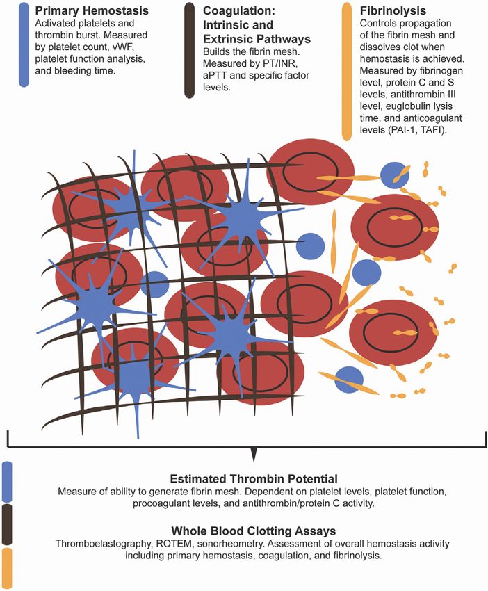
recurrent frustration for the clinician. Figure 1 graphically
the conversion of fibrinogen to fibrin by the enzyme thrombin.
shows the phases of the hemostasis system and some of the
The process involves 3 phases: (1) primary hemostasis or the
laboratory testing available to describe each phase of the
initial plugging of the vascular breach by activated platelets; (2)
process.
coagulation, fibrin mesh construction, and clot fortification by
the plasma procoagulant proteins; and once vascular repair is
complete, (3) fibrinolysis or breakdown of the fibrin mesh by Changes in Liver Disease
plasma anticoagulant proteins.1 These processes are highly ad- Liver disease is marked by changes in all phases of
vanced and rapidly responsive to endothelial factors in the local hemostasis caused by hepatic synthetic dysfunction and portal
environment and are typically triggered by exposure of tissue hypertension (with portosystemic shunting and endothelial
Figure 1. Phases of coagula-
tion and laboratory testing avail-
able for analysis of each phase.1066 NORTHUP AND CALDWELL CLINICAL GASTROENTEROLOGY AND HEPATOLOGY Vol. 11, No. 9
dysfunction). There are compensatory mechanisms that coun- protein deficiencies in this patient population. Research in
terbalance these changes and lead to a “rebalancing”2 of the hemostasis during the past decade has revealed this as a
coagulation system in patients with liver disease. For example, misconception.
synthetic dysfunction and impaired protein production occur Despite decreased levels of procoagulant factors and abnor-
in both procoagulant and anticoagulant proteins, thereby mal INR measurements, cirrhosis patients do not typically have
counterbalancing each other.3 This compensatory rebalancing spontaneous bleeding in sites where patients with congenital
enables even the patient with advanced liver disease to uncom- clotting factor deficiencies experience, such as hemarthroses,
monly have spontaneous bleeding or clotting (without another which argues that patients with liver disease have counterbal-
specific trigger) and remain in a relatively balanced state of ancing forces. In fact, there are strong in vitro data showing
hemostasis. that in the presence of adequate platelet counts and thrombo-
modulin, an endothelial-derived cofactor in the anticoagulant
Platelets system, cirrhosis patients have a normal capacity to generate
Primary hemostasis occurs when tissue factor is sensed thrombin12,14 and the fundamental building blocks of the fibrin
on the luminal side of the vascular endothelium by platelets. mesh. The reason for this normal clotting potential is largely
Platelets respond to tissue factor by activating, developing pseu- due to a decrease in synthesis of a potent anticoagulant protein
dopods, and forming a platelet plug in the vascular breach. C, coupled with increased endothelial-derived factor VIII.15
Activated platelets enable the rapid production of a fibrin mesh Therefore, the compensatory mechanisms in the patient with
by exposing activated clotting factors on their surface and advanced liver disease effectively rebalance the hemostatic sys-
producing a “thrombin burst” through a positive feedback tem.
mechanism.4 The principal abnormality in chronic liver disease Despite a rebalanced system, there are a variety of perturba-
patients is a numeric decrease in the circulating platelet count. tions that can predispose an individual liver disease patient to
The etiology is generally thought to be multifactorial. There is bleeding or clotting. With the progression of disease, the activ-
undoubtedly pooling of platelets and sequestration in the ity of the procoagulant proteins drops to as low as 20%– 46%.15
spleen, and some authors have suggested a role of antiplatelet Although compensatory mechanisms are active, the capacity to
glycoprotein IIb-IIIa antibodies5 in cirrhosis patients, but an adjust to insults to the system is diminished, and small pertur-
immunologic mechanism appears to be a minor contributor. bations can overcome the compensatory mechanisms. The re-
There is also evidence for more rapid turnover and shorter balanced state can be likened to a tightrope crossing a gorge
half-life of platelets because of splenomegaly6 and decreased compared with a highway; there is much less room for upsets.
production because of lower levels of hepatic thrombopoietin.7 Indeed, some patients with advanced liver disease have a loss of
Although thrombopoietin levels recover after liver transplanta- balance toward the clotting side, which is similar to patients
tion,8 the effect on pretransplant thrombocytopenia appears to with congenital protein C deficiency,15 and conversely, some
be small.6 patients are clearly more prone to bleeding. These tendencies
There is controversy regarding a qualitative defect in platelet are not static and may change with the clinical status of the
function in liver disease. Platelet adherence to endothelial sur- patient, perhaps over hours during a hospital stay for decom-
faces is mediated largely through von Willebrand factor (vWF), pensation. Acute events such as infection,16 variceal bleeding,17
which is a large-molecular-weight glycoprotein that becomes and uremia18 lead to acute changes in coagulation in patients
active when cleaved into smaller subunits by the endothelial with liver disease, and these changes are at least partially revers-
derived protease ADAMTS13. As a result of endothelial dys- ible with proper treatment but are not well reflected in conven-
function, levels of vWF are elevated in proportion to the severity tional tests such as the INR. The clinical challenge is determin-
of liver disease.9 Under simulated flow conditions in vitro, ing which patients fall into which group before an event
platelets from cirrhosis patients have decreased adherence, but (bleeding or clotting) occurs.
adherence increases in the presence of cirrhosis patients’
plasma,10 an effect attributed to the excess vWF.11 Despite these Common Laboratory Tests: Strengths
compensatory changes, the presence of a minimum number of and Weaknesses
platelets is required to fully support and initiate the thrombin It is important to understand that the basic clinical
burst and produce adequate end products for coagulation. laboratory tests available to the practitioner do not measure the
Experiments with platelets from cirrhosis patients compared compensatory mechanisms such as protein C activity or vWF
with healthy controls show that a level of around 50 – 60 ⫻ levels. These tests only give information on small portions of
109/L is the relative floor for adequate thrombin generation, the coagulation system that they were designed to analyze. This
and levels above 100 ⫻ 109/L show little extra benefit compared is the major difficulty with the clinical estimation of bleeding or
with controls.12 clotting risk in this population, the lack of comprehensive
testing to show the “big picture” in an individual patient. Table
Coagulation Cascade 1 shows the strengths and weaknesses of some laboratory tests
Liver disease, especially cirrhosis, is characterized by available for measuring hemostasis and their applicability in
reduced synthesis of the procoagulant proteins II, VII, IX, X, as liver disease patients.
well as factor V and factor XI.13 These factor deficiencies directly The INR is possibly the most misunderstood laboratory test
affect the standard coagulation measures available in clinical ordered in the evaluation of liver disease patients. The progres-
laboratories, mainly the PT and its standardization, the INR, sion of protein synthetic dysfunction is inexorably linked with
and to a lesser extent, the activated partial thromboplastin progression of liver disease and prognosis, whether in ALF or
time. Therefore, the initial response of the practitioner is to chronic liver disease. The inclusion of the INR (or PT) in
assume a high risk of bleeding that is due to coagulation multiple prognostic mortality equations for a broad array ofSeptember 2013 COAGULATION IN LIVER DISEASE 1067
Table 1. Characteristics of Diagnostic Tests of Coagulation in Liver Disease: Their Weaknesses and Strengths
Test name System analyzed Strengths Weaknesses
INR and PT Classic procoagulant extrinsic Widely available High interlaboratory variability
pathway only Inexpensive Narrow measure of procoagulant
Quick system only
Good correlation with severity of Not predictive of bleeding
liver disease
Activated partial Classic procoagulant intrinsic Widely available Usually does not reflect hepatic
thromboplastin time pathway only Inexpensive dysfunction
Quick Narrow measure of procoagulant
Can detect congenital factor system only
deficiencies Usually normal or nearly normal in
liver disease
Platelet count Platelet Widely available Does not reflect platelet function
Inexpensive Is not useful in predicting bleeding at
Quick higher levels
Reproducible and has some
clinical correlate with bleeding at
low levels (⬍60 ⫻ 109/L)
Platelet function assays Platelets and primary Can give some evidence of Most assays assume normal platelet
hemostasis generalized platelet function counts and are not calibrated for
compared with normal thrombocytopenia
Not universally available
Not studied extensively in liver
disease
Bleeding time Mucosal and skin hemostasis Can give a better view of whole Generally does not predict
system hemostasis procedural bleeding
Not available in many centers
Time-consuming to perform
Patient discomfort
vWF complex levels Primary hemostasis Reflective of severity of liver Generally not validated in predicting
disease and offers prognostic bleeding in cirrhosis
value in liver disease Laboratory turnaround can be slow
Low levels in liver disease might Complex relationship with
indicate need for platelet ADAMTS13 and platelets not
transfusion for prophylaxis understood in cirrhosis
Fibrinogen Fibrinolysis Low levels suggestive of Acute phase reactant
hyperfibrinolysis Low levels are common in stable
Levels ⬎100 mg/dL suggest nonbleeding cirrhosis patients
adequate fibrinogen for initiation Not predictive of disseminated
of coagulation intravascular coagulation in
cirrhosis
Factor levels Procoagulant and Can give a relative sense for factor Significant laboratory variation
anticoagulant pathways deficiencies on either Factor levels are affected by acute
procoagulant or anticoagulant clotting and other disease
system processes
No clear relationship to bleeding or
clotting risks
Euglobulin lysis time Fibrinolysis Validated measure of fibrinolysis Not widely available
Can be used as a measure of
treatment efficacy in
hyperfibrinolysis
Thromboelastography Universal hemostasis Used for decades for intraoperative Whole blood test requiring near
transfusion guidance immediate turnaround
Can show defects in multiple No standardization of most
components of hemostasis to parameters
guide therapies Requires experience to interpret
tracings
Not validated in predicting bleeding
or clotting in nonsurgical patients
May be insensitive in the
hypercoagulation population1068 NORTHUP AND CALDWELL CLINICAL GASTROENTEROLOGY AND HEPATOLOGY Vol. 11, No. 9
Table 1. Continued
Test name System analyzed Strengths Weaknesses
Sonorheometry Universal hemostasis Can show defects in multiple Experimental use only
components of hemostasis to Not clinically validated
guide therapies
May be more sensitive than
thromboelastography in the
hypercoagulation population
More automated and less
technically difficult than
thromboelastography
Endogenous thrombin Procoagulant and Gives better view of the balance Experimental only
potential anticoagulant pathways between procoagulation and Addition of thrombomodulin not
anticoagulation standardized
Dependent on platelet count
Procoagulant Procoagulant pathway May describe tendency for Highly experimental and not
microparticle assays hypercoagulation validated
liver diseases is evidence of the INR’s close relationship to renal biopsy, central venous catheter placement, arteriogra-
hepatic synthetic dysfunction. Despite this, there are so many phy,25 and coronary artery catheterization.31 It is also docu-
problems with INR as a measure of hemostasis in cirrhosis that mented that when clinicians attempt to “correct” the INR by
its position as an indicator of plasma transfusion is inexplica- using fresh frozen plasma (FFP) before procedures, the clini-
ble, and its use in guidelines at specific values amounts to cians use inadequate doses of FFP and rarely achieve the INR
something of a religious belief. goals with transfusion alone.32
The INR was originally developed in the early 1980s for In recent years, some professional societies have recognized
standardization of therapeutic anticoagulation with vitamin K the weakness of the INR as a measure of bleeding risk. Specif-
antagonists (VKAs), and the calibration of the test is computed ically, the American Association for the Study of Liver Diseases
from healthy volunteers. The INR has been successful in im- practice guideline for the performance of liver biopsy33 states
proving the management of therapeutic anticoagulation in this that there is no specific PT-INR level that clearly predicts
role. In fact, the INR is simply a reflection of the PT ratio bleeding during or after biopsy. In contrast, the Society of
compared with controls by using a correction factor that is Interventional Radiology Standards of Practice Document for
based on the specific thromboplastin used in the PT measure- management of coagulation status before image-guided inter-
ment. However, the common INR is not calibrated for use in ventions34 acknowledges that moderately elevated coagulation
liver disease, especially cirrhosis patients. This is most evident times should not be assumed to represent increased bleeding
in the remarkably high interlaboratory variability in liver dis- risk but recommends correction with FFP (or vitamin K) to an
ease patients, depending on which thromboplastin reagent is INR of 2.0 for low-risk procedures and 1.5 for moderate-risk or
purchased for the assay.19,20 Some authors have argued for a higher-risk procedures. Confusingly, this recommendation does
“liver calibrated” INR21 that uses cirrhosis patients as controls not differentiate between liver disease patients and therapeuti-
and a standard thromboplastin reagent, but the potential eco- cally anticoagulated patients with VKAs and is based on only a
nomics of this recalibration and logistical difficulties with its Delphi consensus recommendation of a committee of interven-
widespread adoption make the INR(liver) more of a theoretical tional radiologists.
option than a reality and does not resolve the limitations of the
test as a measure of hemostasis. Other Measures of Bleeding Risk
There are other common and uncommon laboratory
Risk Assessment for Procedures tests of the hemostatic system in patients with liver disease that
A frequent clinical need is predicting bleeding during or can be used for bleeding and clotting prediction. The blood
after procedures. The INR gives a very narrow view of a single platelet count is an effective screening tool for detecting high-
aspect of hemostasis in cirrhosis, and one would predict that it risk bleeding patients only at the extreme levels. There is phys-
would not offer much use in the prediction of bleeding in liver iological evidence that a peripheral platelet count of 50 – 60 ⫻
disease patients. Despite this, the INR is currently in wide- 109/L is adequate to promote the thrombin burst and kindle
spread use for this purpose by proceduralists and surgeons, the coagulation cascade, whereas 100 ⫻ 109/L is a ceiling above
perhaps because of its availability, the lack of an established which little more thrombin potential is extracted. Retrospective
alternative, and a misunderstanding of its meaning. Interest- clinical studies have suggested that there is a relatively higher
ingly, there are numerous published studies affirming the lack risk for bleeding after percutaneous liver biopsy at platelet
of utility of INR in accurately predicting procedural bleeding in counts below the 60 ⫻ 109/L level, although it should be noted
liver disease patients in a very broad array of procedures includ- that most bleeding cases in this study occurred at relatively
ing percutaneous,22 laparoscopic,23,24 and transjugular liver high platelet counts.35,36 There are commercial platelet function
biopsy,25,26 therapeutic paracentesis,27 colonoscopy with assays such as the platelet function analyzer 100,37 but their
polypectomy,28 percutaneous endoscopic gastrostomy,29 den- clinical availability is limited, and their applicability to liver
tal extractions,30 bronchoscopy, transjugular and percutaneous disease patients is currently unknown because most of theseSeptember 2013 COAGULATION IN LIVER DISEASE 1069
assays depend on normal platelet counts to give accurate plate- nician. There are a number of other experimental methods
let function estimates. Because of the significantly elevated vWF under investigation as measures of hemostasis in liver disease.
factor levels in cirrhosis, some have advocated the use of vWF– Endogenous thrombin potential,14 factor VIII/protein C activ-
factor VIII complex levels as markers for bleeding risk in cir- ity49 ratio, and plasma microparticle activity50 have all been
rhosis patients. The vWF levels have been implicated as a critically important in experimental and research settings to
marker of endothelial dysfunction in cirrhosis patients,38 and help elucidate bleeding and clotting risks, but none have shown
vWF levels have been independently linked to portal hyperten- clinical utility at this point or are still in investigational stages.
sion complications9 but not specifically to bleeding alone.
In regard to the fibrinolytic pathway, currently available
diagnostic testing is inadequate or unavailable to the clinician. Common Clinical Issues in Coagulation
Bleeding time has historically been a measure of bleeding risk in Variceal Bleeding
cirrhosis patients and has been linked to hyperfibrinolysis, but Variceal bleeding is one of the most common bleeding
this test has fallen out of favor because of its lack of sensitivity events experienced by patients with advanced liver disease. Pa-
in predicting bleeding,39 its limited utility relative to laboratory tients with cirrhosis will experience the development of varices
time consumption, patient discomfort, and blood exposure to at a rate of about 8% per year after the onset of cirrhosis.51 Once
laboratory personnel associated with the test. Fibrinogen levels formed, risk factors for bleeding are predominantly related to
are an indirect marker of clot-making capacity or rapid clot hemodynamic and mechanical parameters such as hepatic vein–
breakdown. However, reports of fibrinogen levels in cirrhosis portal pressure gradient,52 varix size, their appearance (red
patients are variable,40 and there appears to be no clear rela- marks and purple color), and the severity of the underlying liver
tionship to fibrinogen levels and bleeding in liver disease except disease.53 Aside from the relationship to severity, there have
in patients with a clear disseminated intravascular coagulation been few data to support the notion that coagulopathy is
syndrome, usually in the setting of multisystem organ failure or directly related to variceal bleeding risk, although increased
sepsis or less commonly during the anhepatic and post-reper- markers of fibrinolysis were predictive of eventual variceal
fusion state during liver transplant operations.41 Fibrinogen is bleeding in one study.54 However, this relationship may reflect
also an acute phase reactant, and levels can fluctuate signifi- worsening portal pressure rather than the direct effect of fibri-
cantly. Supplementation (usually through cryoprecipitate) is an nolysis. In addition, the existence of the platelet plug (nipple
accepted practice in controlling bleeding that is due to massive sign) as a high-risk marker for variceal bleeding indicates at least
hemorrhage or trauma,42 but it has not been studied for bleed- some transient role of primary hemostasis in acute bleeding.
ing or prophylaxis in nontransplant liver disease. The euglob- Despite specific practice guidelines on management of
ulin lysis time has been validated as a measure of hyperfibrin- esophageal varices by the American Association for the Study of
olysis in hospitalized decompensated cirrhosis patients43 and Liver Diseases, the American College of Gastroenterology,55 and
correlates with mucocutaneous bleeding, but the test is not the American Society for Gastrointestinal Endoscopy,56 there
readily available in most clinical laboratories. are no specific recommendations on coagulation parameters for
The above tests evaluate narrow aspects of the hemostatic prophylactic esophageal variceal band ligation (EVBL). Because
system but suffer in applicability for that very reason. They do there is little evidence that coagulation disorders increase the
not measure the capacity of the system to accommodate risk for post-EVBL bleeding57 and no evidence supporting pro-
changes and adapt by using other unmeasured pathways. phylactic transfusion before EVBL, this practice cannot be en-
Thromboelastography has been used for many years as a whole dorsed as routine. In those with coagulation disorders that
blood measure of the clotting system, mainly in the operating prohibit the practitioner from safely performing EVBL, then
room during surgeries involving massive transfusion or bleed- nonselective -blockers alone would be the preferred therapy
ing44 or in cardiovascular disease.45 By using whole blood, these for primary bleeding prophylaxis.
devices measure clot stiffness from the point of primary hemo- For acute variceal bleeding, there is little consensus regard-
stasis through stabilization through fibrinolysis and produce a ing coagulation management during the immediate bleeding
graphical representation of the process.46 A similar device called episode. Blood resuscitation should aim for a target hemoglo-
rotational thromboelastometry is also in use. Because the mea- bin in the 7– 8 g/dL range.58 Overtransfusion of red cells or
surements are made continuously during clot formation and large volumes of plasma should be avoided because of resultant
breakdown and because whole blood is used, theoretically all increases in portal pressures59 and increased rebleeding rates.60
components of the hemostatic system are analyzed by these A recent large randomized controlled trial of transfusion strat-
devices including platelet function, hypercoagulability, and fi- egies in acute gastrointestinal bleeding supports this notion
brinolysis. Although used frequently in the operative setting, and suggests a hemoglobin target of 7 g/dL.61 Optimal platelet
especially in Europe, direct applicability to bleeding risk and counts remain uncertain, although on the basis of adequate
assessment of clinical hypercoagulability syndromes in liver thrombin production, levels exceeding 56 ⫻ 109/L are recom-
disease patients are preliminary and have not been prospectively mended. A fibrinogen level above 100 –150 mg/dL is sometimes
validated.47 Newer technology using ultrasonic pulses to assess recommended by using cryoprecipitate transfusion but remains
clot strength that may be more sensitive at the extremes of clot to be adequately tested in clinical trials, and “adequate” levels
stiffness are in development but not available for clinical use.48 are uncertain. FFP is problematic because of the large volume
Weaknesses of all currently available whole blood testing de- needed to actually replenish coagulation factors62 (usually
vices are the need for fresh whole blood samples (usually less 20 – 40 mL/kg). This dose usually requires several liters of FFP
than 4 hours from phlebotomy), a time-consuming and tech- to “correct” the INR and thus may worsen portal pressure,
nically rigorous operation, and a certain amount of expertise in precipitate anasarca, and expose the patient to events such as
subjective graphical output interpretation required by the cli- transfusion-related acute lung injury, cardiogenic pulmonary1070 NORTHUP AND CALDWELL CLINICAL GASTROENTEROLOGY AND HEPATOLOGY Vol. 11, No. 9
edema, and iatrogenic increases in portal pressures raising re- patients with well-defined cirrhosis and as high as 36% at the
bleeding risk. Because of the negative clinical trial data and high time of autopsy.66 There is also mounting evidence that the
expense, recombinant activated factor VII cannot be recom- development of occlusive PVT is a milestone in the progression
mended for general use, although its role as a true rescue factor of liver disease and portends worsening portal hypertension
during active bleeding with obscured endoscopic fields remains and increased risk of death. In the setting of acute PVT without
to be adequately studied. Other factor concentrates and spe- underlying liver disease, the presentation may be more ominous
cialty products are under development but not studied in this or life-threatening, and aggressive anticoagulation should be
situation. initiated. In patients with chronic liver disease, PVT is typically
a more chronic or subacute development and is frequently
Other Invasive Procedures accompanied by no symptoms or by only a worsening of the
There are no good measures of bleeding risk before complications of portal hypertension.
invasive procedures that are currently available to the practicing There is mounting evidence that treatment or prevention of
clinician, and practice guidelines vary wildly in recommenda- PVT with low-molecular-weight heparin (LMWH) in selected
tions that are based on the issuing authority. One practical cirrhosis patients can avert intrahepatic thrombotic disease and
recommendation that can be made is that truly elective proce- recannulate or resorb early clots.67 A preemptive treatment
dures should be delayed during acute events that might upset strategy in cirrhosis patients at high risk for PVT was evaluated
the rebalanced hemostatic system in cirrhosis patients: acute in a randomized nonblinded clinical trial.68 In this trial, during
infection, severe acute alcoholic hepatitis, and uremia. Deci- a period of 48 weeks, the LMWH treatment arm showed a
sions to proceed with elective or semielective procedures should significant benefit in the prevention of PVT formation, a lower
not be solely based on the INR and platelet counts but on the chance of first hepatic decompensation, and better survival.
severity of comorbidities of the patient, the urgency of the These thought-provoking data raise interest in further study
procedure, the accessibility of the procedural site to mechanical of anticoagulation in this patient population as a means of
hemostasis maneuvers, and the ability to detect bleeding at the preventing progression of stable cirrhosis to the decompensated
site early in the hemorrhage. state. More comprehensive studies to address the efficacy and
There are very few randomized, double-blind controlled clin- the risk-benefit of this approach are needed. Although hemo-
ical trials assessing the effectiveness of prophylactic transfusion static mechanisms have not clearly been associated with variceal
before low-risk invasive procedures such as endoscopic exami- or other portal hypertensive bleeding, these are obviously a
nations, superficial biopsies, or paracenteses. As an indication concern, and varices should be resolved before undertaking
of the lack of adequate study in this field, we are aware of only anticoagulation. The clinician should be clear on the criteria
one that was a controlled trial investigating bleeding after used in these trials. In general, these studies have focused on
dental extractions30 that randomized patients to prophylactic subacute or acute thrombus rather than chronic PVT with
FFP transfusion vs intranasal desmopressin. The researchers cavernous transformation. The use of LMWH has been com-
found that desmopressin was as effective as transfusion, more mon in these trials, although specific dosing has not been
convenient, better tolerated, and less expensive. Acknowledging extensively studied, and the patients should have adequate renal
this important study, there is little theoretical, laboratory, or function to tolerate LMWH. VKAs have been studied in this
clinical benefit to prophylactic FFP transfusion, even though population and have a narrow therapeutic window with high
this is the most commonly performed transfusion practice complication rates.69 Whether prophylactic or therapeutic, the
before procedures.27 Despite this, if the proceduralist decides on practice of treating PVT is still a developing standard, and
prophylactic transfusion, in cirrhosis patients the INR should further data are needed to prove efficacy and safety.
not be used as a sole measure of bleeding risk, and physiolog-
ically it would be more appropriate to achieve a platelet count Hypercoagulation and Venous
greater than 50 – 60 ⫻ 109/L for high-risk procedures by using Thromboembolism in Cirrhosis
platelet transfusions. Because of the coagulation imbalance in some patients
Although Model for End-Stage Liver Disease is a validated with cirrhosis due to relative protein C deficiency and factor
tool for predicting surgical mortality in cirrhosis patients,63 VIII excess,49 there are patients with chronic liver disease who
coagulation and bleeding complications are rare causes of mor- are prone to hypercoagulation. Although many of these pa-
tality in the cirrhosis patient undergoing elective surgery. For tients remain asymptomatic or have a higher risk of PVT, there
nonhepatic surgical procedures, there are sparse data, and are multiple studies confirming the risk of peripheral throm-
much of the transfusion practice is based on nonvalidated boembolic disease such as deep vein thrombosis and pulmonary
protocol-driven anesthesiology recommendations or on surgi- embolism in cirrhosis patients,70,71 despite abnormal INR val-
cal assessment of bleeding in the field. Using thromboelastog- ues and thrombocytopenia. Commonly available clinical vari-
raphy in the operating room could be helpful in targeting ables that can predict the increased risk for venous thrombo-
transfusion practice, but wider training and experience are embolism (VTE) are not well elucidated, although a low protein
needed for this to become routine.64 Surgical teams should be state as reflected by a serum albumin less than 2.8 g/dL is a
aware that persistent postoperative wound, mucosal, or punc- rough indicator. Preliminary reports indicate that standard
ture site bleeding could be a sign of hyperfibrinolysis, which pharmacologic prophylaxis is safe,72 but effectiveness and the
warrants more specific therapy. proper agents remain to be proven. The clinician should con-
sider the risks and benefits of VTE prophylaxis in each individ-
Portal Vein Thrombosis ual patient, but with the current lack of definitive treatment
Portal vein thrombosis (PVT) is a common occurrence data and a significant defined incidence of in-hospital VTE, it
in patients with cirrhosis, with a prevalence of at least 11%65 in would seem reasonable to offer standard pharmacologic VTESeptember 2013 COAGULATION IN LIVER DISEASE 1071
Table 2. Opportunities and Pitfalls in Management of Some Common Clinical Issues Related to Coagulation in Liver Disease
Clinical issue Successful coagulation management opportunity Potential pitfalls
Bleeding esophageal Rapid endoscopic and medical therapy should be Avoid overtransfusion with packed red blood cells,
varices applied aim for target hemoglobin or 7 g/dL
Transfuse platelet concentrates with target of at least Avoid use of empiric FFP unless clear indications
56 ⫻ 109/L are evident
Maintain fibrinogen ⬎100 mg/dL with cryoprecipitate
Goal should be to resuscitate as needed but avoid
increase in portal pressure
Performance of invasive Weigh the risks of severe procedural bleeding (and the Do not use a moderately elevated INR (⬍3) as a
procedures ability to stop it) against the need for prophylaxis measure of procedural bleeding risk
If high-risk procedure, transfuse prophylactic platelets Avoid using FFP for prophylaxis, but if used, recall
to a target of at least 50–60 ⫻ 109/L or closer to that adequate dosing to replace factors is 20–
100 ⫻ 109/L for very high risk 40 mL/kg
If postprocedural bleeding occurs in mucosal sites or rFVIIa should be avoided for prophylaxis in all but
from puncture wounds, consider hyperfibrinolysis the highest-risk procedures
Treat underlying disorders aggressively before elective
procedures (infection, renal failure, etc)
Intranasal DDAVP may be an effective and economical
alternative prophylactic measure in procedures such
as dental extractions
PVT Acute or subacute PVT can be treated with therapeutic Currently available VKAs have a very narrow
anticoagulation (LMWH) and may prolong survival in therapeutic window in cirrhosis patients and are
these patients especially problematic in patients with baseline
Esophageal varices should be treated aggressively elevated INR
endoscopically before anticoagulation Patients with chronic PVT and cavernous
transformation are less likely to benefit from
anticoagulation
Premature discontinuation of anticoagulation
(before transplant) is likely to result in
thrombus recurrence
Deep vein thrombosis or Consider medical prophylaxis in all hospitalized Do not assume the hospitalized cirrhosis patient
pulmonary embolus cirrhosis patients as with any medical inpatient is “auto-anticoagulated” because the INR is
Medical therapy for acute VTE should be with LMWH in elevated
therapeutic doses similar to PVT treatment unless Presence of nonbleeding esophageal varices
contraindicated should not preclude VTE prophylaxis
ALF Despite highly abnormal traditional coagulation Do not use prophylactic transfusion of FFP or
indexes, most ALF patients have reached a whole platelets in ALF without clinically evident
body hemostatic balance bleeding
A single dose of rFVIIa (40 g/kg) can facilitate Do not use continuous infusions of rFVIIa in ALF
performance of intracranial pressure monitor patients because of the potential for thrombotic
placement in ALF patients complications and high cost
rFVIIa, recombinant activated factor VII.
prophylaxis in hospitalized cirrhosis patients unless an obvious rarely accompanied by significant clinical bleeding. Because of the
contraindication is present independent of standard coagula- relative rarity of the syndrome and the typical acute presentation,
tion laboratory values. Treatment for new diagnoses of VTE ALF is difficult to study, and only recently have large multicenter
should essentially follow the same protocol as for PVT, except trials been feasible.73 Recent work that used data from the U.S.
if a delay in treatment is expected because of high-risk esoph- ALF Study Group has revealed that despite the profound elevation
ageal varices eradication, a temporary inferior vena cava filter in INR in most patients with ALF, there appear to be minimal
may be considered, although their use is controversial in many global effects on hemostasis as measured by whole blood clotting
situations. analyses74 and a general hypofibrinolytic state that is due to de-
creased plasminogen and elevated plasminogen activator type 1.75
Acute Liver Failure This results in the clinical state of relative hemostasis despite great
ALF is an uncommon but catastrophic destruction of liver disturbances in INR. This finding solidifies the recommendation
tissue in a patient with previously normal liver function. Profound that prophylactic correction of the coagulopathy in ALF patients
hepatic synthetic dysfunction is the hallmark of the disorder, with should not be attempted without clear clinical bleeding. For high-
resulting metabolic disarray, highly abnormal INR and typical risk invasive procedures with potential catastrophic bleeding con-
laboratory markers of coagulation, alterations in immunologic sequences such as intracranial pressure monitor placement, a sin-
function, and ultimately cerebral edema. Despite the clear relation- gle dose of recombinant activated factor VII given at 40 g/kg
ship of the prolonged PT/INR to poor prognosis and eventual within minutes of the procedures can correct the INR to near
death or requirement for liver transplantation, the syndrome is normal levels.76 This practice should be used only for the highest-1072 NORTHUP AND CALDWELL CLINICAL GASTROENTEROLOGY AND HEPATOLOGY Vol. 11, No. 9
risk procedures, and continuous factor infusions should not be 15. Tripodi A, Primignani M, Chantarangkul V, et al. An imbalance of
used because of the risk for thrombotic complications related to pro- vs anti-coagulation factors in plasma from patients with
this agent. Table 2 summarizes some management issues related cirrhosis. Gastroenterology 2009;137:2105–2111.
to coagulation in specific clinical liver diseases. 16. Ferro D, Basili S, Lattuada A, et al. Systemic clotting activation by
low-grade endotoxaemia in liver cirrhosis: a potential role for
endothelial procoagulant activation. Ital J Gastroenterol Hepatol
Summary and Future Directions 1997;29:434 – 440.
Despite intense study of the hemostatic system in con- 17. Goulis J, Armonis A, Patch D, et al. Bacterial infection is inde-
genital diseases with factor deficiencies, medical knowledge has pendently associated with failure to control bleeding in cirrhotic
patients with gastrointestinal hemorrhage. Hepatology 1998;27:
only recently begun to expand in the area of hemostasis in liver
1207–1212.
disease. In this population, the traditionally used measures of 18. Sagripanti A, Cupisti A, Baicchi U, et al. Plasma parameters of
coagulopathy are insufficient to describe the complex changes the prothrombotic state in chronic uremia. Nephron 1993;63:
in primary hemostasis and platelet function, coagulation, and 273–278.
fibrinolysis. The clinician caring for patients with advanced liver 19. Trotter JF, Brimhall B, Arjal R, et al. Specific laboratory method-
disease suffers from a lack of accurate, reliable, and clinically ologies achieve higher model for endstage liver disease (MELD)
available testing methods to properly assess the true state of scores for patients listed for liver transplantation. Liver Transpl
hemostasis because some patients with chronic liver disease are 2004;10:995–1000.
predisposed for bleeding, some for hypercoagulation, and some 20. Lisman T, van Leeuwen Y, Adelmeijer J, et al. Interlaboratory
in a tenuous but stable balance. More translational and clinical variability in assessment of the model of end-stage liver disease
score. Liver Int 2008;28:1344 –1351.
research is clearly needed to optimize the care for this growing
21. Tripodi A, Chantarangkul V, Primignani M, et al. The international
population of patients with liver disease.
normalized ratio calibrated for cirrhosis (INR(liver)) normalizes
prothrombin time results for model for end-stage liver disease
References calculation. Hepatology 2007;46:520 –527.
1. Tripodi A, Primignani M, Mannucci PM. Abnormalities of hemo- 22. Gilmore IT, Burroughs A, Murray-Lyon IM, et al. Indications, meth-
stasis and bleeding in chronic liver disease: the paradigm is ods, and outcomes of percutaneous liver biopsy in England and
challenged. Intern Emerg Med 2010;5:7–12. Wales: an audit by the British Society of Gastroenterology and the
2. Tripodi A, Mannucci PM. The coagulopathy of chronic liver dis- Royal College of Physicians of London. Gut 1995;36:437– 441.
ease. N Engl J Med 2011;365:147–156. 23. Ewe K. Bleeding after liver biopsy does not correlate with indices
3. De Caterina M, Tarantino G, Farina C, et al. Haemostasis unbal- of peripheral coagulation. Dig Dis Sci 1981;26:388 –393.
ance in Pugh-scored liver cirrhosis: characteristic changes of 24. Denzer U, Helmreich-Becker I, Galle PR, et al. Liver assessment
plasma levels of protein C versus protein S. Haemostasis 1993; and biopsy in patients with marked coagulopathy: value of mini-
23:229 –235. laparoscopy and control of bleeding. Am J Gastroenterol 2003;
4. Tripodi A. Hemostasis in chronic liver disease. J Thromb Haemost 98:893–900.
2006;4:2064 –2065. 25. Segal JB, Dzik WH, Transfusion Medicine/Hemostasis Clinical
5. Kajihara M, Kato S, Okazaki Y, et al. A role of autoantibody- Trials Network. Paucity of studies to support that abnormal co-
mediated platelet destruction in thrombocytopenia in patients agulation test results predict bleeding in the setting of invasive
with cirrhosis. Hepatology 2003;37:1267–1276. procedures: an evidence-based review. Transfusion 2005;45:
6. Kajihara M, Okazaki Y, Kato S, et al. Evaluation of platelet kinet- 1413–1425.
ics in patients with liver cirrhosis: similarity to idiopathic throm- 26. Bruzzi JF, O’Connell MJ, Thakore H, et al. Transjugular liver
bocytopenic purpura. J Gastroenterol Hepatol 2007;22:112– biopsy: assessment of safety and efficacy of the Quick-Core
118. biopsy needle. Abdom Imaging 2002;27:711–715.
7. Pradella P, Bonetto S, Turchetto S, et al. Platelet production and 27. Grabau CM, Crago SF, Hoff LK, et al. Performance standards for
destruction in liver cirrhosis. J Hepatol 2011;54:894 –900. therapeutic abdominal paracentesis. Hepatology 2004;40:484 –
8. Martin TG 3rd, Somberg KA, Meng YG, et al. Thrombopoietin 488.
levels in patients with cirrhosis before and after orthotopic liver 28. Jeon JW, Shin HP, Lee JI, et al. The risk of postpolypectomy
transplantation. Ann Intern Med 1997;127:285–288. bleeding during colonoscopy in patients with early liver cirrhosis.
9. La Mura V, Reverter JC, Flores-Arroyo A, et al. Von Willebrand Surg Endosc 2012;26:3258 –3263.
factor levels predict clinical outcome in patients with cirrhosis 29. Baltz JG, Argo CK, Al-Osaimi AM, et al. Mortality after percutane-
and portal hypertension. Gut 2011;60:1133–1138. ous endoscopic gastrostomy in patients with cirrhosis: a case
10. Lisman T, Bongers TN, Adelmeijer J, et al. Elevated levels of von series. Gastrointest Endosc 2010;72:1072–1075.
Willebrand factor in cirrhosis support platelet adhesion despite 30. Stanca CM, Montazem AH, Lawal A, et al. Intranasal desmopres-
reduced functional capacity. Hepatology 2006;44:53– 61. sin versus blood transfusion in cirrhotic patients with coagulopa-
11. Lisman T, Adelmeijer J, de Groot PG, et al. No evidence for an thy undergoing dental extraction: a randomized controlled trial.
intrinsic platelet defect in patients with liver cirrhosis: studies J Oral Maxillofac Surg 2010;68:138 –143.
under flow conditions. J Thromb Haemost 2006;4:2070 –2072. 31. Townsend JC, Heard R, Powers ER, et al. Usefulness of interna-
12. Tripodi A, Primignani M, Chantarangkul V, et al. Thrombin gener- tional normalized ratio to predict bleeding complications in pa-
ation in patients with cirrhosis: the role of platelets. Hepatology tients with end-stage liver disease who undergo cardiac cathe-
2006;44:440 – 445. terization. Am J Cardiol 2012;110:1062–1065.
13. Sleisenger MH, Feldman M, Friedman LS, et al. Sleisenger and 32. Youssef WI, Salazar F, Dasarathy S, et al. Role of fresh frozen
Fordtran’s gastrointestinal and liver disease: pathophysiology, plasma infusion in correction of coagulopathy of chronic liver
diagnosis, management. Philadelphia: Saunders/Elsevier, disease: a dual phase study. Am J Gastroenterol 2003;98:
2010. 1391–1394.
14. Tripodi A, Salerno F, Chantarangkul V, et al. Evidence of normal 33. Rockey DC, Caldwell SH, Goodman ZD, et al. Liver biopsy. Hepa-
thrombin generation in cirrhosis despite abnormal conventional tology 2009;49:1017–1044.
coagulation tests. Hepatology 2005;41:553–558. 34. Patel IJ, Davidson JC, Nikolic B, et al. Consensus guidelines forSeptember 2013 COAGULATION IN LIVER DISEASE 1073
periprocedural management of coagulation status and 54. Violi F, Leo R, Basili S, et al. Association between prolonged
hemostasis risk in percutaneous image-guided interventions. J bleeding time and gastrointestinal hemorrhage in 102 patients
Vasc Interv Radiol 2012;23:727–736. with liver cirrhosis: results of a retrospective study. Haemato-
35. Seeff LB, Everson GT, Morgan TR, et al. Complication rate of logica 1994;79:61– 65.
percutaneous liver biopsies among persons with advanced 55. Garcia-Tsao G, Sanyal AJ, Grace ND, et al. Prevention and man-
chronic liver disease in the HALT-C trial. Clin Gastroenterol Hepa- agement of gastroesophageal varices and variceal hemorrhage in
tol 2010;8:877– 883. cirrhosis. Hepatology 2007;46:922–938.
36. Caldwell S, Northup PG. Bleeding complication with liver biopsy: 56. Qureshi W, Adler DG, Davila R, et al. ASGE guideline: the role of
is it predictable? Clin Gastroenterol Hepatol 2010;8:826 – endoscopy in the management of variceal hemorrhage, updated
829. July 2005. Gastrointest Endosc 2005;62:651– 655.
37. Chang YW, Liao CH, Day YJ. Platelet function analyzer (PFA-100) 57. Vieira da Rocha EC, D’Amico EA, Caldwell SH, et al. A prospective
offers higher sensitivity and specificity than thromboelastography study of conventional and expanded coagulation indices in pre-
(TEG) in detection of platelet dysfunction. Acta Anaesthesiol dicting ulcer bleeding after variceal band ligation. Clin Gastroen-
Taiwan 2009;47:110 –117. terol Hepatol 2009;7:988 –993.
38. Ferro D, Quintarelli C, Lattuada A, et al. High plasma levels of von 58. de Franchis R, Baveno V faculty. Revising consensus in portal
Willebrand factor as a marker of endothelial perturbation in cir- hypertension: report of the Baveno V consensus workshop on
rhosis: relationship to endotoxemia. Hepatology 1996;23:1377– methodology of diagnosis and therapy in portal hypertension.
1383. J Hepatol 2010;53:762–768.
39. Basili S, Ferro D, Leo R, et al. Bleeding time does not predict 59. Kravetz D, Sikuler E, Groszmann RJ. Splanchnic and systemic
gastrointestinal bleeding in patients with cirrhosis: the CALC hemodynamics in portal hypertensive rats during hemorrhage
group—Coagulation Abnormalities in Liver Cirrhosis. J Hepatol and blood volume restitution. Gastroenterology 1986;90:1232–
1996;24:574 –580. 1240.
40. Arif S, Khan AS, Khan AR. Changes in fibrinogen level in liver 60. Castañeda B, Morales J, Lionetti R, et al. Effects of blood volume
cirrhosis. J Ayub Med Coll Abbottabad 2002;14:19 –21. restitution following a portal hypertensive-related bleeding in
41. Kang YG, Martin DJ, Marquez J, et al. Intraoperative changes in anesthetized cirrhotic rats. Hepatology 2001;33:821– 825.
61. Villanueva C, Colomo A, Bosch A, et al. Transfusion strategies for
blood coagulation and thrombelastographic monitoring in liver
acute upper gastrointestinal bleeding. N Engl J Med 2013;368:
transplantation. Anesth Analg 1985;64:888 – 896.
11–21.
42. Ranucci M, Solomon C. Supplementation of fibrinogen in ac-
62. Holland LL, Brooks JP. Toward rational fresh frozen plasma trans-
quired bleeding disorders: experience, evidence, guidelines, and
fusion: the effect of plasma transfusion on coagulation test
licences. Br J Anaesth 2012;109:135–137.
results. Am J Clin Pathol 2006;126:133–139.
43. Gunawan B, Runyon B. The efficacy and safety of epsilon-amino-
63. Northup PG, Wanamaker RC, Lee VD, et al. Model for End-Stage
caproic acid treatment in patients with cirrhosis and hyperfibrin-
Liver Disease (MELD) predicts nontransplant surgical mortality in
olysis. Aliment Pharmacol Ther 2006;23:115–120.
patients with cirrhosis. Ann Surg 2005;242:244 –251.
44. Wang JS, Lin CY, Hung WT, et al. Thromboelastogram fails to
64. Chitlur M, Sorensen B, Rivard GE, et al. Standardization of throm-
predict postoperative hemorrhage in cardiac patients. Ann Thorac
boelastography: a report from the TEG-ROTEM working group.
Surg 1992;53:435– 439.
Haemophilia 2011;17:532–537.
45. De Matteis F, Barbano G. The thromboelastogram in acute myo-
65. Amitrano L, Guardascione MA, Brancaccio V, et al. Risk factors
cardial infarction not treated with anticoagulants. Panminerva
and clinical presentation of portal vein thrombosis in patients
Med 1961;3:124 –126.
with liver cirrhosis. J Hepatol 2004;40:736 –741.
46. Salooja N, Perry DJ. Thrombelastography. Blood Coagul Fibrino- 66. Wanless IR, Wong F, Blendis LM, et al. Hepatic and portal vein
lysis 2001;12:327–337. thrombosis in cirrhosis: possible role in development of paren-
47. Tripodi A, Primignani M, Chantarangkul V, et al. The coagulopathy chymal extinction and portal hypertension. Hepatology 1995;21:
of cirrhosis assessed by thromboelastometry and its correlation 1238 –1247.
with conventional coagulation parameters. Thromb Res 2009; 67. Senzolo M, Sartori TM, Rossetto V, et al. Prospective evaluation
124:132–136. of anticoagulation and transjugular intrahepatic portosystemic
48. Viola F, Kramer MD, Lawrence MB, et al. Sonorheometry: a shunt for the management of portal vein thrombosis in cirrhosis.
noncontact method for the dynamic assessment of thrombosis. Liver Int 2012;32:919 –927.
Ann Biomed Eng 2004;32:696 –705. 68. Villa E, Cammà C, Marietta M, et al. Enoxaparin prevents portal
49. Tripodi A, Primignani M, Lemma L, et al. Detection of the imbal- vein thrombosis and liver decompensation in patients with ad-
ance of procoagulant versus anticoagulant factors in cirrhosis by vanced cirrhosis. Gastroenterology 2012;143:1253–1260.
a simple laboratory method. Hepatology 2010;52:249 –255. 69. García-Fuster MJ, Abdilla N, Fabiá MJ, et al. [Venous thromboem-
50. Rautou PE, Bresson J, Sainte-Marie Y, et al. Abnormal plasma bolism and liver cirrhosis]. Rev Esp Enferm Dig 2008;100:259 –
microparticles impair vasoconstrictor responses in patients with 262.
cirrhosis. Gastroenterology 2012;143:166 –176. 70. Northup PG, McMahon MM, Ruhl AP, et al. Coagulopathy does
51. Groszmann RJ, Garcia-Tsao G, Bosch J, et al. Beta-blockers to not fully protect hospitalized cirrhosis patients from peripheral
prevent gastroesophageal varices in patients with cirrhosis. venous thromboembolism. Am J Gastroenterol 2006;101:1524 –
N Engl J Med 2005;353:2254 –2261. 1528, quiz 1680.
52. Garcia-Tsao G, Groszmann RJ, Fisher RL, et al. Portal pressure, 71. Søgaard KK, Horváth-Puhó E, Grønbaek H, et al. Risk of venous
presence of gastroesophageal varices and variceal bleeding. thromboembolism in patients with liver disease: a nationwide
Hepatology 1985;5:419 – 424. population-based case-control study. Am J Gastroenterol 2009;
53. North Italian Endoscopic Club for the Study and Treatment of 104:96 –101.
Esophageal Varices. Prediction of the first variceal hemorrhage in 72. Intagliata N, Henry Z, Shah NL, et al. Prophylactic anticoagulation
patients with cirrhosis of the liver and esophageal varices: a for deep venous thrombosis in hospitalized cirrhosis patients is
prospective multicenter study. N Engl J Med safe and does not lead to increased bleeding events. Hepatology
1988;319:983–989. 2011;54:1253A.You can also read