The rostro caudal gradient in the prefrontal cortex and its modulation by subthalamic deep brain stimulation in Parkinson's disease
←
→
Page content transcription
If your browser does not render page correctly, please read the page content below
www.nature.com/scientificreports
OPEN The rostro‑caudal gradient
in the prefrontal cortex and its
modulation by subthalamic deep
brain stimulation in Parkinson’s
disease
F. Konrad Schumacher1,2,3,4,10,11, Lena V. Schumacher1,3,5,10, Florian Amtage1,3,10,11,
Andreas Horn6, Karl Egger2,3,10, Tobias Piroth1,7,10, Cornelius Weiller1,3,10,11,
Björn O. Schelter8,11, Volker A. Coenen9,10,11 & Christoph P. Kaller1,2,3,10,11*
Deep brain stimulation of the subthalamic nucleus (STN-DBS) alleviates motor symptoms in
Parkinson’s disease (PD) but also affects the prefrontal cortex (PFC), potentially leading to cognitive
side effects. The present study tested alterations within the rostro-caudal hierarchy of neural
processing in the PFC induced by STN-DBS in PD. Granger-causality analyses of fast functional near-
infrared spectroscopy (fNIRS) measurements were used to infer directed functional connectivity from
intrinsic PFC activity in 24 PD patients treated with STN-DBS. Functional connectivity was assessed
ON stimulation, in steady-state OFF stimulation and immediately after the stimulator was switched
ON again. Results revealed that STN-DBS significantly enhanced the rostro-caudal hierarchical
organization of the PFC in patients who had undergone implantation early in the course of the disease,
whereas it attenuated the rostro-caudal hierarchy in late-implanted patients. Most crucially, this
systematic network effect of STN-DBS was reproducible in the second ON stimulation measurement.
Supplemental analyses demonstrated the significance of prefrontal networks for cognitive functions
in patients and matched healthy controls. These findings show that the modulation of prefrontal
functional networks by STN-DBS is dependent on the disease duration before DBS implantation and
suggest a neurophysiological mechanism underlying the side effects on prefrontally-guided cognitive
functions observed under STN-DBS.
Many aspects of human behavior rely on the prefrontal cortex (PFC) and its interactions with a variety of other
cortical and subcortical brain structures1. The PFC is directly connected to the subthalamic nucleus (STN)2–4,
a prime target of deep brain stimulation (DBS) in Parkinson’s disease. Although STN-DBS is primarily used to
target the motor circuit of cortex-basal ganglia interactions, changes of activity and behavior in associative loops
have been widely observed, studied and described5–8. Specifically, STN-DBS intervenes in a complex network
of cortico-subcortical pathways, providing a variety of potential routes to interfere with remote brain areas9.
Given that STN-DBS is assumed to shift the balance between inhibitory and excitatory network activity to restore
functionality of the diseased motor system10,11, currents spreading beyond the stimulation target can likely com-
promise the balance in associative and limbic cortico-basal ganglia loops connected with the STN12. Although the
1
Department of Neurology, Medical Center, University of Freiburg, Breisacher
Str. 64, 79106 Freiburg, Germany. 2Department of Neuroradiology, Medical Center, University of Freiburg,
Freiburg, Germany. 3Freiburg Brain Imaging Center, University of Freiburg, Freiburg, Germany. 4Faculty of
Biology, University of Freiburg, Freiburg, Germany. 5Medical Psychology and Medical Sociology, University of
Freiburg, Freiburg, Germany. 6Department of Neurology, Movement Disorders and Neuromodulation Unit,
Charité, University Medicine Berlin, Berlin, Germany. 7Kantonsspital Aarau, Aarau, Switzerland. 8Institute for
Complex Systems and Mathematical Biology, University of Aberdeen, Aberdeen, UK. 9Department of Stereotactic
and Functional Neurosurgery, Medical Center, University of Freiburg, Freiburg, Germany. 10Faculty of Medicine,
University of Freiburg, Freiburg, Germany. 11BrainLinks‑BrainTools Cluster of Excellence, University of Freiburg,
Freiburg, Germany. *email: christoph.kaller@uniklinik‑freiburg.de
Scientific Reports | (2021) 11:2138 | https://doi.org/10.1038/s41598-021-81535-7 1
Vol.:(0123456789)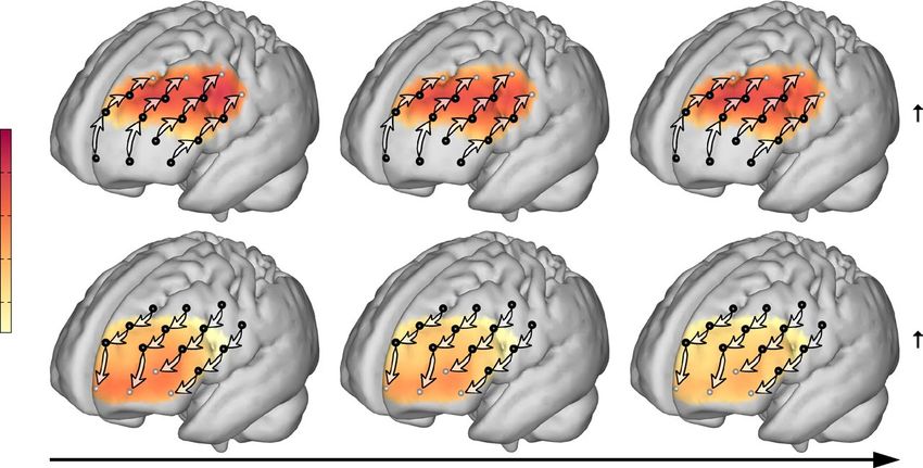
www.nature.com/scientificreports/
benefits of STN-DBS for the alleviation of motor symptoms are unquestioned13–15, cognitive and psychiatric side
effects like impaired verbal fl uency16–18, impulsive b
ehavior19 and even a possibly increased suicidal t endency20
have been reported. These impairments indicate far-reaching implications of modulating basal-ganglia networks,
suggesting that STN-DBS may impact on the functional integrity of the PFC12,21. Directly assessing DBS-induced
neurophysiological changes in the PFC may hence substantially advance our understanding of remote effects
beyond the targeted stimulation sites.
Several models of human PFC functioning have suggested a rostro-caudal hierarchical o rganization22–24. In
these, it is assumed that information processing in rostral PFC precedes and determines processing in caudal
PFC23. Indeed, as shown by functional magnetic resonance imaging (fMRI) and lesion studies, rostral parts of the
lateral PFC get gradually more involved when cognitive demands become increasingly abstract, interdependent,
and temporally extended25–27. Moreover, recent evidence suggests that the upper end of this processing cascade
resides in the mid-dorsolateral PFC rather than in the frontal p ole28–31.
The PFC directly projects into the STN by the associative hyperdirect pathway: Fibers of the corticospinal
tract send axon collaterals to the associative part of the nucleus3,32,33. Moreover, with disease progression, frontal
areas involved in both motor and associative loops show increased atrophy which has recently been linked to the
degree of clinical improvement of Parkinson’s patients under STN-DBS34. Thus, the outcome of STN-DBS signifi-
cantly relies on the integrity of the PFC, which is possibly influenced by the stimulation of the limbic hyperdirect
pathway that was recently found to be confluent with the superolateral medial forebrain bundle in h umans2. In
consequence, the remote impact of STN-DBS on cortical regions is likely to differ between Parkinson’s patients
that undergo surgery early and late after disease onset.
Based on the assumption of rostro-caudally directed interactions within the P FC22–24, we therefore directly
assessed STN-DBS effects on the integrity of this prefrontal hierarchical network by comparing the rostro-caudal
gradient of neural processing in the lateral PFC, estimated from intra-individual functional measurements of
Parkinson’s patients in different DBS states—initially in the ON state, followed by steady-state OFF, and thereafter
again ON DBS. To this end, we used a recently established approach on estimating directed functional connectiv-
ity based on multi-channel near-infrared spectroscopy (fNIRS) measurements that provide a high temporal and
a sufficient spatial resolution to reveal the spatiotemporal evolution of directed neural activity within the lateral
PFC (e.g. Refs31,35,36). Thus, the present approach evaluates the functional integrity of those cortical regions that
are fundamental for higher-order cognitive functions. In addition, neuropsychological assessments were used
to establish a link between the estimated prefrontal hierarchical network properties and cognitive functioning.
Results
The analysis workflow, including the measurement protocol and illustration of fNIRS channel positions, is sum-
marized in Fig. 1. The main analyses were conducted by means of two linear mixed-effects models; the first model
investigated whether STN-DBS (compared ON vs. OFF) exerted an influence on the rostro-caudal connectivity
in the lateral PFC, while the second model (additionally including data from the second ON measurement, cf.
Figure 1A) served as an experimental validation of the main finding of a stimulation-induced alteration of the
rostro-caudal connectivity. An overview of the fixed and random effects structures of Models 1 and 2 is sum-
marized in Table 1. The concise report of the results below is complemented by a comprehensive report of the
respective statistical indices underlying significant effects, estimates, and contrasts in the Supplementary Table S2.
Additionally, control analyses including healthy adults (Supplementary Model S1, Supplementary Fig. S2), assess-
ing the role of the rostro-caudal gradient for cognitive functions (Supplementary Model S2, Supplementary
Fig. S3), and investigating the role of gray matter volume as a proxy of disease-induced cortical atrophy for
directed functional connectivity (Supplementary Model S3) are available in the Supplementary Information.
STN‑DBS modulates hierarchical processing in the PFC (Model 1). In Model 1 we tested the
hypothesis that STN-DBS has an impact on rostro-caudally directed interactions in the PFC. Understand-
ing remote effects of STN-DBS is however challenged by the multitude of variables that add to the outcome
of the treatment, including e.g. the patient’s preoperative clinical s tatus14, stimulation parameters37, electrode
positions38, and concomitant drug treatment39,40. Moreover, higher age and longer disease duration have been
discussed to be disadvantageous for the clinical outcome of STN-DBS34. The initial fixed effects structure of
model 1 (Table 1) therefore included the following factors of interest: stimulation state (ON vs. OFF), direction
of influences (rostro-caudal vs. caudo-rostral), and hemisphere (ipsilateral vs. contralateral with respect to the
hemisphere of disease onset) and all possible interactions between this set of within-subject-factors as well as
each of the following covariates: (I) age at disease onset (initial diagnosis), (II) disease duration before DBS
implantation, (III) time since DBS implantation (I–III summing up to the chronological age), (IV) dopamin-
ergic medication in terms of the levodopa equivalent daily dose (LEDD, calculated according to Tomlinson
et al.41), and (V) stimulation intensity in terms of the volume of activated tissue (VAT). This extensive model was
successively reduced by non-significant fixed effects terms to yield a parsimonious m odel42. Multicollinearity
was controlled using the variance inflation factor (VIF, all VIF < 3.6)43,44. Correlation coefficients for the covari-
ates are provided in Supplementary Table S3.
Confirming the assumed rostro-caudal hierarchical organization of the PFC, the significant main effect for
direction revealed that influences in rostro-caudal direction were generally higher than caudo-rostral influences
(F(1,1995.4) = 110.9, p < 0.001, Fig. 2). As STN-DBS is known to have side effects on cognitive functions subserved
by the P FC16,19, we hypothesized that the prefrontal rostro-caudal gradient could be compromised ON compared
to OFF stimulation. While the respective two-way interaction between stimulation state (ON vs. OFF) and the
direction of influences (rostro-caudal/caudo-rostral) was not statistically significant on its own (F(1,1995.4) = 0.6,
p = 0.454), it was dependent on the disease duration before DBS implantation (F(1,1995.4) = 5.8, p = 0.016). This
Scientific Reports | (2021) 11:2138 | https://doi.org/10.1038/s41598-021-81535-7 2
Vol:.(1234567890)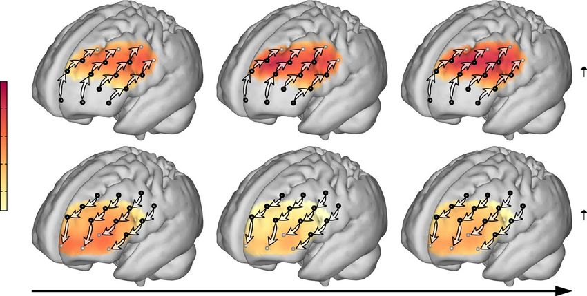
www.nature.com/scientificreports/
a Multichannel fNIRS b Pre- c Spectral d Peak extraction
measurements processing estimation of between .06
oxy directed and .12 Hz
3c e
m urc deoxy correlation- interactions
so based artifact
Connectivity matrix
ector
correction
det
Channels
DC
Channels
standardization
Healthy controls: 12 min. resting-state
PD patients: 3 x 12 min. resting-state: Channels
DBS ON 2h DBS OFF DBS ON Channels
g Visualization f Linear mixed e Selection of directed connections
effects modeling along rostro-caudal axis
rostral caudal
rostral caudal caudal rostral
Influences
DC
caudal rostral
Influences
Averaging across
participants
Figure 1. Scheme of the analysis workflow followed in the present study. Patients watched muted parts of a
nature documentary during fNIRS measurements (a). Artifact correction67 was applied, resulting in perfectly
anticorrelated signals of oxygenated and deoxygenated hemoglobin (b). The time-series were standardized
before estimation of directed coherence (DC) as a measure of directed interactions between fNIRS c hannels69
(c). The maximum DC value in the frequency band between 0.06 and 0.12 Hz was extracted to yield a
2-dimensional, directed connectivity matrix for each measurement (d). According to models postulating a
predominant flow of information from rostral to caudal PFC the influences between PFC regions should be
significantly stronger in rostral-to-caudal direction than in caudal-to-rostral direction. This prediction was
tested by analyzing connections between directly neighboring channels along four rostro-caudal streams
comprising four channels each (black and gray arrows in panel e) and covering most of the lateral surface of
the PFC in each hemisphere (e). The distinction between rostro-caudal and caudo-rostral connections was
represented in the model by the factor direction of influences (f). Influences in both directions were averaged
across patients and projected separately on a standard brain surface (g).
Model Fixed effects terms Random intercepts
dir. × hemisphere × stim. × age at disease onset
+ dir. × hemisphere × stim. × disease duration before DBS implantation
Stream × participant
1: ON and OFF measurements + dir. × hemisphere × stim. × time since DBS implantation
Level × participant
+ dir. × hemisphere × stim. × LEDD
+ dir. × hemisphere × stim. × VAT
Stream × participant
2: ON, OFF & 2nd ON measurements dir. × stim. × disease duration before DBS implantation
Level × participant
Table 1. Initial fixed and random effects structure of each model. N.B. Exhaustive models were successively
reduced by non-significant higher-order terms to yield a parsimonious model (see Supplementary Table S2
for significant effects). Continuous predictors are underlined. All lower-order terms were included. Random
effects were not reduced. dir. direction of influences, LEDD levodopa equivalent daily dose, stim. stimulation
state (ON, OFF, and, in model 2, 2nd ON), VAT volume of activated tissue.
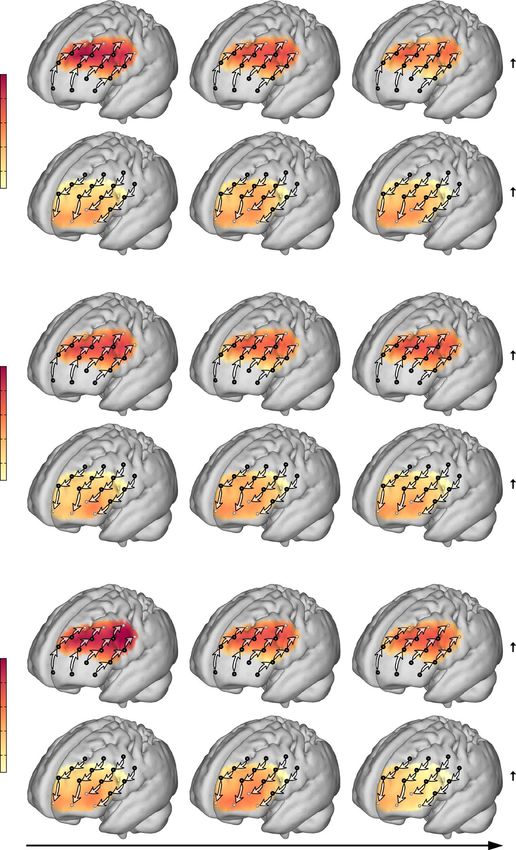
three-way interaction was primarily driven by influences in rostro-caudal direction (Fig. 3 top row, Supplemen-
tary Table S2): Patients who underwent DBS implantation early after disease onset showed stronger rostro-caudal
influences and a steeper gradient ON compared to OFF stimulation. In contrast, in patients who received DBS
implantation at longer disease durations the rostro-caudal gradient was diminished ON stimulation. Moreover,
there was no significant relation between the disease duration before DBS implantation and the gradient in the
OFF state (Fig. 3 middle row).
Model 1 further revealed that the prefrontal gradient heavily depended on the stimulation intensity
(F(1,1995.4) = 15.6, p < 0.001), with a larger VAT being associated with both stronger rostro-caudal and weaker
caudo-rostral influences (Fig. 4, Supplementary Table S2). This two-way interaction between VAT and the direc-
tion of influences was independent of whether STN-DBS was turned ON or OFF and persisted after stimulation
was switched off for 2 h.
Scientific Reports | (2021) 11:2138 | https://doi.org/10.1038/s41598-021-81535-7 3
Vol.:(0123456789)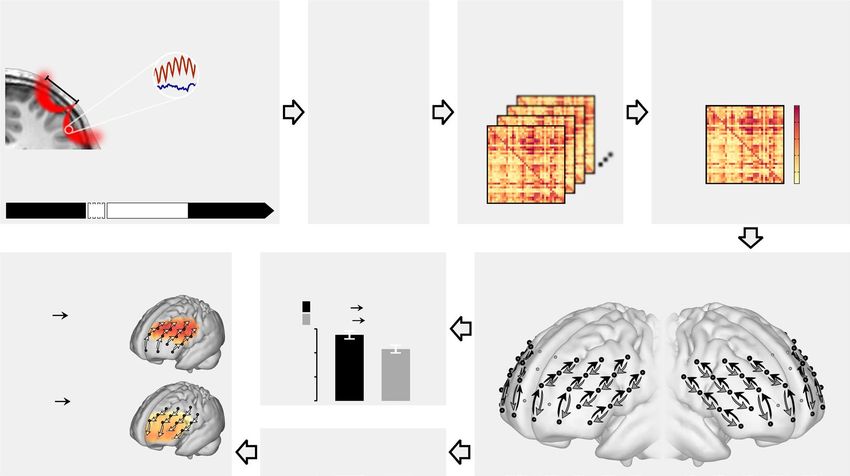
www.nature.com/scientificreports/
Effect of Direction
.6 p < .001
Directed Coherence (DC)
.4
.2
.0
rostral caudal caudal rostral
.5
.4
DC
.3
.2
Figure 2. Granger-causality analysis of directed interactions reveals the rostro-caudal hierarchical organization
in the PFC. The significant main effect for direction in the mixed effects model showed that influences from
rostral to caudal PFC were stronger than from caudal to rostral (top panel). For cortical projections (bottom
panel), directed coherence (DC) values were averaged across hemispheres and projected onto the cortical
surface to represent the influences from channels (black dots) toward caudally (left brain) and rostrally (right
brain) neighboring channels as indicated by arrows. Darker red colors signify stronger influences in terms
of higher DC values. Bars in the top panel represent least square means; error bars indicate 95% confidence
intervals; n = 24.
Besides VAT, LEDD was also positively associated with the strength of the rostro-caudal gradient
(F(1,1995.4) = 9.6, p = 0.002). However, this two-way interaction between LEDD and the direction of influences
was mostly driven by the simple effect of influences in one direction, i.e. LEDD was negatively correlated with
caudo-rostrally directed influences, while the relationship between influences in rostro-caudal direction and
LEDD failed to reach significance (Fig. 5, Supplementary Table S2).
Albeit weak, a third two-way interaction was present between hemisphere and the direction of influences
(F(1,1995.4) = 4.6, p = 0.033). The rostro-caudal gradient appeared to be steeper in the ipsilateral than in the
contralateral hemisphere with respect to the hemisphere of disease onset. This within-patient effect hence cor-
roborates the enhancing between-patient effect of the VAT on the rostro-caudal gradient, as VATs were larger in
the ipsilateral than in the contralateral hemisphere (mean VAT in ipsilateral hemisphere: 230 mm3; mean VAT
in contralateral hemisphere: 174 m m3, t(22) = 2.0, p = 0.053). Thus, the hemispheric differences in the gradient
may be introduced by the asymmetric stimulation intensities.
Taken together, the key finding of Model 1 constituted a significant modulation effect of STN-DBS on the
rostro-caudally directed interactions in the PFC that was moderated by the patients’ disease duration before
DBS implantation. Given that disease duration before DBS implantation was highly correlated with the overall
disease duration (Pearson’s r = 0.79, p < 0.001), we conducted a control analysis and refitted the model with (I)
the variables disease duration before DBS implantation and time since DBS implantation substituted by the overall
disease duration and (II) with all three time-related covariates (age at disease onset, disease duration before DBS
implantation and time since DBS implantation) substituted by chronological age. Neither overall disease duration
nor chronological age exerted any significant moderator effect (all p > 0.05) on the directed interactions in the
PFC, demonstrating that the impact of STN-DBS on the rostro-caudal gradient was indeed specifically depend-
ent on the disease duration before DBS implantation.
Remote effects of STN‑DBS on PFC are reproducible (Model 2). As an experimental validation, in
Model 2 we addressed the reproducibility of the modulation effect of STN-DBS on the rostro-caudally directed
interactions in the PFC and its dependence on the disease duration before DBS implantation. To this end, the
examination followed a fixed protocol with the initial ON measurements always being followed by the subse-
quent OFF measurements. Precluding that the stimulation-dependent results in Model 1 were simply driven
by carry-over effects would hence require to demonstrate that the dependence of the STN-DBS effects on the
disease duration before DBS implantation was reversible and would reappear after stimulation was switched on
again. In consequence, we fitted Model 2 to the data of the ON, OFF, and ON2 measurements and, as a fixed
Scientific Reports | (2021) 11:2138 | https://doi.org/10.1038/s41598-021-81535-7 4
Vol:.(1234567890)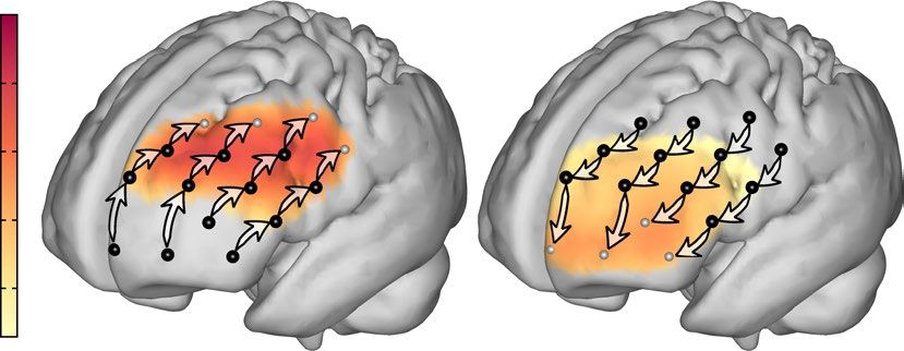
www.nature.com/scientificreports/

                                                    Model estimates                                         Averaged estimates of directed interactions

                                                                                                                                                                         rostral caudal
                                        .5
                   Directed Coherence (DC)

                                        .4
         DBS ON

                                                                                        .5
                                                                                        .4

                                                                                   DC
                                        .3                                              .3

                                                                                                                                                                         caudal rostral
                                                                                        .2

                                        .2
                                                                rostral caudal
                                             Direction
                                        .1                      caudal rostral
         2h

                                                                                                                                                                         rostral caudal
                                        .5
                   Directed Coherence (DC)

                                        .4
                                                                                        .5
                                                                                        .4
         DBS OFF

                                                                                   DC

                                        .3                                              .3

                                                                                                                                                                         caudal rostral
                                                                                        .2

                                        .2

                                        .1

                                                                                                                                                                         rostral caudal
                                        .5
                   Directed Coherence (DC)

                                        .4
                                                                                        .5
         DBS ON

                                                                                        .4
                                                                                   DC

                                        .3                                              .3

                                                                                                                                                                         caudal rostral
                                                                                        .2

                                        .2

                                        .1
                                              5          10        15        20                     1st third                  2nd third               3rd third
                                             Disease duration before DBS                        [3.9, 8.2] years          [8.9, 13.2] years        [13.9, 20] years
                                                 implantation (years)                                   Disease duration before DBS implantation (years)

                                                         Figure 3.  The effect of DBS on rostro-caudally directed interactions in the PFC depends on the disease
                                                         duration before DBS implantation. The relationship between the rostro-caudal gradient and the disease duration
                                                         before DBS implantation in the ON stimulation state (top row, n = 24) shows that patients who received DBS
                                                         early in the course of their disease had a strong gradient that was even stronger than in the OFF stimulation
                                                         state (middle row, n = 22). The later in the individual course of the disease patients underwent implantation,
                                                         the more influences in rostro-caudal direction declined, such that patients who received DBS late after disease
                                                         onset had a clearly diminished gradient ON stimulation compared to OFF stimulation. The replication of this
                                                         time-dependent impact of DBS stimulation on the rostro-caudal hierarchical organization of the PFC in the
                                                         second ON stimulation measurement (bottom row, n = 18) further confirmed this dependence (Model 2, see
                                                         below). The projection of directed coherence (DC) values on the cortical surface was done analogous to Fig. 2
                                                         but separately for three sub-groups, split at the terciles of the disease duration before DBS implantation. Model
                                                         predictions are plotted with 95% non-simultaneous confidence bands.

Scientific Reports |                         (2021) 11:2138 |                    https://doi.org/10.1038/s41598-021-81535-7                                                      5

                                                                                                                                                                      Vol.:(0123456789)
www.nature.com/scientificreports/
Model estimates Averaged estimates of directed interactions
rostral caudal
Direction
rostral caudal
.5 caudal rostral
Directed Coherence (DC)
.4
.5
.4
DC
.3 .3
caudal rostral
.2
.2
.1
100 200 300 400 1st third 2nd third 3rd third
Volume of activated tissue [78, 113] mm³ [117, 213] mm³ [216, 425] mm³
averaged across hemispheres (mm³) Volume of activated tissue averaged across hemispheres (mm³)
Figure 4. Rostro-caudally directed interactions in the PFC increase with stimulation intensity. The prediction
by the linear mixed effects model showed a steep increase of influences from rostral to caudal prefrontal cortex
(PFC) (black line) with larger volumes of activated tissue (VAT) and stimulation strength. At the same time,
caudo-rostrally directed influences weakened with stimulation strength (gray line). These diverging trends
can also be observed from the projected directed coherence (DC) values, where colors indicate the strength
of influences between neighboring channels in rostro-caudal (top row) and caudo-rostral direction (bottom
row). Sources of influences are marked by black dots. The projection of DC values was done separately for three
sub-groups, split at the terciles of VAT. The most striking difference between patients with small and large VATs
appears in influences exerted by the mid-lateral on the caudal PFC. Model predictions in the left panel are
shown with 95% non-simultaneous confidence bands; n = 23.
Model estimates Averaged estimates of directed interactions
rostral caudal
Direction
rostral caudal
.5 caudal rostral
Directed Coherence (DC)
.4
.5
.4
DC
.3 .3
caudal rostral
.2
.2
.1
200 600 1000 1400 1st third 2nd third 3rd third
Levodopa equivalent [150, 510] mg/d [520, 661] mg/d [680, 1606] mg/d
daily dose (mg/d) Levodopa equivalent daily dose (mg/d)
Figure 5. Dopaminergic medication increases the rostro-caudal gradient in the PFC by diminishing rostrally
directed influences. The enhancing effect of the levodopa equivalent daily dose (LEDD) on the rostro-caudal
gradient was mainly driven by a strong decrease of influences in caudo-rostral direction (bottom row of
projections). Analogous to Figs. 3 and 4, the projection of directed coherence (DC) values was done separately
for three sub-groups, split at the terciles of LEDD. Model predictions are plotted with 95% non-simultaneous
confidence bands; n = 24.
effect, included the respective three-way interaction between stimulation state, direction of influences and dis-
ease duration before DBS implantation as well as all corresponding lower-order terms. As depicted in Fig. 3, the
negative correlation between the disease duration before DBS implantation and the rostro-caudal gradient was
present in the ON state and absent in the OFF state. Most importantly, it reemerged in the ON2 state (Fig. 3,
bottom row), within only a few minutes after the stimulation was switched on again. As the three-way interac-
Scientific Reports | (2021) 11:2138 | https://doi.org/10.1038/s41598-021-81535-7 6
Vol:.(1234567890)www.nature.com/scientificreports/
tion closely resembled the pattern observed in Model 1, it hence demonstrated its immediate reproducibility
(F(2,2949.6) = 3.2, p = 0.041). Post-hoc comparisons further confirmed that for influences in rostro-caudal direc-
tion the effect of disease duration before DBS implantation in the ON and ON2 state was significantly different
from the OFF state, while there was no significant difference between the ON and the ON2 state (Fig. 3, Supple-
mentary Table S2). Regarding caudo-rostral influences, there was no significant simple effect of disease duration
before DBS implantation in any stimulation state.
In summary, the modulation of directed influences between PFC sub-regions by STN-DBS and its modera-
tion by disease duration before DBS implantation was not only replicable, but also specific to the predominant
rostro-caudal direction within the PFC and specific for ON states, arguing for a significant stimulation-induced
network effect of STN-DBS on PFC integrity.
Supplemental control analyses. Healthy participants that were matched to PD patients in terms of age
and sex (Supplementary Fig. S1) were assessed in two supplemental analyses. Supplementary Model S1 revealed
that the rostro-caudal connectivity gradient was also apparent in healthy controls (Supplementary Fig. S2). Sup-
plementary Model S2 included both PD patients and matched healthy controls and demonstrated that the rest-
ing-state connectivity gradient within the rostral PFC was associated with performance in a prefrontally-guided
cognitive planning task as well as a more general measure of global cognitive ability (Supplementary Fig. S3).
Thus, the present approach consistently detects a rostro-caudal functional network in the PFC that underlies
higher cognitive abilities. Another supplemental control analysis testing the predictability of the directed func-
tional connectivity by gray matter volume in the PFC revealed no significant interaction effects, precluding that
the variance in the rostro-caudal gradients within the functional networks was significantly driven by differences
in cortical atrophy (Supplementary Model S3).
Discussion
The present study revealed that STN-DBS in Parkinson’s patients significantly modulates the hierarchical organi-
zation of the PFC. Most crucially, this remote network effects of STN-DBS on the PFC depended on disease
progression and hence possibly on the level of sustained integrity of nigro-striatal and fronto-striatal circuits:
Patients who underwent DBS implantation earlier after disease onset had a stronger rostro-caudal gradient of
directed interactions ON versus OFF stimulation. In contrast, patients who received a DBS implantation after
longer disease durations showed diminished rostro-caudally directed interactions ON compared to OFF stimula-
tion. Importantly, this effect was clearly reproducible: In the transition when stimulation was switched ON again
after steady-state OFF, the dependence of the rostro-caudal gradient in the PFC on the disease duration before
DBS implantation re-emerged in the same systematic fashion as before (Fig. 3).
The role of the basal ganglia for the rostro‑caudal hierarchy in the PFC. One hypothesis is that
the STN—as a major input nucleus of the basal ganglia—plays an important role in integrating sensorimotor
information and regulating the activity of two opposing signaling pathways: the direct, movement-facilitating
pathway and the indirect, movement-inhibiting pathway9,45. Loss of dopaminergic neurons in the substantia
nigra pars compacta in Parkinson’s disease leads to an excessive activity of the STN and an imbalance in favor
of the indirect pathway causing hypokinetic symptoms9. DBS is known to elicit action potentials in afferent
and efferent a xons37 thus not only exciting downstream neurons in basal ganglia output nuclei but also anti-
dromically modulating cortical neurons projecting (as part of the hyperdirect pathway) to the STN46. Although
the STN was previously assumed to receive cortical projections only from motor cortices47 a recent tracer study
in macaques revealed topographic projections from various PFC regions (including the dorsolateral PFC) to the
dorsomedially located associative functional zone of the S TN48 which could also be reproduced in human imag-
ing studies2,3,49. Antidromic activation of the limbic hyperdirect pathway may therefore contribute to the pre-
sent stimulation-induced changes in the hierarchical organization of the PFC, affecting cognitive functions4,50.
Alternatively, remote STN-DBS effects on the PFC may be mediated through the downstream nigro-thalamo-
cortical pathway37—in particular as the impact of STN-DBS on rostro-caudally directed prefrontal interactions
depended on the disease duration before DBS implantation and therefore on the disease progression at the time
STN-DBS was initially administered. In Parkinson’s disease dopamine depletion often progresses from sensori-
motor towards associative/cognitive basal ganglia-thalamocortical circuits and affects cortical afferents in caudal
PFC earlier than in rostral PFC5,39. The enhancement of directed interactions within lateral PFC by STN-DBS
may thus be mediated through the intact associative circuits in early-stage Parkinson’s disease, while STN-DBS
has debilitating effects on PFC integrity as soon as disease progression has reached associative circuits.
Disease progression impacts on prefrontal effects of STN‑DBS. The present results provide new,
complementary evidence suggesting that implantation at an earlier stage of Parkinson’s disease may reduce the
risk of DBS-induced detrimental cognitive and psychiatric side effects on prefrontal functioning. As the STN-
DBS effect on directed prefrontal interactions depended neither on the duration of stimulation, nor on the over-
all disease duration, nor on the chronological age but only on the disease duration before DBS implantation, this
raises the question whether STN-DBS itself or the reduction of dopaminergic medication following STN-DBS
may exert preserving effects on the associative basal ganglia-thalamocortical loops51. The stimulation-induced
decrease of the rostro-caudal gradient with increasing disease duration before DBS implantation suggests that
STN-DBS intervention at early-stage Parkinson’s disease may contribute to sustained PFC integrity and that the
timing of implantation is of essence for the overall outcome of D BS52. When patients notice first symptoms of
Parkinson’s disease, at least 50% of neurons in the substantia nigra are already lost53. Animal models of Parkin-
son’s disease further show that STN-DBS can have neuroprotective effects in the substantia n igra51. Although
Scientific Reports | (2021) 11:2138 | https://doi.org/10.1038/s41598-021-81535-7 7
Vol.:(0123456789)www.nature.com/scientificreports/
not confirmed in humans so far52, it has been discussed that DBS implantation may have a slowing effect on
the progression of n eurodegeneration51. Finally, a brain in a healthier state may simply cope with and adapt to
an extensive intervention like DBS better than a more depleted one as, for instance, the motor, cognitive, and
psychiatric symptoms of Parkinson’s disease are alleviated by STN-DBS when applied early in the course of the
disease13,15,52. In line with this, younger patients are known to recover better and have less cognitive decline after
surgery than the elderly54.
As STN-DBS electrodes were implanted through prefrontal entry points the penetration of the PFC during
STN-DBS surgery may also impact on PFC network integrity. However, the present moderation of the rostro-
caudal gradient by disease duration before DBS implantation was reversible and only present under active
stimulation. It is hence unlikely to be introduced by the surgery itself. Nevertheless, the lesion of the STN caused
by electrode implantation is known to have a temporary effect that clinically mimics the effect of stimulation55.
This so-called stun effect is a transient phenomenon which is apparent within the first postoperative weeks but
usually regresses before initial activation of the stimulator. Preliminary results from a follow-up study indicate
that the stun effect caused by STN-DBS surgery indeed attenuates the rostro-caudal gradient post- compared
to pre-DBS surgery only in patients with long disease durations but not in patients with short disease durations
(F. K. Schumacher, V. A. Coenen, C. P. Kaller, unpublished data) reproducing the moderating effect of disease
duration before DBS implantation on the rostro-caudal gradient ON stimulation in the current study.
Effects of stimulation intensities and dopamine level. VAT and LEDD both had a general amplify-
ing effect on the strength of the prefrontal gradient. However, the effect of VAT was driven by both increas-
ing rostro-caudal influences and decreasing caudo-rostral influences, while the effect of LEDD solely relied on
decreasing influences in caudo-rostral direction (Figs. 4, 5). The mechanisms behind both effects are hence likely
to be distinct40. Moreover, shifts in network states induced by the systemic application of dopamine are mainly
dosage dependent, whereas the VAT effect on cortical networks is probably transduced through parts of the basal
ganglia-cortical loops and may also rely on resonance mediated by the direct, indirect, and limbic hyperdirect
pathway2,45.
Interpreting the effects of VAT and LEDD is limited by the lack of data from patients OFF medication or
before DBS implantation. A stronger gradient may in fact be the cause for rather than the result of stronger
stimulation amplitudes (i.e. patients having a stronger gradient might need a stronger stimulation to alleviate
motor symptoms). The persistent VAT effect OFF stimulation may support this argument but it may likewise
reflect plastic changes in network organization or sustained shifts in neurochemical h omeostasis56.
Finally, the magnitude of the VAT estimates (Supplementary Table S1) indicates that the stimulation directly
affected subcortical structures other than the STN in some patients. Besides stimulation intensities, taking loca-
tions of active electrode contacts into account in future studies is therefore vital to understand the role of this
excessive stimulation.
Limitations. A possible limitation of the present approach constitutes its reliance on hemodynamic low-fre-
quency oscillations as systemic physiological oscillations (e.g. Mayer-waves57) may partly contribute to the signal
variance in the frequency band used here. However, there is increasing evidence that this signal component
reflects neuronal a ctivity58 and conveys information about functional connectivity59,60. In addition, we recently
demonstrated the robustness of the present approach against physiological noise35. The network connections
reconstructed here are hence unlikely to reflect mere physiological artifacts but instead represent (direct or indi-
rect) signaling pathways between neuronal populations.
A pivotal question that remains is whether the disruption of the prefrontal hierarchy by STN-DBS is indeed
causal for cognitive decline. The present findings suggest that receiving STN-DBS in a more advanced stage of
Parkinson’s disease may increase the risk of cognitive side-effects because the stimulation disturbs the hierarchical
organization in the PFC. Furthermore, the relevance of this hierarchical organization in the PFC for cognitive
functioning is demonstrated in the Supplementary Model S2. Yet, the present data do not allow to conclude that
effects of STN-DBS on cognitive performance are mediated by its remote effects on the PFC. Resolving this issue
would require to simultaneously conduct cognitive assessments and fNIRS measurements repeatedly ON and
OFF STN-DBS, which would however likely be confounded by the patients’ motor symptoms and challenged
by psychometric i ssues61.
Although PD patients were assessed on medication adhering to their usual medication to ensure dopamine
levels being as stable and as physiological as possible, it cannot be fully excluded that changes in dopamine over
time may have at least partly contributed to the differences ON vs. OFF STN-DBS observed in Model 1. However,
these STN-DBS effects on the functional network structure in the PFC were replicated in the second (later) ON
stimulation measurement (Model 2; cf. Fig. 3 top and bottom row), thus strongly suggesting that the modulation
of the rostro-caudal gradient in the PFC was driven by the experimental variation of the STN stimulation and
not by changes in dopamine levels over time.
Conclusion
The present study not only provides novel insights into the remote network effects of STN-DBS but also offers a
completely new perspective on the potential neurophysiological mechanisms underlying cognitive and psychiat-
ric side effects of STN-DBS in Parkinson’s disease. Specifically, we demonstrated that the rostro-caudal hierarchy
in the PFC is compromised by STN stimulation in patients who underwent electrode implantation after longer
disease duration. In contrast, stimulation enhanced the prefrontal hierarchy in patients who received DBS early
in the course of their disease. In addition, stimulation intensities and dopaminergic drug dosages predicted the
strength of the prefrontal hierarchy. Taken together, by allowing to directly monitor the remote network effects
Scientific Reports | (2021) 11:2138 | https://doi.org/10.1038/s41598-021-81535-7 8
Vol:.(1234567890)www.nature.com/scientificreports/
of STN-DBS the novel approach applied here might provide a promising opportunity for future refinements of
DBS in the STN and beyond in terms of a methodological foundation for individually tailored optimization and
adjustment of stimulation parameters.
Methods and materials
The study protocol was approved by the ethics committee of the University of Freiburg (vote 410/11) and reg-
istered at German Clinical Trials Register (DRKS, www.drks.de, identifier DRKS00003530, date of registration:
10/02/2012). The study was carried out in accordance with the Declaration of Helsinki and the guidelines of
the local ethics committee on research involving human participants. All participants gave written informed
consent prior to participation.
Participants and procedures. Twenty-six patients with idiopathic Parkinson’s disease and implanted
STN-DBS participated in the present study. To control for hemispheric differences in disease severity, two
patients that had no record of the side of disease onset were excluded from the present analyses. Following
a neuropsychological assessment (including the Tower of London task, see below), patients were rated by the
UPDRS-III and underwent the fNIRS measurements. After the first fNIRS measurement (12 min, ON stimu-
lation), the stimulator was switched off for approximately 2 h. Of the 24 included patients (6 females, mean
age ± SD 61.5 ± 9.9 years), 2 dropped out during this period. The second fNIRS measurement was conducted
OFF stimulation for 12 min and was preceded by another UPDRS-III assessment. Four further patients dropped
out after the measurement OFF stimulation. In order to capture the transition from OFF to ON stimulation, we
conducted a third measurement immediately after the stimulator was switched on again. Taken together, three
12-min fNIRS measurements were acquired (Fig. 1a): first ON stimulation (n = 24), thereafter in steady-state
OFF stimulation (after 2 h of rest without stimulation; n = 22) and finally in the transition state immediately after
the stimulator was switched back on (ON2, n = 18). During all measurements, patients were watching muted
movie parts from the nature documentary ‘Earth’62; the order of the movie parts was balanced across patients. To
ensure dopamine levels to be as stable and as physiological as possible, patients were told to adhere to their usual
medication on the day of study participation and were examined during medical ON periods (see Supplemental
Table S1 for patient characteristics; cf. Fig. 1a for experimental workflow). As a control group, 24 healthy adults
were matched to the patients in terms of age and sex (Supplementary Fig. S1) and underwent a 12-min fNIRS
measurement during passive watching of a nature documentary (see Supplementary Model S1 for results and
details of healthy controls). Cognitive functioning of patients and healthy controls was assessed with the Tower
of London planning task63 (see Supplementary Methods for details) which substantially relies on the P FC64;
planning accuracy was used to predict resting-state directed functional connectivity in Supplementary Model S2
to address the role of prefrontal networks for cognitive abilities.
Acquisition of fNIRS data. Multi-channel fNIRS was used to record brain activity in the PFC (Fig. 1a)
and to explicitly test the hypothesis of rostro-caudally directed interactions as it sampled activation in the PFC
z65 and at a spatial resolution that ensured separation of rostral, mid-
at a sufficient temporal resolution of 10 H
dle, and caudal PFC (Fig. 1e). FNIRS data was acquired using an ETG-4000 optical topography system (Hitachi
Medical Systems, Japan). Spatial optode arrangement was derived from the system’s 3 × 11 grid configuration
consisting of 17 emitters and 16 detectors. We modified this probe set by placing 12 emitters and 13 detectors
on the forehead (interoptode distance of 3 cm resulting in a diagonal channel distance of 2.1 cm), resulting in
38 channels evenly distributed over the PFC (Fig. 1e). Unused emitter optodes were covered by black caps to
avoid crosstalk; recordings were performed in a room without windows and the room lights were switched off
during measurements. Grid placement over PFC was standardized across patients (I) by aligning the grid center
to the sagittal midline and (II) by positioning the lower center optode at a distance of about 1.5 cm above the
nasion. Presentation of the nature video and on-/offset of simultaneous fNIRS recordings were controlled by
NBS Presentation software (version 12.2; Neurobehavioral Systems Inc., CA). To prevent artifacts during fNIRS
measurement due to head movements, participants’ heads were stabilized using a chin rest. Raw data of light
intensity changes were converted into hemoglobin concentration changes by in-house Matlab software (version
2012b, The MathWorks, Natick, MA, USA, unpublished tool box) using the modified Beer-Lambert law66. Due
to the absorption of interfering hairs or saturation of detectors some channels did not contain any signal. The
respective time series were interpolated from the surrounding channels using the Matlab 4 griddata method.
This affected a total of 15 channels in 9 datasets (out of 2432 channels in 64 datasets; 0.6% of channels). In
order to remove motion-induced artifacts, we applied the correlation-based correction method developed by
Cui et al.67. This method not only effectively removes motion artifacts, but also increases the contrast-to-noise
ratio. The resulting data for oxygenated and deoxygenated hemoglobin are perfectly anticorrelated and therefore
have identical spectral properties. The time series were standardized for Granger-causality analysis. We refrained
from applying further artifact correction methods like filtering or resampling, because such data preprocessing
has been shown to produce false positives in Granger-causality a nalysis68. Furthermore, we used a frequency-
domain measure of Granger-causality (see below) that allowed avoiding frequency bands prone to physiological
noise introduced by respiration and heart-beat (see also Refs.31,35). Further information on the validity of the
here applied fNIRS approach on directed functional connectivity can be found in Refs.31,35.
Analyses of directed interactions. Directed coherence (DC)69 as an implementation of Granger-cau-
sality70 was used to capture the intrinsic functional organization of the PFC and directed interactions between
its rostral and caudal parts. DC was estimated using the frequency domain multivariate analyses toolbox (www.
fdm.uni-freiburg.de/Toolboxes/fdma-toolbox). For DC calculation, a vector autoregressive model was fitted
Scientific Reports | (2021) 11:2138 | https://doi.org/10.1038/s41598-021-81535-7 9
Vol.:(0123456789)www.nature.com/scientificreports/
with a model order of 20, corresponding to the past 2 s of the time-series and providing a frequency resolution
of the DC estimate of 0.5 Hz. Note, however, that for the Fourier transformation the autoregression coefficients
were zero-padded to the length of the time series in order to smooth the spectral estimate. As functional con-
scillations59, we chose the frequency band
nectivity between different brain areas is apparent in low frequency o
between 0.06 and 0.12 Hz and used the maximum DC value in this band for further analysis (see also Refs.31,35).
DBS electrode localizations. Preoperative T2-weighted MRI and CT scans as well as postoperative axial
slabs of CT scans focusing on the subcortical volume around the electrode tips were acquired. Details on imag-
ing parameters varied across patients and can be requested from the authors. Using BRAINSfit software71 as
implemented in 3D slicer (version 4.6.0; www.slicer.org), postoperative CTs were linearly co-registered and
fused to preoperative CTs. The fused images were then again linearly co-registered to preoperative MRIs. Pre-
operative MRIs were nonlinearly warped into International Consortium for Brain Mapping (ICBM) 152 2009b
nonlinear asymmetric space using the SyN approach as implemented in Advanced Normalization T ools72. Using
73
the Lead-DBS software (version 1.5.1; www.leadsuite.io), electrode positions were reconstructed hybridly in
native and standard stereotactic space. Renderings of electrode positions in relation to the STN are provided in
Supplementary Fig. S4, showing that implanted electrodes mainly resided within the posterior, i.e. the motor
subdivision of the STN.
Estimation of the volume of activated tissue (VAT). To construct a conductor model of the DBS elec-
trode and surrounding tissue, the Medtronic DBS electrode 3391 model was discretized into voxels of 0.22 mm
isotropic size. The electrode defined the mid-axis of a volume 101 × 101 × 90 voxels in size. The rest of the space
was filled with gray and white matter. Gray matter was defined by the structures STN, red nucleus and internal/
external pallidum as specified by the DISTAL atlas3 which exactly corresponds to the ICBM 2009b nonlin-
ear template used for normalization. The remaining space was declared as white matter. The volume was con-
verted into a hexaedral mesh using FieldTrip software74; www.fieldtriptoolbox.org). Conductivities of 0.33 and
0.14 S/m were assigned to gray and white matter respectively. These values are commonly used in neuroscientific
modeling studies75. For the platinum/iridium contacts and insulated parts of the electrodes, values of 1 08 S/m
and 10–16 S/m were used, respectively. A forward model to obtain the voltage distribution was solved using
the SimBio toolbox (Ref.76; https://www.mrt.uni-jena.de/simbio/index.php/Main_Page). In case of monopolar
stimulation, the surface of the cubic model was used as the cathode. This generated a gradient denoting the volt-
age distribution for each hexaedral element of the model. The gradient was thresholded for magnitudes > 0.2 V/
mm2 to define the VAT. The VATs in both hemispheres were substantially correlated (Pearson’s r = 0.55, p = 0.007)
and were therefore averaged and used as a single predictor in the statistical analysis.
Statistical analyses. DC and between-subject covariates were analyzed in linear mixed effects models
using the lme4 p ackage77 (version 1.1–14) for R statistics (version 3.4.2; http://cran.r-project.org). According
to extant models of prefrontal organization22,23, we expected influences predominantly in rostro-caudal direc-
tion and therefore selected the connections between 4 rostro-caudally adjacent fNIRS channels in 4 parallel
rows within each hemisphere for statistical analyses (i.e., resulting in 12 connections per hemisphere; Fig. 1e).
Hemispheres were classified into ‘ipsilateral’ and ‘contralateral’ with respect to the hemisphere of disease onset.
Two linear mixed effects models were computed as summarized in Table 1. Models were analyzed following a
top-down procedure42: Higher-order fixed-effects interaction terms that did not reach significance (i.e. p > 0.05)
were identified using the anova method (Type III F-statistics with Satterthwaite’s approximation of degrees of
freedom) implemented in the lmerTest p ackage78 (version 2.0-33) and successively removed to yield a parsimo-
nious model42 (see Table 1 for the highest-order terms of the initial models and Supplementary Table S2 for the
significant highest-order terms of the parsimonious models). Models were fitted with random intercepts for (I)
the interaction between participant and stream (identifying channel rows in rostro-caudal direction) and (II)
the interaction between participant and level (identifying adjacent channel pairs—i.e. connections—along the
rostro-caudal axis) using maximum likelihood estimation. Post-hoc comparisons and calculation of confidence
bands were performed using the lsmeans package79 (version 2.27-2). Multiplicity adjustment was applied using
Tukey’s method.
Received: 28 May 2020; Accepted: 28 December 2020
References
1. Graybiel, A. M. & Mink, J. W. The basal ganglia and cognition. In The Cognitive Neurosciences (ed. Gazzaniga, M. S.) 565–585
(MIT Press, Cambridge, 2009).
2. Coenen, V. A. et al. The anatomy of the human medial forebrain bundle: Ventral tegmental area connections to reward-associated
subcortical and frontal lobe regions. NeuroImage Clin. 18, 770–783 (2018).
3. Ewert, S. et al. Toward defining deep brain stimulation targets in MNI space: A subcortical atlas based on multimodal MRI, histol-
ogy and structural connectivity. Neuroimage 170, 271–282 (2018).
4. Kelley, R. et al. A human prefrontal-subthalamic circuit for cognitive control. Brain 141, 205–216 (2018).
5. Redgrave, P. et al. Goal-directed and habitual control in the basal ganglia: Implications for Parkinson’s disease. Nat. Rev. Neurosci.
11, 760–772 (2010).
6. Rodriguez-Oroz, M. C. et al. Initial clinical manifestations of Parkinson’s disease: Features and pathophysiological mechanisms.
Lancet. Neurol. 8, 1128–1139 (2009).
Scientific Reports | (2021) 11:2138 | https://doi.org/10.1038/s41598-021-81535-7 10
Vol:.(1234567890)www.nature.com/scientificreports/
7. Horn, A. et al. Connectivity Predicts deep brain stimulation outcome in Parkinson disease. Ann. Neurol. 82, 67–78 (2017).
8. Vanegas-Arroyave, N. et al. Tractography patterns of subthalamic nucleus deep brain stimulation. Brain 139, 1200–1210 (2016).
9. Albin, R. L., Young, A. B. & Penney, J. B. The functional anatomy of basal ganglia disorders. Trends Neurosci. 12, 366–375 (1989).
10. Jahanshahi, M., Obeso, I., Rothwell, J. C. & Obeso, J. A. A fronto-striato-subthalamic-pallidal network for goal-directed and
habitual inhibition. Nat. Rev. Neurosci. 16, 719–732 (2015).
11. Kahan, J. et al. Resting state functional MRI in Parkinson’s disease: The impact of deep brain stimulation on ‘effective’ connectivity.
Brain 137, 1130–1144 (2014).
12. Volkmann, J., Daniels, C. & Witt, K. Neuropsychiatric effects of subthalamic neurostimulation in Parkinson disease. Nat. Rev.
Neurol. 6, 487–498 (2010).
13. Schüpbach, W. M. M. et al. Neurostimulation for Parkinson’s disease with early motor complications. N. Engl. J. Med. 368, 610–622
(2013).
14. Bronstein, J. M. et al. Deep brain stimulation for Parkinson disease. Arch. Neurol. 68, 165–171 (2011).
15. Lhommée, E. et al. Behavioural outcomes of subthalamic stimulation and medical therapy versus medical therapy alone for Par-
kinson’s disease with early motor complications (EARLYSTIM trial): Secondary analysis of an open-label randomised trial. Lancet.
Neurol. 17, 223–231 (2018).
16. Parsons, T. D., Rogers, S. A., Braaten, A. J., Woods, S. P. & Tröster, A. I. Cognitive sequelae of subthalamic nucleus deep brain
stimulation in Parkinson’s disease: A meta-analysis. Lancet Neurol. 5, 578–588 (2006).
17. Combs, H. L. et al. Cognition and depression following deep brain stimulation of the subthalamic nucleus and globus pallidus
pars internus in Parkinson’s disease: A meta-analysis. Neuropsychol. Rev. 25, 439–454 (2015).
18. Witt, K. et al. Neuropsychological and psychiatric changes after deep brain stimulation for Parkinson’s disease: A randomised,
multicentre study. Lancet Neurol. 7, 605–614 (2008).
19. Frank, M. J., Samanta, J., Moustafa, A. A. & Sherman, S. J. Hold your horses: Impulsivity, deep brain stimulation, and medication
in parkinsonism. Science 318, 1309–1312 (2007).
20. Voon, V. et al. A multicentre study on suicide outcomes following subthalamic stimulation for Parkinson’s disease. Brain 131,
2720–2728 (2008).
21. Castrioto, A., Lhommée, E., Moro, E. & Krack, P. Mood and behavioural effects of subthalamic stimulation in Parkinson’s disease.
Lancet Neurol. 13, 287–305 (2014).
22. Koechlin, E., Ody, C. & Kouneiher, F. The architecture of cognitive control in the human prefrontal cortex. Science 302, 1181–1185
(2003).
23. Badre, D. & D’Esposito, M. Is the rostro-caudal axis of the frontal lobe hierarchical?. Nat. Rev. Neurosci. 10, 659–669 (2009).
24. Fuster, J. M. The Prefrontal Cortex (Academic Press/Elsevier, Amsterdam, 2008).
25. Christoff, K., Keramatian, K., Gordon, A. M., Smith, R. & Mädler, B. Prefrontal organization of cognitive control according to
levels of abstraction. Brain Res. 1286, 94–105 (2009).
26. Azuar, C. et al. Testing the model of caudo-rostral organization of cognitive control in the human with frontal lesions. Neuroimage
84, 1053–1060 (2014).
27. Badre, D. & D’Esposito, M. Functional magnetic resonance imaging evidence for a hierarchical organization of the prefrontal
cortex. J. Cogn. Neurosci. 19, 2082–2099 (2007).
28. Badre, D. & Nee, D. E. Frontal cortex and the hierarchical control of behavior. Trends Cogn. Sci. 22, 170–188 (2018).
29. Nee, D. E. & D’Esposito, M. The hierarchical organization of the lateral prefrontal cortex. Elife 5, e12112 (2016).
30. Margulies, D. S. et al. Situating the default-mode network along a principal gradient of macroscale cortical organization. Proc.
Natl. Acad. Sci. U. S. A. 113, 12574–12579 (2016).
31. Schumacher, F. K., Schumacher, L. V., Schelter, B. O. & Kaller, C. P. Functionally dissociating ventro-dorsal components within
the rostro-caudal hierarchical organization of the human prefrontal cortex. Neuroimage 185, 398–407 (2019).
32. McIntyre, C. C. & Hahn, P. J. Network perspectives on the mechanisms of deep brain stimulation. Neurobiol. Dis. 38, 329–337
(2010).
33. Accolla, E. A. et al. Brain tissue properties differentiate between motor and limbic basal ganglia circuits. Hum. Brain Mapp. 35,
5083–5092 (2014).
34. Muthuraman, M. et al. Effects of DBS in parkinsonian patients depend on the structural integrity of frontal cortex. Sci. Rep. 7,
43571 (2017).
35. Schumacher, F. K. et al. The impact of physiological noise on hemodynamic-derived estimates of directed functional connectivity.
Brain Struct. Funct. 224, 3145–3157 (2019).
36. Medvedev, A. V. Does the resting state connectivity have hemispheric asymmetry? A near-infrared spectroscopy study. Neuroimage
85, 400–407 (2014).
37. Kringelbach, M. L., Jenkinson, N., Owen, S. L. F. & Aziz, T. Z. Translational principles of deep brain stimulation. Nat. Rev. Neurosci.
8, 623–635 (2007).
38. Min, H.-K. et al. Dopamine release in the nonhuman primate caudate and putamen depends upon site of stimulation in the sub-
thalamic nucleus. J. Neurosci. 36, 6022–6029 (2016).
39. Cools, R. Dopaminergic modulation of cognitive function-implications for L-DOPA treatment in Parkinson’s disease. Neurosci.
Biobehav. Rev. 30, 1–23 (2006).
40. Zaidel, A., Bergman, H., Ritov, Y. & Israel, Z. Levodopa and subthalamic deep brain stimulation responses are not congruent. Mov.
Disord. 25, 2379–2386 (2010).
41. Tomlinson, C. L. et al. Systematic review of levodopa dose equivalency reporting in Parkinson’s disease. Mov. Disord. 25, 2649–2653
(2010).
42. West, B. T., Welch, K. B. & Galecki, A. T. Linear Mixed Models: A Practical Guide Using Statistical Software (Chapman & Hall/CRC,
Boca Raton, 2014).
43. Lüdecke, D., Makowski, D., Waggoner, P. & Patil, I. Performance: Assessment of regression models performance. CRAN https://
doi.org/10.5281/zenodo.3952174 (2020).
44. James, G., Witten, D., Hastie, T. & Tibshirani, R. An Introduction to Statistical Learning. (Springer, New York, 2013). https://doi.
org/10.1007/978-1-4614-7138-7.
45. Lee, H. J. et al. Activation of direct and indirect pathway medium spiny neurons drives distinct brain-wide responses. Neuron 91,
412–424 (2016).
46. Gradinaru, V., Mogri, M., Thompson, K. R., Henderson, J. M. & Deisseroth, K. Optical deconstruction of parkinsonian neural
circuitry. Science 324, 354–359 (2009).
47. Nambu, A., Takada, M., Inase, M. & Tokuno, H. Dual somatotopical representations in the primate subthalamic nucleus: Evidence
for ordered but reversed body-map transformations from the primary motor cortex and the supplementary motor area. J. Neurosci.
16, 2671–2683 (1996).
48. Haynes, W. I. A. & Haber, S. N. The organization of prefrontal-subthalamic inputs in primates provides an anatomical substrate
for both functional specificity and integration: Implications for Basal Ganglia models and deep brain stimulation. J. Neurosci. 33,
4804–4814 (2013).
49. Coenen, V. A. et al. Medial forebrain bundle stimulation as a pathophysiological mechanism for hypomania in subthalamic nucleus
deep brain stimulation for Parkinson’s disease. Neurosurgery 64, 1106–1114 (2009) (discussion 1114–1115).
Scientific Reports | (2021) 11:2138 | https://doi.org/10.1038/s41598-021-81535-7 11
Vol.:(0123456789)You can also read