Proline oxidation fuels mitochondrial respiration during dark-induced leaf senescence in Arabidopsis thaliana
←
→
Page content transcription
If your browser does not render page correctly, please read the page content below
Journal of Experimental Botany, Vol. 70, No. 21 pp. 6203–6214, 2019
doi:10.1093/jxb/erz351 Advance Access Publication August 27, 2019
This paper is available online free of all access charges (see https://academic.oup.com/jxb/pages/openaccess for further details)

RESEARCH PAPER

Proline oxidation fuels mitochondrial respiration during
dark-induced leaf senescence in Arabidopsis thaliana

                                                                                                                                                               Downloaded from https://academic.oup.com/jxb/article-abstract/70/21/6203/5555536 by Fachbereichsbibliothek user on 30 July 2020
Alban Launay1, Cécile Cabassa-Hourton1, Holger Eubel2, Régis Maldiney1, Anne Guivarc’h1, Emilie Crilat1,
Séverine Planchais1, Jérôme Lacoste1,†, Marianne Bordenave-Jacquemin1, Gilles Clément3, Luc Richard1,
Pierre Carol1, Hans-Peter Braun2, , Sandrine Lebreton1,* and Arnould Savouré1,*,
1
  Sorbonne Université, CNRS, IRD 242, INRA, PARIS 7, UPEC, Institut d’Ecologie et des Sciences de l’Environnement de Paris,
iEES, F-75005 Paris, France
2
  Institute of Plant Genetics, Plant Proteomics, Leibniz University Hannover, Herrenhäuser Str. 2, 30419 Hannover, Germany
3
  Institut Jean-Pierre Bourgin, UMR 1318, INRA-AgroParisTech, Centre INRA Versailles, 78026 Versailles Cedex, France

†
  Present address: Sorbonne Université, CNRS, Institut de Biologie Paris Seine, IBPS, F-75005 Paris, France
* Correspondence: sandrine.lanfranchi@upmc.fr or arnould.savoure@upmc.fr

Received 9 April 2019; Editorial decision 17 July 2019; Accepted 18 July 2019

Editor: Ariel Vicente, CONICET- National University of La Plata, Argentina

Abstract
Leaf senescence is a form of developmentally programmed cell death that allows the remobilization of nutrients and
cellular materials from leaves to sink tissues and organs. Among the catabolic reactions that occur upon senescence,
little is known about the role of proline catabolism. In this study, the involvement in dark-induced senescence of pro-
line dehydrogenases (ProDHs), which catalyse the first and rate-limiting step of proline oxidation in mitochondria,
was investigated using prodh single- and double-mutants with the help of biochemical, proteomic, and metabolomic
approaches. The presence of ProDH2 in mitochondria was confirmed by mass spectrometry and immunogold label-
ling in dark-induced leaves of Arabidopsis. The prodh1 prodh2 mutant exhibited enhanced levels of most tricarb-
oxylic acid cycle intermediates and free amino acids, demonstrating a role of ProDH in mitochondrial metabolism.
We also found evidence of the involvement and the importance of ProDH in respiration, with proline as an alternative
substrate, and in remobilization of proline during senescence to generate glutamate and energy that can then be ex-
ported to sink tissues and organs.

Keywords: Arabidopsis thaliana, dark-induced leaf senescence, mitochondria, primary metabolism, proline dehydrogenase,
proline metabolism.

Introduction
Leaf senescence is the final stage of leaf development and is                    chloroplasts, which leads to a general coordinated remodelling
critical for converting cellular materials into exportable nu-                   of cellular metabolism. In order to fuel the energetic demand
trients that can be transported to sink tissues and organs (Lim                  of cells after the loss of the photosynthetically active chloro-
et al., 2007). Senescence starts with programmed dismantling of                  plasts, mitochondria persist long after chloroplast degradation.

Abbreviations:AOX, alternative oxidase; CP, cytochrome pathway; DIS, dark-induced senescence; DLS, developmental leaf senescence; P5C, pyrroline-5-
carboxylate; P5CDH, P5C dehydrogenase; P5CR, pyrroline-5-carboxylate reductase; P5CS, pyrroline-5-carboxylate synthetase; ProDH, proline dehydrogenase;
TCA, tricarboxylic acid.
© The Author(s) 2019. Published by Oxford University Press on behalf of the Society for Experimental Biology.
This is an Open Access article distributed under the terms of the Creative Commons Attribution Non-Commercial License (http://creativecommons.org/licenses/
by-nc/4.0/), which permits non-commercial re-use, distribution, and reproduction in any medium, provided the original work is properly cited. For commercial
re-use, please contact journals.permissions@oup.com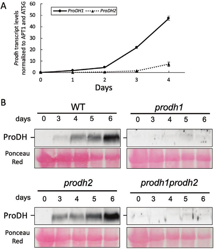
6204 | Launay et al.

Mitochondria are involved in oxidative energy metabolism and          expression in flowers, particularly in pollen and stigma (Funck
release energy to allow the maintenance of primary metab-             et al., 2010). In contrast with ProDH1, low expression is gener-
olism prior to cell death. Mitochondria numbers have been             ally observed for ProDH2 with the exception of vascular tissues
shown to decrease by 30% during developmental leaf senes-             and the abscission zones of floral organs and senescent leaves,
cence, although the integrity of the remaining mitochondria is        where high expression levels have been recorded (Funck et al.,
preserved (Keech et al., 2007; Chrobok et al., 2016). It has also     2010; Faës et al., 2015).
been postulated that mitochondria acquire additional func-               ProDH1 subcellular localization has unambiguously been
tions, such as selective catabolism of lipids and amino acids,        shown to be mitochondrial (Mani et al., 2002; Schertl and
that compensate the loss of energy supplied by chloroplasts           Braun, 2014; Cabassa-Hourton et al., 2016). In response to
and maintain cell viability until the completion of nutrient          proline treatment, ProDH1 has been shown to be part of low
remobilization from the cell (Keech et al., 2007).                    molecular mass complexes in mitochondria and to play an es-

                                                                                                                                                   Downloaded from https://academic.oup.com/jxb/article-abstract/70/21/6203/5555536 by Fachbereichsbibliothek user on 30 July 2020
   Glutamate, which is released from several mitochondrial cata-      sential role in mitochondrial proline oxidation, which may pro-
bolic pathways, is proposed to play a crucial role in nitrogen        vide reducing power, energy, and nitrogen sources for growth
remobilization during leaf senescence (Chrobok et al., 2016). It      (Cabassa-Hourton et al., 2016). The function of ProDH2 was
is one of the main amino acids present in phloem sap in senes-        addressed later because its gene had for a long time been con-
cing Arabidopsis (Masclaux-Daubresse et al., 2010) and in poplar      sidered to be a pseudo gene. The use of ProDH2-GFP fusion
(Couturier et al., 2010). Glutamate may originate from proline        proteins suggests either mitochondrial or plastid localization
catabolism and from conversion through arginase activity of ar-       (Van Aken et al., 2009; Funck et al., 2010). Arabidopsis ProDH2
ginine to ornithine, which in turn enters in the proline catabolic    has been shown to complement a yeast ∆put1 mutant and, when
pathway (Funck et al., 2008). Taken together, although little is      overexpressed in a GFP-tagged form, to rescue an Arabidopsis
known about its role at this developmental stage, these findings      prodh1 mutant using proline as sole source of nitrogen (Funck
suggest that proline catabolism could play a role in recycling pro-   et al., 2010), demonstrating that ProDH2 as well as ProDH1 can
line and arginine during leaf senescence. Proline accumulation        mediate proline oxidation. However, in Arabidopsis supplied
has been described in excised rice leaves during dark-induced         with an excess of proline and other nitrogen sources, ProDH2
senescence (Wang et al., 1982; Mondal et al., 1985), although a       is neither able to overcome the proline sensitivity of the prodh1
decrease in proline content has been reported during develop-         mutant (Funck et al., 2010; Cabassa-Hourton et al., 2016) nor
mental leaf senescence in Arabidopsis (Chrobok et al., 2016).         to restore the lack of ProDH1 in mitochondrial proline oxida-
   Energy deprivation triggers dramatic reprogramming of              tion (Cabassa-Hourton et al., 2016).Therefore the two ProDHs
transcription, which supports metabolic adjustment. Proline           appear to have distinct and non-redundant physiological func-
metabolism and its regulation have been the subject of nu-            tions in proline oxidation, at least in Arabidopsis (Funck et al.,
merous studies in plants (Szabados and Savouré, 2010; Sharma          2010; Rizzi et al., 2017; Cabassa-Hourton et al., 2016).
et al., 2011; Servet et al., 2012). Anabolism and catabolism are         All of these data suggest that remobilization of proline and
each composed of two steps and take place in different cel-           its oxidized derivatives could participate in the senescence
lular compartments. In response to environmental stresses such        process; however, the role of ProDHs remains to be demon-
as drought and salinity, proline is synthesized from glutamate        strated. Therefore, in this study we investigated the contri-
in the cytosol and most probably in chloroplasts via the ac-          bution of ProDHs during dark-induced leaf senescence in
tivities of bifunctional pyrroline-5-carboxylate synthetase           Arabidopsis. Our analyses of the prodh1 prodh2 double-mutant
(P5CS) and P5C reductase (P5CR) (Székely et al., 2008).               in comparison to the wild-type revealed an important func-
Upon recovery from stress, proline is rapidly oxidized to glu-        tion of ProDHs in proline oxidation and its contribution to
tamate in mitochondria via sequential action of proline de-           respiration during senescence. Distinct metabolome patterns
hydrogenase (ProDH) and P5C dehydrogenase (P5CDH). In                 demonstrated a link between the tricarboxylic acid cycle and
Arabidopsis, proline oxidation occurs via two non-redundant           ProDH, demonstrating a key role of proline oxidation for pro-
ProDH isoforms (Funck et al., 2010). ProDH1 and ProDH2                viding energy and glutamate during senescence.
share high sequence homology, with 75% identical amino acids
(Servet et al., 2012). ProDH1 expression has been shown to            Materials and methods
be repressed during water stress but is induced by rehydration
(Kiyosue et al., 1996;Verbruggen et al., 1996). In addition to its    Plant growth and senescence treatment
developmental regulation, ProDH1 expression appears to be             All the Arabidopsis thaliana lines tested were in the Columbia-0 (Col-0)
triggered by hypo-osmolarity and exogenously applied proline          background.The prodh1-4 (SALK_119334) and prodh2-2 (GABI_328G05)
(Verbruggen et al., 1996; Nakashima et al., 1998). Expression of      insertion mutants were obtained from the Salk Institute, La Jolla, CA, USA
                                                                      (Alonso and Stepanova, 2003) and from the Center for Biotechnology,
ProDH1 in response to proline or hypo-osmolarity is controlled        Universität Bielefeld, Germany (Kleinboelting et al., 2012), respectively,
by a proline-responsive element (PRE) cis-acting element via          and genetically characterized (Cabassa-Hourton et al., 2016). A double-
direct binding of the S1-bZIP transcriptional activator to its        mutant prodh1-4 prodh2-2 (hereafter called prodh1 prodh2) was obtained
promoter (Satoh et al., 2002, 2004; Weltmeier et al., 2006).          by crossing prodh1-4 and prodh2-2 as described by Cabassa-Hourton et al.
In addition bZIP1, bZIP2, and bZIP53 have been shown to               (2016). Seedlings were sown and grown in soil under a 16/8-h light/dark
                                                                      cycle at 80–100 µmol photons m−2 s−1 at 21 °C for 4 weeks.
regulate ProDH1 transcript levels and proline content during             Senescence experiments were carried out on excised leaves that were
dark-induced starvation (Dietrich et al., 2011; Pedrotti et al.,      left to age in the dark. Leaves were collected from 4-week-old plants and
2018). Transcriptional analysis of ProDH1 indicates abundant          placed on wet Whatman® paper in Petri dishes. The dishes were then
Involvement of ProDHs in dark-induced leaf senescence | 6205

wrapped in aluminum foil and kept in the growth chamber until further         steps as described by Cabassa-Hourton et al. (2016). The mitochondrial
analysis. For mitochondrial respiration, ProDH activity, transcript and       protein concentration was determined according to Lowry et al. (1951)
metabolomic analyses, leaves 7, 8, and 9 from the base of the plant were      using BSA as a standard.
harvested from 4-week-old plants and were processed as indicated above           Oxygen consumption was measured on 120 µg of crude mitochon-
to trigger dark-induced senescence.                                           dria using a Clark-type oxygen electrode (Hansatech) in 1 ml of air-
                                                                              saturated electrode medium (0.3 M sucrose, 5 mM KH2PO4, 10 mM TES
                                                                              buffer pH 7.4, 10 mM KCl, 2 mM MgCl2, and 0.1 % fatty-acid free BSA
Immunocytochemical studies                                                    from Sigma-Aldrich) at 25 °C. The respiratory chain was first activated
For electron microscopy, 2×5-mm pieces of leaves were cut and placed          with 50 µM of ADP whichever substrate was being used. Respiration
in a fixation solution containing 3% (v/v) formaldehyde and 0.5%              was measured from three entry points independently. A mix of 10 mM
(v/v) glutaraldehyde in 1× PBS (137 mM NaCl, 2.7 mM KCl, 10 mM                malate, 1 mM glutamate, and 5 mM pyruvate was used as substrates for
Na2HPO4, 1.8 mM KH2PO4, pH 7.4). The samples were then subjected              investigating respiration from complex I, 10 mM succinate for respiration
                                                                              from complex II, and 6 mM L-proline for respiration from proline. From

                                                                                                                                                          Downloaded from https://academic.oup.com/jxb/article-abstract/70/21/6203/5555536 by Fachbereichsbibliothek user on 30 July 2020
to a low vacuum for 20 min, and washed twice in PBS and once in
distilled water for 20 min each. A dehydration procedure was then per-        these entry points, electron flux through the cytochrome pathway (CP)
formed with 25% and 50% ethanol successively for 20 min each under            was measured as the rate of oxygen uptake sensitive to cyanide and was
gentle shaking. The samples were then stored in 70% ethanol overnight         then inhibited with 1.5 mM of KCN. We refer to it here as CP capacity.
at 4 °C. Dehydration was achieved by successive incubation in 80%             For complex II and proline, the capacity for electron flux through the
and 90% ethanol for 1 h each. Embedding was started in a mix of 25%           alternative oxidase (AOX) pathway was obtained as the rate of oxygen
London Resin White (LR) in 90% ethanol for 1 h under gentle shaking,          uptake in the presence of 1.5 mM KCN.The AOX pathway was activated
followed by incubation in 50% LR in 90% ethanol overnight, 75% LR             by a mix of 5 mM pyruvate and 1 mM dithiotreitol (DTT) and then in-
in 90% ethanol for 2 h, and finally in 100% LR for 3 h. The embedded          hibited with 0.1 mM n-propyl gallate.We refer to it here as AOX capacity.
samples were then placed in gelatine capsules at 55 °C for 48 h. Ultra-          ProDH activity was measured using the 2,6-dichlorophenolindophenol
thin sections were then cut with a diamond knife to a thickness of 70 nm.     (DCIP-)based assay as described by Cabassa-Hourton et al. (2016).
Sections were collected on 200 mesh Formwar-coated nickel grids.
   For immunological detection of ProDH, the grids were firstly incu-
bated in goat serum 5% (v/v) in T1 buffer [0.05 M Tris-HCl, 2% (w/v)          Measurement of proline and chlorophyll contents
NaCl, 0.1% (w/v) BSA grade V, and 0.05% (v/v) Tween-20, pH 7.4] for           Measurement of the free proline content in 50–70 mg samples of fresh
1 h. Antibodies raised against the ProDH1 protein were diluted 50-fold        rosette leaf tissues was carried out using the procedure described by Ben
in T1 buffer and applied to the grids overnight at 4 °C. The grids were       Rejeb et al. (2015), which was adapted from the method developed by
then washed five times with T1 buffer and three times with T2 buffer          Bates et al. (1973). The absorbance of the proline–ninhydrin complex
[0.02 M Tris-HCl, 2.5% (w/v) NaCl, 0.1% (w/v) BSA grade V, and 0.05%          in toluene was measured at 520 nm using a NP80 Nanophotometer®
Tween-20 (v/v), pH 8] for 5 min each. The grids were incubated with a         (Implen). A standard curve of known L-proline quantities allowed the
10-nm colloidal gold–goat anti-rabbit IgG whole molecule complex in           calculation of the proline content in the samples.
T2 buffer for 2 h at room temperature and were then washed five times            For measurement of chlorophyll contents, 100 mg of frozen leaves
with T2 and five times with ultra-pure water for 5 min each. The grids        were ground to powder in liquid nitrogen and resuspended in 1 ml of
were finally contrasted with 5% uranyl acetate in water for 5 min, washed     92% acetone. Samples were centrifuged for 10 min at 10 000 g. The
three times with ultra-pure water and then with lead citrate tri-hydrate in   supernatants were collected and the volume adjusted to 2 ml with 80%
water for 30 s, and washed 10 times with ultra-pure water. Observations       acetone. The absorbance was read at 663 nm and 645 nm to determine
were made using a Zeiss 912 TEM with Omega filter.                            chlorophyll concentrations using the equations of Arnon (1949).

RNA extraction and droplet digital PCR analysis                               Protein extraction and western blotting
RNAs were extracted from three leaves at the same developmental stage         Leaves (100–150 mg) were ground in a fine powder in liquid nitrogen.
(~100 mg of plant tissue) that were ground in liquid nitrogen using a mixer   The frozen powder was then suspended into 2 volumes of extraction
mill (MM301; Retsch, Haan, Germany) according to Ben Rejeb et al.             buffer containing 8 M urea, 5% (w/v) SDS, 40 mM Tris-HCl pH 6.8,
(2015). RNA quality and concentration were determined using a NP80            0.1 mM EDTA, and 0.4 mg ml–1 bromophenol blue, supplemented with
Nanophotometer® (Implen). A mixture of cDNAs corresponding to four            1× protease inhibitor cocktail (Merck). Samples were shaken three times
independent biological experiments was prepared for each time point and       for 5 min with 5 min intervals on ice.The samples were then centrifuged
used for droplet digital PCR (ddPCR). ddPCR was conducted and data            at 18 000 g for 10 min at 10 °C and the supernatants (30 µl) were loaded
were analysed using a QX200 ddPCR system (Bio-Rad) according to               and separated onto a 8% polyacrylamide gel. The proteins were then
the manufacturer’s protocol. The 20-μl reaction was conducted using 2×        transferred onto nitrocellulose membranes (Amersham Hybond-ECL®
ddPCR supermix, and the cDNA sample was at a concentration of 20 ng           from GE Healthcare) and were immunoblotted using purified antibodies
µl−1. The sample mix and 70 μl of droplet generation oil were added into      directed against either Arabidopsis recombinant ProDH1, ProDH2,
their corresponding wells of the DG8TM cartridge in order to be emul-         P5CDH, or P5CS proteins. Antibody binding was detected using a sec-
sioned into 20 000 oil encapsulated nanodroplets. Data analysis was per-      ondary antibody conjugated with a horseradish peroxidase and the ECL
formed using the Quantasoft™ software (Bio-Rad) to calculate the number       prime system from GE Healthcare using the G:BOX® system (Syngene).
of copies per cDNA. The absolute expression quantitation of the ProDH
genes was normalized using adenine phosphoribosyltransferase 1 (APT1,
At1g27450) and the ubiquinol-Cyt c reductase iron–sulfur subunit (AT5G,       Identification of ProDH2 by MS analysis
At5g13440) as housekeeping genes (Ben Rejeb et al., 2015). All the primers    Preparation of samples and identification of proteins by MS was per-
used for ddPCR are given in Supplementary Table S1 at JXB online.             formed as described by Fromm et al. (2016). Briefly, 50 µg of proteins
                                                                              from crude mitochondria isolated from leaves of prodh1 seedlings were
Crude mitochondria extracts, and measurement of respiration                   loaded onto polyacrylamide gels (stacking gel 10% acrylamide, separating
                                                                              gel 14% acrylamide). Gel runs were stopped when the bromophenol blue
and ProDH activity                                                            dye front reached the end of the stacking gel, and were subsequently
Mitochondria were isolated at 4 °C from 3 g of fresh leaves (num-             stained with Coomassie Brilliant Blue G250. Gel pieces containing the
bers 7, 8, and 9 from the base of the plant) of 4-week-old Arabidopsis        proteins that concentrated at the stacking gel–separation gel interface
Col-0 lines and the double-mutant prodh1 prodh2 after either 0 d or 5         were subjected to in-gel digestion followed by peptide extraction. MS
d of dark-induced leaf senescence. The harvested leaves were ground in        analyses were performed using a Dionex Ultimate 3000 (ThermoFisher
a cold mortar with 30 ml of grinding buffer, and crude mitochondria           Scientific) equipped with a 50-cm reverse-phase analytical column (C18,
were obtained by filtration followed by several differential centrifugation   ID 75 µm, particle diameter 3 µm, 100Å pore size) in line to a 2-cm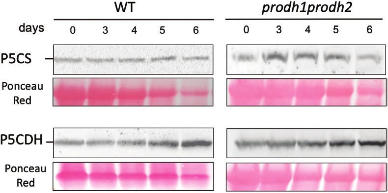
6206 | Launay et al.
reverse-phase pre-column (C18, ID 100 µm, particle diameter 5 µm, 100Å time. As shown in Fig. 1A, chlorophyll contents progressively
pore size). Peptides were eluted using a 1-h non-linear acetonitrile gra- decreased during DIS in all genotypes to less than 0.2 mg
dient and submitted to tandem MS employing a top-10 duty cycle. MS/
MS data were subsequently evaluated using the Mascot (Matrix Science,
chlorophyll g−1 FW at Day 6 (contents could not be deter-
London, UK) search engine querying against an in-house Arabidopsis mined at 7 d due to degradation of the leaf tissue). The kin-
(TAIR10) protein database. etics of chlorophyll degradation were similar to the WT in the
both prodh single- and double- mutants. Chrobok et al. (2016)
Metabolic profiling using GC-MS determined five time periods of senescence from T0, which
Sample preparation, analysis, and data processing were performed as pre-
corresponds to leaf expansion, to T4, which corresponds to the
viously described by Fiehn (2006). Ground, frozen samples (100 mg) mature leaf at the late stage of senescence. Based on this refer-
from harvested leaves (7, 8, and 9 from the base of the plant) exposed to ence, the chlorophyll contents indicated that Day 3 and Day 4
5 d of either dark or light conditions were resuspended in 1 ml of frozen in our study were equivalent to Chrobok’s T2 period, and Day
Downloaded from https://academic.oup.com/jxb/article-abstract/70/21/6203/5555536 by Fachbereichsbibliothek user on 30 July 2020
(−20 °C) water:acetonitrile:isopropanol (2:3:3) containing ribitol at 4 μg 5 and Day 6 were equivalent to the T3 period. We also moni-
ml−1 and extracted by shaking for 10 min at 4 °C. Insoluble material was
removed by centrifugation.Three blank tubes were subjected to the same
tored the proline content, and no significant variations were
steps as the samples. A quality control was made by pooling an equal observed in the WT and the single-mutants up to Day 6 of
volume from each light/dark condition. A sample of 30 μl was collected DIS (Fig. 1B). However, significant increases in proline accu-
from each tube and dried overnight at 35 °C in a vacuum centrifuge. mulation were measured in the prodh1 prodh2 double-mutant
Derivatization was performed according to Clément et al. (2018), compared to the WT at Day 4 and Day 5, suggesting a putative
4 h after the end of which the whole sample series was first injected
in splitless mode and then in split mode (1/30). Four different standard
role of ProDH in the oxidation of proline during senescence.
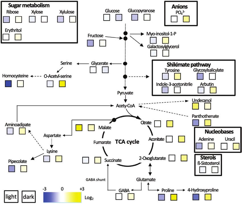
mixes were injected at the beginning and the end of the analysis as well
as three independent derivatizations of the quality control (beginning, Expression of ProDH during DIS
middle, and end) for monitoring the derivatization stability. Samples were
randomized. The instrument was an Agilent 7890A gas chromatograph ProDH expression was investigated in dark-induced senescent
coupled to an Agilent 5977B mass spectrometer. The column was a Rxi- leaves in a time-course experiment. Leaves from 4-week-old
5SilMS from Restek (30 m with 10-m integraguard column). The liner
(Restek # 20994) was changed before analysis. The oven temperature Arabidopsis WT and prodh1 prodh2 plants were collected during
ramp was 70 °C for 7 min then 10 °C min−1 to 330 °C for 5 min (run senescence.Transcript analyses using the highly sensitive droplet
length 38 min). The helium constant flow was 0.7 ml min−1. The fol- digital PCR (ddPCR) method revealed very low transcript
lowing temperatures were used: injector, 250 °C; transfer line, 290 °C; levels of both ProDH1 and ProDH2 in the WT at Day 0 (Fig.
source, 250 °C; and quadripole, 150 °C. Five scans per second were ac- 2A), although the ProDH1 transcript level was 5-fold higher
quired spanning a 50–600 Da range. The instrument was tuned with
perfluorotributylamine (PFTBA) with the 69 m/z and 219 m/z of equal than ProDH2. During the 4 d of the senescence experiment, a
intensities. The split mode conditions were 70 °C for 2 min, then 30 °C consistent increase of ProDH1 transcripts (575-fold induction
min–1 to 330 °C for 5 min. The helium constant flow was 1 ml min−1. between 0–4 d), and to a lesser extent ProDH2, occurred in the
The data were processed as follows. The raw Agilent data files were WT (at Day 5, ProDH transcript levels could not be determined
converted into NetCDF format and analysed with AMDIS (http:// due to high degradation of RNAs). At Day 4, the ProDH1 tran-
chemdata.nist.gov/mass-spc/amdis/). A home retention indices/mass
spectra library built from the NIST, Golm (http://gmd.mpimp-golm. script level was 6.5-fold higher than that of ProDH2. These re-
mpg.de/), and Fiehn databases and standard compounds was used for sults prompted us to monitor ProDH protein accumulation in
metabolite identification. Peak areas were also determined with the a time-course experiment using an antibody directed against
Targetlynx software (Waters) after conversion of the NetCDF files into ProDH1 recombinant protein (Cabassa-Hourton et al., 2016).
masslynx format. AMDIS, Target Lynx in splitless, and split 30 mode data In the WT total protein extracts, no ProDH signal was detected
were compiled in one single Excel file for comparisons. After subtraction
of the blank mean, peak areas were normalized to ribitol and expressed at Day 0 (Fig. 2B). A faint signal appeared at Day 3, increased
on a fresh weight basis. Only metabolites showing repeatable and signifi- at Day 4 and Day 5, and reached a maximum at the end of the
cant differences (based on t-tests) with respect to light and dark condi- senescence period. A similar progression of ProDH accumula-
tions are presented in heat maps. A complete list of all the metabolites that tion was observed in the prodh2 mutant, but no ProDH signal
were detected can be found in Supplementary Data Set S1. could be detected in either the prodh1 or prodh1 prodh2 mu-
tants. These results suggested that at least the ProDH1 isoform
Statistical analysis accumulated during DIS. ProDH2 could not be detected in
Two-way ANOVAs were performed for the qPCR analysis. The effects the total protein extracts but an increase of ProDH2 transcripts
of senescence on chlorophyll and proline contents in wild-type and mu- was observed. As the antibody directed against ProDH1 could
tants, including time effects, as well as across days was assessed by ANOVA.
Tukey tests were performed for comparisons of ProDH activities.
detect recombinant ProDH2 but with a lower sensitivity than
recombinant ProDH1 (Cabassa-Hourton et al., 2016), the lack
of detection of ProDH2 in the total protein extracts could have
Results been due to the fact that ProDH2 is a very low abundant en-
Knockout Arabidopsis prodh mutants accumulate zyme, as postulated by Cabassa-Hourton et al. (2016).
proline during dark-induced senescence ProDH2 was therefore investigated by western blotting using
a new antibody directed against ProDH2 recombinant protein.
Dark-induced senescence (DIS) was triggered by cutting leaves Leaves from 4-week-old plants were sampled after 5 d of DIS
of 4-week-old Arabidopsis Col-0 (wild-type, WT), prodh1, and crude mitochondria were isolated (Cabassa-Hourton et al.,
prodh2, and prodh1 prodh2 plants at the same developmental 2016). Western blots showed different signal patterns with anti-
stage and placing them in the dark for 6 d. The progression of bodies directed against either ProDH1 or ProDH2 (Fig. 3).The
DIS was followed by measuring chlorophyll degradation over anti-ProDH1 antibody showed a signal at an apparent molecular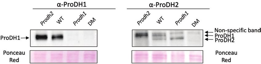
Involvement of ProDHs in dark-induced leaf senescence | 6207

                                                                                                                                                            Downloaded from https://academic.oup.com/jxb/article-abstract/70/21/6203/5555536 by Fachbereichsbibliothek user on 30 July 2020
Fig. 1. Chlorophyll contents and proline accumulation during dark-induced leaf senescence in Arabidopsis. (A) Total chlorophyll content was measured
in a time-course experiment over 6 d of dark-induced senescence in Col-0 (wild-type, WT), prodh1, prodh2, and the prodh1 prodh2 double-mutant.
Data are means (±SE) of four independent rosettes from two independent experiments. The time periods T0, T2, and T3 are delineated by the progression
of senescence in leaves according to Chrobok et al. (2016). Variations within each time-point were not statistically different (ANOVA, P>0.05). Different
letters indicate significant differences in overall chlorophyll content between time-points (ANOVA, P
6208 | Launay et al.
present at Day 0. The level of P5CS protein was not really af- could be measured in the prodh1 prodh2 double-mutant what-
fected during the time-course experiment in the WT and the ever the growth conditions, as has previously been reported
prodh1 prodh2 double-mutant. On the other hand, the P5CDH by Cabassa-Hourton et al. (2016). Taken together, these results
content slightly increased during DIS. These results showed that indicated that ProDH was functional during DIS.
DIS enhanced proline catabolism but not proline biosynthesis. Oxygen consumption by crude mitochondria extracts was
investigated in the WT and the prodh1 prodh2 double-mutant.
DIS triggers ProDH activity and proline respiration Complex I capacity through the cytochrome pathway increased
by 2.5-fold after 5 d of DIS in both the WT and the double-
The enzymatic activity of ProDH was investigated in crude mutant (Fig. 8A). Complex II capacity increased in the same
mitochondria extracts during DIS. After 5 d of DIS, ProDH range for the WT but to a lower extent for the double-mutant
activity increased by 5-fold in the WT (Fig. 7), but no activity (Fig. 8B). The potential contribution of the AOX pathway was
Downloaded from https://academic.oup.com/jxb/article-abstract/70/21/6203/5555536 by Fachbereichsbibliothek user on 30 July 2020
determined by completely inhibiting the CP with KCN and
by activating the AOX pathway with DTT in the presence of
excess of pyruvate. The capacity for electron flux through the
AOX pathway increased by 2-fold in the WT during DIS when
succinate was used as the substrate (Fig. 8C). In the WT, proline
respiration increased through both the CP and AOX pathway
by 14- and 3-fold, respectively, after 5 d of DIS (Fig. 8D).
Interestingly, the potential contribution of the AOX pathway
upon proline respiration was higher than the CP at Day 0 while
the opposite was observed after 5 d of DIS, when 65% of the
electron flux passed through the CP. No proline respiration was
measured in the double-mutant (data no shown), as was previ-
ously observed by Cabassa-Hourton et al. (2016).
Taken together, these results indicated that there was a
higher proline oxidation rate during DIS, making it a potential
respiratory substrate, together with a shift of the major electron
pathway for proline respiration from AOX to cytochrome.
Metabolic changes in detached leaves subjected
to DIS
To gain more insights into ProDH function during senescence,
a metabolic profiling analysis was carried out in detached leaves
of the WT and prodh1 prodh2 double-mutant exposed for 5 d
to either light or dark conditions. A GC-MS untargeted ap-
proach identified 63 metabolites, of which 32 were unknown,
Fig. 2. ProDH expression during dark-induced leaf senescence in that were differentially accumulated in the double-mutant
Arabidopsis. (A) ProDH1 and ProDH2 expression in the Col-0 wild-type compared with the WT (Fig. 9). Clearly opposite behavior was
(WT) was quantified by droplet digital PCR after 0–4 d of dark-induced generally observed mainly for differentially accumulated me-
senescence. Data are means (±SE) of four biological replicates. (B) tabolites of the primary metabolism in the two genotypes ex-
Western blots of total proteins extracted from 100 mg fresh leaves from
posed to light or dark conditions. Under light conditions, the
Col-0 (WT), prodh1, prodh2, and the prodh1 prodh2 double-mutant in a
time-course experiment over 6 d of dark-induced senescence. Proteins identified metabolites were generally accumulated to a higher
were separated by SDS/PAGE and blots were probed with an anti-ProDH1 level in the WT than in the double-mutant, which was in con-
antibody. (This figure is available in colour at JXB online.) trast to DIS where the opposite response was observed. In the
Fig. 3. ProDH expression in Arabidopsis leaf mitochondria after 5 d of dark-induced senescence. Antibodies raised against recombinant ProDH1 and
ProDH2 isoforms were used for western blots of proteins from crude mitochondria extracted from Col-0 (wild-type, WT), prodh1, prodh2 and the prodh1
prodh2 double-mutant (DM). The blots were loaded with 5 µg (ProDH1) and 10 µg (ProDH2) of crude mitochondrial extracts. (This figure is available in
colour at JXB online.)Involvement of ProDHs in dark-induced leaf senescence | 6209
Downloaded from https://academic.oup.com/jxb/article-abstract/70/21/6203/5555536 by Fachbereichsbibliothek user on 30 July 2020
Fig. 4. Identification of the ProDH2 isoform in Arabidopsis mitochondria by mass spectrometry using crude mitochondrial extracts from leaves of prodh1
plants after 5 d of dark-induced senescence. (A) Summary of MS results. (B) The ProDH2 protein sequence with the identified peptides marked in red.
Fig. 5. Immunolocalization using antibodies directed against ProDH during dark-induced senescence in leaves of Arabidopsis. The electron micrographs
show sections of leaves after 5 d of dark-induced senescence for (A) Col-0 (wild-type, WT), (B) prodh1, (C) prodh2, and (D) the prodh1 prodh2 double-
mutant, and show typical labelling within the mitochondria. Arrows indicate the presence of ProDH. No signal was detected in prodh1 prodh2. m,
mitochondrion; cp, chloroplast.
TCA cycle, levels of citrate, aconitate, 2-oxoglutarate, succinate, WT in dark conditions. In contrast, the glutamate level was not
and malate were significantly higher in prodh1 prodh2 than in significantly changed between light and dark conditions in the
the WT under DIS. This was also the case with some amino WT and the double-mutant.
acids such as proline, hydroxyproline, lysine, acetyl-serine, tyro-
sine, GABA, aminoadipate, and homocysteine, with nucleobases Discussion
such as adenine and uracil, and with sugars such as ribose, xy-
lose, xylulose, erythritol, and fructose. Interestingly, proline and The remobilization of metabolites during stress and senescence
its derivatives such as hydroxyproline and GABA followed the plays an important role in plant adaptation to the environment.
same trend. Only glycerate and glucose were present at lower Here, we hypothesized a requirement for ProDHs for alterna-
levels in the prodh1 prodh2 double-mutant compared to the tive respiration and for the remobilization of proline during6210 | Launay et al.
c
25
nmol reduced DCPIP
min-1 mg protein-1
20
15
10
b
5
0 a a
WT DM WT DM
Days 0 5
Downloaded from https://academic.oup.com/jxb/article-abstract/70/21/6203/5555536 by Fachbereichsbibliothek user on 30 July 2020
Fig. 6. Analysis of the proline metabolism enzymes P5CS and P5CDH in Fig. 7. ProDH activity in Arabidopsis leaves in response to dark-induced
Arabidopsis leaves over 5 d of dark-induced senescence. Western blots senescence. Activity was measured in crude mitochondria extracts from
of total proteins extracts taken from Col-0 (wild-type, WT) and the prodh1 leaves 7, 8, and 9 from the base of the plant that were sampled at 0 d and
prodh2 double-mutant. The blots were obtained using antibodies raised 5 d from Col-0 (wild-type, WT) and the prodh1 prodh2 double-mutant.
against P5CS and P5CDH recombinant enzymes. (This figure is available Data are means (±SE) of at least for independent biological experiments.
in colour at JXB online.) Different letters indicate significant differences between means as
determined by ANOVA followed by Tukey's test (PInvolvement of ProDHs in dark-induced leaf senescence | 6211
Downloaded from https://academic.oup.com/jxb/article-abstract/70/21/6203/5555536 by Fachbereichsbibliothek user on 30 July 2020
Fig. 8. Oxygen consumption in mitochondria isolated from Arabidopsis leaves from Col-0 (wild-type, WT) and the prodh1 prodh2 double-mutant (DM) in
response to dark-induced senescence. Oxygen consumption was determined using a Clark-type electrode in crude mitochondria extracts from leaves 7,
8, and 9 from the base of the plant sampled at 0 d and 5 d of dark-induced senescence. The capacity of the cytochrome pathway (CP) was measured
from (A) complex I, (B) complex II, and (D) proline. The capacity of the alternative oxidase pathway (AOX) was measured in WT mitochondria from (C)
complex II and (D) proline. Data are means (±SE) of at least four biological replicates. Different letters indicate significant differences between means as
determined using ANOVA (P6212 | Launay et al.
Thibodeau, 1995). Interestingly, our work showed for the first amino acid, also accumulated more in the double-mutant
time that senescence stimulated a striking increase in proline than in the WT during DIS (Fig. 9). Interestingly, Law et al.
respiration (Fig. 8D). Cabassa-Hourton et al. (2016) demon- (2018) have shown that genes encoding proteins involved in
strated that treatment of Arabidopsis seedlings with proline primary energy production (respiration, fermentation, and
triggers ProDH1 accumulation and activity. Concomitantly, ß-oxidation), amino acids, lipid or nucleotide catabolism, sulfur
proline respiration increases but to a significantly lesser ex- metabolism, and the shikimate pathway are overexpressed
tent than during senescence: it was two-fold higher in DIS during senescence of individually darkened leaves. ProDH
than in proline-treated seedlings. To our knowledge, our data and proline may thus be crucial actors of this metabolic switch
represents one of the highest rates of proline respiration that under senescence.
has been measured. Electron fluxes generated from proline res- Proline generated by DIS can be metabolized by ProDH
piration in proline-treated seedlings (Cabassa-Hourton et al., enzymes to produce P5C. This can also be synthesized by or-
Downloaded from https://academic.oup.com/jxb/article-abstract/70/21/6203/5555536 by Fachbereichsbibliothek user on 30 July 2020
2016), as well as in leaves before senescence (Day 0, this study), nithine aminotransferase, the mRNA levels of which are also
are mainly directed through the AOX pathway and do not ef- up-regulated during senescence (Buchanan-Wollaston et al.,
ficiently contribute to ATP synthesis. However, upon DIS in 2005; van der Graaff et al., 2006; Chrobok et al., 2016). We
our study, the electron flux from proline oxidation was prefer- also found that the P5CDH protein, which allows the con-
entially directed through the CP, allowing a better coupling to version of P5C to glutamate, was up regulated in DIS (Fig.
ATP synthesis. Taken together, these observations suggest the 6). Previous results for DLS have shown either no response or
importance of proline as an alternative respiratory substrate up-regulation (van der Graaff et al., 2006). No induction of
during senescence. Proline oxidation may contribute to the P5CS was observed under our conditions, which is consistent
generation of ATP that is necessary for the maintenance of with previous results reported for leaves subjected to DIS (van
leaf viability while the remobilization of nutrients is carried der Graaff et al., 2006). Thus, DIS triggers proline catabolism
out. It has previously been reported that the total oxidation of rather than proline biosynthesis and allows the generation of
one proline molecule can generate reductants that can fuel the glutamate, which is the metabolic precursor of glutamine and
mitochondrial electron transfer chain and support the forma- asparagine that are involved in nitrogen remobilization during
tion of 30 ATP molecules (Atkinson, 1977). DIS leads to inten- senescence (Finnemann and Schjoerring, 2000; Lin and Wu,
sive metabolite recycling through protein degradation and the 2004; Masclaux-Daubresse et al., 2010). Proline content has
generation of amino acids that are used as alternative substrates been shown to decrease during developmental senescence in
for respiration. More generally, energy-limited conditions re- wild-type Arabidopsis and in tobacco (Masclaux et al., 2000;
quire alternative pathways of respiration to provide the ne- Chrobok et al., 2016), which indicates an important role of
cessary respiratory function (Barros et al., 2017). Interestingly, ProDH in proline oxidation to release glutamate. Moreover,
proline has also been shown to be the main energy substrate to it has been shown that proline regulates the expression of
fuel flight in some insects (Gäde and Auerswald, 2002). Glutamine Synthase 1 (GS1) and Glutamate Dehydrogenase
In addition to its role in respiratory electron flux, ProDH (GDH), which encode two enzymes involved in nitrogen
is more widely involved in mitochondrial metabolism. Our remobilization during senescence (Masclaux-Daubresse et al.
untargeted metabolomic approach based on GC-MS provided (2005), thus supporting an important role of this amino acid as
a more comprehensive view of the role of ProDH in primary a signal molecule during senescence.
metabolic pathways during senescence (Fig. 9). Interestingly, Proline could originate from the degradation of cellular
this analysis identified increased levels of citrate, aconitate, and cell wall proteins, which are notably rich in proline and
2-oxoglutarate, succinate, malate, proline, and hydroxyproline hydroxy (Ihsan et al., 2017), as has also been described for
in the prodh1 prodh2 double-mutant in detached leaves sub- the mammalian extracellular matrix (Pandhare et al., 2009).
jected to DIS when compared to the WT. These metabolites Degradation of cell wall proteins could therefore lead to an
are linked to the TCA cycle and associated pathways. The in- increase of free proline concentration in the cell that would
crease of TCA intermediates in the double-mutant may indi- act as an activator of ProDH transcription and as a substrate for
cate a lower availability of alternative substrates such as proline, both ProDH isoforms, contributing to nitrogen remobilization
which could no longer be oxidized to supply the carbon back- through glutamine generation from glutamate and its subse-
bone in the form of glutamate or 2-oxoglutarate to sustain the quent transport to sinks. In conclusion, our results indicate
TCA cycle. An increase in TCA intermediates has previously a dual role of ProDH under DIS. Proline oxidation allows it
been observed in plants lacking D-2-hydroxyglutarate de- to be used as an alternative respiratory substrate and contrib-
hydrogenase or isovaleryl-CoA dehydrogenase, demonstrating utes to the production of glutamate and energy that can sub-
the participation of lysine and branched amino acids as alterna- sequently participate in the remobilization of nutrients from
tive substrates for respiration during senescence (Araújo et al., senescent tissues to developing plant organs.
2010; Engqvist et al., 2011). More generally, a reduced flow
of carbon into the TCA cycle seems to lead to an increase of Supplementary data
intermediates, probably due to a reduced overall activity of the
TCA cycle (Araujo et al., 2010; Yu et al., 2012; Huang et al., Supplementary data are available at JXB online.
2013; Pires et al., 2016). Table S1. List of primers used for ddPCR.
Metabolites from sugar metabolism and the shikimate Data Set S1. Raw data and statistical analysis of metabolite
pathway together with homocysteine, a sulfur-containing profiles in the leaf experiments.Involvement of ProDHs in dark-induced leaf senescence | 6213
Acknowledgements Finnemann J, Schjoerring JK. 2000. Post-translational regulation of
cytosolic glutamine synthetase by reversible phosphorylation and 14-3-3
We are indebted to Dr Dietmar Funck for kindly providing an anti- protein interaction. The Plant Journal 24, 171–181.
body directed against P5CDH. We thank the electron microscopy team Fromm S, Senkler J, Eubel H, Peterhänsel C, Braun HP. 2016. Life
of the IBPS Imaging Facility, Sorbonne University for their help with without complex I: proteome analyses of an Arabidopsis mutant lacking
the mitochondrial NADH dehydrogenase complex. Journal of Experimental
the experiments. This study received financial support from Sorbonne
Botany 67, 3079–3093.
University.
Funck D, Eckard S, Müller G. 2010. Non-redundant functions of two pro-
line dehydrogenase isoforms in Arabidopsis. BMC Plant Biology 10, 70.
Funck D, Stadelhofer B, Koch W. 2008. Ornithine-delta-aminotransferase
References is essential for arginine catabolism but not for proline biosynthesis. BMC
Allu AD, Soja AM, Wu A, Szymanski J, Balazadeh S. 2014. Salt stress Plant Biology 8, 40.
and senescence: identification of cross-talk regulatory components. Journal Gäde G, Auerswald L. 2002. Beetles’ choice—proline for energy output:
Downloaded from https://academic.oup.com/jxb/article-abstract/70/21/6203/5555536 by Fachbereichsbibliothek user on 30 July 2020
of Experimental Botany 65, 3993–4008. control by AKHs. Comparative Biochemistry and Physiology Part B:
Alonso JM, Stepanova AN. 2003. T-DNA mutagenesis in Arabidopsis. Biochemistry & Molecular Biology 132, 117–129.
Methods in Molecular Biology 236, 177–188. Huang S, Taylor NL, Ströher E, Fenske R, Millar AH. 2013. Succinate
Araújo WL, Ishizaki K, Nunes-Nesi A, et al. 2010. Identification of the dehydrogenase assembly factor 2 is needed for assembly and activity of
2-hydroxyglutarate and isovaleryl-CoA dehydrogenases as alternative elec- mitochondrial complex II and for normal root elongation in Arabidopsis. The
tron donors linking lysine catabolism to the electron transport chain of Plant Journal 73, 429–441.
Arabidopsis mitochondria. The Plant Cell 22, 1549–1563. Ihsan MZ, Ahmad SJ, Shah ZH, Rehman HM, Aslam Z, Ahuja I,
Arnon DI. 1949. Copper enzymes in isolated chloroplasts. Bones AM, Ahmad JN. 2017. Gene mining for proline based signaling pro-
polyphenoloxidase in Beta vulgaris. Plant Physiology 24, 1–15. teins in cell wall of Arabidopsis thaliana. Frontiers in Plant Science 8, 233.
Atkinson DE. 1977. Cellular energy metabolism and its regulation. New Keech O, Pesquet E, Ahad A, Askne A, Nordvall D, Vodnala SM,
York: Academic Press. Tuominen H, Hurry V, Dizengremel P, Gardeström P. 2007. The different
fates of mitochondria and chloroplasts during dark-induced senescence in
Barros JAS, Cavalcanti JHF, Medeiros DB, Nunes-Nesi A, Avin- Arabidopsis leaves. Plant, Cell & Environment 30, 1523–1534.
Wittenberg T, Fernie AR, Araújo WL. 2017. Autophagy deficiency com-
promises alternative pathways of respiration following energy deprivation in Kiyosue T, Yoshiba Y, Yamaguchi-Shinozaki K, Shinozaki K. 1996. A
Arabidopsis thaliana. Plant Physiology 175, 62–76. nuclear gene encoding mitochondrial proline dehydrogenase, an enzyme
involved in proline metabolism, is upregulated by proline but downregulated
Bates LS, Waldren RP, Teare ID. 1973. Rapid determination of free pro- by dehydration in Arabidopsis. The Plant Cell 8, 1323–1335.
line for water-stress studies. Plant and Soil 39, 205–207.
Kleinboelting N, Huep G, Kloetgen A, Viehoever P, Weisshaar B.
Ben Rejeb K, Lefebvre-De Vos D, Le Disquet I, Leprince AS, 2012. GABI-Kat SimpleSearch: new features of the Arabidopsis thaliana
Bordenave M, Maldiney R, Jdey A, Abdelly C, Savouré A. 2015. T-DNA mutant database. Nucleic Acids Research 40, D1211–D1215.
Hydrogen peroxide produced by NADPH oxidases increases proline accu-
mulation during salt or mannitol stress in Arabidopsis thaliana. The New Klodmann J, Senkler M, Rode C, Braun HP. 2011. Defining the protein
Phytologist 208, 1138–1148. complex proteome of plant mitochondria. Plant Physiology 157, 587–598.
Buchanan-Wollaston V, Page T, Harrison E, et al. 2005. Comparative Law SR, Chrobok D, Juvany M, et al. 2018. Darkened leaves use dif-
transcriptome analysis reveals significant differences in gene expression and ferent metabolic strategies for senescence and survival. Plant Physiology
signalling pathways between developmental and dark/starvation-induced 177, 132–150.
senescence in Arabidopsis. The Plant Journal 42, 567–585. Lim PO, Kim HJ, Nam HG. 2007. Leaf senescence. Annual Review of
Cabassa-Hourton C, Schertl P, Bordenave-Jacquemin M, et al. 2016. Plant Biology 58, 115–136.
Proteomic and functional analysis of proline dehydrogenase 1 link proline Lin J-F, Wu S-H. 2004. Molecular events in senescing Arabidopsis leaves.
catabolism to mitochondrial electron transport in Arabidopsis thaliana. The The Plant Journal 39, 612–628.
Biochemical Journal 473, 2623–2634. Lowry OH, Rosebrough NJ, Farr AL, Randall RJ. 1951. Protein meas-
Chrobok D, Law SR, Brouwer B, et al. 2016. Dissecting the meta- urement with the Folin phenol reagent. The Journal of Biological Chemistry
bolic role of mitochondria during developmental leaf senescence. Plant 193, 265–275.
Physiology 172, 2132–2153. Mani S, Van De Cotte B, Van Montagu M, Verbruggen N. 2002. Altered
Clément G, Moison M, Soulay F, Reisdorf-Cren M, Masclaux- levels of proline dehydrogenase cause hypersensitivity to proline and its
Daubresse C. 2018. Metabolomics of laminae and midvein during leaf analogs in Arabidopsis. Plant Physiology 128, 73–83.
senescence and source-sink metabolite management in Brassica napus Masclaux C, Valadier MH, Brugière N, Morot-Gaudry JF, Hirel B.
L. leaves. Journal of Experimental Botany 69, 891–903. 2000. Characterization of the sink/source transition in tobacco (Nicotiana
Collier DE, Thibodeau BA. 1995. Changes in respiration and chemical tabacum L.) shoots in relation to nitrogen management and leaf senes-
content during autumnal senescence of Populus tremuloides and Quercus cence. Planta 211, 510–518.
rubra leaves. Tree Physiology 15, 759–764. Masclaux-Daubresse C, Carrayol E, Valadier MH. 2005. The two ni-
Couturier J, Doidy J, Guinet F, Wipf D, Blaudez D, Chalot M. 2010. trogen mobilisation- and senescence-associated GS1 and GDH genes are
Glutamine, arginine and the amino acid transporter Pt-CAT11 play important controlled by C and N metabolites. Planta 221, 580–588.
roles during senescence in poplar. Annals of Botany 105, 1159–1169. Masclaux-Daubresse C, Daniel-Vedele F, Dechorgnat J, Chardon F,
Dietrich K, Weltmeier F, Ehlert A, Weiste C, Stahl M, Harter K, Dröge- Gaufichon L, Suzuki A. 2010. Nitrogen uptake, assimilation and
Laser W. 2011. Heterodimers of the Arabidopsis transcription factors remobilization in plants: challenges for sustainable and productive agricul-
bZIP1 and bZIP53 reprogram amino acid metabolism during low energy ture. Annals of Botany 105, 1141–1157.
stress. The Plant Cell 23, 381–395. Mondal WA, Dey BB, Choudhuri MA. 1985. Proline accumulation as a
Engqvist MK, Kuhn A, Wienstroer J, Weber K, Jansen EE, Jakobs C, reliable indicator of monocarpic senescence in rice cultivars. Experientia 41,
Weber AP, Maurino VG. 2011. Plant D-2-hydroxyglutarate dehydrogenase 346–348.
participates in the catabolism of lysine especially during senescence. The Nakashima K, Satoh R, Kiyosue T, Yamaguchi-Shinozaki K,
Journal of Biological Chemistry 286, 11382–11390. Shinozaki K. 1998. A gene encoding proline dehydrogenase is not only
Faës P, Deleu C, Aïnouche A, et al. 2015. Molecular evolution and tran- induced by proline and hypoosmolarity, but is also developmentally regu-
scriptional regulation of the oilseed rape proline dehydrogenase genes sug- lated in the reproductive organs of Arabidopsis. Plant Physiology 118,
gest distinct roles of proline catabolism during development. Planta 241, 1233–1241.
403–419. Narsai R, Howell KA, Millar AH, O’Toole N, Small I, Whelan J. 2007.
Fiehn O. 2006. Metabolite profiling in Arabidopsis. Methods in Molecular Genome-wide analysis of mRNA decay rates and their determinants in
Biology 323, 439–447. Arabidopsis thaliana. The Plant Cell 19, 3418–3436.6214 | Launay et al.
Pandhare J, Donald SP, Cooper SK, Phang JM. 2009. Regulation Sharma S, Villamor JG, Verslues PE. 2011. Essential role of tissue-
and function of proline oxidase under nutrient stress. Journal of Cellular specific proline synthesis and catabolism in growth and redox balance at
Biochemistry 107, 759–768. low water potential. Plant Physiology 157, 292–304.
Parre E, Ghars MA, Leprince AS, Thiery L, Lefebvre D, Bordenave M, Szabados L, Savouré A. 2010. Proline: a multifunctional amino acid.
Richard L, Mazars C, Abdelly C, Savouré A. 2007. Calcium signaling Trends in Plant Science 15, 89–97.
via phospholipase C is essential for proline accumulation upon ionic but Székely G, Abraham E, Cseplo A, et al. 2008. Duplicated P5CS genes of
not nonionic hyperosmotic stresses in Arabidopsis. Plant Physiology 144, Arabidopsis play distinct roles in stress regulation and developmental con-
503–512. trol of proline biosynthesis. The Plant Journal 53, 11–28.
Pedrotti L, Weiste C, Nägele T, et al. 2018. Snf1-RELATED KINASE1- Van Aken O, Zhang B, Carrie C, Uggalla V, Paynter E, Giraud E,
controlled C/S1-bZIP signaling activates alternative mitochondrial metabolic Whelan J. 2009. Defining the mitochondrial stress response in Arabidopsis
pathways to ensure plant survival in extended darkness. The Plant Cell 30, thaliana. Molecular Plant 2, 1310–1324.
495–509.
van der Graaff E, Schwacke R, Schneider A, Desimone M, Flügge UI,
Pires MV, Pereira Júnior AA, Medeiros DB, et al. 2016. The influence Kunze R. 2006. Transcription analysis of Arabidopsis membrane trans-
Downloaded from https://academic.oup.com/jxb/article-abstract/70/21/6203/5555536 by Fachbereichsbibliothek user on 30 July 2020
of alternative pathways of respiration that utilize branched-chain amino porters and hormone pathways during developmental and induced leaf
acids following water shortage in Arabidopsis. Plant, Cell & Environment senescence. Plant Physiology 141, 776–792.
39, 1304–1319.
Verbruggen N, Hua XJ, May M, Van Montagu M. 1996. Environmental
Rao RS, Salvato F, Thal B, Eubel H, Thelen JJ, Møller IM. 2017. The and developmental signals modulate proline homeostasis: evidence for a
proteome of higher plant mitochondria. Mitochondrion 33, 22–37. negative transcriptional regulator. Proceedings of the National Academy of
Rizzi YS, Cecchini NM, Fabro G, Alvarez ME. 2017. Differential con- Sciences, USA 93, 8787–8791.
trol and function of Arabidopsis ProDH1 and ProDH2 genes on infection Wang CY, Cheng SH, Kao CH. 1982. Senescence of rice leaves. VII.
with biotrophic and necrotrophic pathogens. Molecular Plant Pathology 18, Proline accumulation in senescing excised leaves. Plant Physiology 69,
1164–1174. 1348–1349.
Satoh R, Fujita Y, Nakashima K, Shinozaki K, Yamaguchi-Shinozaki K. Weltmeier F, Ehlert A, Mayer CS, Dietrich K, Wang X, Schütze K,
2004. A novel subgroup of bZIP proteins functions as transcriptional ac- Alonso R, Harter K, Vicente-Carbajosa J, Dröge-Laser W. 2006.
tivators in hypoosmolarity-responsive expression of the ProDH gene in Combinatorial control of Arabidopsis proline dehydrogenase transcription
Arabidopsis. Plant & Cell Physiology 45, 309–317. by specific heterodimerisation of bZIP transcription factors. The EMBO
Satoh R, Nakashima K, Seki M, Shinozaki K, Yamaguchi-Shinozaki K. Journal 25, 3133–3143.
2002. ACTCAT, a novel cis-acting element for proline- and hypoosmolarity- Yu H, Du X, Zhang F, Zhang F, Hu Y, Liu S, Jiang X, Wang G, Liu D.
responsive expression of the ProDH gene encoding proline dehydrogenase 2012. A mutation in the E2 subunit of the mitochondrial pyruvate dehydro-
in Arabidopsis. Plant Physiology 130, 709–719. genase complex in Arabidopsis reduces plant organ size and enhances the
Schertl P, Braun HP. 2014. Respiratory electron transfer pathways in plant accumulation of amino acids and intermediate products of the TCA cycle.
mitochondria. Frontiers in Plant Science 5, 163. Planta 236, 387–399.
Servet C, Ghelis T, Richard L, Zilberstein A, Savouré A. 2012. Proline Yu J, Zhang Y, Di C, et al. 2016. JAZ7 negatively regulates dark-induced
dehydrogenase: a key enzyme in controlling cellular homeostasis. Frontiers leaf senescence in Arabidopsis. Journal of Experimental Botany 67,
in Bioscience 17, 607–620. 751–762.You can also read