Structure of the N-WASP EVH1 Domain-WIP Complex: Insight into the Molecular Basis of Wiskott-Aldrich Syndrome
←
→
Page content transcription
If your browser does not render page correctly, please read the page content below
Cell, Vol. 111, 565–576, November 15, 2002, Copyright 2002 by Cell Press
Structure of the N-WASP EVH1 Domain-WIP
Complex: Insight into the Molecular Basis
of Wiskott-Aldrich Syndrome
Brian F. Volkman,1,4 Kenneth E. Prehoda,2,4,5 verrier et al., 2001). Similarly, a knockout mutation of
Jessica A. Scott,2,6 Francis C. Peterson,1 the more ubiquitously expressed N-WASP gene results
and Wendell A. Lim2,3 in embryonic lethality and defects in many but not all
1
Department of Biochemistry actin-based motility processes (Snapper et al., 2001).
Medical College of Wisconsin These proteins have also been hijacked by pathogens
Milwaukee, Wisconsin 53226 including shigella and vaccinia virus as required compo-
2
Department of Cellular and Molecular nents for their own actin-based intracellular motility (Su-
Pharmacology zuki et al., 1998; Moreau et al., 2000).
University of California, San Francisco Recent studies have begun to uncover the molecular
San Francisco, California 94143 mechanism by which WASP/N-WASP regulates actin
polymerization. WASP and N-WASP have a highly mod-
ular domain structure (Figure 1A). A ⵑ100 residue
Summary C-terminal “output” domain containing a verprolin-cofi-
lin-acidic motif (VCA) directly binds to and activates the
actin-related protein (Arp)2/3 actin nucleating complex
Missense mutants that cause the immune disorder
(Machesky and Insall, 1998; Rohatgi et al., 1999),
Wiskott-Aldrich Syndrome (WAS) map primarily to the
whereas the remaining regions of WASP/N-WASP,
Enabled/VASP homology 1 (EVH1) domain of the actin
which encompass multiple domains, are involved in tar-
regulatory protein WASP. This domain has been impli-
geting and regulating the activity of the output domain
cated in both peptide and phospholipid binding. We
in response to upstream signaling inputs. An important
show here that the N-WASP EVH1 domain does not
goal is to understand the function of the regulatory do-
bind phosphatidyl inositol-(4,5)-bisphosphate, as pre-
mains found in WASP/N-WASP in order to understand
viously reported, but does specifically bind a 25 resi-
what signals regulate WASP and the mechanism of this
due motif from the WASP Interacting Protein (WIP).
regulation. Several of the domains, including the basic
The NMR structure of the complex reveals a novel
motif (B), the GTPase binding domain (GBD), and the
recognition mechanism—the WIP ligand, which is far
proline-rich region, have been found to participate di-
longer than canonical EVH1 ligands, wraps around the
rectly or indirectly in autoinhibitory interactions that re-
domain, contacting a narrow but extended surface.
press or block the activity of the VCA output domain
This recognition mechanism provides a basis for un-
(Miki et al., 1998; Kim et al., 2000; Prehoda et al., 2000).
derstanding the effects of mutations that cause WAS.
These autoinhibitory interactions are specifically re-
lieved by interaction with upstream activators including
Introduction the phospholipid phosphatidyl inositol-(4,5)-bisphos-
phate (PIP2), the GTPase Cdc42, and Src homology 3
Spatial and temporal regulation of the actin cytoskeleton (SH3) domain-containing proteins such as Nck (Higgs
is essential for controlling cell shape and movement. and Pollard, 2000; Prehoda et al., 2000; Rohatgi et al.,
The Wiskott-Aldrich Syndrome Protein (WASP) and its 2000, 2001; Carlier et al., 2000).
homolog N-WASP are signal transduction proteins that One of the most important domains in WASP is the
have emerged as central players that promote actin Enabled/VASP Homology 1 domain (also known as
polymerization in response to upstream intracellular sig- WASP Homology 1 or WH1 domain) located at the N
nals (Carlier et al., 1999; Snapper and Rosen, 1999; terminus of the protein (Gertler et al., 1996; Symons et
Higgs and Pollard, 2001; Millard and Machesky, 2001; al., 1996; Callebaut et al., 1998; Renfranz and Beckerle,
Pollard et al., 2000). Mutation of WASP, which is primar- 2002). The functional importance of the WASP EVH1
ily expressed in hematopoetic cells, results in Wiskott- domain is underscored by the fact that 28 of the 35
Aldrich Syndrome (WAS), an X-linked recessive disorder identified missense mutations that lead to the disease
characterized by immunodeficiency, eczema, and WAS are concentrated within this domain (Derry et al.,
thrombocytopenia (Derry et al., 1994; Kolluri et al., 1995; 1995; El-Hakeh et al., 2002; Greer et al., 1996; Kolluri et
Villa et al., 1995; Greer et al., 1996; Zhu et al., 1997). al., 1995; Zhu et al., 1997). In addition, this domain is
These symptoms are consistent with cytoskeletal de- required for N-WASP-dependent intracellular motility of
fects in hematopoetic cells (Zicha et al., 1998) and a vaccinia virus (Moreau et al., 2000).
possible role of WASP/N-WASP in motility, thrombogen- EVH1 domains are found in several other proteins,
esis, endocytosis, phagocytosis, and other actin-based including the cytoskeletal regulatory proteins Enabled
processes (Snapper et al., 1998; Snapper and Rosen, and VASP and the neuronal signaling protein Homer
1999; Cannon et al., 2001; Coppolino et al., 2001; Le- (Callebaut et al., 1998; Gertler et al., 1996). In these
cases, the EVH1 domain appears to serve as a modular
3
protein-peptide docking unit that can participate in pro-
Correspondence: wlim@itsa.ucsf.edu
4
tein targeting. These previously characterized EVH1 do-
These authors contributed equally to this work.
5
Present address: Institute of Molecular Biology, University of Ore-
mains all utilize a conserved aromatic binding surface
gon, Eugene, Oregon 97403. to recognize specific, 6–10 residue proline-rich peptide
6
Present address: University of Wisconsin Medical School, Madison, motifs (Prehoda et al., 1999; Fedorov et al., 1999; Carl
Wisconsin 53706. et al., 1999; Ball et al., 2000; Beneken et al., 2000; Barzik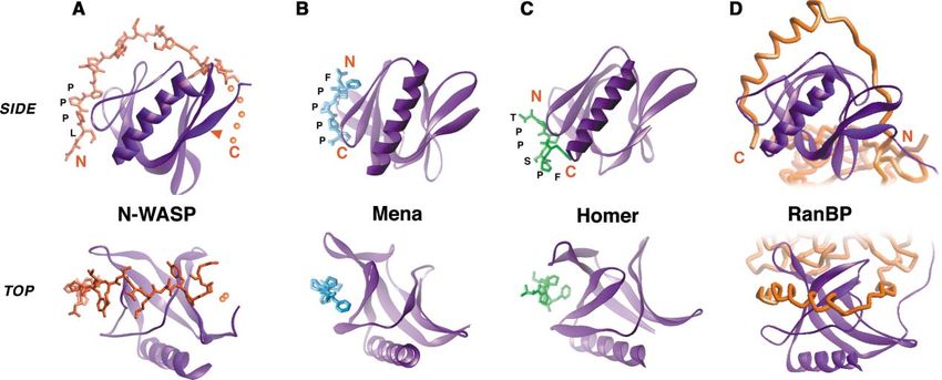
Cell
566

Figure 1. The N-Terminal Region of N-WASP Is an EVH1 Domain that Binds WIP via an Extended Peptide Ligand
(A) Proposed domain structure of N-WASP, as well as putative ligands for each domain.
(B) Example PIP2 vesicle binding assays. S indicates supernatant, P indicates pellet. Bound protein is retained in the pellet. The PLC␦ PH
domain and the N-WASP EVH1 fragment were expressed as fusions to glutathione-S-transferase (GST). Remaining proteins were expressed
as His6-tag fusions.
(C) Example far-Western protein binding assays. Probe proteins were biotinylated by expressing them as fusions to the Promega Pinpoint
vector (see Experimental Procedures). Each protein was tested for binding to a GST-fusion to WASP Interacting Protein (WIP) fragment
containing residues 457–490 and to the ActA sequence DFPPPTDEEL, which is a ligand for the Mena EVH1 domain.
(D) Summary of PIP2 and WIP (457–490) binding using various N-WASP constructs. None of the constructs bind PIP2, but all those that precisely
encompass the predicted EVH1 region can bind the WIP fragment.
(E) Deletion mapping reveals that a 25 residue polypeptide from WIP (residues 461–485) is the minimal fragment required for binding to the
N-WASP EVH1 domain (residues 26–147) in a far-Western gel overlay binding assay.

et al., 2001). Thus, it has been postulated that the WASP/                proline-rich sequences that may be putative EVH1 do-
N-WASP EVH1 domain may also serve a targeting                             main binding motifs (Martinez-Quiles et al., 2001; Mo-
function.                                                                 reau et al., 2000; Naqvi et al., 1998; Ramesh et al., 1997;
   Despite its importance in the pathology of WAS, the                    Savoy et al., 2000). It has not been determined, however,
precise molecular function of the WASP EVH1 domain                        if these motifs are sufficient to mediate the interaction
has remained unclear (Insall and Machesky, 1999). Sev-                    between WASP and WIP. Furthermore, homology mod-
eral functions have been ascribed to the domain, includ-                  eling based on known EVH1 domain structures fails to
ing binding to the acidic phospholipid, phosphatidyl ino-                 explain the behavior of several of the most common
sitol (4,5) bisphosphate (PIP2) (Imai et al., 1999; Miki et               WAS-causing mutations—specifically, the most signifi-
al., 1996) and binding to the WASP interacting protein                    cant mutational hotspot associated with severe WAS
(WIP) (Ramesh et al., 1997), both of which are known                      is located at a surface that is predicted to lie directly
regulators of N-WASP-mediated actin polymerization.                       opposite the canonical peptide binding surface (Be-
EVH1 domains share the same overall fold with Pleck-                      neken et al., 2000; Prehoda et al., 1999). These inconsis-
strin Homology (PH) domains (Fedorov et al., 1999; Pre-                   tent findings have several possible explanations: (1) the
hoda et al., 1999; Ball et al., 2000; Barzik et al., 2001;                N-WASP/WASP EVH1 domain may differ significantly in
Beneken et al., 2000), many of which specifically bind                    structure from other EVH1 domains; (2) these domains
phosphoinositides (Rebecchi and Scarlata, 1998). Thus,                    may have multiple binding sites—one that interacts with
it has been suggested that EVH1 domains may be bi-                        peptides and another that interacts with other ligands
functional, interacting with both peptides and phospho-                   such as PIP2; or (3) these domains may use a mechanism
inositides (Prehoda et al., 1999).                                        of peptide interaction that is significantly different from
   WIP is an actin binding protein that contains several                  that of other EVH1 domains.
Structure of the N-WASP EVH1-WIP Complex
567
Figure 2. N-WASP EVH1 Domain Sequence
(A) Constructs of the N-WASP EVH1 domain fused to the minimal WIP peptide. Fusions are linked via a (Gly-Ser-Gly-Ser-Gly) linker. Only the
top fusion yielded highly soluble protein amenable to structural analysis.
(B) Structure-based alignment of the Rat N-WASP EVH1 domain with its close homologs WASP (H. sapiens) and Bee1p (S. cerevisiae). Also
shown are sequences of other more distantly related, but structurally characterized EVH1 domains. Secondary structure elements in the
N-WASP structure are indicated above the sequence. Sites that show a direct NOE contact with the WIP peptide are marked by filled triangles.
Sites in WASP where missense mutation cause the disease WAS are circled (lavender, strong/moderate phenotype; green, mild phenotype).
A conserved charged residue array in the WASP family EVH1 domains, described in Figure 5, are indicated in red (negative) or blue (positive).
Here, we have sought to clarify what ligands the generated several fragments encompassing the mouse
N-WASP EVH1 domain can specifically bind and how N-WASP EVH1 domain and tested them for phospholipid
WAS-causing mutants may disrupt these interactions. binding. Phospholipid vesicle binding assays show that
We find that the N-WASP EVH1 domain and other EVH1 none of the N-WASP fragments are capable of binding
domains cannot bind PIP2 with significant affinity, con- PIP2 with detectable affinity using this assay (Figure 1B).
trary to a previous report. However, we find that the In contrast, in the same assay, strong PIP2 binding is
N-WASP EVH1 domain can specifically interact with a observed for two other known lipid binding modules:
25 residue peptide from WIP. We have determined the the Pleckstrin Homology (PH) domain from phospholi-
structure of the EVH1-WIP peptide complex using NMR pase C-delta (PLC-␦) and a fragment of N-WASP (resi-
spectroscopy, revealing that the domain adopts the dues 178–274) that includes a highly basic region (B
same overall fold as other EVH1 domains, but it utilizes domain) that has recently been shown to mediate PIP2
a novel mechanism of peptide recognition. The WIP regulation in N-WASP (Prehoda et al., 2000; Rohatgi et
peptide, which is more than twice as long as typical al., 2001). We also show that a canonical EVH1 domain,
EVH1 ligands, wraps around the entire EVH1 domain from the protein Mena, is also not capable of binding
like a piece of string around a spool, contacting regions PIP2 in this assay. Thus, despite a high degree of struc-
on the domain surface extending far beyond the canoni- tural homology with PH domains, EVH1 domains do not
cal EVH1 peptide binding site. The structure reveals a generally appear to share the function of high-affinity
novel class of domain-mediated protein-protein interac- phosphoinositide binding, at least at a level comparable
tions and suggests how WAS-causing mutations are to that observed for canonical PH domains (Kd ⵑ1 M).
likely to disrupt the WASP/WIP interaction.
The Minimal EVH1 Ligand Is a 25 Residue
Results and Discussion Peptide from WIP
In contrast, in far-Western protein binding assays, we
The EVH1 Domain of N-WASP Does Not Bind PIP2 found that N-WASP fragments encompassing the EVH1
It was previously reported that the N-terminal region of region were responsible for binding to a fragment from
N-WASP including the EVH1 domain was a PH or PH- WIP (residues 416–488) that had previously been shown
like domain capable of PIP2 binding (Miki et al., 1996). We to interact with N-WASP (Figure 1C). The fragment of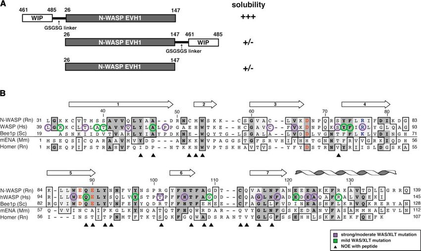
Cell
568
N-WASP required for WIP binding correlates precisely Table 1. Structural Statistics for 20 WIP-EVH1 Structures
with the EVH1 domain boundaries (Figure 1D).
The 72 residue WIP fragment contains many proline- NOE Constraints Number
rich putative EVH1 binding motifs, so we used deletion Long 803
analysis to delineate the minimal recognition require- Medium 278
ments (Figure 1E). A 25 amino acid fragment of WIP Short 455
Intraresidue 348
(residues 461–485) is minimally required for recognition.
Totala 1884
This motif is well conserved in certain isoforms of the WIP/EVH1 NOEsb 135
recently identified WIP homolog CR16 (Ho et al., 2001)
and the yeast homolog verprolin (Naqvi et al., 1998), and Ramachandran Statisticsc
its presence appears to correlate with ability to bind Most favored 76.4%
N-WASP. The WIP motif encompasses a proline-rich Additionally allowed 20.7%
motif (DLPPPEP) similar to that recognized by the EVH1 Generously allowed 2.0%
Disallowed 0.9%
domain from Mena (DFPPPPT). In our assays, the WIP
sequence, in fact, crossreacts with the Mena EVH1 do- Parameter Family Minimized Average
main, although the N-WASP EVH1 domain does not 2
Target function (Å ) 0.86 ⫾ 0.17 0.76
crossreact with a 10 residue ligand for the Mena EVH1 Upper limit violations
domain from the protein ActA (Niebuhr et al., 1997; Pre- Number ⬎ 0.1 Å 10 ⫾ 3 4
hoda et al., 1999). Sum of violations (Å) 5.1 ⫾ 0.7 3.8
The most striking feature of the minimal N-WASP Maximum violation (Å) 0.24 ⫾ 0.07 0.19
Torsion angle violations
EVH1 domain binding motif is that at 25 residues, it is
Number ⬎ 5⬚ 0⫾0 0
more than twice as long as the minimal 6–10 residue Sum of violations (⬚) 0.4 ⫾ 0.1 0.4
peptides sufficient for Mena, VASP, or Homer EVH1 rec- Maximum violation (⬚) 0.05 ⫾ 0.01 0.03
ognition (Niebuhr et al., 1997; Prehoda et al., 1999; Ball Van der Waals violations
et al., 2000; Barzik et al., 2001; Beneken et al., 2000), Number ⬎ 0.2 Å 0⫾1 0
suggesting that the binding energy is well-distributed Sum of violations (Å) 3.4 ⫾ 0.5 3.9
across this motif. Interestingly, the isolated N-WASP Maximum violation (Å) 0.17 ⫾ 0.07 0.16
EVH1 domain is highly insoluble and is poorly ex- Atomic Rmsdsd (Å): Family of 20 Structures versus Mean
pressed, precluding any high-resolution structural or
Backbone 0.50 ⫾ 0.06
biochemical studies. However, we found that when we Heavy atom 0.94 ⫾ 0.08
fused the 25 residue WIP motif to the N terminus of the a
Unique, nontrivial constraints derived from a total of 4305 NOEs
EVH1 domain via a 5 residue linker, the protein now
observed in four separate 2D and 3D NOESY spectra using the
expressed extremely well in bacteria and was highly CALIBA function of the DYANA program.
soluble (Figure 2A). In contrast, however, a similar fusion b
Constraints between residues of the WIP sequence and residues
of the WIP peptide to the C terminus did not yield soluble of the N-WASP EVH1 domain.
protein, thereby indicating a dependence on stereo- c
Only residues observed by NMR, corresponding to WIP residues
chemical orientation. 461–480 and N-WASP residues 34–137, were included in Ramachan-
dran analysis.
d
Disordered linker and loop residues were excluded. Rmsd calcula-
Structure of the EVH1 Domain-WIP Peptide
tions included WIP residues 462–468 and 474–479
Complex: A Novel “Wrapping”
Mechanism of Recognition
The unusual length of the WIP polypeptide required for
N-WASP EVH1 binding suggests a novel mechanism of The overall fold of the N-WASP EVH1 domain is similar
interaction that could, for example, depend on higher- to that observed for the Mena, Homer, and VASP do-
order secondary or tertiary structure of the ligand. To mains (Fedorov et al., 1999; Prehoda et al., 1999; Be-
understand how the WIP polypeptide interacts with the neken et al., 2000; Barzik et al., 2001). The structure-
N-WASP EVH1 domain, we determined the NMR struc- based alignment of several EVH1 domain sequences is
ture of the highly soluble fusion protein encompassing shown in Figure 2B.
the interaction pair described above (Table 1). The structure reveals, however, a novel mode of
Structural analysis of the EVH1-WIP complex utilized EVH1-mediated interaction—the WIP polypeptide exists
the single-chain construct described above. However, in a largely extended conformation and wraps around
to confirm that the covalent linker did not affect the much of the domain, like a piece of string around a
mode of WIP binding, we also produced a second analo- concave spool (Figure 3A). Because of the extended
gous construct with a thrombin cleavage site in the nature of the ligand, the surface of the EVH1 domain
linker. After purification, this construct could be cleaved that is contacted for recognition is far larger (ⵑ900 Å2 )
to completion and remained as a soluble complex. No than the canonical binding surfaces utilized by the Mena
differences were observed in backbone triple-reso- and Homer EVH1 domains to recognize their much
nance or NOESY NMR spectra acquired on samples of shorter peptide ligands (ⵑ300 Å2 ) (Figures 3B and 3C).
each version of the complex (cleaved or tethered), aside Nonetheless, as a part of its interaction, the N-WASP
from new residues introduced in the thrombin recogni- EVH1 domain does use the conserved canonical binding
tion site (see Supplemental Figure S1 at http://www.cell. surface to recognize a highly proline-rich portion of its
com/cgi/content/full/111/4/565/DC1), illustrating that cognate motif (discussed below).
the presence of a covalent link has no effect on WIP The overall interaction of the N-WASP EVH1 domain
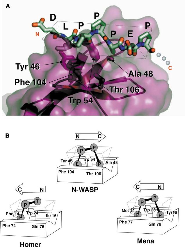
peptide binding mode. with this extended WIP polypeptide has unexpectedStructure of the N-WASP EVH1-WIP Complex 569 Figure 3. Structure of the N-WASP/WIP Complex Determined by NMR and Comparison to Other EVH1 Domain-Mediated Complexes (A) N-WASP EVH1 domain in complex with WIP residues 461–485. Note that residues 480–485 are required for binding but are poorly defined in the structure because of a lack of assignments and constraints. The likely path of those residues is indicated by the dotted line. The red arrow indicates the site of the most common severe WAS missense mutation, Arg76 [86]. This site falls at the opposite surface from the canonical EVH1 binding surface. (B) Structure of the Mena EVH1 domain in complex with an 8 residue peptide from ActA. (C) Structure of the Homer EVH1 in complex with a 5 residue peptide from mGluR. (D) Structure of the RanBP EVH1 domain in complex with an extended, ⬎25 residue polypeptide from Ran. Note that contacts between the EVH1 domain and other parts of Ran are made in the complex. Top and bottom images show two views rotated by 90⬚ around a horizontal axis in the plane of the page. Note that in the N-WASP complex, the peptide ligand is recognized in the reverse N- to C-terminal orientation from other EVH1 complexes. similarity to the interaction of the Ran Binding Protein to continue wrapping further around the N-WASP EVH1 (RanBP) EVH1 domain with a fragment from the nuclear domain, along a continued path similar to that observed import protein Ran (Figure 3D). In the crystal structure in the RanBP/Ran interaction. of the Ran-RanBP complex, a ⵑ25 residue polypeptide The extensive interaction interface in the N-WASP/ sequence from Ran wraps around the entire RanBP WIP interaction may explain the biochemical behavior of EVH1 domain, following the same path as that observed the domain—this exposed, largely hydrophobic binding for the WIP peptide when binding the N-WASP EVH1 surface may lead to the poor solubility of the unliganded domain (Vetter et al., 1999). This interaction, however, is domain. Association of N-WASP with WIP or a related only one of several extensive regions of contact between ligand may be obligatory for proper folding of the pro- the Ran and RanBP proteins, and it is unknown if this tein—consistent with the observation that the N-WASP/ wrapping interaction alone is sufficient for binding, as WIP interaction appears to be constitutive in vivo. is the case for the N-WASP/WIP interaction. In the current NMR structure, the conformation of Common Elements of Proline-Rich residues 479–485 (C-terminal end of the WIP peptide) Peptide Binding are poorly defined because of a lack of resonance as- The most N-terminal portion of the WIP polypeptide, signments. The difficulty in assigning these residues which contains the sequence LPPP, binds at the same may be a result of several possibilities: first, the main conserved aromatic surface used by other EVH1 do- chain amide protons may have relatively high solvent mains for proline peptide binding (Figure 4). As is ob- accessibility and exchange rates; second, the residues served in other EVH1-peptide complexes, this region of may exist in multiple conformations that interconvert the peptide adopts a polyproline II (PPII) helical confor- within the intermediate exchange regime; and third, the mation, a left-handed helix with three residues per turn. region may be highly mobile. Dynamics of the polypep- Other proline-rich motif binding domains, including SH3 tide backbone were probed using the 15N-1H hetero- and WW domains, and profilin also recognize their li- nuclear NOE. For residues 477–480, decreasing NOE gands in a PPII conformation. The PPII helix docks values are observed moving from N- to C-terminal, a against a complementary surface of largely aromatic trend consistent with enhanced mobility for the adjacent side chains in a manner very similar to that observed in unobserved residues (data not shown). Nonetheless, ex- other EVH1 domains. A universal element of this recogni- tension of the peptide C terminus beyond residue 480 tion is a central, conserved tryptophan residue (Trp 54), is energetically important for binding (Figure 1E). We which forms a ridge over which the PPII helix packs, hypothesize that side chain atoms in this region of the making favorable van der Waals contact. In addition, peptide make primary contacts with the EVH1 domain, the tryptophan indole nitrogen proton forms a hydrogen while the main chain atoms may be relatively mobile. bond with a carbonyl oxygen of the PPII ligand. Less well As shown in Figure 3A, these residues are most likely conserved but largely hydrophobic residues comprise
Cell
570

                                                                                 Figure 4. The N-WASP EVH1 Surface Used
                                                                                 to Recognize the Proline-Rich Element in the
                                                                                 WIP Ligand Is Similar to that Used by Other
                                                                                 EVH1 Domains
                                                                                 (A) Close-up view of region of N-WASP EVH1
                                                                                 domain that contacts residues 481–487 of the
                                                                                 WIP ligand. The ligand adopts a polyproline
                                                                                 II (PPII) helical conformation that docks over
                                                                                 Trp 54. The carbonyl oxygen between the api-
                                                                                 cal ligand prolines also makes a hydrogen
                                                                                 bond to the Trp nitrogen. Pockets sur-
                                                                                 rounding Trp 54 are made by residues 46 and
                                                                                 104 or 48 and 106.
                                                                                 (B) Corresponding residues in the Homer and
                                                                                 Mena EVH1 domains are also used in recogni-
                                                                                 tion of the proline-rich peptide ligand. In these
                                                                                 structures, however, the PPII helix binds in
                                                                                 the opposite N- to C-terminal orientation.

residue binding pockets that flank the central tryp-           Buried Mutations Linked to Wiskott-Aldrich
tophan.                                                        Syndrome
                                                               Because of the extremely high homology between
                                                               WASP and N-WASP (Figure 2), this EVH1 domain struc-
Two Orientations for EVH1-Peptide Binding                      ture and its novel mode of peptide binding provides
One of the most striking features of this structure is that    the first opportunity to accurately interpret missense
the WIP polypeptide docks on the EVH1 domain in an             mutations that result in the disease WAS. WAS is an
N- to C-terminal orientation that is the reverse of that       X-linked recessive disorder that is characterized by
observed in all previous EVH1-peptide complexes (Fig-          thrombocytopenia (platelet deficiency), eczema, and re-
ures 3 and 4B). Thus, the EVH1 domain, like other pro-         currence of bacterial and viral infections. A mild form
line-rich binding modules (including SH3 and WW do-            of the disease is known as X-linked thrombocytopenia
mains) and profilin, can bind PPII ligands in two possible     (XLT). WAS/XLT-causing missense mutations occur at
orientations. This orientational flexibility, first explored   ⵑ30 residues within the WASP EVH1 domain. For the
in depth for SH3 domains, is due largely to the 2-fold         discussion below we will describe sites of interaction
rotational pseudosymmetry of the PPII structure, both          using the residue numbering for N-WASP. However, for
in terms of shape and hydrogen bonding moieties (Lim           clarity and comparison, the corresponding residue num-
et al., 1994; Feng et al. 1994). This rotational symmetry      ber from WASP will also be given in square brackets.
allows the relatively isoenergetic docking of the PPII           Several of the common WAS-causing mutations map
helix in two orientations against a single compatible          to the hydrophobic core of the EVH1 domain, especially
surface. It is interesting to note that in the RanBP EVH1      at residues that are absolutely conserved between
structure, the extended peptide from Ran binds in the          WASP and N-WASP (Table 2A, Figure 5A). Mutations at
opposite orientation from that observed in the current         buried positions often destabilize a protein and indi-
structure.                                                     rectly decrease function. However, it is noteworthy that
Structure of the N-WASP EVH1-WIP Complex
571
Table 2. WAS Missense Mutations Localized to the EVH1 Domain and Their Role in the N-WASP-WIP Complex
Type of Position in N-WASP Disease
Mutation [WASP numbering] Substitution Severity Description of Position Probable Effect
(A) BURIED Trp87 [97] Cys strong core destabilization/conform. change
His105 [115] Tyr strong core; H bond to Tyr97 destabilization/conform. change
Phe118 [128] Ser strong core; beneath pept. bind. surf. destabilization/conform. change
Ala124 [134] Thr strong core destabilization/conform. change
Tyr73 [83] Cys mild core; beneath pept. bind. surf. destabilization/conform. changea
Tyr97 [107] Cys mild core; H bond to His 105 destabilization/conform. changea
(B) SURFACE Leu31 [39] Pro strong near peptide C term. disrupt WIP interaction
Cys63 [73] Arg strong near peptide N term. disrupt WIP interaction
Val65 [75] Met strong near peptide C term. disrupt WIP interaction
Ser72 [82] Phe strong near peptide C term. (NOE) disrupt WIP interaction
Arg76 [86] Cys, His, Leu, Pro strong near peptide C term. disrupt WIP interactiona
Arg101 [111] Pro strong near peptide C term. (NOE) disrupt WIP interaction
Ala115 [Gly125] Arg strong near peptide N term. (NOE) disrupt WIP interaction
Glu123 [133] Lys strong away from peptide structural salt bridge?
Thr37 [45] Met mild (XLT) near peptide C term. disrupt WIP interaction
Gln44 [52] His mild (XLT) near peptide N term. disrupt WIP interaction
Lys33 [Arg41] Gly mild near peptide C term. disrupt WIP interaction
Ser39 [47] Asp mild near peptide C term. disrupt WIP interaction
Ser40 [48] Ile mild near peptide C term. disrupt WIP interaction
Ala48 [56] Val mild near peptide N term. (NOE) disrupt WIP interaction
Phe74 [84] Leu mild near peptide C term. disrupt WIP interaction
Gln89 [99] Arg mild near peptide C term. (NOE) disrupt WIP interaction
Gly109 [119] Glu mild near peptide N term. (NOE) disrupt WIP interaction
Glu121 [131] Lys mild salt bridge with Lys125 destabilization/conform. change
Arg128 [138] Pro strong H bond to Ser99 destabilization/conform. change
Data compiled from WASP database (http://www.tmd.ac.jp/med/ped/WASPbase.html)
“NOE” indicates observed NOE contact between WIP peptide and this residue.
a
Mutation experimentally observed to disrupt WASP-WIP interaction by yeast two-hybrid assay (Stewart et al., 1999).
many of these buried mutations cluster into a single tation of Trp54 has been shown to disrupt proper local-
packing unit (Trp87 [97], Tyr97 [107], His105 [115], ization to vaccinia virus (Moreau et al., 2000). In fact,
Phe118 [128], and Ala124 [134]) that includes a buried the most dramatic hotspot of disease mutations is on
hydrogen bond between residues Tyr97 [107] and the face of the EVH1 domain directly opposite the pro-
His105 [115]. Even fairly subtle mutations at these posi- line-rich peptide binding surface (Beneken et al., 2000).
tions (i.e., Tyr97 [107]-Cys, His105 [115]-Tyr, Ala124 For example, the most frequent mutated residue re-
[134]-Thr), which maintain hydrophobicity and thus sulting in a severe disease phenotype is Arg76 [86].
might not be expected to be very destabilizing, result Every possible single base change missense mutation
in the disease. Interestingly, this packing unit lies directly (Cys, His, Leu, Pro) has been found in the WAS patient
beneath the proline-rich peptide binding surface. In fact, population (Table 2B). Mutations are also found at other
one of these residues, His105 [115], is the covalent surface residues that flank Arg76 [86], including Leu31
neighbor of two residues that directly contact this seg- [39], Thr37 [45], Ser40 [Thr48], Cys63 [73], and Phe74
ment of the WIP ligand (Phe104 [114], Thr106 [116]). [84]. Given the distance of this mutational hotspot from
Thus, it seems possible that subtle mutations in this the canonical peptide binding site identified in pre-
packing unit perturb the precise stereochemistry of the viously characterized EVH1 domain complexes, it has
proline-rich peptide binding surface and thereby impair been difficult to reconcile how these mutations in WASP
WIP binding. This hypothesis is consistent with several might disrupt EVH1 domain interactions and function
previous findings: first, the Tyr97 [107]-Cys and Ala124 (Beneken et al., 2000).
[134]-Thr mutations were found to impair interaction In the context of this new structure, however, we now
with WIP in an in vitro yeast two-hybrid binding assay know that the WIP ligand wraps around the N-WASP
(Stewart et al., 1999); and second, studies of patient EVH1 domain, extending from the canonical proline-rich
samples reveal no significant decrease of endogenous peptide-docking site all the way to mutational hotspot
expression levels of the WASP protein in a patient bear- surrounding Arg76 [86]. Contacts made in this region
ing a mutation at Tyr97 [107]. are therefore likely to be critical for WIP binding. Unfortu-
nately, because of a lack of assignments, we do not
Surface Mutations Linked to Wiskott-Aldrich know the precise conformation of the last seven resi-
Syndrome dues of the WIP polypetide and exactly how these resi-
Approximately 20 of the reported WAS-causing muta- dues interact with the hotspot centered around Arg76
tions map to the EVH1 domain surface (Table 2B, Figure [86]. However, these seven residues are required for
5B). Oddly, there are no naturally occurring mutations binding and therefore must be involved in energetically
at the proline-rich peptide binding surface (although mu- important interactions.Cell
572
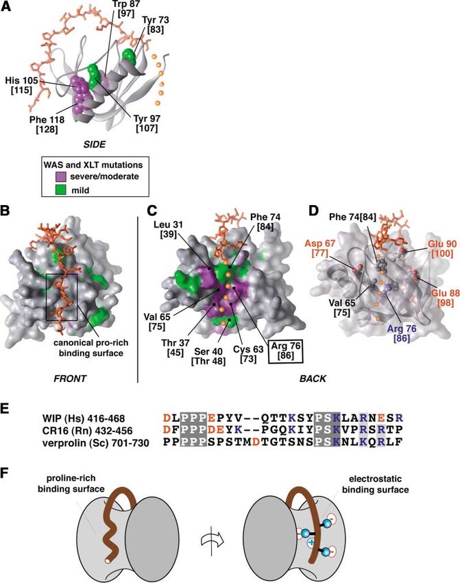
Figure 5. Structural Basis of Mutations that
Cause the Disorder WAS
(A) Residues in the core of the N-WASP EVH1
domain that when mutated lead to WAS are
shown in space-filling depiction. Severe/
moderate mutations are colored in lavender;
mild mutation are colored in green. Residue
numbering is that of N-WASP, with the corre-
sponding numbering from WASP given in
square brackets. Peptide is colored orange,
with the postulated path of the peptide C ter-
minus indicated by small orange spheres.
(B) Front face of N-WASP EVH1 domain indi-
cating surface mutations associated with
WAS. Residues are colored as above.
(C). Back face of N-WASP EVH1 domain, indi-
cating surface mutations associated with
WAS. Molecular has been rotated 180⬚ from
the view in (B).
(D) Transparent surface view identical to that
in (C) reveals a conserved network of charged
residues, centered around Arg76 [86] (see
Figure 2 for conservation).
(E) Alignment of WIP homologs in the WASP
binding peptide reveals two regions of high
conservation, including the C-terminal
PSKxxR/K motif that is likely to dock at the
surface shown in (D). Charged residues are
colored in red (negative) or blue (positive).
(F) Cartoon of how the extended WIP peptide
and its homologs may wrap around the
N-WASP EVH1 domain to contact two distant
but energetically important binding surfaces.
Based on the evidence given below, we postulate that conserved in all WIP homologs, including mouse WIP,
the C-terminal end of the WIP peptide binds at this human CR16, and yeast verprolin—all of which have
mutational hotspot and participates in an energetically been shown to interact with WASP or WASP homologs
critical network of electrostatic interactions. First, the (Figure 5E). This cluster of basic residues is within the
proposed binding path for the remaining portions of conserved motif: P-S-K-x-x-R/K-x(1-3)-R. In summary, we
the peptide correlates exactly with the binding groove therefore propose a model in which the conserved basic
utilized by the RanBP EVH1 domain to recognize one motif found at the C terminus of the WIP ligand specifi-
terminus of the Ran peptide (see Figure 3D). Second, cally interacts with a complementary network of con-
mutation of the central Arg76 [86] to His has been shown served acidic residues that are properly positioned by
to impair interaction with WIP in an in vitro yeast two- the central Arg76 [86] residue (Figure 5F). Such an inter-
hybrid assay (Stewart et al., 1999). Third, Arg76 [86] is action may have a fairly high degree of mobility, consis-
the central residue in a conserved electrostatic network tent with the difficulty in observing assignable reso-
presented on this surface of the EVH1 domain (Figure nances, despite its apparent energetic importance.
5D). Surrounding Arg76 [86] are a ring of acidic residues, Overall, this structure is therefore consistent with a
Asp67 [77], Glu88 [98], and Glu90 [100] (Figure 5D), all simple model for the cause of WAS: the major defect in
of which are conserved from human WASP/N-WASP to these diverse WAS missense mutations is likely to be
the yeast ortholog, Bee1p (see Figure 2). In the current disruption of the interaction with WIP. Disruption of this
structure, Arg76 [86] is very well ordered for an arginine interaction may prevent proper subcellular targeting of
residue—several interresidue NOEs are observed for the WASP. The reason that mutations are found at sites
Arg side chain, and its epsilon NH proton exchanges widely spread over the EVH1 domain surface, well be-
very slowly. This observation is consistent with Arg76 yond the canonical EVH1 peptide binding surface, is
[86] playing a central role in coordinating and orienting because this EVH1 domain utilizes a novel mode of inter-
this otherwise repulsive network of surrounding acidic action—the WIP ligand wraps around almost two-thirds
residues. Fourth, the C terminus of the WIP peptide of the entire circumference of the EVH1 domain, con-
contains a cluster of basic residues that is very well tacting many distant surfaces.Structure of the N-WASP EVH1-WIP Complex
573
Conclusions cated and centrifuged lysate was used directly as probe, after esti-
This structure of the N-WASP EVH1 domain in complex mating fusion protein concentration by SDS-PAGE. Washing and
chemiluminescence detection was performed as described (Pre-
with a peptide ligand from WIP reveals a novel mecha-
hoda et al., 1999).
nism of peptide recognition by EVH1 domains and high-
lights the versatility of EVH1 domains as a recognition
platform. While the majority of EVH1 domains and other Protein Labeling and Purification
The WIP-EVH1 sequence (residues 461–485 of human WIP and resi-
similar protein-protein modules bind short 6–12 residue
dues 26–147 of rat N-WASP, linked by the intervening sequence
motifs, the N-WASP EVH1 domain binds a single long GSGSG) was ligated into the pBH4 expression vector, which con-
25 residue motif. Comparison of the WIP ligand and its tains a His6-tag followed by a Tobacco Etch Virus (TEV) protease
homologs from different species suggest that there are cleavage site. For uniform labeling with 15N and 13C, BL21(DE3) cells
two conserved submotifs that are recognized: the freshly transformed with the WIP-EVH1 expression plasmid were
N-terminal PPP submotif, similar to other EVH1 ligands, grown in M9 minimal media containing [13C]glucose (2 g/l) and
[15N]ammonium chloride (1 g/l) as the respective sole carbon and
and the C-terminal PSKxxR submotif. These two sub-
nitrogen sources. Expression was induced by the addition of IPTG
motifs interact on opposite faces of the EVH1 domain, to a final concentration of 1 mM. After expression for 3 hr, the cells
leading to this very extensive wrapping mechanism of were harvested by centrifugation, resuspended in Lysis Buffer (50
recognition (Figures 5E and 5F). The extensive binding mM PO4, 300 mM NaCl, 10 mM imidazole [pH 8.0]), disrupted by
surface employed provides an explanation for the spa- sonication, and centrifuged, and the soluble fraction bound in batch
tially dispersed set of WAS-causing mutants that map to Ni-NTA agarose. After extensive washing with lysis buffer, bound
protein was eluted with lysis buffer ⫹ 300 mM imidazole. The His6-
to the EVH1 domain.
tag was removed by incubation with TEV protease for 2 hr at room
This interaction represents a novel or hybrid class of temperature. Cleaved WIP-EVH1 protein was further purified on a
protein-protein interactions. Most protein-protein inter- ResourceS (Pharmacia) column at pH 7.5 with a 1–500 mM NaCl
actions are divided into two major classes: (1) interac- gradient. Pure protein was pooled and concentrated to ⵑ1 mM.
tions that involve the docking of two large folded protein Protein size was confirmed by mass spectroscopy before and after
surfaces, and (2) interactions that involve the docking NMR data collection.
A cleavable version of the linked WIP-EVH1 protein was generated
of a short, largely linear peptide epitope on a protein
as described above, but with an intervening linker sequence
surface (Stanfield and Wilson, 1995). Like the second GGLVPRGSGG (underlined sequence is thrombin cleavage site).
class of interactions, the N-WASP EVH1-WIP peptide 13
C/15N-labeled protein was prepared as described above and
interaction involves recognition of a linear epitope. How- cleaved at a concentration of 0.5 mM by adding 50 nM of bovine
ever, because the epitope is extremely long, the contact thrombin (Sigma). Cleavage was complete in 1 hr as monitored by
area involved is as large as that observed in the first SDS-PAGE.
class of interactions. It will be interesting to see if other
EVH1 domains also have the ability to recognize longer NMR Spectroscopy
peptides with higher specificity by utilizing more of their NMR experiments were carried out on Bruker DRX600 spectrome-
surface binding potential. It is also possible that in some ters, either at the Medical College of Wisconsin (with triple-reso-
nance Z-axis gradient CryoProbe威) or at the National Magnetic Res-
cases EVH1 domains may be able to use their extensive
onance Facility at Madison (with conventional probe). Samples
binding surfaces to simultaneously interact with two or contained ⵑ1 mM WIP-EVH1 protein in 90% H2O/10% D2O with 20
more ligands, recognizing short, proline-rich peptides mM sodium phosphate (pH 7.0), 20 mM sodium chloride, 1 mM
with the canonical binding surface and recognizing other dithiothreitol, and 0.05% sodium azide. Resonance assignments
yet unidentified ligands at remaining portions of the do- and distance constraints were derived from the following experi-
main surface. ments: 3D 15N NOESY-HSQC (Talluri and Wagner, 1996), 3D 15N SE
TOCSY-HSQC (Zhang et al., 1994), 3D HCCH-TOCSY (Kay et al.,
1993), 3D SE CCONH (Grzesiek et al., 1993), 3D SE HNCA (Grzesiek
Experimental Procedures
and Bax, 1992; Kay et al., 1994), 3D SE HNCOCA (Grzesiek and Bax,
1992), 3D SE HNCO (Grzesiek and Bax, 1992; Muhandiram and Kay,
Lipid Vesicle Binding Assays
1994), 3D 13C SE NOESY-HSQC (one each for the aromatic and
Cosedimentation lipid vesicle binding assays were performed as
aliphatic 13C regions) (Kay et al., 1993), and 13C constant-time SE
described by Kavran et al. (1998). Specifically, 3 M protein and
HSQC (Santoro and King, 1992). NOESY mixing times were 80 ms.
vesicles (ⵑ20 M PIP2) were mixed in 20 mM HEPES (pH 7.0), 100
Isotropic mixing periods were 22 ms (CCONH) and 60 ms (15N
mM NaCl, 1 mM DTT (total assay volume 85 l), incubated at 25⬚C
TOCSY-HSQC). Heteronuclear NOEs were measured from an inter-
for 5 min, and centrifuged at 100,000 ⫻ g for 15 min. The vesicles leaved pair of 2D 15N-1H gradient sensitivity enhanced correlation
comprised PC:PS:PIP2 at ratios of 48:48:2. Apparent concentrations
spectra (Farrow et al., 1994) recorded with and without a 3.5 s proton
of PIP2 were calculated as 50% of total. The vesicle pellet was
saturation period using a total recycle delay of 5.5 s. Data were
washed with 1000 l of buffer three times and suspended in a
processed using NMRPipe (Delaglio et al., 1995) and analyzed using
volume of buffer equal to the supernatant. Bound (pellet) and un-
XEASY (Bartels et al., 1995). Chemical shifts were referenced to
bound proteins (supernatant) were identified by SDS-PAGE. This
2,2-dimethylsilapentane-5-sulfonic acid (DSS) directly for 1H and
assay can detect binding of the PLC-␦ PH domain, which has an
indirectly for 15N and 13C (Markley et al., 1998). The CSI program
affinity for PIP2 of Kd ⫽ 2 M.
was used for determining the secondary structure from the chemical
shift index (Wishart and Sykes, 1994; Wishart et al., 1992). For the
Far-Western Protein Binding Assays thrombin-cleaved protein, resonance assignments and NOE pat-
Assays were performed as described (Prehoda et al., 1999). Putative terns were determined for the cleaved WIP-EVH1 complex using 3D
EVH1 peptide ligands were generated as fusions to glutathione SE HNCA, 3D CCONH, 3D 15N NOESY-HSQC, and 3D 13C NOESY-
S-transferase (GST), electrophoresed on a 10%–20% SDS-PAGE HSQC spectra, under the same conditions. No significant chemical
gel, and transferred to nitrocellulose by electroblotting, and the blot shift differences were observed apart from residues of the linker
was blocked for 1 hr at 4⬚C in Superblock (Pierce). Biotinylated that differ in the two constructs (see Supplemental Figure S1 at
EVH1 domains were produced as fusions to a naturally biotinylated http://www.cell.com/cgi/content/full/111/4/565/DC1), and both
E. coli protein using the PIN-POINT system (Promega). The soni- samples displayed the same pattern of WIP-EVH1 NOEs.Cell
574
Structure Calculation and Analysis Derry, J.M., Ochs, H.D., and Francke, U. (1994). Isolation of a novel
Structures were iteratively refined using the simulated annealing gene mutated in Wiskott-Aldrich syndrome. Cell 78, 635–644.
protocol (10,000 steps total) of the torsion angle dynamics program Derry, J.M., Kerns, J.A., Weinberg, K.I., Ochs, H.D., Volpini, V., Esti-
DYANA (Güntert et al., 1997). NOE peak intensities were converted vill, X., Walker, A.P., and Francke, U. (1995). WASP gene mutations
into upper distance bounds with the CALIBA function of DYANA. A in Wiskott-Aldrich syndrome and X-linked thrombocytopenia. Hum.
total of 143 φ and dihedral angle constraints were generated from Mol. Genet. 4, 1127–1135.
1 ␣ 13 ␣ 13  13
H , C , C , C’, and 15N secondary shifts using TALOS (Cornilescu
El-Hakeh, J., Rosenzweig, S., Oleastro, M., Basack, N., Berozdnik,
et al., 1999). Of the final 50 structures calculated, the 20 conformers
L., Molina, F., Rivas, E.M., Zelazko, M., and Danielian, S. (2002).
with the lowest target function values were selected for analysis.
Wiskott-Aldrich syndrome in Argentina: 17 unique, including nine
The mean structure, calculated from this ensemble of 20 structures
novel, mutations. Hum. Mutat. 19, 186–187.
in MOLMOL (Koradi et al., 1996), was minimized in DYANA using
8000 steps of conjugate gradient minimization. Coordinates (mini- Farrow, N.A., Muhandiram, R., Singer, A.U., Pascal, S.M., Kay, C.M.,
mized average structure and final 20 structures) have been depos- Gish, G., Shoelson, S.E., Pawson, T., Forman-Kay, J.D., and Kay,
ited in the RCSB database (accession code 1MKE). Residues in the L.E. (1994). Backbone dynamics of a free and a phosphopeptide-
PDB file are numbered consecutively with the following residue complexed src homology 2 domain studeid by 15N NMR relaxation.
number translation: PDB(1–25) ⫽ WIP(461–485); PDB(26–30) ⫽ Biochemistry 33, 5984–6003.
GSGSG linker; PDB(31–144) ⫽ N-WASP(26–139) (note: residues Fedorov, A.A., Fedorov, E., Gertler, F., and Almo, S.C. (1999). Struc-
140–147 of N-WASP were not observed by NMR and are not included ture of EVH1, a novel proline-rich ligand-binding module involved
in the PDB file). in cytoskeletal dynamics and neural function. Nat. Struct. Biol. 6,
661–665.
Received: April 17, 2002 Feng, S., Chen, J.K., Yu, H., Simon, J.A., and Schreiber, S.L. (1994).
Revised: September 4, 2002 Two binding orientations for peptides to the Src SH3 domain: devel-
opment of a general model for SH3-ligand interactions. Science 266,
References 1241–1247.
Gertler, F.B., Niebuhr, K., Reinhard, M., Wehland, J., and Soriano,
Ball, L.J., Kuhne, R., Hoffmann, B., Hafner, A., Schmieder, P., Volk- P. (1996). Mena, a relative of VASP and Drosophila Enabled, is impli-
mer-Engert, R., Hof, M., Wahl, M., Schneider-Mergener, J., Walter, cated in the control of microfilament dynamics. Cell 87, 227–239.
U., et al. (2000). Dual epitope recognition by the VASP EVH1 domain Greer, W.L., Shehabeldin, A., Schulman, J., Junker, A., and Simino-
modulates polyproline ligand specificity and binding affinity. EMBO vitch, K.A. (1996). Identification of WASP mutations, mutation hot-
J. 19, 4903–4914. spots and genotype-phenotype disparities in 24 patients with the
Bartels, C., Xia, T.-H., Billeter, M., Güntert, P., and Wüthrich, K. Wiskott-Aldrich syndrome. Hum. Genet. 98, 685–690.
(1995). The program XEASY for computer-supported NMR spectral Grzesiek, S., and Bax, A. (1992). Inproved 3D triple-resonance NMR
analysis of biological macromolecules. J. Biomol. NMR 5, 1–10. techniques applied to a 31 kDa protein. J. Magn. Reson. 96, 432–440.
Barzik, M., Carl, U.D., Schubert, W.D., Frank, R., Wehland, J., and Grzesiek, S., Anglister, J., and Bax, A. (1993). Correlation of back-
Heinz, D.W. (2001). The N-terminal domain of Homer/Vesl is a new bone amide and aliphatic side-chain resonances in 13C/
class II EVH1 domain. J. Mol. Biol. 309, 155–169. 15N-enriched proteins by isotropic mixing of 13C magnetization. J.
Beneken, J., Tu, J.C., Xiao, B., Nuriya, M., Yuan, J.P., Worley, P.F., Magn. Reson. 101, 114–119.
and Leahy, D.J. (2000). Structure of the Homer EVH1 domain-peptide Güntert, P., Mumenthaler, C., and Wüthrich, K. (1997). Torsion angle
complex reveals a new twist in polyproline recognition. Neuron 26, dynamics for NMR structure calculation with the new program
143–154. DYANA. J. Mol. Biol. 273, 283–298.
Callebaut, I., Cossart, P., and Dehoux, P. (1998). EVH1/WH1 domains Higgs, H.N., and Pollard, T.D. (2000). Activation by Cdc42 and PIP(2)
of VASP and WASP proteins belong to a large family including Ran- of Wiskott-Aldrich syndrome protein (WASp) stimulates actin nucle-
binding domains of the RanBP1 family. FEBS Lett. 441, 181–185. ation by Arp2/3 complex. J. Cell Biol. 150, 1311–1320.
Cannon, J.L., Labno, C.M., Bosco, G., Seth, A., McGavin, M.H., Higgs, H.N., and Pollard, T.D. (2001). Regulation of actin filament
Siminovitch, K.A., Rosen, M.K., and Burkhardt, J.K. (2001). Wasp network formation through ARP2/3 complex: activation by a diverse
recruitment to the T cell:APC contact site occurs independently of array of proteins. Annu. Rev. Biochem. 70, 649–676.
Cdc42 activation. Immunity 15, 249–259. Ho, H.Y., Rohatgi, R., Ma, L., and Kirschner, M.W. (2001). CR16
Carl, U.D., Pollmann, M., Orr, E., Gertlere, F.B., Chakraborty, T., and forms a complex with N-WASP in brain and is a novel member of
Wehland, J. (1999). Aromatic and basic residues within the EVH1 a conserved proline-rich actin-binding protein family. Proc. Natl.
domain of VASP specify its interaction with proline-rich ligands. Acad. Sci. USA 98, 11306–11311.
Curr. Biol. 9, 715–718. Imai, K., Nonoyama, S., Miki, H., Morio, T., Fukami, K., Zhu, Q.,
Carlier, M.F., Ducruix, A., and Pantaloni, D. (1999). Signalling to actin: Aruffo, A., Ochs, H.D., Yata, J., and Takenawa, T. (1999). The pleck-
the Cdc42-N-WASP-Arp2/3 connection. Chem. Biol. 6, R235–R240. strin homology domain of the Wiskott-Aldrich syndrome protein is
Carlier, M.F., Nioche, P., Broutin-L’Hermite, I., Boujemaa, R., Le involved in the organization of actin cytoskeleton. Clin. Immunol.
Clainche, C., Egile, C., Garbay, C., Ducruix, A., Sansonetti, P., and 92, 128–137.
Pantaloni, D. (2000). GRB2 links signaling to actin assembly by en- Insall, R., and Machesky, L. (1999). PH domains in WASP—a bug
hancing interaction of neural Wiskott-Aldrich syndrome protein in the system? Wiskott-Aldrich syndrome protein. Trends Cell Biol.
(N-WASp) with actin-related protein (ARP2/3) complex. J. Biol. 9, 211–212.
Chem. 275, 21946–21952. Kavran, J.M., Klein, D.E., Lee, A., Falasca, M., Isakoff, S.J., Skolnik,
Coppolino, M.G., Krause, M., Hagendorff, P., Monner, D.A., Trimble, E.Y., and Lemmon, M.A. (1998). Specificity and promiscuity in phos-
W., Grinstein, S., Wehland, J., and Sechi, A.S. (2001). Evidence for phoinositide binding by pleckstrin homology domains. J. Biol. Chem.
a molecular complex consisting of Fyb/SLAP, SLP-76, Nck, VASP 273, 30497–30508.
and WASP that links the actin cytoskeleton to Fc␥receptor signalling Kay, L.E., Xu, G.-Y., Singer, A.U., Muhandiram, D.R., and Forman-
during phagocytosis. J. Cell Sci. 114, 4307–4318. Kay, J.D. (1993). A gradient-enhanced HCCH-TOCSY experiment
Cornilescu, G., Delaglio, F., and Bax, A. (1999). Protein backbone for recording sidechain 1H and 13C correlations in H2O samples of
angle restraints from searching a database for chemical shift and proteins. J. Magn. Reson. 101, 333–337.
sequence homology. J. Biomol. NMR 13, 289–302. Kay, L.E., Xu, G.Y., and Yamazaki, T. (1994). Enhanced-sensitivity
Delaglio, F., Grzesiek, S., Vuister, G.W., Zhu, G., Pfeifer, J., and Bax, triple-resonance spectroscopy with minimal H2O saturation. J.
A. (1995). NMRPipe: a multidimensional spectral processing system Magn. Reson. 109, 129–133.
based on UNIX pipes. J. Biomol. NMR 6, 277–293. Kim, A.S., Kakalis, L.T., Abdul-Manan, N., Liu, G.A., and Rosen, M.K.Structure of the N-WASP EVH1-WIP Complex
575
(2000). Autoinhibition and activation mechanisms of the Wiskott- Renfranz, P.J., and Beckerle, M.C. (2002). Doing (F/L)PPPPs: EVH1
Aldrich syndrome protein. Nature 404, 151–158. domains and their proline-rich partners in cell polarity and migration.
Kolluri, R., Shehabeldin, A., Peacocke, M., Lamhonwah, A.M., Teich- Curr. Opin. Cell Biol. 14, 88–103.
ert-Kuliszewska, K., Weissman, S.M., and Siminovitch, K.A. (1995). Rohatgi, R., Ma, L., Miki, H., Lopez, M., Kirchhausen, T., Takenawa,
Identification of WASP mutations in patients with Wiskott-Aldrich T., and Kirschner, M.W. (1999). The interaction between N-WASP
syndrome and isolated thrombocytopenia reveals allelic heteroge- and the Arp2/3 complex links Cdc42-dependent signals to actin
neity at the WAS locus. Hum. Mol. Genet. 4, 1119–1126. assembly. Cell 97, 221–231.
Koradi, R., Billeter, M., and Wüthrich, K. (1996). MOLMOL: a program
Rohatgi, R., Ho, H.Y., and Kirschner, M.W. (2000). Mechanism of
for display and analysis of macromolecular structures. J. Mol. Graph.
14, 51–55. N-WASP activation by CDC42 and phosphatidylinositol 4, 5-bispho-
sphate. J. Cell Biol. 150, 1299–1310.
Leverrier, Y., Lorenzi, R., Blundell, M.P., Brickell, P., Kinnon, C.,
Ridley, A.J., and Thrasher, A.J. (2001). Cutting edge: the Wiskott- Rohatgi, R., Nollau, P., Ho, H.Y., Kirschner, M.W., and Mayer, B.J.
Aldrich syndrome protein is required for efficient phagocytosis of (2001). Nck and phosphatidylinositol 4,5-bisphosphate synergisti-
apoptotic cells. J. Immunol. 166, 4831–4834. cally activate actin polymerization through the N-WASP-Arp2/3
Lim, W.A., Richards, F.M., and Fox, R.O. (1994). Structural determi- pathway. J. Biol. Chem. 276, 26448–26452.
nants of peptide-binding orientation and of sequence specificity in Santoro, J., and King, G.C. (1992). A constant-time 2D overboden-
SH3 domains. Nature 372, 375–379. hausen experiment for inverse correlation of isotopically enriched
Machesky, L.M., and Insall, R.H. (1998). Scar1 and the related species. J. Magn. Reson. 97, 202–207.
Wiskott-Aldrich syndrome protein, WASP, regulate the actin cy-
Savoy, D.N., Billadeau, D.D., and Leibson, P.J. (2000). Cutting edge:
toskeleton through the Arp2/3 complex. Curr. Biol. 8, 1347–1356.
WIP, a binding partner for Wiskott-Aldrich syndrome protein, coop-
Markley, J.L., Bax, A., Arata, Y., Hilbers, C.W., Kaptein, R., Sykes, erates with Vav in the regulation of T cell activation. J. Immunol.
B.D., Wright, P.E., and Wüthrich, K. (1998). Recommendations for 164, 2866–2870.
the presentation of NMR structures of proteins and nucleic acids.
Pure Appl. Chem. 70, 117–142. Snapper, S.B., and Rosen, F.S. (1999). The Wiskott-Aldrich syn-
drome protein (WASP): roles in signaling and cytoskeletal organiza-
Martinez-Quiles, N., Rohatgi, R., Anton, I.M., Medina, M., Saville,
tion. Annu. Rev. Immunol. 17, 905–929.
S.P., Miki, H., Yamaguchi, H., Takenawa, T., Hartwig, J.H., Geha,
R.S., and Ramesh, N. (2001). WIP regulates N-WASP-mediated actin Snapper, S.B., Rosen, F.S., Mizoguchi, E., Cohen, P., Khan, W., Liu,
polymerization and filopodium formation. Nat. Cell Biol. 3, 484–491. C.H., Hagemann, T.L., Kwan, S.P., Ferrini, R., Davidson, L., et al.
Miki, H., Miura, K., and Takenawa, T. (1996). N-WASP, a novel actin- (1998). Wiskott-Aldrich syndrome protein-deficient mice reveal a
depolymerizing protein, regulates the cortical cytoskeletal re- role for WASP in T but not B cell activation. Immunity 9, 81–91.
arrangement in a PIP2-dependent manner downstream of tyrosine
Snapper, S.B., Takeshima, F., Anton, I., Liu, C.H., Thomas, S.M.,
kinases. EMBO J. 15, 5326–5335.
Nguyen, D., Dudley, D., Fraser, H., Purich, D., Lopez-Ilasaca, M., et
Miki, H., Sasaki, T., Takai, Y., and Takenawa, T. (1998). Induction al. (2001). N-WASP deficiency reveals distinct pathways for cell
of filopodium formation by a WASP-related actin-depolymerizing surface projections and microbial actin-based motility. Nat. Cell
protein N-WASP. Nature 391, 93–96. Biol. 3, 897–904.
Millard, T.H., and Machesky, L.M. (2001). The Wiskott-Aldrich syn-
Stanfield, R.L., and Wilson, I.A. (1995). Protein-peptide interactions.
drome protein (WASP) family. Trends Biochem. Sci. 26, 198–199.
Curr. Opin. Struct. Biol. 5, 103–113.
Moreau, V., Frischknecht, F., Reckmann, I., Vincentelli, R., Rabut,
G., Stewart, D., and Way, M. (2000). A complex of N-WASP and WIP Stewart, D.M., Tian, L., and Nelson, D.L. (1999). Mutations that cause
integrates signalling cascades that lead to actin polymerization. Nat. the Wiskott-Aldrich syndrome impair the interaction of Wiskott-
Cell Biol. 2, 441–448. Aldrich syndrome protein (WASP) with WASP interacting protein. J.
Muhandiram, D.R., and Kay, L.E. (1994). Gradient-enhanced triple- Immunol. 162, 5019–5024.
resonance three-dimensional NMR experiments with improved sen- Suzuki, T., Miki, H., Takenawa, T., and Sasakawa, C. (1998). Neural
sitivity. J. Magn. Reson. 103, 203–216. Wiskott-Aldrich syndrome protein is implicated in the actin-based
Naqvi, S.N., Zahn, R., Mitchell, D.A., Stevenson, B.J., and Munn, motility of Shigella flexneri. EMBO J. 17, 2767–2776.
A.L. (1998). The WASp homologue Las17p functions with the WIP
Symons, M., Derry, J.M., Karlak, B., Jiang, S., Lemahieu, V., McCor-
homologue End5p/verprolin and is essential for endocytosis in
mick, F., Francke, U., and Abo, A. (1996). Wiskott-Aldrich syndrome
yeast. Curr. Biol. 8, 959–962.
protein, a novel effector for the GTPase CDC42Hs, is implicated in
Niebuhr, K., Ebel, F., Frank, R., Reinhard, M., Domann, E., Carl, U.D., actin polymerization. Cell 84, 723–734.
Walter, U., Gertler, F.B., Wehland, J., and Chakraborty, T. (1997). A
novel proline-rich motif present in ActA of Listeria monocytogenes Talluri, S., and Wagner, G. (1996). An optimized 3D NOESY-HSQC.
and cytoskeletal proteins is the ligand for the EVH1 domain, a protein J. Magn. Reson. B 112, 200–205.
module present in the Ena/VASP family. EMBO J. 16, 5433–5444. Vetter, I.R., Nowak, C., Nishimoto, T., Kuhlmann, J., and Witting-
Pollard, T.D., Blanchoin, L., and Mullins, R.D. (2000). Molecular hofer, A. (1999). Structure of a Ran-binding domain complexed with
mechanisms controlling actin filament dynamics in nonmuscle cells. Ran bound to a GTP analogue: implications for nuclear transport.
Annu. Rev. Biophys. Biomol. Struct. 29, 545–576. Nature 398, 39–46.
Prehoda, K.E., Lee, D.J., and Lim, W.A. (1999). Structure of the
Villa, A., Notarangelo, L., Macchi, P., Mantuano, E., Cavagni, G.,
enabled/VASP homology 1 domain-peptide complex: a key compo-
Brugnoni, D., Strina, D., Patrosso, M.C., Ramenghi, U., Sacco, M.G.,
nent in the spatial control of actin assembly. Cell 97, 471–480.
et al. (1995). X-linked thrombocytopenia and Wiskott-Aldrich syn-
Prehoda, K.E., Scott, J.A., Mullins, R.D., and Lim, W.A. (2000). Inte- drome are allelic diseases with mutations in the WASP gene. Nat.
gration of multiple signals through cooperative regulation of the Genet. 9, 414–417.
N-WASP-Arp2/3 complex. Science 290, 801–806.
Wishart, D.S., and Sykes, B.D. (1994). The 13C chemical shift index:
Ramesh, N., Anton, I.M., Hartwig, J.H., and Geha, R.S. (1997). WIP,
a protein associated with wiskott-aldrich syndrome protein, induces a simple method for the identification of protein secondary structure
actin polymerization and redistribution in lymphoid cells. Proc. Natl. using 13C chemical shift data. J. Biomol. NMR 4, 171–180.
Acad. Sci. USA 94, 14671–14676. Wishart, D.S., Sykes, B.D., and Richards, F.M. (1992). The chemical
Rebecchi, M.J., and Scarlata, S. (1998). Pleckstrin homology do- shift index: a fast and simple method for the assignment of protein
mains: a common fold with diverse functions. Annu. Rev. Biophys. secondary structure through NMR spectroscopy. Biochemistry 31,
Biomol. Struct. 27, 503–528. 1647–1651.You can also read