Molecular structures of the eukaryotic retinal importer ABCA4 - eLife
←
→
Page content transcription
If your browser does not render page correctly, please read the page content below
RESEARCH ARTICLE

                                        Molecular structures of the eukaryotic
                                        retinal importer ABCA4
                                        Fangyu Liu1,2†, James Lee1, Jue Chen1,3*
                                        1
                                         Laboratory of Membrane Biology and Biophysics, The Rockefeller University, New
                                        York, United States; 2Tri-Institutional Training Program in Chemical Biology, New
                                        York, United States; 3Howard Hughes Medical Institute, Chevy Chase, United States

                                        Abstract The ATP-binding cassette (ABC) transporter family contains thousands of members
                                        with diverse functions. Movement of the substrate, powered by ATP hydrolysis, can be outward
                                        (export) or inward (import). ABCA4 is a eukaryotic importer transporting retinal to the cytosol to
                                        enter the visual cycle. It also removes toxic retinoids from the disc lumen. Mutations in ABCA4
                                        cause impaired vision or blindness. Despite decades of clinical, biochemical, and animal model
                                        studies, the molecular mechanism of ABCA4 is unknown. Here, we report the structures of human
                                        ABCA4 in two conformations. In the absence of ATP, ABCA4 adopts an outward-facing
                                        conformation, poised to recruit substrate. The presence of ATP induces large conformational
                                        changes that could lead to substrate release. These structures provide a molecular basis to
                                        understand many disease-causing mutations and a rational guide for new experiments to uncover
                                        how ABCA4 recruits, flips, and releases retinoids.

*For correspondence:                    Introduction
juechen@rockefeller.edu                 ABCA4, also known as the Rim protein or ABCR, is an ATP-binding cassette (ABC) transporter essen-
                    †
                                        tial to vision. Over 800 mutations in the ABCA4 gene cause Stargardt disease, the most common
Present address: Department
                                        form of inherited macular degeneration (Lin et al., 2016; Ran et al., 2014). Several other diseases,
of Pharmaceutical Chemistry,
                                        including age-related macular degeneration, autosomal-recessive retinitis pigmentosa, and cone-rod
University of California, San
Francisco, San Francisco, United        dystrophy, are also linked to defective ABCA4 (Allikmets et al., 1997). Currently, no cure nor effec-
States                                  tive treatment exists for these diseases. Understanding the structure and function of ABCA4 is
                                        important to uncovering the underlying mechanisms and facilitating therapy development.
Competing interests: The
                                            The molecular properties of ABCA4 are unique and intriguing. It belongs to the large family of
authors declare that no
                                        ABC transporters, which include thousands of ATP-powered pumps translocating different sub-
competing interests exist.
                                        strates across the membrane (Molday, 2015). Prokaryotic ABC transporters include both importers
Funding: See page 14                    that bring substrates into the cytosol and exporters that move substrates out of the cytosol. In
Received: 28 September 2020             eukaryotes, however, the vast majority of ABC transporters are exporters. ABCA4 is an exception; it
Accepted: 18 February 2021              functions as an importer in photoreceptor cells, where light stimuli are translated into electric signals
Published: 19 February 2021             (Figure 1A; Illing et al., 1997; Papermaster et al., 1982). In the dark, 11-cis retinal binds the inert
Reviewing editor: David Drew,
                                        opsin. Light catalyzes the isomerization of 11-cis retinal to all-trans retinal, which is subsequently
Stockholm University, Sweden            released from the activated opsin. For photoreceptor cells to function continuously, all-trans retinal
                                        must be converted back to 11-cis retinal through a series of enzymatic reactions known as the visual
    Copyright Liu et al. This article
                                        cycle (Figure 1A). ABCA4 enables this process by moving all-trans retinal into the cytoplasm, likely
is distributed under the terms of
                                        in the form of its phosphatidylethanolamine (PE) conjugates, N-retinylidene-PE (Figure 1A). In addi-
the Creative Commons
Attribution License, which              tion, ABCA4 also clears excess trans and 11-cis retinal out of the disc lumen to prevent potential tox-
permits unrestricted use and            icity (Quazi and Molday, 2014; Weng et al., 1999).
redistribution provided that the            Retinoids have been shown to bind to purified ABCA4 and stimulate its ATPase activity
original author and source are          (Ahn et al., 2000; Sun et al., 1999). Transporter assays have demonstrated that ABCA4 actively
credited.                               transports N-retinylidene-PE across disc membranes (Quazi et al., 2012; Quazi and Molday, 2014).

Liu et al. eLife 2021;10:e63524. DOI: https://doi.org/10.7554/eLife.63524                                                                 1 of 18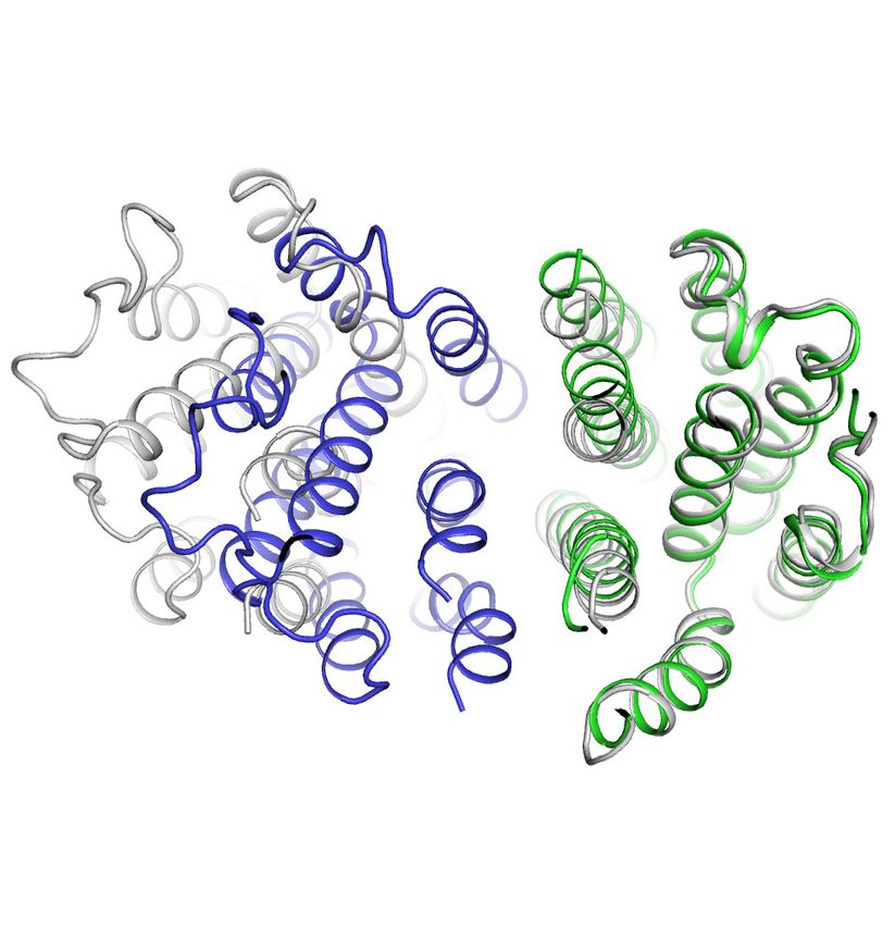
Research article Structural Biology and Molecular Biophysics
Fig. 1
A
Outer
segment Disc lumen
11-cis retinal all-trans retinal
11-cis retinal all-trans retinal
Opsin Opsin 4
CA
ABCA4 AB
Inner ADP
11-cis retinal all-trans retinal ATP
segment ATP
ADP Light
The Visual Cycle
A photoreceptor cell
kDa 670 15844 17 1.35
C 4
B C D
A4
AB CA
EQ AB
1000 30 20 + Retinal
ATP turnover rate [min-1]
ATP turnover rate [min-1]
T
+ Retinoic Acid
W
800 kDa WT ABCA4 + Retinal/DOPE
15 + Retinoic Acid/DOPE
A280 (mAU)
600 250 20
150
400 100 10
75
10
200 50
5
0 EQ ABCA4
0 0
0.0 5.0 10.0 15.0 20.0 25.0 0 1 2 3 4 0 10 20 30 40 50
Elution Volume (mL) ATP [mM] Substrate (µM)
Figure 1. Biochemical characterization of ABCA4. (A) An illustration showing a rod photoreceptor cell and a zoomed-in view of the outer segment disc,
where ABCA4 is located. ATP-hydrolysis enables ABCA4 to transport all-trans retinal and 11-cis retinal from the disc lumen into cytosol. (B) Size
exclusion profile of purified wild-type (WT) ABCA4 and the E1087Q/E2096Q mutant (EQ). (C) Basal ATPase activity measured in 0.06% digitonin at 28˚C.
Data points represent the means and standard deviations (SDs) of three measurements. The WT ABCA4 has a Km of 0.08 ± 0.01 mM and specific
turnover rate of 28.1 ± 0.7 ATP per minute, corresponding to a maximal ATPase activity of 112.5 ± 2.8 nmol/mg/min. (D) The ATPase activity of purified
ABCA4 as a function of all-trans retinal or all-trans retinoic acid in the presence or absence of 0.1 mg/mL 1,2-dioleoyl-sn-glycero-3-
phosphoethanolamine (DOPE) and 100 mM ATP. Data points represent the means and SDs of three measurements from the same protein preparation.
These studies provide direct evidence that ABCA4 is an ATP-driven importer, transporting N-retinyli-
dene-PE from the luminal to the cytoplasmic side of the disc membrane in photoreceptor cells.
Although significant progress was made in understanding the physiological role of ABCA4, struc-
tural information is limited. Previously, the overall architecture of bovine ABCA4 was characterized
by electron microscopy (EM) to 18 Å resolution (Tsybovsky et al., 2013). More recently, the struc-
ture of a related transporter, ABCA1, was determined to be 4.1 Å (Qian et al., 2017). ABCA1 is a
lipid exporter sharing 51% sequence identity with ABCA4 but transports substrate in the opposite
direction.
In this study, we used cryo-electron microscopy (cryo-EM) to determine the structures of human
ABCA4 in two conformations. These structures, both determined at 3.3 Å resolution, reveal ATP-
dependent conformational changes different from those observed in any other ABC transporters.
Results
Recombinant ABCA4 functions similarly to protein purified from native
source
The wild-type (WT) ABCA4 and a double-mutant E1087Q/E2096Q (EQ) were overexpressed in mam-
malian cells and purified to homogeneity (Figure 1B). The E1087 and E2096 residues were con-
served as the general bases catalyzing ATP hydrolysis. Mutation of the corresponding residues in
many other ABC transporters abolished ATP hydrolysis but not ATP binding (Kim and Chen, 2018;
Zhang et al., 2017; Zhang et al., 2018). The size-exclusion profiles of the purified EQ mutant and
Liu et al. eLife 2021;10:e63524. DOI: https://doi.org/10.7554/eLife.63524 2 of 18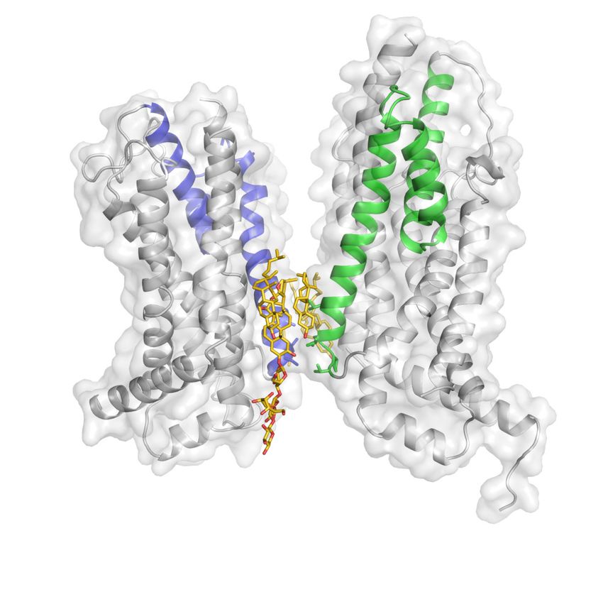
Research article                                                                           Structural Biology and Molecular Biophysics

                                    the WT protein are comparable, suggesting that the EQ mutant folds similarly to the WT protein
                                    (Figure 1B).
                                       The ATPase activity of ABCA4 was measured in detergent solution as a function of ATP concen-
                                    tration (Figure 1C). The recombinant WT protein shows a Km of 80 ± 10 mM for ATP and a maximal
                                    hydrolysis activity of 28.1 ± 0.7 ATP per minute (or 112.5 ± 2.8 nmol/mg/min), comparable to those
                                    of the native protein purified from rod photoreceptor cells (75 ± 18 mM and 190 ± 36 nmol/mg/min,
                                    respectively) (Ahn et al., 2000). The EQ mutant, on the other hand, shows no measurable ATPase
                                    activity (Figure 1C). Also consistent with literature (Quazi and Molday, 2014), in the presence of
                                    PE, retinal stimulates the ATPase activity of WT ABCA4 by up to threefold (Figure 1D). No stimula-
                                    tion was observed with retinoic acid or in the absence of PE (Figure 1D). These data indicate that
                                    the recombinant ABCA4 exhibits biochemical properties very similar to the ABCA4 purified from
                                    native sources.

                                    ABCA4 adopts an outward-facing conformation in the absence of ATP
                                    The human ABCA4 is a single polypeptide of 2273 residues (Figure 2). It consists of two homolo-
                                    gous halves, each containing a transmembrane domain (TMD), an exocytoplasmic domain (ECD), a
                                    cytoplasmic nucleotide binding domain (NBD), and a regulatory domain (RD) (Bungert et al., 2001).
                                    The structure of WT ABCA4 was determined by single-molecule cryo-EM in the absence of ATP and
                                    substrate to an overall resolution of 3.3 Å (Figure 2—figure supplement 1, Table 1). The EM den-
                                    sity is of high quality (Figure 2—figure supplement 2), permitting de novo building of most resi-
                                    dues, except for a few loops and a 134-residue region in ECD1 (Figure 2B, blue density). In
                                    addition, residues 118–137 and 479–496 were built as polyalanine as their side chain densities were
                                    invisible. The final model, consisting of 1941 residues, 14 lipids, 1 detergent molecule, and 14 sugar
                                    molecules, was refined to excellent stereochemistry (Table 1).
                                        ABCA4 is an elongated molecule, extending approximately 240 Å across the lipid bilayer
                                    (Figure 2B). The two ECDs pack closely with each other, protruding 130 Å into the lumen
                                    (Figure 2B). The two TMDs adopt an outward-facing configuration, forming a large hydrophobic
                                    cavity that is continuous with both the luminal solution and, through a lateral opening, to the lipid
                                    bilayer itself. In the absence of ATP, the two NBDs are apart from each other and the two RDs form
                                    extensive interdomain interactions (Figure 2B). It was previously suggested that the highly specific
                                    localization of ABCA4 to the rim region of the disc membrane (Figure 1A) might be due to the large
                                    size of the ECD domains (Molday, 2015). The structure of ABCA4 supports this notion as the spac-
                                    ing between the flattened regions of the disc membranes (Nickell et al., 2007) may be insufficient
                                    to accommodate the elongated ECDs.

                                    An extended groove of the ECDs
                                    The luminal region, composed of 600 residues in ECD1 and 290 residues in ECD2, does not contain
                                    any known enzymatic or structural motifs of known function. However, more than 300 disease-caus-
                                    ing mutations map to this region (Figure 3—figure supplement 1) (Human Gene Mutations Data-
                                    base: http://www.hgmd.cf.ac.uk), underscoring the essential nature of the ECDs.
                                        In contrast to the rest of the molecule, where the N- and C-terminal halves have similar structures,
                                    pseudo twofold symmetry does not exist in the ECDs. Instead, the ECDs assemble to form one unit
                                    with a large extended hollow interior, accessible from the luminal space (Figure 3). The structure, as
                                    previously described for ABCA1 (Qian et al., 2017), can be separated into three layers: base, tunnel,
                                    and lid (Figure 3A). The base has a global fold docked onto the luminal surface of the two TMDs.
                                    The tunnel is an -helical barrel with an inner diameter of approximately 15 Å (Figure 3A, D). The
                                    density corresponding to the lid region does not permit amino acid assignment, suggesting that the
                                    lid region is flexible (Figure 3A, C). Nevertheless, it is clear that the interior of the lid is also hollow
                                    (Figure 3C). Strong densities that may correspond to lipid or detergent molecules were observed
                                    inside the extended groove in the base layer (Figure 3A, B, Figure 2—figure supplement 2).
                                        Consistent with biochemical analysis (Bungert et al., 2001), the structure of the ECDs is stabilized
                                    by six disulfide bonds (Figure 3E). Seven missense mutations of the cysteines involved in disulfide
                                    bonding (C54Y, C75G, C519R, C641S, C1455R, C1488R, and C1490Y) have been identified in
                                    patients with retinal diseases (Fumagalli et al., 2001; Hu et al., 2019; Lewis et al., 1999;
                                    Rosenberg et al., 2007). Previously, mass spectrometry (Tsybovsky et al., 2011) and mutagenesis

Liu et al. eLife 2021;10:e63524. DOI: https://doi.org/10.7554/eLife.63524                                                                 3 of 18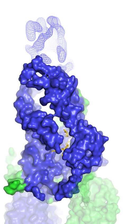
Research article                                                                                                                                                            Structural Biology and Molecular Biophysics

    A

                                  ECD1
                                                                                                                       ECD2
                                                                                                                                                                        1862
                                                                                                                                       1665
                                                                                     814
      Disc                                       648
                                                                       782     788                                                                      1815     1820
     Lumen              45                             722       730                             836
                                                                                                               1393                      1749        1760                             1874
                                                                                     EH2                                                                                EH4
                                                                               EH1                                                                               EH3

                                                                               796    799
                   TMD1 1                    2          3       4       5                    6                        7            8          99    10    11     1834 1839     12 TMD2
                                         3

   Cytoplasm   N       IH1        22
                                                               752     759
                                                                                     861                       IH3     1370                        1783   1790
                             20                          693                                                                            1720                                   1898
                   3                                                                                      1348 1368
                                       679       IH2
                                                                                                                            1706       IH4
                                                                                           914                                                                                 1916

                                                                                                 NBD1                                                                   NBD2
                                                                                                                     1280
                                                                                                              RD1                                           RD2                        C
                                                                                                                                                                  VFVNFA              2273
                                                                                                                                                                           2253

    B

                                  ECD2                                       ECD1

          Lumen
          ~130 Å

          Disc
        membrane                  TMD1                                         TMD2                     90o

          ~40 Å

        Cytoplasm
                             NBD1                                                  NBD2
          ~70 Å

                                       RD2                                   RD1

Figure 2. The overall structure of ABCA4 in the absence of ATP. (A) The domain structure of ABCA4. The N- and C-terminal halves of the molecule are
shown in blue and green. (B) Two orthogonal views of ABCA4 in ribbon presentation. Also shown is the electron microscopy density corresponding to
residues 138–271 in ECD1. Ordered detergents, lipids, and N-linked glycans are shown as stick model. The position of the membrane is indicated by
two gray lines.
The online version of this article includes the following figure supplement(s) for figure 2:
Figure supplement 1. Cryo-electron microscopy reconstructions of the ATP-free, wild-type ABCA4.
Figure supplement 2. Local density of the ATP-free, wild-type ABCA4 reconstruction.

                                                         studies (Bungert et al., 2001) have identified multiple N-linked glycosylation sites in the ECDs. Den-
                                                         sities corresponding to sugar moieties were observed on residues N98, N415, N444, N504, N1469,
                                                         N1529, N1588, and N1662. It is likely that glycosylation plays an important role in the folding or traf-
                                                         ficking of ABCA4,as missense mutations at positions 415, 504, and 1588 are linked to retinal dis-
                                                         eases (Consugar et al., 2015; Khan et al., 2019; Kim et al., 2019).

Liu et al. eLife 2021;10:e63524. DOI: https://doi.org/10.7554/eLife.63524                                                                                                                                                  4 of 18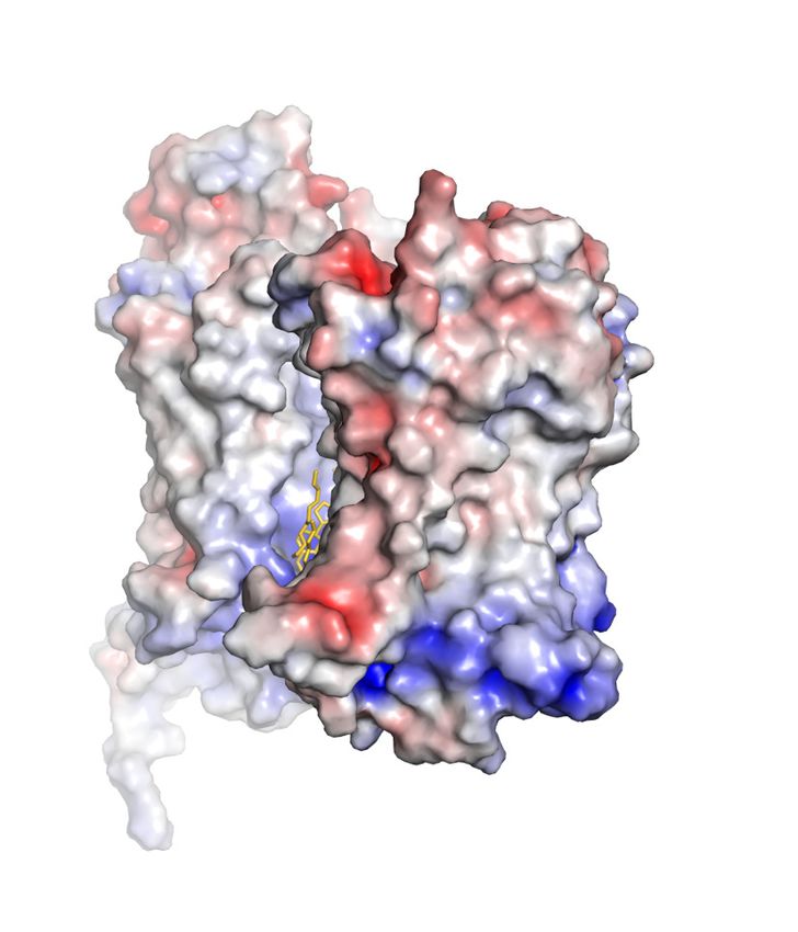
Research article                                                                       Structural Biology and Molecular Biophysics

                                    Table 1. Summary of electron microscopy data and structure refinement statistics for wild-type
                                    ABCA4.
                                    Data collection
                                    Microscope                                                                            Titan Krios (FEI)
                                    Voltage (kV)                                                                          300
                                    Detector                                                                              K2 Summit (Gatan)
                                    Pixel size (Å)                                                                       1.03
                                    Defocus range (mm)                                                                    0.8–2.5
                                    Movies                                                                                5226
                                    Frames/movie                                                                          50
                                     DDose rate (electrons/pixel/s)                                                       8
                                                            2
                                    Total dose (electrons/Å )                                                            75
                                    Number of particles                                                                   1,375,284
                                    Model composition
                                    Non-hydrogen atoms                                                                    15,789
                                    Protein residues                                                                      1941
                                    Lipids                                                                                14
                                    Digitonin                                                                             1
                                    Sugar molecules                                                                       14
                                    Refinement
                                    Resolution (Å)                                                                       3.3
                                    Sharpening B-factor (Å2)                                                                 99.8
                                    Root-mean-square deviations
                                    Bond lengths (Å)                                                                     0.006
                                    Bond angles (˚)                                                                       1.185
                                    Validation
                                    Molprobity score                                                                      2.16
                                    Clashscore, all atoms                                                                 18.86
                                    Favored rotamers (%)                                                                  97.10
                                    Ramachandran plot (%)
                                    Favored                                                                               94.28
                                    Allowed                                                                               5.72
                                    Outliers                                                                              0.0

                                       Prokaryotic ABC importers often contain an extracytoplasmic binding protein that functions as a
                                    receptor for substrates (Davidson et al., 2008). Currently it is unknown if the ECDs of ABCA4 have
                                    an analogous function. Previously it was shown that isolated ECD2 binds to all-trans retinal (Biswas-
                                    Fiss et al., 2010), but how this interaction relates to the transport cycle remains unclear.

                                    The transmembrane region contains a hydrophilic cleft
                                    The structure of the TMD belongs to the newly classified type V fold (Thomas and Tampé, 2020)
                                    previously observed only in ABC exporters (Lee et al., 2016; Qian et al., 2017; Taylor et al., 2017;
                                    Bi et al., 2018). The two TMDs come in contact with each other through two helical turns at the
                                    cytoplasmic end of TM5 and TM11, burying only 124 2 surface per subunit (Figure 4A). The gap
                                    between the TMDs at the level of the inner leaflet is filled with ordered lipids (Figure 4A, B). Thus,
                                    the cytoplasmic gate of the transmembrane pathway is formed partially by residues in TM5 and
                                    TM11 and partially by lipid molecules. Mutations of many lipid-interacting residues are found in
                                    patients with vision diseases (Human Gene Mutations Database: http://www.hgmd.cf.ac.uk). These

Liu et al. eLife 2021;10:e63524. DOI: https://doi.org/10.7554/eLife.63524                                                             5 of 18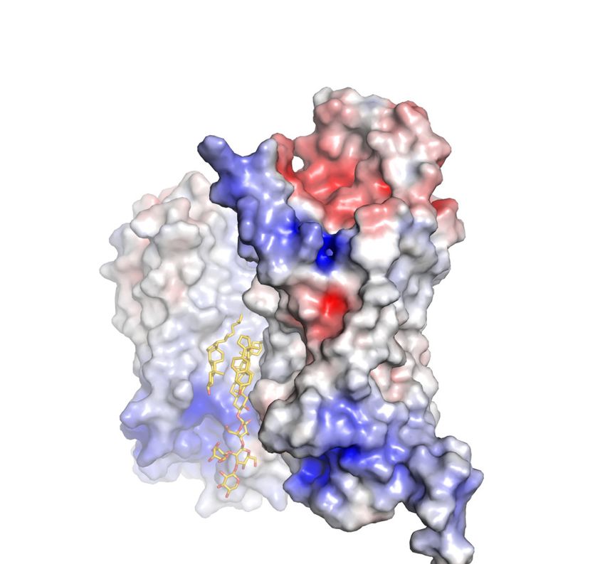
Research article                                                                                Structural Biology and Molecular Biophysics

               A                                 B                                 C                Lid              E                Base

     Lid
                                                                                                                                                      C81

                                                                                                                                             C54
                                                                                                                             C1488
                                                                                                                                     C641

   Tunnel                                                                                                                            C1490
                                                                                                                         C1502

                                                                                   D              Tunnel

    Base                                                                                                                                     C519

                                                                                                                         C324                       C370
                                                                                                 ~15 Å
                                                                                                                             C75

Figure 3. A three-tiered structure of the exocytoplasmic domains (ECDs). (A) Ribbon and (B) surface representation of the ECDs, together with the
electron microscopy (EM) density of the lid region. Bound detergents and lipids are shown as yellow sticks. (C) A luminal view of the lid; the EM density
is shown as blue mesh. (D) A cross section of the tunnel region. (E) The ECDs are stabilized by inter- and intra-domain disulfide bonds.
The online version of this article includes the following figure supplement(s) for figure 3:
Figure supplement 1. Disease-causing mutations (orange) are widely distributed throughout the structure.

                                     observations indicate that lipids form an integral part of the retinoid transport system, possibly by
                                     regulating the folding and function of ABCA4 (Figure 1, Ahn et al., 2000).
                                        A prominent feature of the type V fold is that each TMD contains two exocytoplasmic helices
                                     (EHs) forming a EH-turn-EH insertion halfway into the membrane (Figure 4B, C). Each EH exposes a
                                     helical end to the low dielectric membrane center, a thermodynamically unfavorable configuration.
                                     In ABCA4, this configuration is stabilized by an acidic residue and a tyrosine residue on the neigh-
                                     boring TM helices (Figure 4B, C). In TMD1, D846 and Y850 form hydrogen bonds with mainchain
                                     carbonyl oxygen and amide atoms of EH1 and EH2, respectively (Figure 4B, right panel). In TMD2,
                                     E1885 and Y1889 neutralize the ionizable atoms at the ends of EH3 and EH4 (Figure 4C, right
                                     panel). Mutating E1885 to lysine causes Stargardt disease (Rivera et al., 2000), likely due to folding
                                     defects. The EH3-turn-EH4 insertion leads to a large hydrophilic cleft on the surface of TMD2
                                     (Figure 4C). A cluster of charged residues from EH3, EH4, and TM12, including the clinically relevant
                                     residue R1843 (Lewis et al., 1999; Rivera et al., 2000), are located as deep as 10 below the
                                     expected membrane surface (Figure 4C).
                                        In some K+ or Cl- channels, helices with one end exposed to the membrane contribute to the
                                     binding and selection of conducting ions (Doyle, 1998; Faraldo-Gómez et al., 2004). Although the
                                     EH-turn-EH insertion is highly conserved in type V transporters, the functional significance of this
                                     motif is still unknown.

                                     The RDs exhibit ACT-like folds
                                     In the cytoplasm, the two NBDs and two RDs form a domain-swapped dimer (Figure 2B). RD1
                                     crosses the dimer interface to interact with NBD2, and RD2 crosses back over to pack against NBD1.
                                     The two RDs have very similar structures; both exhibit a fold similar to the ACT domain (Figure 4D),
                                     which is a conserved structural motif that typically binds small regulatory molecules (Chipman and
                                     Shaanan, 2001; Grant, 2006). Sequence alignment indicates that the ACT fold is conserved among
                                     all members of the ABCA subfamily (Figure 4—figure supplement 1).

Liu et al. eLife 2021;10:e63524. DOI: https://doi.org/10.7554/eLife.63524                                                                             6 of 18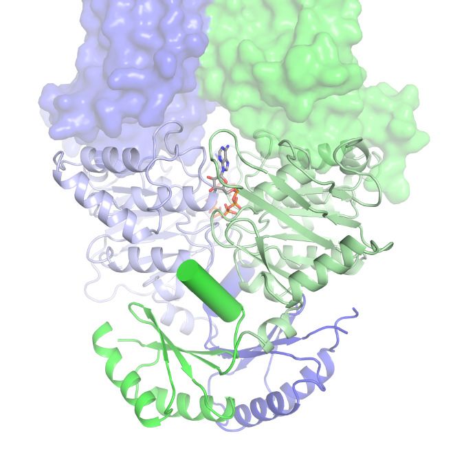
Research article                                                                                    Structural Biology and Molecular Biophysics

                                       Fig. 4
                                        A

                                                                                              90o

                                                      TMD1                 TMD2
                                                              140o

                                        B                                                                              EH2

                                                                                                      EH1

                                                                                                                 D846

                                                                                                                Y850

                                                      TMD2           TMD1

                                                              180o                                                          EH4

                                        C

                                                                                                    EH3

                                                                                                                    R1843

                                                                                                                     E1885

                                                                                                                    Y1889

                                                      TMD1           TMD2

                                                                          Į2   Į1
                                                      Į1     Į2
                                        D
                                                                                                                Į2          Į1
                                            R2                                           R1

                                                       ȕ3    ȕ1 ȕ4   ȕ4   ȕ1   ȕ3                         ȕ4                ȕ3
                                                 ȕ2                                 ȕ2                         ȕ1                 ȕ2

                                                                                                                    ACT fold

                                    Figure 4. The structures of the transmembrane domains (TMDs) and regulatory domains (RDs). (A) Two views of
                                    the TMDs. EH1, EH2, and TM5 are highlighted in blue, and EH3, EH4, and TM11 are in green. The side chain of
                                    residues making inter-subunit contacts is shown as stick model. Lipids are shown in yellow. (B) Electrostatic surface
                                    representation of the TMDs, calculated assuming a pH of 7 and a concentration of 0.15 M of both (+1) and ( 1)
                                    Figure 4 continued on next page

Liu et al. eLife 2021;10:e63524. DOI: https://doi.org/10.7554/eLife.63524                                                                          7 of 18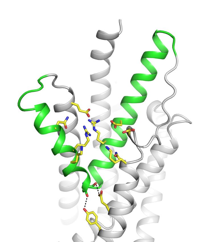
Research article                                                                                        Structural Biology and Molecular Biophysics

                                    Figure 4 continued
                                    ions. Scale: red, negative ( 5 kT/e); blue, positive (+5 kT/e). The EH1-turn-EH2 insertion is highlighted as blue
                                    ribbon. (C) An orthogonal view of (B), highlighting the hydrophilic indentation formed by the EH3-turn-EH4
                                    insertion. (D) The RDs exhibit the ACT-like fold with the babbab topology. EH: exocytoplasmic helices.
                                    The online version of this article includes the following figure supplement(s) for figure 4:
                                    Figure supplement 1. The ACT motifs and the pinning helices (PHs) are conserved in the ABCA subfamily.

                                       In the ABC transporter superfamily, the bacterial methionine importer MetNI also contains two
                                    RDs with ACT-like folds. The RDs of MetNI play an important role in ‘trans-inhibition’: at high intra-
                                    cellular concentration, methionine binds to the RDs to inhibit further uptake of methionine
                                    (Kadaba et al., 2008). For ABCA4, it was reported that an NBD1-R1 construct binds retinal with
                                    high affinity (Biswas-Fiss et al., 2012). Whether retinal binding is mediated through the ACT motif
                                    and the physiological relevance of such interaction remain to be investigated.

                                    Structure of ABCA4 in the presence of ATP
                                    To capture ABCA4 in its ATP-bound state, the structure of the hydrolysis-deficient mutant ABCA4-
                                    EQ was determined in the presence of 10 mM ATP-Mg2+ to 3.3 Å resolution (Figure 5, Figure 5—
                                    figure supplements 1 and 2, Table 2). Both 2D and 3D image classes from cryo-EM data show that
                                    most particles are in the NBD-dimerized configuration. The final model contains 2 ATP molecules, 8
                                    lipid molecules, and 1920 residues. Similar to that of the WT protein, the lid region is highly mobile.

     A                                                   B

                     ECD2                   ECD1

     Lumen
                                                                          EH3   EH4
     ~130 Å

     Disc
   membrane          TMD1                 TMD2
                                                         C         ATPase Site 1                     ATPase Site 2
     ~40 Å

                                                                  T2070
                                                                                                   D1061
    Cytoplasm
                                                                                           K1054
                     NBD1                    NBD2
     ~70 Å
                                                                                Walker A
                                                             Signature Motif                                     Walker A
                                                                                           Signature Motif

                        RD2              RD1

Figure 5. The structure of ABCA4 in the presence of ATP. (A) Ribbon diagram of the ATP-bound ABCA4 EQ mutant, together with electron
microscopy density of the lid region. (B) Surface representation of the exocytoplasmic domains. The EH3-turn-EH4 motif is shown in green ribbon; lipids
are shown as stick model. (C) Structure of the two ATPase sites. ATP is shown as sticks. Dashed lines indicate the non-consensus hydrogen bonds
stabilizing the ribose moiety. EH: exocytoplasmic helices.
The online version of this article includes the following figure supplement(s) for figure 5:
Figure supplement 1. Cryo-electron microscopy reconstructions of the ATP-bound, EQ ABCA4.
Figure supplement 2. Local density of the ATP-bound, EQ ABCA4 reconstruction.

Liu et al. eLife 2021;10:e63524. DOI: https://doi.org/10.7554/eLife.63524                                                                                 8 of 18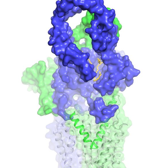
Research article                                                                      Structural Biology and Molecular Biophysics

                                    Table 2. Summary of electron microscopy data and structure refinement statistics for EQ ABCA4.
                                    Data collection
                                    Microscope                                                                           Titan Krios (FEI)
                                    Voltage (kV)                                                                         300
                                    Detector                                                                             K2 Summit (Gatan)
                                    Pixel size (Å)                                                                      1.03
                                    Defocus range (mm)                                                                   0.8–2.5
                                    Movies                                                                               9070
                                    Frames/movie                                                                         50
                                    Dose rate (electrons/pixel/s)                                                        8
                                                            2
                                    Total dose (electrons/Å )                                                           75
                                    Number of particles                                                                  1,136,006
                                    Model composition
                                    Non-hydrogen atoms                                                                   15,513
                                    Protein residues                                                                     1920
                                    Lipids                                                                               8
                                    ATP                                                                                  2
                                    Mg2+                                                                                 2
                                    Sugar molecules                                                                      14
                                    Refinement
                                    Resolution (Å)                                                                      3.3
                                    Sharpening B-factor (Å2)                                                                93.8
                                    Root-mean-square deviations
                                    Bond lengths (Å)                                                                    0.007
                                    Bond angles (˚)                                                                      0.898
                                    Validation
                                    Molprobity score                                                                     2.10
                                    Clashscore, all atoms                                                                12.42
                                    Favored rotamers (%)                                                                 97.99
                                    Ramachandran plot (%)
                                    Favored                                                                              91.68
                                    Allowed                                                                              8.32
                                    Outliers                                                                             0.0

                                        In the presence of ATP and absence of substrate, ABCA4 exhibits a conformation very different
                                    from that of the ATP-free structure (Figure 5). The two NBDs form a closed dimer. The two TMDs
                                    are also in close contact, leaving no cavity at their interface (Figure 5A). The ECDs are positioned
                                    differently from that of the ATP-free form, placing the hollow channel directly above the EH3-turn-
                                    EH4 cleft (Figure 5B).
                                        The ATP-binding signature motifs of ABCA4 deviate from the consensus LSGGQ sequence. The
                                    signature motif of NBD1 is LSGGM and that of NBD2 is YSGGN. In a typical ATPase site, Q in the
                                    LSGGQ signature motif positions the ribose moiety of ATP through an H-bond and van der Waals’
                                    contact (Davidson and Chen, 2004). In ABCA4, this role is fulfilled by the mainchain carboxyl group
                                    of T2070 in ATPase site 1 and the side chain of K1054 and mainchain carboxyl of D1061 in ATPase
                                    site 2 (Figure 5C). Consequently, the two ATP molecules are bound in a manner typical of ABC
                                    transporters.
                                        ABCA4 binds and hydrolyzes GTP as effectively as ATP (Ahn et al., 2000; Illing et al., 1997). The
                                    structure indicates that both ATPase sites can bind GTP without steric hindrance. As rod

Liu et al. eLife 2021;10:e63524. DOI: https://doi.org/10.7554/eLife.63524                                                            9 of 18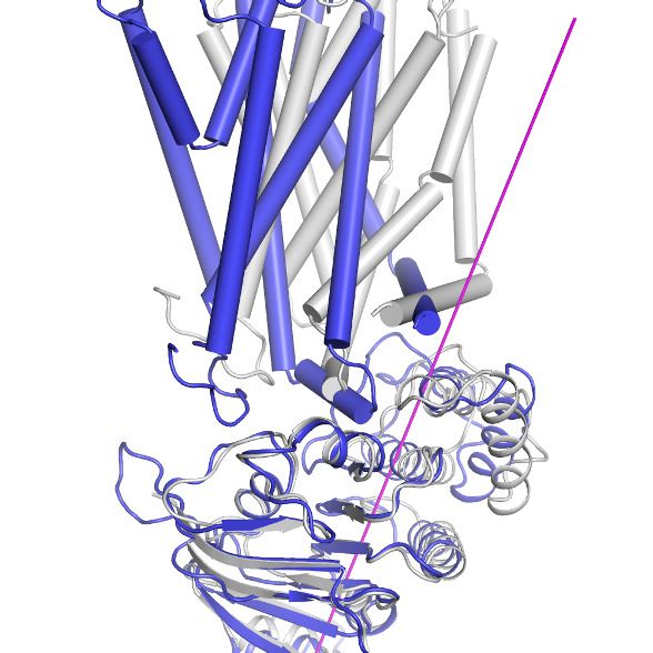
Research article Structural Biology and Molecular Biophysics
photoreceptor cells contain similar levels of ATP and GTP (Biernbaum and Bownds, 1985), these
observations suggest that ABCA4 might utilize either nucleotide to power retinal translocation.
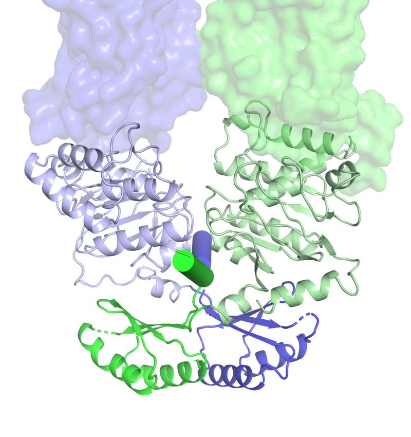
Nature of the conformational changes
The large conformational changes of ABCA4 are initiated in the NBDs where two ATP molecules
bind and stabilize a closed NBD dimer. Using the RD dimer as a reference, the two NBDs move like
a pair of tweezers akin to that of the maltose transporter (Oldham et al., 2007; Figure 6A, B). At
the base of the tweezers, two short helices pin the two NBDs together from opposing sides of the
molecule (Figure 6A, B, cylinders). One of the pinning helices (PHs) is formed by residues following
RD1 and the other PH contains the C-terminal, highly conserved, VFVNFA motif (Fitzgerald et al.,
2004; Patel et al., 2019). In the absence of ATP, each PH simultaneously interacts with the H loop
of one NBD and the D loop of the other NBD (Figure 6A). Upon ATP-binding, the NBDs rotate
toward each other, forming additional contacts with the RDs and PHs that are functionally important
(Figure 6B). For example, in the ATP-bound conformation, D1102 in NBD1 forms a hydrogen bond
with H2202 in RD2, and R2107 in NBD2 forms salt bridges with E1270 in PH1 (Figure 6B). Mutations
D1102Y and R2107H, which disrupt these interactions, are observed in patients with vision diseases
(Fritsche et al., 2012; Zernant et al., 2014). Notably, the R2107H mutation is the most frequent
mutation found in African Americans (Zernant et al., 2014).
How does NBD dimerization in the cytosol affect the TMDs to alter access of the translocation
pathway? In opposing contrast to typical ABC transporters that are inward-facing when their NBDs
are separated, the resting state of ABCA4 is outward-facing (Figure 2). Thus, the nature of the con-
formational change in ABCA4 is qualitatively different from the ‘rocker-switch’ motion described for
many transporters (Abramson et al., 2003; Huang et al., 2003). Transition from the ATP-free to
ATP-bound conformation involves two motions of the TMDs. The first is a movement in concert with
the NBDs toward the molecular center, bringing the TMDs closer to each other. The second is a 30˚
twisting motion of TMD1 relative to the RecA-like subdomain (Figure 6C). The helical subdomain of
NBD1 moves together with TMD1. This twisting motion, not seen in any other ABC transporter, pla-
ces helices TM1 and TM2 within van der Waals’ contact with TM8 and TM11, closing the TM cavity
completely (Figure 6D).
Discussion
Although a large body of clinical data indicates that ABCA4 plays a vital role in vision, we are still at
an early stage in understanding its molecular mechanism. In this work, we present the structures of
ABCA4 in two different conformations. These structures provide a molecular basis to understand
why many clinically relevant mutations (Figure 3—figure supplement 1) could lead to misfolding or
malfunction of ABCA4. They also reveal several intriguing features of ABCA4 that could be impor-
tant to its function. For example, the ECDs contain an extended lipid-binding groove that changes
its position in the presence of ATP. The EH3-turn-EH4 cleft, unique in ABCA4, seems well-positioned
to lower the barrier for lipid headgroups to flip across the membrane. The ACT-fold of the RDs
suggests that they may interact with small molecules to regulate the activity of ABCA4. Taken
together, the structural information provides a molecular basis to design new experiments to
uncover how ABCA4 recruits, flips, and releases retinoids and how these processes are regulated in
the vision cycle.
The two structures of ABCA4 support an alternating access model for retinal import (Figure 7). In
the resting state, ABCA4 adopts an outward-facing conformation, poised to recruit substrate from
the exocytoplasmic side of the membrane. ATP binding induces a major conformational change that
closes the transmembrane pathway to release substrate. ATP hydrolysis resets the transporter to
begin a new cycle. Several lines of evidence support this working model. In the absence of ATP,
N-retinylidene-PE and all-trans retinal bind ABCA4 with micromolar affinity (Beharry et al., 2004;
Zhong and Molday, 2010) consistent with substrate recruitment to the outward-facing resting state.
The addition of ATP, but not ADP, releases substrate (Beharry et al., 2004; Zhong and Molday,
2010), likely due to the closure of the TM cavity in the NBD-dimerized conformation (Figure 6).
Many details of the ABCA4 transport cycle, such as whether an inward-facing conformation exists
and how substrate is recruited and released, remain to be determined.
Liu et al. eLife 2021;10:e63524. DOI: https://doi.org/10.7554/eLife.63524 10 of 18Research article Structural Biology and Molecular Biophysics
ATP-free ATP-bound
A B
NBD1 NBD2 NBD1 NBD2
RD2 RD1 RD2 RD1
NBD1 NBD2 NBD1 NBD2
D loop H2128
H2128
D1102
D1102
H2202
H2202
180o 180o
NBD2 NBD1 NBD2 NBD1
H1119 180o H1119
R2107 R2107
E1270 E1270
C D
30o
1 7
1 5
3 2
3
2 8
IH1
2
Helical subdomain
RecA-like subdomain
11
1
Figure 6. Conformational changes upon ATP binding. (A, B) The tweezer-like motion of the nucleotide binding
domains (NBDs). Transmembrane domains (TMDs) are shown as surface representations; NBDs are displayed as
ribbons. The two pinning helices are highlighted as cylinders. ATPs are shown as sticks. (C) The twisting motion of
TMD1. The two structures are superpositioned based on NBD1. The ATP-bound form is shown in blue and ATP-
Figure 6 continued on next page
Liu et al. eLife 2021;10:e63524. DOI: https://doi.org/10.7554/eLife.63524 11 of 18Research article Structural Biology and Molecular Biophysics
Figure 6 continued
free conformation in gray. The rotation axis of TMD1 and the helical subdomain is indicated in magenta. (D)
Closing of the TM cavity upon TMD1 twisting. The structures are aligned based on TMD2. ATP-bound TMDs are
shown in blue and green; ATP-free conformation is shown in gray.
As more ABC transporter structures become available, it is now evident that one cannot differen-
tiate importers from exporters based on their structures. For example, the previously defined ‘type I
exporter fold’ has now been observed in importers YbtPQ (Wang et al., 2020), IrtAB (Arnold et al.,
2020), and ABCD4 (Xu et al., 2019). The structures of ABCA1 and ABCA4 are also very similar (Fig-
ure 7—figure supplement 1). Their TMDs are essentially superimposable, and the ECDs also share
a common fold. The two structures differ significantly in the cytoplasmic region: ABCA1 does not
exhibit the domain-swapped configuration as observed in ABCA4 (Figure 7—figure supplement 1);
however, this difference is unlikely to be the determinant of their functional difference. The transport
directionality of ABCA1 and ABCA4 is likely to be governed by when the substrates are recruited
and released.
Materials and methods
Cell culture
Sf9 cells were cultured in Sf-900 II SFM medium (GIBCO) 5% FBS and 1% antibiotic-antimycotic.
HEK293S GnTl-cells were cultured in Freestyle 293 (GIBCO) supplemented with 2% FBS and 1% anti-
biotic-antimycotic.
Mutagenesis
Mutations (E1087Q, E2096Q) were introduced using QuikChange Site-Directed Mutagenesis System
(Stratagene).
Protein expression and purification
DNA encoding the human ABCA4 gene was synthesized and codon-optimized for expression in
mammalian cells (BioBasic) and subcloned into a BacMam expression vector (Goehring et al., 2014)
with a C-terminal green fluorescent protein (GFP) tag. The resulting plasmid was transformed to
DH10Bac Escherichia coli cells to produce bacmid DNA. Recombinant baculoviruses were first gen-
erated in Sf9 cells using Cellfectin II reagents (Invitrogen). The resulting P1 viruses were amplified for
two more generations. To express the recombinant proteins, P3 virus (10% v/v) was used to infect
ABCA4
ATP binding
Outward-facing
ATP-bound
ATP-free
substrate ATP hydrolysis
ATP
Figure 7. A working transport model. For simplicity, the extracellular domains and the substrate-binding proteins
are omitted from the schematic drawings.
The online version of this article includes the following figure supplement(s) for figure 7:
Figure supplement 1. Structural comparison of ABCA4 and ABCA1.
Liu et al. eLife 2021;10:e63524. DOI: https://doi.org/10.7554/eLife.63524 12 of 18Research article Structural Biology and Molecular Biophysics
HEK293S GnTI- suspension cells at 3 106 cells/mL. Infected cells were cultured at 37˚C for 12 hr
before the temperature was decreased to 30˚C. Protein expression was induced by adding 10 mM
sodium butyrate at 30˚C (Goehring et al., 2014). To purify the protein, cells were harvested after 48
hr post induction, resuspended in lysis buffer (50 mM HEPES pH 8, 2 mM MgCl2, 200 mM NaCl,
20% Glycerol), and supplemented with protease inhibitors (1 mg/mL leupepetin, 1 mg/mL pepstatin,
1 mg/mL aprotonin, 100 mg/mL trypsin inhibitor, 1 mM benzamidine, and 1 mM phenylmethylsulfonyl
fluoride [PMSF]) and DNase (3 mg/mL). Membranes were solubilized with 1.25% (w/v) 2,2-didecylpro-
pane-1,3-bis-b-D-maltopyranoside and 0.25% (w/v) cholesteryl hemisuccinate for 2 hr at 4˚C. The cell
lysates were centrifuged at 70,000 g for 1 hr, and the supernatant was applied to CNBR-activated
Sepharose resin (GE Healthcare) conjugated with anti-GFP nanobodies (Kirchhofer et al., 2010).
The resin was washed with buffer A (20 mM HEPES pH 8, 200 mM NaCl, 2 mM MgCl2, 0.06% digito-
nin) and then incubated with PreScission protease (5:1 w/w ratio) at 4˚C for 3 hr to remove the C-ter-
minal GFP tag. The protein was eluted with buffer A and further purified with gel filtration
chromatography using a Superose 6 10/300 column (GE Healthcare) equilibrated with buffer A. The
EQ ABCA4 sample was purified similarly except that 1 mM ATP was supplemented to the lysis buffer
and buffer A.
EM data acquisition and processing
Protein eluted from the gel filtration column was concentrated to 6 mg/mL. The EQ ABCA4 sample
was further incubated with ATP and MgCl2 (10 mM final concentration) on ice for 15 min. About 3
mM (final concentration) fluorinated Fos-choline-8 was added to samples right before freezing on
Quantifoil R1.2/1.3 400 mesh Au grids using Vitrobot Mark IV (FEI). EM images were collected on a
300 kV Titian Krios (FEI) with a K2 Summit detector (Gatan) in super-resolution mode using Serial
EM. The defocus ranged from 0.8 to 2.5 mm, and the dose rate was 8 e-/pixel/s.
For WT ABCA4, 5226 images were collected. The movie frames were first corrected for gain ref-
erence and binned by 2 to obtain a physical pixel size of 1.03 A˚. Beam-induced sample motion was
corrected using MotionCor2 (Zheng et al., 2017). CTF estimation was performed using Gctf
(Zhang, 2016). In total, 1,375,284 particles were picked automatically using Gautomatch (http://
www.mrc-lmb.cam.ac.uk/kzhang) and imported in cryoSPARC v2 (Punjani et al., 2017) for 2D classi-
fication. After 2D classification, 425,348 particles were selected for ab initio reconstruction in cryo-
SPARC. The best class (320,102 particles) was chosen for non-uniform (NU) refinement to yield a
map of 3.64 Å. The half maps, masks, and particles from cryoSPARC were imported to RELION 3
(Scheres, 2012; Zivanov et al., 2018) through pyem suite (https://github.com/asarnow/pyem;
Asarnow et al., 2019) for postprocessing and polishing. The polished particles were then imported
to cryoSPARC for another round of NU refinement to yield a final map of 3.27 Å.
The EQ ABCA4 datasets were processed similarly with the following differences, and the best
map was obtained by combining two separate datasets. For the first dataset, 6766 images were col-
lected and 857,074 particles were picked by Gautomatch. 2D classification yielded 455,456 particles
for ab initio reconstruction, and the best class (287,299 particles) was chosen for NU refinement to
obtain a 3.57 Å map. Further postprocessing and polishing in RELION 3 yielded a map of 3.37 Å .
For the second dataset, 2304 images were collected and 278,932 particles were picked. 2D classifi-
cation yielded 121,894 particles for ab initio reconstruction, and the best class (46,432 particles) was
chosen for NU refinement to obtain a 3.83 Å map. Further postprocessing and polishing in RELION
3 yielded a map of 3.52 Å . The best classes from both datasets (287,299 particles and 46,432 par-
ticles) were combined after polishing and imported into cryoSPARC to obtain a final map of 3.27 Å
with NU refinement.
Model building, refinement, and analysis
Model building and refinement were carried out as previously described (Zhang and Chen, 2016).
In brief, each dataset was randomly split into two halves, one half for model building and refinement
and the other half for validation. Iterative model building and real space refinement were performed
in Coot (Emsley et al., 2010) and Phenix (Afonine et al., 2018; Liebschner et al., 2019), respec-
tively. The final model of WT ABCA4 includes 1941 residues: 3–117, 118–137 (polyalanine), 272–468,
479–496 (polyalanine), 497–873, 916–937, 947–1162, 1201–1277, 1340–1900, 1911–2172, and 2178–
2253; 14 lipids, one detergent (digitonin), and 14 sugar molecules (10 2-acetamido-2-deoxy-beta-D-
Liu et al. eLife 2021;10:e63524. DOI: https://doi.org/10.7554/eLife.63524 13 of 18Research article Structural Biology and Molecular Biophysics
glycopyranose molecules and 4 beta-D-mannopyranose molecules). The final model of EQ ABCA4
includes 1920 residues: 3–117, 118–137 (polyalanine), 273–279 (polyalanine), 280–303, 308–331,
339–350, 362–448, 449–457 (polyalanine), 458, 459–468 (polyalanine), 498–502 (polyalanine), 503–
881, 914–1164, 1199–1280, 1342–1902, 1916–2173, 2178–2252, 2 Mg2+, 2 ATPs, 8 lipids, and 14
sugar molecules. Structural model validation was done using Phenix and MolProbity (Chen et al.,
2010). The local resolutions were estimated using RELION 3 (Scheres, 2012; Zivanov et al., 2018)
by using half maps from cryoSPARC (Punjani et al., 2017).
Domain movements were analyzed with Dyndom (http://dyndom.cmp.uea.ac.uk/dyndom/). Fig-
ures were generated with PyMOL (Schrödinger, LLC) and UCSF Chimera (Pettersen et al., 2004).
ATPase assay
The basal ATPase activity (Figure 1C) was measured using an ATP/Nicotinamide adenine
dinucleotide (NADH) consuming coupled method (Scharschmidt et al., 1979) in reaction buffer (50
mM HEPES pH 8.0, 150 mM KCl, 0.06% digitonin, 2 mM MgCl2, 60 mg/mL pyruvate kinase, 32 mg/
mL lactate dehydrogenase, 9 mM phosphoenolpyruvate, 0.15 mM NADH). WT ABCA4 or EQ
ABCA4 were diluted to 0.2 mM in reaction buffer. A control buffer was also prepared without
ABCA4. Different concentrations of ATPs were added to initiate the reaction, and the fluorescence
changes of NADH were recorded by a Tecan Infinite M1000 microplate reader at excitation wave-
length of 340 nm and emission wavelength of 445 nm. To quantify the ATPase activity, mean values
and standard deviation from three independent measurements were calculated. The values for Km
and the specific turnover rates were determined by nonlinear regression of the Michaelis–Menten
equation using GraphPad Prism 8. The maximal ATPase activities were calculated assuming a molec-
ular weight of 256 kDa for human ABCA4.
Retinal stimulation of ATPase activity of ABCA4 (Figure 1D) was measured using a Transcreener
ADP2 FI Assay (Bellbrook Labs) in kinetic mode according to the manufacturer’s recommendations.
Reactions were assembled in triplicate in black, round-bottom, 384-well plates (Corning #5414)
under dim red light. Also, 10 mL reactions were prepared by incubating ABCA4 with varying concen-
trations of all-trans retinal or all-trans retinoic acid for 15 min on ice, then adding ATP and 10 mL of
ADP2 detection mix containing tracer and antibody. The final reaction mixture contains 50 nM
ABCA4, 100 mM ATP, 20 mM HEPES, 75 mM NaCl, 3 mM MgCl2, 1 mM Dithiothreitol (DTT),
0.005% glyco-diosgenin (GDN), 0.001% Brij-35, 1% ethanol, and the concentration of substrate as
indicated. Fluorescence changes were recorded by a Tecan Infinite M1000 microplate reader at exci-
tation wavelength of 580 nm and emission wavelength of 620 nm. Standard curves mimicking the
conversion of ATP to ADP were used to convert raw fluorescence measurements to ADP formation.
The data were analyzed by GraphPad Prism 8.
Acknowledgements
We thank Mark Ebrahim and Johanna Sotiris at The Rockefeller Evelyn Gruss Lipper Cryo-Electron
Microscopy Resource Center for assistance in data collection. JL is a fellow of the Helen Hay Whitney
Foundation. We also thank The Rockefeller University and the Howard Hughes Medical Institute for
financial support. The authors declare no competing financial interests.
Additional information
Funding
Funder Author
Howard Hughes Medical Insti- Jue Chen
tute
Helen Hay Whitney Foundation James Lee
The funders had no role in study design, data collection and
interpretation, or the decision to submit the work for publication.
Liu et al. eLife 2021;10:e63524. DOI: https://doi.org/10.7554/eLife.63524 14 of 18Research article Structural Biology and Molecular Biophysics
Author contributions
Fangyu Liu, Conceptualization, Data curation, Investigation, Writing - original draft, Writing - review
and editing; James Lee, Data curation, Writing - review and editing; Jue Chen, Conceptualization,
Supervision, Funding acquisition, Investigation, Writing - original draft, Writing - review and editing
Author ORCIDs
Fangyu Liu https://orcid.org/0000-0001-5022-0106
James Lee https://orcid.org/0000-0002-8551-0258
Jue Chen https://orcid.org/0000-0003-2075-4283
Decision letter and Author response
Decision letter https://doi.org/10.7554/eLife.63524.sa1
Author response https://doi.org/10.7554/eLife.63524.sa2
Additional files
Supplementary files
. Transparent reporting form
Data availability
The cryo-EM maps are deposited in the Electron Microscopy Data Bank (EMDB) under accession
codes: EMD-23409, EMD-23410. The corresponding atomic models are deposited in the Protein
Data Bank (PDB) under accession codes 7LKP and 7LKZ.
The following datasets were generated:
Database and
Author(s) Year Dataset title Dataset URL Identifier
Liu F, Lee J, Chen J 2021 Structure of ATP-free human https://www.ebi.ac.uk/ Electron Microscopy
ABCA4 pdbe/entry/emdb/EMD- Data Bank, EMD-2340
23409 9
Liu F, Lee J, Chen J 2021 Structure of ATP-bound human https://www.ebi.ac.uk/ Electron Microscopy
ABCA4 pdbe/entry/emdb/EMD- Data Bank, EMD-
23410 23410
Liu F, Lee J, Chen J 2021 Structure of ATP-free human https://www.rcsb.org/ RCSB Protein Data
ABCA4 structure/7LKP Bank, 7LKP
Liu F, Lee J, Chen J 2021 Structure of ATP-bound human https://www.rcsb.org/ RCSB Protein Data
ABCA4 structure/7LKZ Bank, 7LKZ
References
Abramson J, Smirnova I, Kasho V, Verner G, Kaback HR, Iwata S. 2003. Structure and mechanism of the lactose
permease of Escherichia coli. Science 301:610–615. DOI: https://doi.org/10.1126/science.1088196, PMID: 12
893935
Afonine PV, Poon BK, Read RJ, Sobolev OV, Terwilliger TC, Urzhumtsev A, Adams PD. 2018. Real-space
refinement in PHENIX for cryo-EM and crystallography. Acta Crystallographica. Section D, Structural Biology
74:531–544. DOI: https://doi.org/10.1107/S2059798318006551, PMID: 29872004
Ahn J, Wong JT, Molday RS. 2000. The effect of lipid environment and retinoids on the ATPase activity of ABCR,
the photoreceptor ABC transporter responsible for stargardt macular dystrophy. Journal of Biological
Chemistry 275:20399–20405. DOI: https://doi.org/10.1074/jbc.M000555200, PMID: 10767284
Allikmets R, Shroyer NF, Singh N, Seddon JM, Lewis RA, Bernstein PS, Peiffer A, Zabriskie NA, Li Y, Hutchinson
A, Dean M, Lupski JR, Leppert M. 1997. Mutation of the stargardt disease gene (ABCR) in age-related macular
degeneration. Science 277:1805–1807. DOI: https://doi.org/10.1126/science.277.5333.1805, PMID: 9295268
Arnold FM, Weber MS, Gonda I, Gallenito MJ, Adenau S, Egloff P, Zimmermann I, Hutter CAJ, Hürlimann LM,
Peters EE, Piel J, Meloni G, Medalia O, Seeger MA. 2020. The ABC exporter IrtAB imports and reduces
mycobacterial siderophores. Nature 580:413–417. DOI: https://doi.org/10.1038/s41586-020-2136-9, PMID: 322
96173
Asarnow D, Palovcak E, Cheng Y. 2019. UCSF pyem. Zenodo. v0.5. https://doi.org/10.5281/zenodo.3576630
Liu et al. eLife 2021;10:e63524. DOI: https://doi.org/10.7554/eLife.63524 15 of 18Research article Structural Biology and Molecular Biophysics
Beharry S, Zhong M, Molday RS. 2004. N-retinylidene-phosphatidylethanolamine is the preferred retinoid
substrate for the photoreceptor-specific ABC transporter ABCA4 (ABCR). Journal of Biological Chemistry 279:
53972–53979. DOI: https://doi.org/10.1074/jbc.M405216200, PMID: 15471866
Bi Y, Mann E, Whitfield C, Zimmer J. 2018. Architecture of a channel-forming O-antigen polysaccharide ABC
transporter. Nature 553:361–365. DOI: https://doi.org/10.1038/nature25190, PMID: 29320481
Biernbaum MS, Bownds MD. 1985. Light-induced changes in GTP and ATP in frog rod
photoreceptors comparison with recovery of dark current and light sensitivity during dark adaptation. Journal
of General Physiology 85:107–121. DOI: https://doi.org/10.1085/jgp.85.1.107, PMID: 3968531
Biswas-Fiss EE, Kurpad DS, Joshi K, Biswas SB. 2010. Interaction of extracellular domain 2 of the human retina-
specific ATP-binding cassette transporter (ABCA4) with all-trans-retinal. Journal of Biological Chemistry 285:
19372–19383. DOI: https://doi.org/10.1074/jbc.M110.112896, PMID: 20404325
Biswas-Fiss EE, Affet S, Ha M, Biswas SB. 2012. Retinoid binding properties of nucleotide binding domain 1 of
the stargardt disease-associated ATP binding cassette (ABC) transporter, ABCA4. Journal of Biological
Chemistry 287:44097–44107. DOI: https://doi.org/10.1074/jbc.M112.409623, PMID: 23144455
Bungert S, Molday LL, Molday RS. 2001. Membrane topology of the ATP binding cassette transporter ABCR and
its relationship to ABC1 and related ABCA transporters: identification of N-linked glycosylation sites. The
Journal of Biological Chemistry 276:23539–23546. DOI: https://doi.org/10.1074/jbc.M101902200,
PMID: 11320094
Chen VB, Arendall WB, Headd JJ, Keedy DA, Immormino RM, Kapral GJ, Murray LW, Richardson JS, Richardson
DC. 2010. MolProbity : all-atom structure validation for macromolecular crystallography . Acta
Crystallographica Section D Biological Crystallography 66:12–21. DOI: https://doi.org/10.1107/
S0907444909042073
Chipman DM, Shaanan B. 2001. The ACT domain family. Current Opinion in Structural Biology 11:694–700.
DOI: https://doi.org/10.1016/S0959-440X(01)00272-X, PMID: 11751050
Consugar MB, Navarro-Gomez D, Place EM, Bujakowska KM, Sousa ME, Fonseca-Kelly ZD, Taub DG, Janessian
M, Wang DY, Au ED, Sims KB, Sweetser DA, Fulton AB, Liu Q, Wiggs JL, Gai X, Pierce EA. 2015. Panel-based
genetic diagnostic testing for inherited eye diseases is highly accurate and reproducible, and more sensitive for
variant detection, than exome sequencing. Genetics in Medicine 17:253–261. DOI: https://doi.org/10.1038/
gim.2014.172, PMID: 25412400
Davidson AL, Dassa E, Orelle C, Chen J. 2008. Structure, function, and evolution of bacterial ATP-binding
cassette systems. Microbiology and Molecular Biology Reviews 72:317–364. DOI: https://doi.org/10.1128/
MMBR.00031-07, PMID: 18535149
Davidson AL, Chen J. 2004. ATP-binding cassette transporters in Bacteria. Annual Review of Biochemistry 73:
241–268. DOI: https://doi.org/10.1146/annurev.biochem.73.011303.073626, PMID: 15189142
Doyle DA. 1998. The structure of the potassium channel: molecular basis of K+ conduction and selectivity.
Science 280:69–77. DOI: https://doi.org/10.1126/science.280.5360.69
Emsley P, Lohkamp B, Scott WG, Cowtan K. 2010. Features and development of coot. Acta Crystallographica.
Section D, Biological Crystallography 66:486–501. DOI: https://doi.org/10.1107/S0907444910007493,
PMID: 20383002
Faraldo-Gómez JD, Forrest LR, Baaden M, Bond PJ, Domene C, Patargias G, Cuthbertson J, Sansom MS. 2004.
Conformational sampling and dynamics of membrane proteins from 10-nanosecond computer simulations.
Proteins: Structure, Function, and Bioinformatics 57:783–791. DOI: https://doi.org/10.1002/prot.20257,
PMID: 15317024
Fitzgerald ML, Okuhira K, Short GF, Manning JJ, Bell SA, Freeman MW. 2004. ATP-binding cassette transporter
A1 contains a novel C-terminal VFVNFA motif that is required for its cholesterol efflux and ApoA-I binding
activities. Journal of Biological Chemistry 279:48477–48485. DOI: https://doi.org/10.1074/jbc.M409848200,
PMID: 15347662
Fritsche LG, Fleckenstein M, Fiebig BS, Schmitz-Valckenberg S, Bindewald-Wittich A, Keilhauer CN, Renner AB,
Mackensen F, Mößner A, Pauleikhoff D, Adrion C, Mansmann U, Scholl HP, Holz FG, Weber BH. 2012. A
subgroup of age-related macular degeneration is associated with mono-allelic sequence variants in the ABCA4
gene. Investigative Opthalmology & Visual Science 53:2112–2118. DOI: https://doi.org/10.1167/iovs.11-8785,
PMID: 22427542
Fumagalli A, Ferrari M, Soriani N, Gessi A, Foglieni B, Martina E, Manitto MP, Brancato R, Dean M, Allikmets R,
Cremonesi L. 2001. Mutational scanning of the ABCR gene with double-gradient denaturing-gradient gel
electrophoresis (DG-DGGE) in Italian stargardt disease patients. Human Genetics 109:326–338. DOI: https://
doi.org/10.1007/s004390100583, PMID: 11702214
Goehring A, Lee CH, Wang KH, Michel JC, Claxton DP, Baconguis I, Althoff T, Fischer S, Garcia KC, Gouaux E.
2014. Screening and large-scale expression of membrane proteins in mammalian cells for structural studies.
Nature Protocols 9:2574–2585. DOI: https://doi.org/10.1038/nprot.2014.173, PMID: 25299155
Grant GA. 2006. The ACT domain: a small molecule binding domain and its role as a common regulatory
element. Journal of Biological Chemistry 281:33825–33829. DOI: https://doi.org/10.1074/jbc.R600024200,
PMID: 16987805
Hu FY, Li JK, Gao FJ, Qi YH, Xu P, Zhang YJ, Wang DD, Wang LS, Li W, Wang M, Chen F, Shen SM, Xu GZ,
Zhang SH, Chang Q, Wu JH. 2019. ABCA4 gene screening in a Chinese cohort with stargardt disease:
identification of 37 novel variants. Frontiers in Genetics 10:773. DOI: https://doi.org/10.3389/fgene.2019.
00773, PMID: 31543898
Liu et al. eLife 2021;10:e63524. DOI: https://doi.org/10.7554/eLife.63524 16 of 18Research article Structural Biology and Molecular Biophysics
Huang Y, Lemieux MJ, Song J, Auer M, Wang DN. 2003. Structure and mechanism of the glycerol-3-phosphate
transporter from Escherichia coli. Science 301:616–620. DOI: https://doi.org/10.1126/science.1087619,
PMID: 12893936
Illing M, Molday LL, Molday RS. 1997. The 220-kDa rim protein of retinal rod outer segments is a member of the
ABC transporter superfamily. Journal of Biological Chemistry 272:10303–10310. DOI: https://doi.org/10.1074/
jbc.272.15.10303, PMID: 9092582
Kadaba NS, Kaiser JT, Johnson E, Lee A, Rees DC. 2008. The high-affinity E. coli methionine ABC transporter:
structure and allosteric regulation. Science 321:250–253. DOI: https://doi.org/10.1126/science.1157987,
PMID: 18621668
Khan M, Cornelis SS, Khan MI, Elmelik D, Manders E, Bakker S, Derks R, Neveling K, van de Vorst M, Gilissen C,
Meunier I, Defoort S, Puech B, Devos A, Schulz HL, Stöhr H, Grassmann F, Weber BHF, Dhaenens CM, Cremers
FPM. 2019. Cost-effective molecular inversion probe-based ABCA4 sequencing reveals deep-intronic variants
in stargardt disease. Human Mutation 40:1749–1759. DOI: https://doi.org/10.1002/humu.23787, PMID: 312123
95
Kim MS, Joo K, Seong MW, Kim MJ, Park KH, Park SS, Woo SJ. 2019. Genetic mutation profiles in Korean
patients with inherited retinal diseases. Journal of Korean Medical Science 34:e161. DOI: https://doi.org/10.
3346/jkms.2019.34.e161, PMID: 31144483
Kim Y, Chen J. 2018. Molecular structure of human P-glycoprotein in the ATP-bound, outward-facing
conformation. Science 359:915–919. DOI: https://doi.org/10.1126/science.aar7389, PMID: 29371429
Kirchhofer A, Helma J, Schmidthals K, Frauer C, Cui S, Karcher A, Pellis M, Muyldermans S, Casas-Delucchi CS,
Cardoso MC, Leonhardt H, Hopfner KP, Rothbauer U. 2010. Modulation of protein properties in living cells
using nanobodies. Nature Structural & Molecular Biology 17:133–138. DOI: https://doi.org/10.1038/nsmb.
1727, PMID: 20010839
Lee JY, Kinch LN, Borek DM, Wang J, Wang J, Urbatsch IL, Xie XS, Grishin NV, Cohen JC, Otwinowski Z, Hobbs
HH, Rosenbaum DM. 2016. Crystal structure of the human sterol transporter ABCG5/ABCG8. Nature 533:561–
564. DOI: https://doi.org/10.1038/nature17666, PMID: 27144356
Lewis RA, Shroyer NF, Singh N, Allikmets R, Hutchinson A, Li Y, Lupski JR, Leppert M, Dean M. 1999. Genotype/
Phenotype analysis of a photoreceptor-specific ATP-binding cassette transporter gene, ABCR, in stargardt
disease. The American Journal of Human Genetics 64:422–434. DOI: https://doi.org/10.1086/302251, PMID:
9973280
Liebschner D, Afonine PV, Baker ML, Bunkóczi G, Chen VB, Croll TI, Hintze B, Hung LW, Jain S, McCoy AJ,
Moriarty NW, Oeffner RD, Poon BK, Prisant MG, Read RJ, Richardson JS, Richardson DC, Sammito MD,
Sobolev OV, Stockwell DH, et al. 2019. Macromolecular structure determination using X-rays, neutrons and
electrons: recent developments in phenix. Acta Crystallographica Section D Structural Biology 75:861–877.
DOI: https://doi.org/10.1107/S2059798319011471, PMID: 31588918
Lin B, Cai XB, Zheng ZL, Huang XF, Liu XL, Qu J, Jin ZB. 2016. Clinical and genetic analyses reveal novel
pathogenic ABCA4 mutations in stargardt disease families. Scientific Reports 6:35414. DOI: https://doi.org/10.
1038/srep35414, PMID: 27739528
Molday RS. 2015. Insights into the molecular properties of ABCA4 and its role in the visual cycle and stargardt
disease. Progress in Molecular Biology and Translational Science 134:415–431. DOI: https://doi.org/10.1016/
bs.pmbts.2015.06.008, PMID: 26310168
Nickell S, Park PS, Baumeister W, Palczewski K. 2007. Three-dimensional architecture of murine rod outer
segments determined by cryoelectron tomography. Journal of Cell Biology 177:917–925. DOI: https://doi.org/
10.1083/jcb.200612010, PMID: 17535966
Oldham ML, Khare D, Quiocho FA, Davidson AL, Chen J. 2007. Crystal structure of a catalytic intermediate of
the maltose transporter. Nature 450:515–521. DOI: https://doi.org/10.1038/nature06264, PMID: 18033289
Papermaster DS, Reilly P, Schneider BG. 1982. Cone Lamellae and red and green rod outer segment disks
contain a large intrinsic membrane protein on their margins: an ultrastructural immunocytochemical study of
frog retinas. Vision Research 22:1417–1428. DOI: https://doi.org/10.1016/0042-6989(82)90204-8, PMID: 6
985105
Patel MJ, Biswas SB, Biswas-Fiss EE. 2019. Functional significance of the conserved C-Terminal VFVNFA motif in
the retina-specific ABC transporter, ABCA4, and its role in inherited visual disease. Biochemical and Biophysical
Research Communications 519:46–52. DOI: https://doi.org/10.1016/j.bbrc.2019.08.121
Pei J, Kim BH, Grishin NV. 2008. PROMALS3D: a tool for multiple protein sequence and structure alignments.
Nucleic Acids Research 36:2295–2300. DOI: https://doi.org/10.1093/nar/gkn072, PMID: 18287115
Pettersen EF, Goddard TD, Huang CC, Couch GS, Greenblatt DM, Meng EC, Ferrin TE. 2004. UCSF chimera–a
visualization system for exploratory research and analysis. Journal of Computational Chemistry 25:1605–1612.
DOI: https://doi.org/10.1002/jcc.20084, PMID: 15264254
Punjani A, Rubinstein JL, Fleet DJ, Brubaker MA. 2017. cryoSPARC: algorithms for rapid unsupervised cryo-EM
structure determination. Nature Methods 14:290–296. DOI: https://doi.org/10.1038/nmeth.4169, PMID: 2
8165473
Qian H, Zhao X, Cao P, Lei J, Yan N, Gong X. 2017. Structure of the human lipid exporter ABCA1. Cell 169:
1228–1239. DOI: https://doi.org/10.1016/j.cell.2017.05.020, PMID: 28602350
Quazi F, Lenevich S, Molday RS. 2012. ABCA4 is an N-retinylidene-phosphatidylethanolamine and
phosphatidylethanolamine importer. Nature Communications 3:925. DOI: https://doi.org/10.1038/
ncomms1927, PMID: 22735453
Liu et al. eLife 2021;10:e63524. DOI: https://doi.org/10.7554/eLife.63524 17 of 18You can also read