Deficiency of myostatin protects skeletal muscle cells from ischemia reperfusion injury - Nature
←
→
Page content transcription
If your browser does not render page correctly, please read the page content below
www.nature.com/scientificreports
OPEN Deficiency of myostatin protects
skeletal muscle cells from ischemia
reperfusion injury
Christoph Wallner1,3, Marius Drysch1,3, Mustafa Becerikli1, Sonja Verena Schmidt1,
Stephan Hahn2, Johannes Maximilian Wagner1, Felix Reinkemeier1, Mehran Dadras1,
Alexander Sogorski1, Maxi von Glinski1, Marcus Lehnhardt1 & Björn Behr1*
Ischemia reperfusion (IR) injury plays a pivotal role in many diseases and leads to collateral damage
during surgical interventions. While most studies focus on alleviating its severity in the context of
brain, liver, kidney, and cardiac tissue, research as regards to skeletal muscle has not been conducted
to the same extent. In the past, myostatin (MSTN), primarily known for supressing muscle growth,
has been implicated in inflammatory circuits, and research provided promising results for cardiac IR
injury mitigation by inhibiting MSTN cell surface receptor ACVR2B. This generated the question if
interrupting MSTN signaling could temper IR injury in skeletal muscle. Examining human specimens
from free myocutaneous flap transfer demonstrated increased MSTN signaling and tissue damage
in terms of apoptotic activity, cell death, tissue edema, and lipid peroxidation. In subsequent in vivo
MstnLn/Ln IR injury models, we identified potential mechanisms linking MSTN deficiency to protective
effects, among others, inhibition of p38 MAPK signaling and SERCA2a modulation. Furthermore,
transcriptional profiling revealed a putative involvement of NK cells. Collectively, this work establishes
a protective role of MSTN deficiency in skeletal muscle IR injury.
Skeletal muscle IR injury is a multidisciplinary occurrence. While many studies have examined potential targets
to alleviate tissue damage caused by IR, focus was often directed towards internal organs or cardiac muscle,
respectively. Considering the high prevalence of skeletal muscle IR injury (e.g. surgery under tourniquet, free
tissue transfer, acute arterial embolisms of extremities), scientific efforts do not seem commensurate with clinical
significance. With cardiovascular diseases being among the leading causes of death in the western w orld1, it is
obvious that most IR research focusses on the heart. However, occurrence of IR as side effect of elective surgery,
may not be underestimated. Regarding microsurgical transfer of free tissue in humans, previous research has
been reporting opposing results. While it was mentioned that usual time frames in free tissue transfer were too
short to induce IR injury on a posttranslational level2, others demonstrated contrary e vidence3. IR injury has
been linked to free flap f ailure4, and, additionally, its importance has been illuminated in the context of successful
vascularized composite allotransplantation5. In general, severe IR injury may even lead to remote organ damage
potentially culminating in systemic inflammation and, at worst, d eath6,7.
Starting point of the pathophysiological cascade of IR injury8–11 is hypoxia that in turn interrupts the electron
chain and hence adenosine triphosphate (ATP) production in mitochondria. The following anaerobic metabolism
further decreases ATP levels as well as antioxidative agents and induces dysfunction of different ion channels/
pumps on mitochondria, endoplasmic reticulum, and on the cell surface. Accumulation of H+, Na+, and C a2+
leads to decreased pH, hyperosmolarity, and cell swelling which impairs enzyme activity and leads to clumping
of nuclear chromatin. Reperfusion then increases radical oxygen species (ROS) generation, facilitated by low-
ered antioxidative agents in ischemic cells. Elevated levels of ROS induce endothelial stress, lipid peroxidation,
cytokine release, DNA damage, complement activation, and cell membrane damage. If cell death occurs in the
setting of IR injury it is generally a combination of concurrent processes; the most common ones are apoptosis,
mitoptosis, necrosis, necroptosis, and autophagy.
Myostatin also known as growth differentiation factor 8 (GDF8) is encoded by the MSTN gene in humans
and is a member of the TGF-β family. In animals, lack or inhibition of MSTN leads to increased muscle mass also
referred to as “double muscling”12. A spontaneous MSTN mutation observed in a human individual had similar
1
Department of Plastic Surgery, BG University Hospital Bergmannsheil, Ruhr University Bochum,
Bürkle‑de‑la‑Camp Platz 1, 44789 Bochum, Germany. 2Department of Molecular Gastrointestinal Oncology,
Ruhr University Bochum, Universitätsstraße 150, 44780 Bochum, Germany. 3These authors contributed equally:
Christoph Wallner and Marius Drysch. *email: bjorn.behr@rub.de
Scientific Reports | (2021) 11:12572 | https://doi.org/10.1038/s41598-021-92159-2 1
Vol.:(0123456789)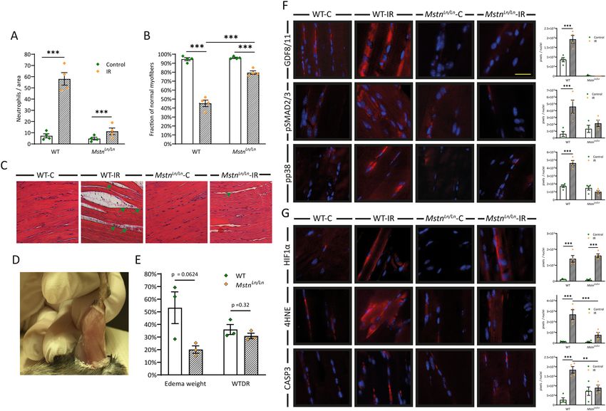
www.nature.com/scientificreports/
Figure 1. Impact of IR injury on human skeletal muscle cells. (A) Harvested free latissimus dorsi
myocutaneous flap from which muscle specimens were obtained. (B) Infiltration of inflammatory cells
into skeletal muscle and (C) magnitude of myofiber deterioration assessed in HE stained specimens. (D)
representative images in longitudinal and cross-sectional views (E) Quantification of protein levels of MSTN
(GDF8) and its downstream targets phospho-SMAD2/3 (pSMAD2/3) and phospho-p38 MAPK (p38 MAPK).
Left panel shows representative images of immunofluorescence analysis. (F) Markers of hypoxia, lipid
peroxidation, and apoptosis quantified using immunofluorescence analysis. Left panel shows representative
images. Control: specimens harvested immediately before blood circulation detachment of the latissimus
dorsi muscle, IR: specimens harvested shortly before finishing surgery (mean ischemia time: 64 ± 9 min, mean
reperfusion time: 77 ± 25 min). Results are shown as means ± SEM. Scale bar 40 μm. p value: *< 0.05, **< 0.01,
***< 0.001 (unpaired two tailed t test).
effects and resulted in gross muscle hypertrophy13. Besides phenotypic effects, MSTN interacts with vascular
smooth muscle and inflammatory cells and has been linked to ROS modulation via interaction with NF-kB,
TNF-α, and the NADPH oxidase s ystem14–17. MSTN can further activate p38 mitogen-activated protein kinase
(p38 MAPK)15, which is known to play a crucial role in IR injury18. In 2019, Magga et al. also demonstrated
myocardial protection from IR injury by systemic blockade of MSTN receptor A CVR2B19. While these effects
have been reported in the heart, pathophysiological considerations suggest similar effects in skeletal muscle.
This work reveals the significance of IR injury in routinely performed free myocutaneous flap surgery and
investigates a potential role of MSTN deficiency in mitigating skeletal muscle cell damage arising from IR utiliz-
ing in vivo knockout studies.
Results
Impact of IR injury on human skeletal muscle. Many studies have characterized IR injuries in different
species leading to different outcomes depending on species, tissue, severity, and time of ischemia. To establish a
baseline of IR injury in human skeletal muscle cells for this work, we sought to determine the extent of IR injury
resulting from free autologous tissue transfer. Therefore, we utilized the free latissimus dorsi myocutaneous flap
in a rare human experiment setting (Fig. 1A). Specimens were collected immediately before flap harvesting (con-
trol) and on the last occasion before final wound closure (IR), resulting in a mean ischemia time of 64 ± 9 min,
Scientific Reports | (2021) 11:12572 | https://doi.org/10.1038/s41598-021-92159-2 2
Vol:.(1234567890)
www.nature.com/scientificreports/
Figure 2. Transcriptional profiling of human IR injury. (A) Volcano plot representing differentially expressed
genes (DEGs) characterized by expression values within the 50th–100th percentile, fold change ≥ 1.7 and
corrected p value (Benjamin and Hochberg, FDR) < 0.05. (B) Heatmap with the 50 DEGs displaying the highest
fold change. (C) Gene set enrichment analysis using MSigDB collections hallmarks gene sets and GO gene
sets BP and MF. (D) Expression of enriched mRNAs in the positive regulation of p38 MAPK cascade gene set.
FDR-values (Benjamini–Hochberg): *< 0.1; **< 0.05; (E) Enrichment plot of Hallmark TNF-α signaling via
NF-κB (p < 0.001, FDR = 0.092). RNA of 4 biological samples per group was pooled considering concentration
differences and analyzed in duplicates via RNA microarray (Agilent Technologies, California, USA).
and mean reperfusion time of 77 ± 25 min. Due to IR injury, inflammatory cell infiltration increased almost
fivefold (Fig. 1B) and fraction of normal myofibers decreased by 42 percentage points (Fig. 1C). Inflammatory
cell infiltration was validated using MPO staining (Fig. S1). Because previous studies demonstrated a connection
between IR injury, MSTN, and its downstream targets19 we sought to investigate these proteins. In particular,
MSTN, SMAD2/3, and p38 MAPK have been described to interact in a mutually dependent manner and upon
cell stress and ischemia20,21. We found all three of them to be activated due to IR injury (Fig. 1E) which is consist-
ent with previous fi ndings22. To further validate an IR injury state in our samples and to grade hypoxia as well as
oxidational and apoptotic processes, HIF-1α, 4-hydroxynonenal, and caspase 3 were measured (Fig. 1F). HIF-1α
is a well characterized protein upregulated in hypoxic conditions and displayed an almost fourfold increase
(p < 0.05). Caspase 3, marking the final pathway of apoptosis, displayed the same increase (fourfold, p < 0.01),
and 4-HNE as marker lipid peroxidation due to oxidative stress23 rose threefold (p < 0.01). Taken together, these
findings clearly demonstrated the extent of IR injury in routinely performed free latissimus dorsi myocutaneous
flap transfer which is paralleled by an increase in MSTN and its downstream targets, noteworthy p38 MAPK.
Transcriptome changes in skeletal muscle following IR injury. Next, we examined the impact of IR
injury on skeletal muscle on a transcriptional level. Microarray analysis revealed 2554 differentially expressed
genes (DEGs, Fig. 2A) of which 1360 were up- and 1194 downregulated due to IR (Supplementary Dataset S1).
Overexpression analysis with ConsensusPathDB mapped 1152 of the upregulated genes to 3102 1- and 2-next
neighbours network neighbourhood-based entity sets (NESTs, filtered for p < 0.001, minimum overlap with
input list = 5, Fig. S2A,B) and 45 annotated Kyoto Encyclopedia of Genes and Genomes (KEGG) pathways24 (fil-
tered for p < 0.01, minimum overlap with input list = 5, Fig. S2C). Regarding the 148 1-next NESTs, most highly
overrepresented set centers were HLA class II related (Fig. S2A), presumably reflecting the immune response
to IR. Further overrepresented set centers included “NFkB.complex” and “p-S423, S425-SMAD3” of which the
Scientific Reports | (2021) 11:12572 | https://doi.org/10.1038/s41598-021-92159-2 3
Vol.:(0123456789)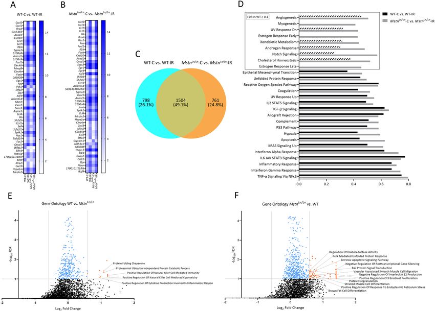
www.nature.com/scientificreports/
latter meets the observed immunofluorescence analysis upregulation (Fig. 1E). Overrepresented 2-next NESTs
included 2954 set centers. Among these, several inflammatory markers (e.g. “IL6”, “NOD2”, “IFNG gene”) and
p38 MAPK related proteins (“p38”, “mapk12”, “mapk13”) were found (Fig. S2B). Overrepresented KEGG path-
ways encompassed immunological pathways, i.e. “Graft-versus-host disease”, “Allograft rejection”, and interest-
ingly, further autoimmune pathways (Fig. S2C), all in all indicating a strong immune response (Supplementary
Dataset S2). The 50 DEGs with the highest fold change between groups (Fig. 2B) are in line with previous obser-
vations. MMP3, IL6, and CXCL9, for instance, have all been described in the realm of IR i njury6,25,26. To further
investigate the transcriptome, we performed Gene Set Enrichment Analysis (GSEA) including hallmark and
Gene Ontology (GO) Biological Process (BP) and Molecular Function (MF) gene sets from MSigDB (Fig. 2C,E).
Gene enrichment and level of significance were highest in gene sets “GO Natural Killer Cell Activation Involved
In Immune Response” (ES: 0.75, p < 0.001, FDR = 0.072) and “Hallmark TNF-α Signaling Via NFkB” (ES: 0.38,
p < 0.001, FDR = 0.091, Fig. 2E). Overall, enriched gene sets with an FDR < 0.1 aligned well with the analysis of
DEGs and previous studies27,28, and convey the impression of a severe immune response, mediated via NK cells,
TNF-α, NFkB, IL6, JAK1/2, STAT3 (Fig. 2C), and potentially p38 MAPK (Fig. 2D) in response to IR injury.
Expression values from selected genes of the GO gene set “positive regulation of p38 MAPK cascade” (IL1B,
XDH, BMP2, GADD45B, GADD45A, and ZC3H12A) are shown in Fig. 2D. Taken together, data obtained from
analyzing human skeletal muscle (Figs. 1, 2) elucidated up-regulation of MSTN (GDF8), SMAD2/3, and p38
MAPK activity (and corresponding transcription), a severe immune response, and cell stress upon IR. Implica-
tions of these observations with particular focus on the interplay between MSTN, p38 MAPK, NFκB, and IL6
will be elaborated in “Discussion” section.
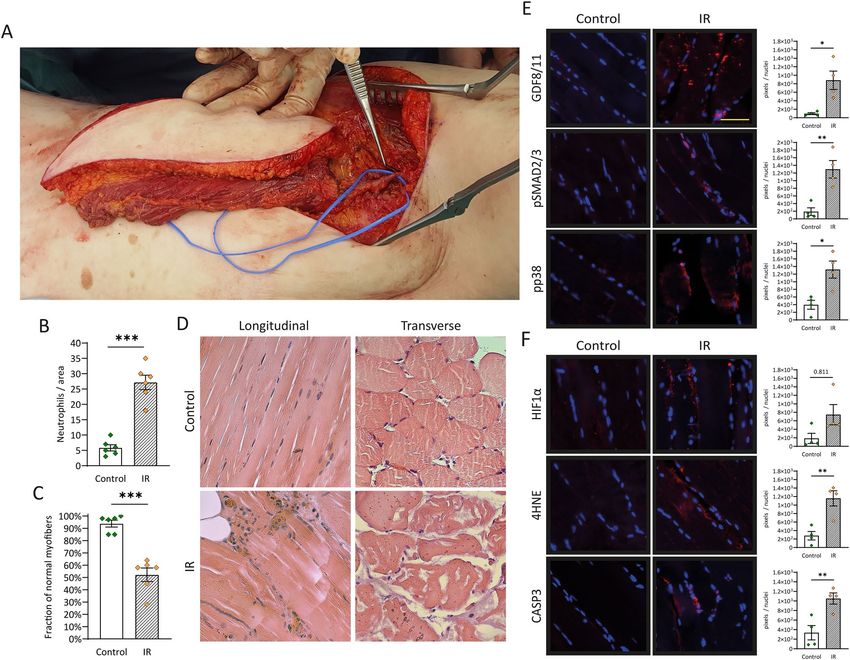
MSTN knockout mitigates IR injury in murine skeletal muscle. To further investigate a potential
role of MSTN in IR injury, we used an in vivo knockout model. Heterozygote C57BL/6J-MstnLn/J mice were
obtained from Jackson Laboratory (Maine, USA, Stock No 009345) and bred to homozygote MstnLn/Ln geno-
types. Ischemia was induced as stated before29. Briefly, hindlimbs of both wildtype and MstnLn/Ln were exposed
to ischemia for 4 h using a low-pressure tourniquet. After reperfusion for 2 h, mice were euthanized, and the
tissue was harvested. Human skeletal muscle had demonstrated neutrophil infiltration and myofiber deteriora-
tion in specimens due to IR injury (Fig. 1A,B). We thus started by examining the same aspects in mice. Number
of neutrophils per area increased eightfold (p < 0.001, Fig. 3A,C) in WT mice; in MstnLn/Ln mice on the other
hand, neutrophil infiltration was not significant. Results were validated using MPO staining (Fig S4). Tissue
deterioration was likewise reduced in MstnLn/Ln mice. While WT mice exhibited signs of cell damage in almost
50% of previously healthy myofibers (p < 0.001), fraction of normal myofibers in MstnLn/Ln mice only decreased
by 16.5 percent points (p < 0.05, Fig. 3B,C). Regarding immunofluorescence analysis, MSTN deficiency further
seemed to exert a protective role. Protein levels of MSTN (GDF8) (twofold), pSMAD2/3 (threefold), and pp38
MAPK (eightfold) were significantly increased in WT mice due to IR while the MstnLn/Ln knockout blunted these
effects (Fig. 3F,G). HIF-1α was substantially elevated in both groups, most certainly in response to hypoxic con-
ditions. In WT mice IR led to increased lipid peroxidation (22-fold increase of 4HNE) and in turn to induction
of apoptosis (sevenfold increase of caspase 3), grossly mimicking the events observed in human skeletal muscle.
MstnLn/Ln mice, however, displayed statistically insignificant enhanced lipid peroxidation (sevenfold increase
of 4HNE), but this did not trigger apoptosis in terms of caspase 3 activity (Fig. 3F,G). To further elucidate
the possibility of reduced apoptotic activity, we performed an apoptosis array (Fig. S3) which supported these
observations. Decreased activity of caspase 3 upstream targets BAD (− 15%, p < 0.05) and BAX (− 27%, p < 0.01)
in MstnLn/Ln-IR mice in comparison to WT-IR mice was accompanied by lowered levels of cleaved caspase 3
itself (− 22%, p < 0.01) (Fig. S3A). In addition, BCL-2 as prominent member of the antiapoptotic protein family
was upregulated in response to IR injury in MstnLn/Ln (+ 19%, p < 0.05) but not in WT mice (Fig. S3B). Besides
apoptosis, another key element of IR injury is the inflammatory response. Here, diapedesis of neutrophils and
upregulation of pro-inflammatory cytokines is regularly accompanied by interstitial fluid l eakage30 which can be
observed both microscopically (Fig. 3C) and macroscopically (Fig. 3D). Thus, in animal models of IR injury, it
is a standard to evaluate edema formation. This was performed as previously d escribed29. In brief, the hindlimb
was cut off in toto, weighed, dried for 48 h, and weighed again. For exact calculation we kindly like to refer to
“Materials and methods” section and supplementary dataset S3. Regarding the wet-to-dry-ratio, no statistically
significant changes could be detected (Fig. 3E, right bars). Considering differences in weight between WT and
muscular MstnLn/Ln mice, the difference in edema increase between both groups is near-significant (p = 0.0624).
Relative edema weight increased by 53% (± 12%) in WT mice and by only 20% (± 3%) in MstnLn/Ln mice (Fig. 3E,
left bars). Evaluating these results, one should also bear in mind the higher muscle mass and increased weight
of MstnLn/Ln mice which would let assume their proneness to IR injury and its sequelae, including edema forma-
tion. Hence, yet the non-distinctness in wet-to-dry-ratio may indicate diminished susceptibility to IR injury in
MstnLn/Ln mice.
Transcriptome changes due to IR injury in WT and MstnLn/Ln mice. Since MstnLn/Ln mice demon-
strated increased tolerance towards IR injury, we sought to explore transcriptional changes using an RNA micro-
array (Agilent Technologies, California, USA). Upon analysis we identified 2303 DEGs in WT and 2261 DEGs in
MstnLn/Ln mice due to IR injury (Supplementary Dataset S4). Regarding the 50 DEGs with the highest fold change
(Fig. 4A,B), results in both groups tied well with our transcriptome data of human IR injury (Fig. 2). DEGs can
be mostly mapped to immunologic (e.g. Cxcl5, Cxcl1, Cxcl10, Ccl7, Ccl2, Il6, Il1b) and/or IR related activity (e.g.
Fos, Fosb, Fosl, Egr1, Mmp8, Acod1)31–34. Considering the entire DEGs (Fig. 4C; Supplementary Dataset S4),
both groups seemed to facilitate compensatory mechanisms in response to IR. Substantially elevated activation
transcription factor 3 (WT: 54-fold change, FDR = 0.001; MstnLn/Ln: 53-fold change, FDR = 0.001), for instance,
Scientific Reports | (2021) 11:12572 | https://doi.org/10.1038/s41598-021-92159-2 4
Vol:.(1234567890)
www.nature.com/scientificreports/
Figure 3. Mitigation of IR injury in MSTN deficient mice. (A) Tissue infiltration of inflammatory cells and
(B) magnitude of myofiber deterioration assessed in HE stained skeletal muscle specimens of C57BL/6J and
C57BL/6J-MstnLn/Ln mice. (C) HE stained skeletal muscle demonstrating myofiber deterioration, neutrophil
immigration (green arrows), and edema formation. (D) Macroscopically visible edema formation due to IR
injury. (E) Changes in the wet-to-dry ratio and edema weight compared between C57BL/6J and C57BL/6J-
MstnLn/Ln mice (n = 3). For calculation of WTDR and edema weight see methods. (F) Immunofluorescence
analysis of MSTN/GDF11-pSMAD2/3-pp38 MAPK pathway and representative images of immunofluorescence
analysis. (G) Markers of hypoxia, lipid peroxidation, and apoptosis. Left panel illustrates representative images.
WT = C57BL/6J mice, MstnLn/Ln = C57BL/6J-MstnLn/Ln mice. Results are shown as means ± SEM. Scale bar 40 μm.
p value: *< 0.5, **< 0.1, ***< 0.001; two-tailed unpaired t test for pairwise analysis. ANOVA followed by multiple
test comparison via Tukey’s post-hoc test (homoscedasticity) or Brown–Forsythe and Welch ANOVA followed
by Dunnett T3 post-hoc test (heteroscedasticity) for multi-group analysis. RNA of four biological samples
per group was pooled considering concentration differences and analyzed in duplicates via RNA microarray
(Agilent Technologies, California, USA).
has been reported to convey protective effects against IR in different tissues35. Another example is Dusp1 (WT:
1.9-fold change, FDR = 0.004; MstnLn/Ln: 3.9-fold change, FDR = 0.003). DUSP1 dephosphorylates members of
the MAPK family and has been shown to ameliorate cardiac IR injury36. Stronger upregulation in MstnLn/Ln
mice also provided a potential explanation for decreased p38 MAPK (Fig. 3F) activity upon IR. To gain a better
understanding of the DEGs, we examined the overlap between both groups (Fig. 4C; Supplementary Dataset S5).
798 genes were differentially expressed only in WT mice and 761 in MstnLn/Ln mice. Shared DEGs added up to
a total of 1504. We then mapped those that were upregulated in only one group to pathways using the Consen-
susPathDB (filtered for p < 0.01, minimum overlap with input list = 5, Supplementary Dataset S6, S7). For WT
mice, this once again verified the observations made during analysis of human IR, namely overrepresentation
of pathways “NF-kappa B signaling pathways” and “Natural killer cell mediated cytotoxicity” (p < 0.001, Sup-
plementary Dataset S6). Additionally, overrepresentation of the majority of the other 11 pathways (e.g., “Signal-
ing by Interleukins”, “Cytokine Signaling in Immune system”, and “TYROBP Causal Network”) was consistent
with pre-existing studies. For MstnLn/Ln mice on the other hand, results differed substantially (Supplementary
Dataset S7). Even though some of the 27 overrepresented pathways were related to enhanced immunologic
activity, the vast majority was not. Instead, several pathways were related to MAPK signaling which might appear
counterintuitive considering that our previous experiments demonstrated decreased expression of p38 MAPK.
The MAPK family however incorporates several other MAP kinases as well as up- and downstream targets. For
instance, further inspection of these gene sets revealed increased Dusp4 and Dusp16 expression. DUSP4 for its
part has been linked to p38 MAPK regulation and is usually degraded under oxidative s tress37. Furthermore, the
Scientific Reports | (2021) 11:12572 | https://doi.org/10.1038/s41598-021-92159-2 5
Vol.:(0123456789)www.nature.com/scientificreports/
Figure 4. Transcriptional profiling of IR injury alleviation in WT MstnLn/Ln mice. Heatmap depicting expression
values of 50 DEGs (expression values within the 50th–100th percentile, fold change ≥ 1.7, FDR (Benjamini–
Hochberg) < 0.05) sorted by descending fold change due to IR injury in (A) WT or (B) MstnLn/Ln mice. (C)
Venn diagram illustrating the overlap between genes that are differentially expressed due to IR injury in WT or
MstnLn/Ln mice, respectively. (D) GSEA with the hallmark gene set comparing enriched gene sets after IR injury
in WT (WT-C vs. WT-IR) and MstnLn/Ln (MstnLn/Ln-C vs. MstnLn/Ln-IR) mice. Enrichment scores inside the box
are non-significant in WT mice at an FDR-threshold of 0.1. (E) GSEA of GO terms BP and MF comparing
WT and MstnLn/Ln mice before and after IR injury. Blue dots mark enrichment scores that are below the FDR-
threshold of 0.1. Orange dots mark values that also meet the fold change cut-off of 1.5 when comparing the
enrichment score of MstnLn/Ln-C vs. MstnLn/Ln-IR mice with the enrichment score of WT-C vs. WT-IR mice for a
given gene set.
set included Rap1b. While RAP1 has been linked to aggravated IR injury in the heart, the B-subunit has been
shown to be imperative for angiogenesis38. Last pathway to emphasize is “VEGFA-VEGFR2 Signaling Pathway”
which aligned well with the results of the GSEA (Fig. 4D–F). To conclude the analysis of DEGs, we filtered for
genes that were differentially expressed in WT and MstnLn/Ln mice but regulated differently, yielding 78 genes of
interest (Fig. S5). Pathway analysis for DEGs up-regulated in WT and down-regulated in KO did not identify
any overrepresentation. Vice versa, we determined 7 pathways containing DEGs down-regulated in WT mice
but up-regulated in MstnLn/Ln mice, mainly associated to muscle contraction (Supplementary Dataset S8). Five
out of these incorporated ATPase Sarcoplasmic/Endoplasmic Reticulum Ca2+ Transporting 2 (Atp2a2). In WT
mice, Atp2a2 decreased 1.5-fold (FDR = 0.0018) upon IR injury; in MstnLn/Ln on the contrary, it increased 2.2-
fold (FDR = 0.0016). Atp2a2 codes for SERCA2a, the most important SERCA genotype. It is mainly expressed
in the heart and slow-twitch skeletal muscle. Upon activation, it can increase its activity to enhance Ca2+ uptake
and thus reduce reperfusion-induced calcium overload. Moreover, studies have demonstrated proteolysis of
SERCA2a, among others, via MMP-2, as part of cardiac IR injury39. Taken together, the increased expression
of Atp2a2 provides another potential mechanism that protects MstnLn/Ln mice from IR injury. Upon deeper
analysis, we identified further differently regulated mRNAs (Krt8, Krt18, Mymx, Myh7b, Npr3) between WR and
MstnLn/Ln mice that have been previously associated with IR injury and shall be examined in “Discussion” sec-
tion. For more thorough transcriptome examination, we performed GSEA using Hallmark (Fig. 4D) and GO BP
and MF (Fig. 4E,F) gene sets. Many enriched hallmark gene sets (Fig. 4D) were revealed before in human skeletal
muscle upon IR, i.e. “TNF-α Signaling Via NFκB”, “Inflammatory response”, and “Allograft Rejection” (Fig. 2C).
Strikingly, enriched hallmark gene sets in MstnLn/Ln mice included “Myogenesis” (ES: 0.4, FDR = 0.088) without
Scientific Reports | (2021) 11:12572 | https://doi.org/10.1038/s41598-021-92159-2 6
Vol:.(1234567890)www.nature.com/scientificreports/
any enrichment in WT mice. Closer inspection of the data also indicated enrichment of gene sets “Angiogenesis”
(ES: 0.5, FDR = 0.09), and “Notch Signaling” (ES: 0.57, FDR = 0.039) in MstnLn/Ln mice but not in WT mice (ES:
0.45, FDR = 0.28 and ES: 0.25, FDR = 1, respectively). For GSEA, we filtered for gene sets displaying at least 1.5-
fold change when comparing enrichment scores between WT-C vs. WT-IR and MstnLn/Ln-C vs. MstnLn/Ln-IR.
FDR threshold was set to 0.1, resulting in 10 gene sets in WT and 76 gene sets in MstnLn/Ln mice (Supplementary
Dataset S9, S10). Interestingly, 3 gene sets enriched in WT mice were related to NK cell activation, one of the
main enriched gene sets in human skeletal muscle (Fig. 2C). In line with this, “Negative Regulation of Inter-
leukin 12 Production” (ES: 0.74, FDR = 0.078) was enriched in MstnLn/Ln mice. Further enriched gene sets in
MstnLn/Ln mice were often related to cell differentiation and growth, all in all creating the impression of enhanced
tissue proliferation in response to IR. Altogether, observed transcriptome changes supported the results pre-
sented in the previous section (Fig. 3) and identified several potential mechanisms of mitigated IR injury arising
from MstnLn/Ln deficiency.
Discussion
This work emphasizes the incidence of skeletal muscle IR injury in routinely performed surgery and demonstrates
protective effects of MSTN deficiency. Despite short time frames (mean ischemia time: 64 ± 9 min, mean reper-
fusion time: 77 ± 25 min), we found unequivocal signs of IR injury when examining skeletal muscle from free
flap surgery. Observed increases in markers of hypoxia, lipid peroxidation, and apoptosis thereby corroborate
previous findings in either skeletal muscle or other tissues40–42. Interplay between MSTN and p38 MAPK as well
as their involvement in IR injury have likewise been stated b efore20,21. Elevated p38 MAPK has been described
in cardiac ischemic p reconditioning43 and p38 MAPK inhibition has been shown to alleviate myocardial IR44.
Moreover, similar observations have been made in the context of other organs45,46. This work, however, is the first
one to demonstrate a positive correlation between elevated levels of MSTN, p38 MAPK, and the presence of IR
injury in human skeletal muscle. Transcriptional assessment revealing upregulation of genes included in the GO
gene set “positive regulation of p38 MAPK cascade” further underlined the observed contribution of p38 MAPK.
But p38 MAPK is not the only mediator linking MSTN to IR injury. NFκB is a transcription factor with basically
ubiquitous occurrence that can be activated by several stimuli. It is also well-established that it can be stimulated
via TNF-α and other chemokines such as IL-6. In 2011, Sriram et al.15 revealed that Myostatin itself can activate
NFκB which in turn enhances transcription of Myostatin and TNF-α, further increasing NFκB activity in the
sense of a positive feedback loop. Additionally, TNF-α as well as IL-6 can also directly activate transcription of
MSTN14. In line with our hypothesis of a protective role of MSTN deficiency, pathways related to TNF-α, IL-6,
and NFκB displayed the highest enrichment scores in transcriptome analysis (Fig. 2C,E). Another important
result emerging from the human data is the activation of NK cells, probably facilitated by IFN-γ (Fig. 2C).
Regarding lymphocytes in terms of IR injury, C D4+ T cells are usually considered most relevant9. Involvement
of NK cells has also been reported9, albeit more rarely. Interestingly, we found transcription of genes related to
NK cell activation being significantly more amplified in WT mice than in mice deficient of functioning MSTN
when exposed to IR. This aligns well with the observed negative transcriptional regulation of IL-12 production
in MstnLnLn mice due to IR. IL-12 physiologically activates NK cells and lymphocytes and stimulates the produc-
tion of IFN-γ and TNF-α.
The main purpose of this work, however, was to investigate the protective capacity of MSTN deficiency in
the setting of IR injury. First of all, experiments conducted with WT mice mirrored results observed after flap
surgery in humans (Figs. 1, 3), rendering the used in vivo IR model highly suitable to examine the impact of the
Mstn knockout. MSTN deficiency reduced neutrophil infiltration, myofiber deterioration, tissue edema, lipid
peroxidation, and apoptosis (Fig. 3). In that regard it should be stated that the used antibody is directed against
myostatin (GDF8) and GDF11. Due to their homology however, differentiation in immunofluorescence is not
expedient. The hypothesized p38 MAPK implication was supported by absence of its up-regulation in MstnLn/Ln
mice after IR. Protective effects of MSTN inhibition have previously been reported in the heart. This was achieved
using a soluble ACVR2B decoy receptor19 or by inhibiting ACTRII/TGFBR signaling47, respectively. Interestingly,
the latter presented an underlying mechanism we identified ourselves during transcriptome analysis in mice:
the mentioned up-regulation of Atp2a2 in MstnLn/Ln mice with concurrent down-regulation in WT mice, which
was paralleled by up-regulation of SERCA2a (translational product of Atp2a2) in the setting of cardioprotection
after myocardial infarction in m ice47. Additionally, Zhu et al.48 had previously linked SERCA2a activity to p38
MAPK signaling: in 2017, they studied the effects of IR on rat cardiac function. It was found that during IR, p38
MAPK pathway activation eventually suppresses the activity of SERCA2a which reinforces calcium overload and
mitochondrial membrane potential imbalance leading to cardiomyocyte apoptosis. This bridges the gap between
myostatin inhibition, p38 MAPK signaling, and enhanced SERCA2a activity. Noteworthy, our findings paralled
previous findings in cardiac muscle.
Scanning for differently regulated DEGs in WT and MsntLn/Ln mice as a result of IR also identified further
mediators (Krt8, Krt18, Myh7b, and Npr3) that may confer protective effects of MSTN deficiency. The first two
ones code for keratin 8 and keratin 18, proteins that are usually found in intermediate filaments of epithelial cells.
Interestingly, their ectopic expression in the heart has been linked to cardioprotective e ffects49. This is in line with
our earlier observations in skeletal muscle, which showed up-regulation of Krt8 and Krt18 in MsntLn/Ln mice but
down-regulation in WT mice after IR. Additionally, keratin 8 is known to interact with p38 M APK50. The protein
product of Myh7b (miR-499) was also up-regulated in Msnt Ln/Ln
mice. In the heart, miR-499 could prevent car-
diomyocyte apoptosis and reduced the infarct size of myocardial infarction in mice51. In murine skeletal muscle,
miR-499 may drive myogenic differentiation of satellite cells and inhibit their adipogenic differentiation52 which
also aligns well with the amplification of hallmark gene set “myogenesis” in MsntLn/Ln mice after IR without any
enrichment in WT mice (Fig. 4D). Another protein potentially capable of inhibiting apoptosis in cardiomyocytes
Scientific Reports | (2021) 11:12572 | https://doi.org/10.1038/s41598-021-92159-2 7
Vol.:(0123456789)www.nature.com/scientificreports/
Figure 5. Timeline of specimen harvest during free flap surgery.
is natriuretic peptide receptor 3 (NRP3). While up-regulated in MsntLn/Ln mice and down-regulated in WT mice
after IR in this work, previous in vitro studies showed enhanced levels of caspase 3, 8, and 9 due to knockdown
of NPR3 in H9C2 cells, and protective properties of NPR3 regarding H 2O2 induced cell d eath53.
In contrast to earlier findings, we could not observe any influence of MSTN deficiency on ROS generation.
Considering the results of this work and presented signaling pathways—including p38 MAPK circuits—one could
hypothesize that inhibition of MSTN mitigates ROS p roduction18. And indeed, this connection has previously
15
been reported . Contrarily, a recent publication stated that myostatin knockout induced apoptosis in human
cervical cancer cells which was mediated via ROS g eneration54. In the presented work, however, ROS related
pathways did not differ significantly between WT and MstnLn/Ln mice. We therefore speculate that the connection
between MSTN and ROS generation is far more complex and highly context dependent. As another important
finding, increased activity of genes related to myogenesis, angiogenesis and notch signaling in vivo arising from
MSTN deficiency suggests enhanced regenerative capacity in addition to reduced susceptibility to IR injury. In
other words, besides causing attenuated tissue deterioration and cell death, MSTN deficiency seems to result in
up-regulation of mechanisms that restore tissue integrity after the damaging process has occurred. Up-regulation
of Mymx in MstnLn/Ln mice and down-regulation in WT mice after IR further supports this assumption. Mymx
codes for myomerger, which, together with myomaker, is one of the two fundamental fusion proteins required for
skeletal muscle d evelopment55. While myomaker is also essential for muscle r egeneration56, the role of myomerger
has not been studied in the same depth. Yet, observed regulation of myomerger supports the idea of enhanced
tissue regeneration emerging from MSTN deficiency.
In conclusion, this study illuminated the occurrence of IR injury in reconstructive free flap surgery and
highlighted potential beneficial effects of MSTN deficiency in these as well as similar scenarios. We further exam-
ined reasons linking absence of myostatin to reduced cell death and enhanced tissue regeneration. Thereby, we
identified p38 MAPK, SERCA2a, keratin 8 and keratin 18, miR-499, NRP3, and myomerger; either individually
or interdependently and collectively as potential targets regarding IR injury. We would like to encourage further
studies to focus on these proteins, even if not in combination with MSTN. Additionally, we identified a potential
role of NK cells in skeletal muscle IR injury that needs further investigation.
Materials and methods
Human specimen collection. All procedures were performed in accordance with the ethical committee
of the Ruhr University Bochum after approval (approval number: 16-5932-BR). Informed consent was obtained
from all subjects participating in the study. Human skeletal muscle tissue was harvested during planned latis-
simus dorsi free flap surgery (Fig. 5). During each surgery two tissue specimens were taken: the first one imme-
diately before blood circulation of the latissimus dorsi was detached (Control) and the second one shortly before
the procedure was finished (Ischemia/Reperfusion). The specimens were immediately placed into liquid nitro-
gen for gene expression analysis or formaline (4%) for histology and immunofluorescence.
Histology and immunofluorescence. Tissue samples were stained with haematoxylin and eosin for
analysis of tissue damage and neutrophil infiltration. For immunofluorescence stainings, sections and chamber
slides were incubated at 58 °C for 1 h and subsequently rehydrated and incubated with 0.125% Proteinase K for
15 min at 37 °C. After a short washing step with PBS, sections and cells on chamber slides were permeabilized
with 0.1% Tween 20 for 4 min and treated with blocking solution for 1 h. Incubation with primary antibodies
(Supplementary Dataset S11) in blocking solution followed overnight at 4 °C. After washing with PBS, a goat
biotinylated secondary antibody was used for detection. For mouse monoclonal antibodies conjugated mouse
IgGκ binding proteins were used instead of the secondary antibody. All sections have been counterstained with
DAPI. Sections were subsequently mounted with Fluoromount Aqueous Mounting Medium (Sigma Aldrich).
Images for immunofluorescence were taken with a fluorescence microscope (Olympus IX3-Series). By using
the Adobe Magic Wand Tool (settings: tolerance 60%; noncontiguous) in Adobe Photoshop CS6 (https://www.
adobe.com/products/photoshop.html), immunohistochemical positive stained pixels were selected automati-
cally and divided by countable nuclei. Afterwards a mean value was calculated. A detailed list of antibodies
Scientific Reports | (2021) 11:12572 | https://doi.org/10.1038/s41598-021-92159-2 8
Vol:.(1234567890)www.nature.com/scientificreports/
used can be found in the supplemental material Supplementary Dataset S11). For measuring the infiltration of
neutrophils, four regions of interest per section (stained with HE) were chosen (1000 × 1000 Px), and neutrophils
were counted by three independent observers. For the morphometric analysis, an unbiased sampling procedure
was applied. The fraction of normal muscle cells was calculated by measuring the cross-sectional diameters
of fibers in four regions of interest in transverse sections (stained with HE) as reported before57. Cells with a
diameter within 10% of the control group values were considered “normal cells”. Percentage of normal cells was
calculated accordingly.
RNA preparation. After harvesting, muscle specimens were immediately stored in cryovials and shock fro-
zen in liquid nitrogen. Total RNA was then isolated using the RNeasy Fibrous Tissue Mini Kit (Qiagen, Hilden,
Germany). RNA-quality was assessed via NanoDrop. A 260/280 ratio ≥ 2 and 260/230 ratio of 1.8–2.2 were con-
sidered sufficient for further analysis. An amount of 100 ng of every total RNA sample was hybridized to an
individual Agilent whole genome expression microarray (Human GE 4 × 44K, v2 G4845A, AMADID 026652,
Agilent Technologies) according to the Agilent single color protocol. RNA labeling, hybridization and washing
were carried out according to the manufacturer’s instructions. Images of hybridized microarrays were acquired
with a DNA microarray scanner (Agilent G2505B) and features were extracted using the Agilent Feature Extrac-
tion image analysis software (AFE) version A.10.7.3.1 with default protocols and settings. The AFE algorithm
generates a single intensity measure for each feature, referred to as the total gene signal (TGS), which was used
for further data analyses using the GeneSpring GX software package version 14.9.1. AFE-TGS were normalized
by the quantile method. Subsequently, data were filtered on normalized expression v alues17. The gene expression
data from our study have been deposited in the NCBI’s Gene Expression Omnibus (GEO) database (accession
number GSE118380).
Breeding and genotyping of the C57BL/6J‑MstnLn/Ln strain. Heterozygote C57BL/6J-MstnLn/J mice
were obtained from Jackson Laboratory (Maine, USA, Stock No 009345). Multidose N-ethyl-N-nitrosourea
(ENU) treatment was used to induce mutations (T → C transition in the second intron two base pairs down-
stream of exon 2 in the myostatin gene) in founder C57BL/6J mice which resulted in improper splicing and a
loss-of-function allele called the lean (Ln) allele of Mstn (MstnLn). Mice were then bred to obtain homozygote
C57BL/6J-MstnLn/Ln offsprings. Homozygote mice were identified using a qPCR genotyping assay consisting of
a forward primer (5′ ACA GCC TGA ATC CAA CTT AGG 3′), a reverse primer (5′ AAA TGT AAC ACG GTT
GCT AGA ATG 3′), a probe for the wild type allele (5′-HEX AGA TGG GCT GGT AAG TGA TAA CT BHQ1-
3′), and a probe for the lean allele (5′-FAM GAT GGG CTG GCA AGT GAT AAC BHQ1-3′).
Animal surgery. All animal experiments were approved by the IACUC LANUV NRW (The Ministry for
Environment, Agriculture, Conservation and Consumer Protection of the State of North Rhine-Westphalia),
Permit Number: 84-02.04.2016.A045, and the study was carried out in accordance with the ARRIVE guidelines.
C57BL/6J and C57BL/6J-Mstnlean mice were obtained (The Jackson Laboratory, Maine, USA) and kept with
unlimited access to water and standard laboratory chow at a 12 h light/dark cycle. Littermates of both sexes at
the median age of 12 (± 1) weeks were used for all experiments. All interventional procedures were performed
under inhalation anesthesia with isoflurane (3% for induction, 0.8–1.8% for maintenance) and buprenorphine
(0.05 mg/kg s.c.). To maintain a body temperature of 37 °C mice were restrained on a heating pad for the whole
duration of the intervention. C57BL/6J wild type mice (24–27 g) and C57BL/6J-MstnLn/Ln mice (28–33 g) were
randomized in an experimental (IR) and a control (C) group resulting in four groups: WT-C (1), WT-IR (2),
MstnLn/Ln-C (3), and MstnLn/Ln-IR (4). In the IR groups, ischemia was induced as previously described28. Hindlimb
perfusion was interrupted for 2 h, followed by 4 h of reperfusion. Subsequently, mice were euthanized in accord-
ance with the American Veterinary Medical Association Guidelines for the Euthanasia of Animals, and the tissue
was harvested. Control group mice were anesthetized for 2 h without any interventions and euthanized 4 h later.
Edema weight and wet to dry ratio. Immediately after euthanasia the leg was cut off in toto, weighed
and placed in an oven at 55 °C for 48 h. Subsequently, the leg was weighed again. From here, two calcula-
tion methods were used. For the first one, the difference in edema weight increase (in mg) within one group
(C57BL/6J or C57BL/6J-MstnLn/Ln) before and after drying was compared with the other group (edema weight).
For the second method, the weight within each group after drying was divided by the weight before drying,
resulting in values with the unit “percent” (wet to dry ratio and wet to dry ratio increase). Then the difference in
wet to dry ratio increase between both groups was calculated. This was done to account for the heavier weight of
C57BL/6J-MstnLn/Ln in comparison to C57BL/6J mice.
Apoptosis array. For cell lysate preparation, equal amounts of 3 samples per group were suspended in Lysis
Buffer 17 provided with the Proteome Profiler Mouse Apoptosis Array (R&D systems, Minneapolis, Minnesota,
USA). After homogenization with the T 10 basic ULTRA-TURRAX (IKA, Staufen im Breisgau, Germany) and
centrifugation at 12,000 rpm, supernatant was transferred to a fresh tube, and protein concentration was deter-
mined by using the BCA Assay (ThermoFisher Scientific, Waltham, USA). Cell lysates (400 µg) were added
to the array membranes and subsequent steps were carried out according to the manufacturer’s instructions.
Membranes were then exposed to X-ray film for 2 min each. For data analysis, X-ray films were scanned, and
pixel density was quantified in Adobe Photoshop after subtracting an average background signal from each spot.
Scientific Reports | (2021) 11:12572 | https://doi.org/10.1038/s41598-021-92159-2 9
Vol.:(0123456789)www.nature.com/scientificreports/
Statistics. After testing for homo- or heteroscedasticity (F Test), p values for pairwise comparisons were
analyzed via two-tailed unpaired t test with (heteroscedasticity) or without (homoscedasticity) Welch’s correc-
tion. Multi-group comparisons were carried out using ANOVA followed by Tukey’s post-hoc test (homoscedas-
ticity) or Brown-Forsythe and Welch ANOVA followed by Dunnett T3 post-hoc test (heteroscedasticity). All
analyses were performed using GraphPad PRISM (version: 8.3.0; Graphpad Software, Inc, California, USA).
Statistical significances were set at a p value < 0.05. For identification of DEGs, only entities where at least 2
out of the total number of samples had values within the selected cut-off (50th–100th percentile) were further
included in the data analysis process. Using the GeneSpring GX software package version 14.5, pairwise com-
parisons of filtered and normalized single color array data were used to identify differentially expressed genes
(moderated t test). The p values were adjusted for multiple testing according to Benjamini and Hochberg (false
detection rate—FDR) and results were considered statistically significant at adjusted p values below 0.05. Lastly,
only mRNAs with a fold change ≥ 1.7 in the microarray analyses were further considered. For Gene Set Enrich-
ment Analysis, FDR significance thresholds are provided within the figure legends. For figure preparation Adobe
Illustrator version 24.1.1 (https://www.adobe.com/products/illustrator.html) was used.
Study approval. The authors certify that they comply with the ethical guidelines for authorship and pub-
lishing. The study was performed after approval from the ethical committee of the Ruhr University Bochum
(approval number: 16-5932-BR) and in accordance with the Declaratation of Helsinki principles. Written
informed consent was obtained from every participant, and participants were identified by numbers. All animal
experiments were approved by the IACUC LANUV NRW (The Ministry for Environment, Agriculture, Conser-
vation and Consumer Protection of the State of North Rhine-Westphalia), Permit Number: 84-02.04.2016.A045.
Received: 4 January 2021; Accepted: 26 May 2021
References
1. Costantini, A. S. et al. Population health and status of epidemiology in Western European, Balkan and Baltic countries. Int. J.
Epidemiol. 44, 300–323 (2015).
2. Dragu, A. et al. Gene expression analysis of ischaemia and reperfusion in human microsurgical free muscle tissue transfer. J. Cell.
Mol. Med. 15, 983–993 (2011).
3. Eisenhardt, S. U. et al. Monitoring molecular changes induced by ischemia/reperfusion in human free muscle flap tissue samples.
Ann. Plast. Surg. 68, 202–208 (2012).
4. Wang, W. Z., Baynosa, R. C. & Zamboni, W. A. Update on ischemia–reperfusion injury for the plastic surgeon. Plast. Reconstr.
Surg. 128, 685e–692e (2011).
5. Robbins, N. et al. Prevention of ischemia––reperfusion injury and chronic rejection in a porcine vascularized composite allotrans-
plantation model. Transplantation 102, S706 (2018).
6. Yassin, M. M. I., Harkin, D. W., BarrosDsa, A. A. B., Halliday, M. I. & Rowlands, B. J. Lower limb ischemia–reperfusion injury
triggers a systemic inflammatory response and multiple organ dysfunction. World J. Surg. 26, 115–121 (2002).
7. Szijártó, A. et al. Rapidly progressing fatal reperfusion syndrome caused by acute critical ischemia of the lower limb. Cardiovasc.
Pathol. 22, 493–500 (2013).
8. Jassem, W. & Heaton, N. D. The role of mitochondria in ischemia/reperfusion injury in organ transplantation. Kidney Int. 66,
514–517 (2004).
9. Wu, M.-Y. et al. Current mechanistic concepts in ischemia and reperfusion injury. Cell. Physiol. Biochem. 46, 1650–1667 (2018).
10. Granger, D. N. & Kvietys, P. R. Reperfusion injury and reactive oxygen species: The evolution of a concept. Redox Biol. 6, 524–551
(2015).
11. Jordan, J. E., Zhao, Z. Q. & Vinten-Johansen, J. The role of neutrophils in myocardial ischemia–reperfusion injury. Cardiovasc.
Res. 43, 860–878 (1999).
12. Aiello, D., Patel, K. & Lasagna, E. The myostatin gene: An overview of mechanisms of action and its relevance to livestock animals.
Anim. Genet. 49, 505–519 (2018).
13. Schuelke, M. et al. Myostatin mutation associated with gross muscle hypertrophy in a child. N. Engl. J. Med. 350, 2682–2688 (2004).
14. Verzola, D., Barisione, C., Picciotto, D., Garibotto, G. & Koppe, L. Emerging role of myostatin and its inhibition in the setting of
chronic kidney disease. Kidney Int. 95, 506–517 (2019).
15. Sriram, S. et al. Modulation of reactive oxygen species in skeletal muscle by myostatin is mediated through NF-κB. Aging Cell 10,
931–948 (2011).
16. Wallner, C. et al. Inhibition of GDF8 (Myostatin) accelerates bone regeneration in diabetes mellitus type 2. Sci. Rep. 7, 1–11 (2017).
17. Wallner, C. et al. Interaction with the GDF8/11 pathway reveals treatment options for adenocarcinoma of the breast. Breast 37,
134–141 (2018).
18. Ashraf, M. et al. A p38MAPK/MK2 signaling pathway leading to redox stress, cell death and ischemia/reperfusion injury. Cell
Commun. Signal. 12, 6 (2014).
19. Magga, J. et al. Systemic blockade of ACVR2B ligands protects myocardium from acute ischemia–reperfusion injury. Mol. Ther.
27, 600–610 (2019).
20. Philip, B., Lu, Z. & Gao, Y. Regulation of GDF-8 signaling by the p38 MAPK. Cell Signal. 17, 365–375 (2005).
21. Hanna, A. & Frangogiannis, N. G. The role of the TGF-β superfamily in myocardial infarction. Front. Cardiovasc. Med. 6, 140
(2019).
22. Castillero, E. et al. Cardiac myostatin upregulation occurs immediately after myocardial ischemia and is involved in skeletal muscle
activation of atrophy. Biochem. Biophys. Res. Commun. 457, 106–111 (2015).
23. Requena, J. R. et al. Lipoxidation products as biomarkers of oxidative damage to proteins during lipid peroxidation reactions.
Nephrol. Dial. Transpl. 11, 48–53 (1996).
24. Kanehisa, M. & Goto, S. KEGG: Kyoto Encyclopedia of Genes and Genomes. Nucleic Acids Res. 28, 27–30 (2000).
25. Hu, T. et al. Regional expression of Act-MMP3 contributes to the selective loss of neurons in ganglion cell layers following acute
retinal ischemia/reperfusion injury. Curr. Eye Res. 45, 591–603 (2020).
26. Kim, N. et al. Reduction in renal ischemia–reperfusion injury in mice by a phosphoinositide 3-kinase p110gamma-specific inhibi-
tor. Transplantation 99, 2070–2076 (2015).
Scientific Reports | (2021) 11:12572 | https://doi.org/10.1038/s41598-021-92159-2 10
Vol:.(1234567890)www.nature.com/scientificreports/
27. Wang, F. et al. Methane attenuates lung ischemia–reperfusion injury via regulating PI3K-AKT-NFκB signaling pathway. J. Recept.
Signal Transduct. 40, 209–217 (2020).
28. Beldi, G. et al. Deletion of CD39 on natural killer cells attenuates hepatic ischemia/reperfusion injury in mice. Hepatology 51,
1702–1711 (2010).
29. Drysch, M. et al. An optimized low-pressure tourniquet murine hind limb ischemia reperfusion model: Inducing acute ischemia
reperfusion injury in C57BL/6 wild type mice. PLoS One 14, 20 (2019).
30. Gute, D. C., Ishida, T., Yarimizu, K. & Korthuis, R. J. Inflammatory responses to ischemia and reperfusion in skeletal muscle. Mol.
Cell. Biochem. 179, 169–187 (1998).
31. Yi, Z. et al. IRG1/Itaconate activates Nrf2 in hepatocytes to protect against liver ischemia–reperfusion injury. Hepatology https://
doi.org/10.1002/hep.31147 (2020).
32. Saddic, L. A. et al. Progression of myocardial ischemia leads to unique changes in immediate-early gene expression in the spinal
cord dorsal horn. Am. J. Physiol. Hear. Circ. Physiol. 315, H1592–H1601 (2018).
33. Daly, M. C. et al. Role of matrix metalloproteinase-8 as a mediator of injury in intestinal ischemia and reperfusion. FASEB J. 30,
3453–3460 (2016).
34. Billah, M. et al. Egr-1 functions as a master switch regulator of remote ischemic preconditioning-induced cardioprotection. Basic
Res. Cardiol. 115, 20 (2020).
35. Yoshida, T. et al. ATF3 protects against renal ischemia–reperfusion injury. J. Am. Soc. Nephrol. 19, 217–224 (2008).
36. Jin, Q. et al. DUSP1 alleviates cardiac ischemia/reperfusion injury by suppressing the Mff-required mitochondrial fission and
Bnip3-related mitophagy via the JNK pathways. Redox Biol. 14, 576–587 (2018).
37. Barajas-Espinosa, A., Basye, A., Angelos, M. G. & Chen, C. A. Modulation of p38 kinase by DUSP4 is important in regulating
cardiovascular function under oxidative stress. Free Radic. Biol. Med. 89, 170–181 (2015).
38. Chrzanowska-Wodnicka, M., Kraus, A. E., Gale, D., White, G. C. & Vansluys, J. Defective angiogenesis, endothelial migration,
proliferation, and MAPK signaling in Rap1b-deficient mice. Blood 111, 2647–2656 (2008).
39. Roczkowsky, A. et al. Myocardial MMP-2 contributes to SERCA2a proteolysis during cardiac ischaemia–reperfusion injury.
Cardiovasc. Res. 116, 1021–1031 (2020).
40. Awasthi, Y. C. et al. Self-regulatory role of 4-hydroxynonenal in signaling for stress-induced programmed cell death. Free Radic.
Biol. Med. 45, 111–118 (2008).
41. Eefting, F. et al. Role of apoptosis in reperfusion injury. Cardiovasc. Res. 61, 414–426 (2004).
42. Zhang, Y. Q. et al. New progress in roles of nitric oxide during hepatic ischemia reperfusion injury. World J. Gastroenterol. 23,
2505–2510 (2017).
43. Steenbergen, C. & Steenbergen, C. INVITED REVIEW The role of p38 mitogen-activated protein kinase in myocardial ischemia/
reperfusion injury; relationship to ischemic preconditioning. Basic Res. Cardiol. 97, 276–285 (2002).
44. Kumphune, S., Chattipakorn, S. & Chattipakorn, N. Role of p38 inhibition in cardiac ischemia/reperfusion injury. Eur. J. Clin.
Pharmacol. 68, 513–524 (2012).
45. Li, Y. et al. 6-Gingerol protects intestinal barrier from ischemia/reperfusion-induced damage via inhibition of p38 MAPK to NF-κB
signalling. Pharmacol. Res. 119, 137–148 (2017).
46. Song, W., Wei, L., Du, Y., Wang, Y. & Jiang, S. Protective effect of ginsenoside metabolite compound K against diabetic nephropathy
by inhibiting NLRP3 inflammasome activation and NF-κB/p38 signaling pathway in high-fat diet/streptozotocin-induced diabetic
mice. Int. Immunopharmacol. 63, 227–238 (2018).
47. Castillero, E. et al. Activin type II receptor ligand signaling inhibition after experimental ischemic heart failure attenuates cardiac
remodeling and prevents fibrosis. Am. J. Physiol. Hear. Circ. Physiol. 318, H378–H390 (2020).
48. Zhu, S. et al. Luteolin enhances sarcoplasmic reticulum C a2+-ATPase activity through p38 MAPK signaling thus improving rat
cardiac function after ischemia/reperfusion. Cell. Physiol. Biochem. 41, 999–1010 (2017).
49. Papathanasiou, S. et al. Tumor necrosis factor-α confers cardioprotection through ectopic expression of keratins K8 and K18. Nat.
Med. 21, 1076–1084 (2015).
50. Wöll, S., Windoffer, R. & Leube, R. E. p38 MAPK-dependent shaping of the keratin cytoskeleton in cultured cells. J. Cell Biol. 177,
795–807 (2007).
51. Wang, J. X. et al. MiR-499 regulates mitochondrial dynamics by targeting calcineurin and dynamin-related protein-1. Nat. Med.
17, 71–78 (2011).
52. Jiang, J. et al. MiR-499/PRDM16 axis modulates the adipogenic differentiation of mouse skeletal muscle satellite cells. Hum. Cell
31, 282–291 (2018).
53. Lin, D., Chai, Y., Izadpanah, R., Braun, S. E. & Alt, E. NPR3 protects cardiomyocytes from apoptosis through inhibition of cytosolic
BRCA1 and TNF-α. Cell Cycle 15, 2414–2419 (2016).
54. Han, Y. Q. et al. Myostatin knockout induces apoptosis in human cervical cancer cells via elevated reactive oxygen species genera-
tion. Redox Biol. 19, 412–428 (2018).
55. Quinn, M. E. et al. Myomerger induces fusion of non-fusogenic cells and is required for skeletal muscle development. Nat. Com-
mun. 8, 20 (2017).
56. Millay, D. P., Sutherland, L. B., Bassel-Duby, R. & Olson, E. N. Myomaker is essential for muscle regeneration. Genes Dev. 28,
1641–1646 (2014).
57. Mars, M. & Gregory, M. A. A model for determining baseline morphometrics of skeletal myofibres. J. S. Afr. Vet. Assoc. 85, 20
(2014).
Acknowledgements
This work was supported by Grants F850-15 and F946N3-20 of FoRUM (research Grant by the Ruhr University
Bochum, School of Medicine). We acknowledge support by the Open Access Publication Funds of the Ruhr
University Bochum.
Author contributions
C.W. and M.D. conceptualized the manuscript, designed experiments, and were responsible for data analysis and
visualization. M.D., M.B., S.V.S., F.R., and S.A.H. performed experiments. C.W., M.D., S.V.S., B.B., and M.L. wrote
the manuscript. C.W., B.B., J.M.W., M.L., M.B., A.S., M.S., and M.D. provided reagents. All authors discussed
the results and edited the final manuscript. Regarding the two co-first authors, C.W. is listed first because he had
the initial idea for the manuscript.
Funding
Open Access funding enabled and organized by Projekt DEAL.
Scientific Reports | (2021) 11:12572 | https://doi.org/10.1038/s41598-021-92159-2 11
Vol.:(0123456789)www.nature.com/scientificreports/
Competing interests
The authors declare no competing interests.
Additional information
Supplementary Information The online version contains supplementary material available at https://doi.org/
10.1038/s41598-021-92159-2.
Correspondence and requests for materials should be addressed to B.B.
Reprints and permissions information is available at www.nature.com/reprints.
Publisher’s note Springer Nature remains neutral with regard to jurisdictional claims in published maps and
institutional affiliations.
Open Access This article is licensed under a Creative Commons Attribution 4.0 International
License, which permits use, sharing, adaptation, distribution and reproduction in any medium or
format, as long as you give appropriate credit to the original author(s) and the source, provide a link to the
Creative Commons licence, and indicate if changes were made. The images or other third party material in this
article are included in the article’s Creative Commons licence, unless indicated otherwise in a credit line to the
material. If material is not included in the article’s Creative Commons licence and your intended use is not
permitted by statutory regulation or exceeds the permitted use, you will need to obtain permission directly from
the copyright holder. To view a copy of this licence, visit http://creativecommons.org/licenses/by/4.0/.
© The Author(s) 2021
Scientific Reports | (2021) 11:12572 | https://doi.org/10.1038/s41598-021-92159-2 12
Vol:.(1234567890)You can also read