A defect in the NOG gene increases susceptibility to spontaneous superficial chronic corneal epithelial defects (SCCED) in boxer dogs - BMC ...
←
→
Page content transcription
If your browser does not render page correctly, please read the page content below
Meurs et al. BMC Veterinary Research    (2021) 17:254
https://doi.org/10.1186/s12917-021-02955-1

 RESEARCH                                                                                                                                            Open Access

A defect in the NOG gene increases
susceptibility to spontaneous superficial
chronic corneal epithelial defects (SCCED)
in boxer dogs
Kathryn M. Meurs1,2, Keith Montgomery1,2, Steven G. Friedenberg3, Brian Williams1,2 and Brian C. Gilger1,2*

  Abstract
  Background: Superficial chronic corneal epithelial defects (SCCEDs) are spontaneous corneal defects in dogs that
  share many clinical and pathologic characteristics to recurrent corneal erosions (RCE) in humans. Boxer dogs are
  predisposed to SCCEDs, therefore a search for a genetic defect was performed to explain this susceptibility. DNA
  was extracted from blood collected from Boxer dogs with and without SCCEDs followed by whole genome
  sequencing (WGS). RNA sequencing of corneal tissue and immunostaining of corneal sections from affected SCCED
  Boxer dogs with a deletion in the NOG gene and affected non-Boxer dogs without the deletion were performed.
  Results: A 30 base pair deletion at a splice site in Noggin (NOG) (Chr 9:31453999) was identified by WGS and was
  significantly associated (P < 0.0001) with Boxer SCCEDs compared to unaffected non-Boxer dogs. NOG, BMP4,
  MMP13, and NCAM1 all had significant fold reductions in expression and SHH was significantly increased in Boxers
  with the NOG deletion as identified by RNA-Seq. Corneal IHC from NOG deletion dogs with SCCEDs had lower NOG
  and significantly higher scores of BMP2.
  Conclusions: Many Boxer dogs with SCCED have a genetic defect in NOG. NOG is a constitutive protein in the
  cornea which is a potent inhibitor of BMP, which likely regulate limbal epithelial progenitor cells (LEPC).
  Dysregulation of LEPC may play a role in the pathogenesis of RCE.
  Keywords: Corneal ulcer, Chronic, Superficial, Recurrent erosion, NOG, Boxer

Background                                                                              epithelial border, a paracentral lesion location, and a lack
Superficial chronic corneal epithelial defects (SCCEDs),                                of infectious organisms [1–6]. Morphologic similarities
also known as indolent corneal ulcers, are spontaneous                                  also include a reduced adhesion of the corneal epithelium
corneal defects in dogs that share many clinical, patho-                                to the underlying extracellular matrix, a deficient epithelial
physiologic, and pathologic characteristics with recurrent                              basement membrane, absence and abnormality of basal
corneal erosions (RCE) in humans [1–3]. These similar-                                  epithelial cell hemidesmosomes, and a loss of anchoring
ities include a spontaneous and recurrent nature, associ-                               fibrils from the basal epithelium to the stroma [2, 3, 6].
ated ocular discomfort, epithelial loss with a non-adherent                             Furthermore, a reduction of corneal innervation was iden-
                                                                                        tified in dogs with SCCEDs, thus resembling neurotrophic
* Correspondence: bgilger@ncsu.edu
1
                                                                                        corneal ulcers in humans [1]. RCEs and SCCEDs may be
 Clinical Sciences, North Carolina State University, 1060 William Moore Drive,
                                                                                        primarily the result of a defect in the epithelial basement
Raleigh, North Carolina 27613, USA
2
 Present address: Upstate Veterinary Specialties, Latham, NY, USA                       membrane or poor epithelial healing from complete or
Full list of author information is available at the end of the article

                                         © The Author(s). 2021 Open Access This article is licensed under a Creative Commons Attribution 4.0 International License,
                                         which permits use, sharing, adaptation, distribution and reproduction in any medium or format, as long as you give
                                         appropriate credit to the original author(s) and the source, provide a link to the Creative Commons licence, and indicate if
                                         changes were made. The images or other third party material in this article are included in the article's Creative Commons
                                         licence, unless indicated otherwise in a credit line to the material. If material is not included in the article's Creative Commons
                                         licence and your intended use is not permitted by statutory regulation or exceeds the permitted use, you will need to obtain
                                         permission directly from the copyright holder. To view a copy of this licence, visit http://creativecommons.org/licenses/by/4.0/.
                                         The Creative Commons Public Domain Dedication waiver (http://creativecommons.org/publicdomain/zero/1.0/) applies to the
                                         data made available in this article, unless otherwise stated in a credit line to the data.
Meurs et al. BMC Veterinary Research (2021) 17:254 Page 2 of 9
partial limbal stem cell or limbal progenitor cell deficien- and data collection. The study was carried out in com-
cies [7–9]. RCEs may also be secondary to corneal degen- pliance with the ARRIVE guidelines.
erative diseases, dry eye, eyelid abnormalities, previous
corneal surgery, and systemic diseases, such as diabetes, Animal selection/phenotyping
among other causes [3, 6]. Whole blood samples were collected from Boxer dogs
Treatment of SCCEDs or RCEs remains challenging either with, or having a documented history (clinical
and therapies for these chronic, painful conditions have cases of the NC State Veterinary Teaching Hospital
not changed substantially in the past several decades. Ophthalmology service) of having SCCED, and Boxer
Conservative treatments, such as epithelial debridement, dogs over 10 years of age (clinical cases of the NC State
topical antibiotics, and artificial tears are commonly used Veterinary Teaching Hospital Cardiology service). with
but rarely effective. Unresponsive patients may benefit no known history of a SCCED and no evidence of previ-
from use of therapeutic bandage contact lenses, amnion ous corneal disease as examined by an NC State Univer-
membrane grafts, matrix metalloproteinase (MMP) in- sity veterinary ophthalmologist. All blood was collected
hibitors (topical or systemic), and/or topical serum eye in EDTA tubes and DNA was extracted using the stand-
drops. Diamond burr debridement, anterior stromal ard protocol of the DNeasy Blood and Tissue Kit
puncture, alcohol delamination, or superficial keratec- (Qiagen, Germantown, MD).
tomy are used in resistant cases, especially when the pa-
tient has suspected epithelial basement membrane Whole genome sequencing
dystrophies [3–6, 10–15]. Effective treatments that target Samples from 8 SCCED Boxer dogs were submitted for
the underlying cause of both SCCEDs and RCEs remain library preparation and whole genome sequencing, using
elusive. a 150 base pair (bp) paired-end read configuration on an
Boxer dogs are highly predisposed to development of Illumina HiSeq 4000 high-throughput sequencing sys-
SCCEDs, however, the underlying cause is unknown [4, tem (Genewiz LLC, South Plainfield, NJ).
16–18]. To determine if there is an underlying genetic Variant calling from next-generation sequencing data
abnormality to describe this predisposition, and to inves- was performed using a standardized bioinformatics pipe-
tigate the underlying pathogenesis of SCCEDs and RCEs line for all samples, as described previously [19]. Se-
(i.e., using the Boxer dog as a naturally-occurring model quence reads were trimmed using Trimmomatic 0.32 to
for RCEs), a search for a genetic defect was performed a minimum phred-scaled base quality score of 30 at the
in Boxer dogs. Using whole genome sequencing (WGS), start and end of each read, with a minimum read length
we identified a 30-base-pair deletion at a splice site in of 70 bp, and aligned to the canFam3 reference sequence
the noggin gene (NOG), which was significantly associ- using BWA 0.7.13 [20–22]. Aligned reads were prepared
ated (P < 0.0001) with Boxer dogs with SCCEDs com- for analysis using Picard Tools 2.8 (http://broadinstitute.
pared to unaffected non-Boxer dogs. Furthermore, RNA github.io/picard) and GATK 3.7 following best practices
sequencing of corneal samples and immunostaining of for base quality score recalibration and indel realignment
keratectomy specimens from SCCED Boxer dogs defi- (Broad Institute, Cambridge, MA, USA) [23, 24]. Variant
cient in NOG revealed reduced expression of NOG and calls were made using GATK’s HaplotypeCaller walker,
alteration of RNA expression of several factors in the and variant quality score recalibration (VQSR) was per-
BMP signaling pathway compared to non-Boxer SCCED formed using sites from dbSNP 146 and the Illumina
dogs that were wild type (WT) for the NOG deletion. 174 K CanineHD BeadChip as training resources. A
These results suggest that NOG deficiency and effect on VQSR tranche sensitivity cutoff of 99.9% to SNPs and
the BMP signaling pathway may impact limbal cell pro- 99% to indels was used for downstream analyses; geno-
genitor cells and play a role in SCCEDs and RCEs. type calls with a phred-scaled quality score < 20 were
flagged but not removed from the variant callset.
Methods Variants (heterozygous or homozygous) present in at
Use of animals in this study adhered to the Association least 7/8 affected Boxers were identified. The resulting
for Research in Vision and Ophthalmology statement on variants were then filtered against a previously estab-
the use of animals in ophthalmic and vision research. lished database of variants from 84 non-Boxer dogs of
The animal use protocol was approved and monitored 17 different dog breeds without a known risk for devel-
by the North Carolina State University Institutional Ani- opment of SCCED (https://cidd.discoveryspace.ca/).
mal Care and Use Committee (IACUC) (Protocol # 18– (Supplemental data Table 1) Any variants with a minor
164-0). The North Carolina State University Veterinary allele frequency greater than 1% in the non-Boxer dogs
Hospital Board approved the protocol for evaluation of were removed. The remaining variants were categorized
clinical patients in this study. Owners of dogs signed by Variant Effect Predictor 91 (https://useast.ensembl.
and provided informed consent for all sample collections org/info/docs/tools/vep/index.html) and prioritized byMeurs et al. BMC Veterinary Research (2021) 17:254 Page 3 of 9
their functional impact (e.g., stop codon, frameshift, configuration. Image analysis and base calling were con-
change in amino acid, etc.). They were manually curated ducted by the HiSeq Control Software. Raw sequence
for potential role in corneal disease and corneal wound data (.bcl files) generated from Illumina HiSeq was con-
healing, such as bone morphogenetic proteins (BMPs), verted into fastq files and de-multiplexed using Illumi-
transforming growth factor beta superfamily, noggin, etc. na’s bcl2fastq 2.17 software. One mis-match was allowed
Missense variants were evaluated for genomic functional for index sequence identification.
significance with Polyphen (http://genetics.bwh.harvard.
edu/pph2/), SIFT (http://sift.jcvi.org/) and Provean RNA-Seq analysis
(http://provean.jcvi.org/index.php). In Silico Splice Site The datasets and transcript counts were estimated using
Analysis was performed with Human Slice Site Finder Salmon (version 0.11.2) in quasi-mapping mode against
(http://www.umd.be/HSF/) to predict the potential im- dog reference genome CanFam3.1 with default settings
pact of splice site changes. [26]. Salmon estimated counters were summarized to
Variants involved with genes associated with a poten- gene level using the tximport package in R (v 3.4.4) for
tial role in corneal disease and/or corneal wound healing use with DESeq2 [27]. DESeq2 was used for differential
were prioritized based on the variant’s impact on the expression testing. Differential gene expression analysis
gene (deleterious missense, stop/start gained or lost, was performed by comparing samples from Boxer SCCE
inframe deletion, frameshift) and were pursued with D cases to non-Boxer SCCED cases. A cutoff of |log2
Sanger Sequencing in the additional affected (62) Boxer fold change| > 1.5 was considered significant [28].
dogs and apparently unaffected (25) Boxer dogs and Ingenuity Pathway Analysis (IPA) (Qiagen, Redwood
tested for allelic association with SCCED using a Fisher’s CA) was used to evaluate key biologic pathways involv-
exact test. A p-value of < 0.05 was considered significant. ing NOG to identify genes that could be impacted by al-
Variants of strongest interest were subsequently fil- tered NOG for further assessment with RNA Seq and
tered against a larger established canine database of 391 immunohistochemical analysis.
non-Boxer dogs of 53 different breeds to assess variant
prevalence in the canine population. Immunohistochemical analysis of corneal specimens from
SCCED dogs
RNA library preparation and sequencing Keratectomies were performed to treat the SCCED in
To assess RNA expression of genes in corneal tissue as- five Boxer dogs with the NOG deletion and three af-
sociated with SCCED, total RNA was extracted from fected non-Boxer dogs without the NOG deletion. Cor-
corneal tissue gently debrided from four SCCED Boxer neal keratectomy specimens were preserved for future
dogs with the NOG deletion and three affected non- analysis and were fixed in 4% buffered paraformaldehyde
Boxer dogs without the NOG deletion (Boston Terrier overnight at 4 °C and then transferred to 70% ethanol
(2), Pomeranian) as has been described previously [25]. before being embedded in paraffin. Tissues were sec-
Using the Qiagen RNeasy Plus Universal mini kit (Qia- tioned at 5 μm and stained with hematoxylin and eosin.
gen, Germantown, MD) RNA was quantified using Immunofluorescence was performed following a previ-
Qubit 2.0 Fluorometer (Life Technologies, Carlsbad, CA) ously described method with immunohistochemistry
and RNA integrity was checked with Agilent TapeSta- antibodies selected for the gene of interest and genes in
tion (Agilent Technologies, Palo Alto, CA). RNA library the biologic pathway predicted to be impacted by altered
preparation, sequencing, and initial bioinformatics ana- gene function [29]. In short, sections were deparaffinized
lysis was conducted at GENEWIZ, LLC (South Plain- by incubating the slides two times in xylene for 10 min
field, NJ). The RNA sequencing library preparation was (min) each, followed by immersing the slides sequen-
performed with the NEBNext Ultra RNA Library Prep tially in two rounds of 100% (3 min each), 95% (1 min),
Kit (NEB, Ipswich, MA, USA) for Illumina (San Diego, and 80% (1 min) ethanol solutions, and finally in water
CA) followed by manufacturer’s instructions. The se- for 5 min. Antigen retrieval procedure was performed by
quencing library was validated on an Agilent TapeSta- heating the slides to 95 °C in citrate-based (pH 6.0) anti-
tion (Agilent Technologies, Palo Alto, CA, USA), and gen unmasking buffer (Vector Laboratories) before stain-
quantified with a Qubit 2.0 Fluorometer (Invitrogen, ing. Non-specific staining was blocked by using PBS
Carlsbad, CA) as well as by quantitative PCR (KAPA containing 10% of normal goat serum, 0.025% Triton X-
Biosystems, Wilmington, MA, USA). The sequencing li- 100 plus 1% BSA before overnight incubation with the
braries were clustered on one lane of a flowcell. After primary antibody. The primary antibodies included
clustering, the flowcell was loaded on the Illumina HiSeq rabbit polyclonal NOG 1:100 (Abcam ab16054); rabbit
4000 high-throughput sequencing system instrument ac- polyclonal BMP2 (Novus NBP1-19751SS), rabbit poly-
cording to manufacturer’s instructions. The samples clonal BMP4 (Abnova PAB3673). Negative controls were
were sequenced using a 2 × 150 Paired End (PE) performed as described above but without use of aMeurs et al. BMC Veterinary Research (2021) 17:254 Page 4 of 9
primary antibody. After the staining, slides were These eight variants were further evaluated by Sanger
mounted and counter stained with ProLong™ Diamond Sequencing. The variants in KIF5C, NOTCH1 and EMCS
Antifade Mountant with DAPI (p36971, Invitrogen) and were inconsistently found in the affected dog population
observed by Olympus IX83 Fluorescence Microscope and did not have a statistically significant association
(Olympus, Tokyo, Japan) or Zeiss LSM 780 inverted with SCCED. In contrast, the variants in Serpine2 and
confocal microscope. Intensity and distribution of IHC NOG were found in all of the SCCED Boxer dogs but
staining was scored by two, blinded, experienced exam- also were found in 23 of 25, and 24 of 25 of the Boxers
iners using the following scoring scheme; 0 = no staining; without SCCED, respectively.
1 = slight, focal corneal staining; 2 = slight, diffuse stain- When evaluated against the larger canine database of
ing; 3 = moderate intensity, focal to diffuse; 4 = high in- 391 non-Boxer dogs of 53 different breeds, the SERP
tensity, diffuse staining. Results of the two observers INE2 variant was also found in 22 dogs of 11 breeds (5%
were averaged to provide a final score for each specimen. of the overall dog population) while the NOG variant
Pairwise Wilcoxon (Mann-Whitney tests) were per- was found in only 4 dogs of one additional breed
formed to evaluate for group differences in IHC scores (Yorkshire Terrier) (less than 1% of the overall dog
using JMP version 14.0 (SAS Institute Cary, NC). population).
The NOG variant was a 30 base pair deletion at a
Results splice site (Chromosome 9:31,453,999-31,454,029, ENSC
Animal selection/phenotyping AFT00000047287.1:c.46 + 13_46 +
Seventy Boxer dogs either with, or having a documented 42delGTGTGTGTGTGTGTGTGAGTGTGTGTGTGT).
history of SCCED and twenty-five Boxer dogs at least In silico splice site analysis predicted the NOG variant to
10 years of age with no known history of SCCED were disrupt a splice site as well as an enhancer motif site.
identified.
RNA-Seq
Whole genome sequencing With a cutoff of |log2 fold change| > 1.5 considered sig-
After filtering, 5013 variants were identified in at least 7 nificant, Serpine2, was not differentially expressed (0.015
of 8 affected Boxers and in less than 1% of the 84 non- log2 fold) between the SCCED groups while.
Boxer dogs from 17 different dog breeds. After filtering NOG expression was reduced (2.8 log2 fold). (Supple-
for genes identified as those likely to have a possible role mental Data Table 2) Four genes within the BMP Signal-
in corneal disease and/or corneal wound healing, eight ing Pathway which were expressed in the cornea were
variants remained in five genes, KIF5C, NOTCH1, found to be differentially expressed including BMP4,
EMCS, Serpine2 and NOG. (Table 1) These variants MMP13, NCAM1 (all reduced) and SHH (increased)
were all pursued by Sanger sequencing. The variants in (Table 2) (Fig. 1A-E).
KIF5C were all predicted to be low impact (intronic,
non-splice site SNPs) but were pursued anyway because Corneal immunohistochemistry
of the apparent involvement of the gene in the cornea. Samples from Boxer SCCED dogs with the NOG dele-
All remaining variants were in genes without a clear tion had a significantly reduced IHC score (p = 0.03)
likely role in corneal disease and/or corneal wound heal- compared to mean IHC scores from corneal samples
ing and were SNPs or small indels located in a 3′ or 5′ from non-Boxer SCCED dogs without the NOG deletion
untranslated, upstream, downstream or intronic region. (Fig. 2). Keratectomy specimens from Boxer SCCED
These were not evaluated further. dogs had significantly higher BMP2 (p = 0.01) scores
Table 1 Variants identified in Boxers dogs with SCCED in genes likely to have a possible role in corneal disease and corneal wound
healing
Chromosomal location Gene Reference allele Variant allele Gene location
19: 50605253 KIF5C C A Intronic
19: 50602128 KIF5C A T Intronic
19: 50543167 KIF5C T A Intronic
19: 50546726 KIF5C C A Intronic
32: 22284093 EMCS A G Missense
37: 29701267 Serpine 2 A G Splice Site
9: 49015931 NOTCH1 G A Missense
9: 31453999 NOG gtgtgtgtgtgtgt Deletion
gtgtgtgtgtgtgtgaMeurs et al. BMC Veterinary Research (2021) 17:254 Page 5 of 9
Table 2 Differential expression of NOG and genes that NOG NCAM1 and MMP13 and increased SHH as would be
regulates in the BMP Signaling Pathway from SCCED Boxer expected with reduced levels of NOG (Table 3). Specific-
dogs with NOG deletion compared to SCCED non-Boxer ally, noggin inhibits BMP2, 4, 5, 7, 13 and 14, but leaves
without the NOG deletion BMP3, 6, 9, 10 and 15 signaling unaffected [30]. Both
Gene Fold Changea BMP2 and BMP4 are also expected to be inhibited by
NOG −2.8 NOG. Here we found BMP2 to have a measurable in-
Alp Not expressed crease according to the corneal stroma IHC analysis, al-
BMP Not expressed though not in the epithelium by RNA, as would be
expected with a reduction in NOG. BMP4, was de-
BMP2 NS
creased at the RNA level. BMP4 is both negatively regu-
BMP4 −1.6
lated by NOG, as well as having a role in regulating
FGF8 Not expressed NOG and its response to reduced NOG may be more
LEF1 NS complex.
MMP13 −2.3 This study demonstrated a significant association be-
NCAM1 −4.4 tween a genetic NOG defect, decreased tissue noggin, al-
terations in the BMP signaling pathway and delayed
RUNX2 Not expressed
corneal epithelial wound healing in dogs diagnosed with
SHH 3.9
SCCED. Several studies have also observed reduced
SMAD1 Not expressed keratinocyte proliferation and delayed dermal wound
VEGFA NS healing associated with reduced noggin and increased
a
Not expressed indicates that measurable amounts were not detected in the BMP activity [30, 32]. BMPs, BMP receptors, and noggin
sample, NS indicates that the fold change was less than 1.5 have been shown to be expressed in human corneal epi-
thelial cells and keratocytes, and they likely regulate lim-
than corneal samples from non-Boxer SCCED dogs bal stem cells and corneal epithelial wound healing [33,
(Wilcoxon Test) although it was not found to be signifi- 34]. Limbal stem cells reside in a cellular complex called
cantly different by RNA Seq. (Supplemental data Table the stem cell niche [35], consisting of the limbal epithe-
2) BMP4 appeared to be reduced but the differences lial progenitor cells (LEPC) and limbal niche cells
were not significant by IHC (Fig. 2 A-H). (LNC), the latter of which controls self-renewal and fate
decisions of LEPCs partially through production of nog-
Discussion gin [34]. Noggin suppresses BMPs in LEPCs through
Our data have demonstrated that many Boxer dogs with suppression of the SMAD signaling pathway and acti-
superficial nonhealing corneal ulcers have a genetic de- vates canonical Wnt signaling which is correlated with
fect in the NOG gene (a 30 base pair deletion at a splice clonal growth of LEPCs [34]. Therefore, Noggin defi-
site in NOG [Chromosome 9:31,453,999-31,454,029]), ciency would allow BMP to bind to BMPR-I, initiate
which encodes the protein noggin, a constitutively- SMAD signaling, reduce Wnt signaling, and suppress
secreted, 46 kDa, disulfide-linked homodimer consisting LEPC growth, all of which may contribute to develop-
of two, 206 amino acid polypeptide chains [30]. Not only ment of SCCEDs.
was the genetic NOG defect highly associated with Although BMPs and Noggin effect on corneal wound
SCCED Boxer dogs, but these NOG deficient dogs also healing has not been well described, BMPs and noggin
had greater than two fold reduction of corneal epithelial have been shown to have an effect on dermal wound re-
noggin RNA signal and significantly reduced noggin pair and healing. BMP2 and BMP4 have been demon-
staining in the stroma of keratectomy samples compared strated to reduce keratinocyte proliferation [36, 37] and
to NOG+ SCCED dogs. Noggin is involved in the bone transgenic mice overexpressing SMAD1 (part of the
morphogenetic protein (BMP) pathway where it serves BMP signaling cascade) have delayed dermal wound
as a potent inhibitor of BMP of the transforming growth healing [32]. Furthermore, exogenous topical administra-
factor-β superfamily. Noggin binds to BMPs receptor tion of BMP4 and 7 to a human ex vivo dermal wound
complex (BMPR-I) preventing its activation [30, 31]. In healing model impaired epithelial wound closure [32].
the Boxer dogs with SCCED evaluated here the reduc- Overall, the effect of BMPs is to suppress keratinocyte
tion of Noggin was identified, as well as alterations in proliferation and increase apoptosis in the dermal
expression of several other key factors in the BMP Sig- wound epithelium, thus negatively regulate keratinocyte
naling pathway including NCAM1, MMP13 and SHH proliferation and migration during wound healing [32].
(Table 2). Noggin regulation should increase expression Alternatively, noggin has been shown to improve dermal
of NCAM1 and MMP13, and decrease expression of wound healing. Transgenic mice that overexpress noggin
SHH. In the dogs reported here, we identified decreased have accelerated dermal wound healing, increasedMeurs et al. BMC Veterinary Research (2021) 17:254 Page 6 of 9
Fig. 1 RNA-Seq of corneal tissue from 4 SCCED Boxer dogs and 3 SSCED non-Boxer dogs demonstrating differential expression of NOG and four
genes within the BMP Signaling Pathway BMP4, MMP13, NCAM1 (all reduced) and SHH (increased). All were found to be differentially expressed
by comparing samples from Boxer SCCED cases to non-Boxer SCCED cases. A cutoff of |log2 fold change| > 1.5 was considered significant. Mean
and standard error is shown. A.NOG B. BMP4 C. MMP13 D. NCAM1 E. SHH
keratinocyte proliferation, and increased wound vascula- In the dogs evaluated here, Boxer dogs without a
ture compared to wild type mice [32]. Exogenous topical known SCCED history also commonly had the NOG de-
treatment with noggin to a human ex vivo dermal letion. If noggin deficiency is associated with suppression
wound healing model resulted in accelerated epithelial of LEPC growth one might hypothesize that many
wound closure [32]. Therefore, inhibiting BMPs or pro- Boxers will have a normal phenotype until they suffer a
viding exogenous noggin may improve dermal and cor- corneal abrasion and at that point the NOG deletion and
neal epithelialization; and provide a potential therapeutic its impact on the BMP signaling pathway could prevent
for SCCED and chronic recurrent erosions. the LEPC growth and normal healing that would occur
NOG mutations have been reported in humans as an in a dog without this deletion. Only at that time would
autosomal dominant disease with variable phenotype, the SCCED phenotype become apparent. Additionally,
commonly referred to as symphalangism spectrum dis- since the deletion is not one that would shorten life span
order (NOG-SSD), involving 5 main autosomal domin- or impact successful reproduction it would not have
ant syndromes: (1) proximal symphalangism; (2) been naturally selected against and could be quite wide-
multiple synostoses syndrome 1; (3) stapes ankylosis spread within the breed. However, the pathogenesis of
with broad thumbs and toes; (4) tarsal-carpal coalition SCCED in dogs has been reported to a reduced adhesion
syndrome; and (5) brachydactyly type B2 [38–40]. Other of the corneal epithelium to the underlying corneal
than hyperopia and strabismus, which is common, ocu- extracellular matrix and not a result of reduced corneal
lar abnormalities have not been reported in these pa- epithelial growth [1, 2, 17]. It is possible that Noggin de-
tients [40]. Boxer dogs with SCCEDs have not been ficiency may negatively regulate stromal keratinocytes, as
reported to have orthopedic or joint disorders, nor did seen in dermal wound healing [32], leading to changes
the dogs with the NOG deletion included in this study. in extracellular matricies or epithelial basement mem-
brane in the cornea. These changes may then lead toMeurs et al. BMC Veterinary Research (2021) 17:254 Page 7 of 9
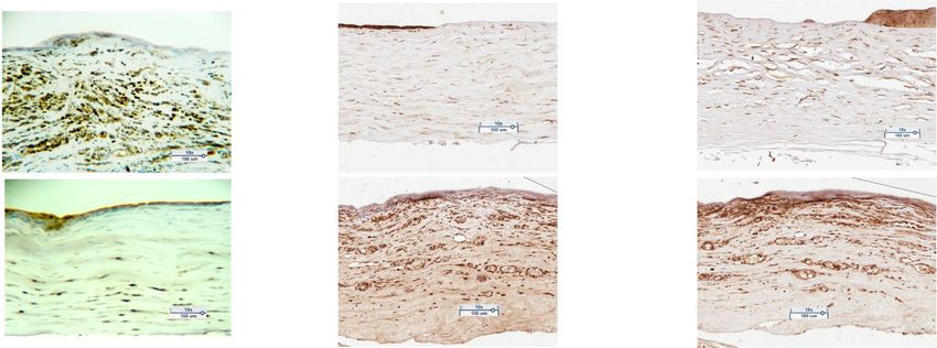
Fig. 2 Immunohistochemistry (IHC) Scores of Keratectomy Specimens of Dogs with Chronic non-healing Corneal ulcers (Scatterplot, median). A
NOG staining. Samples from NOG del/del dogs had a lower mean NOG IHC score than samples from NOG wt/wt, but the difference was not
significant (p = 0.0625). B BMP2, and 4 IHC scores of samples from NOG del/del and NOG wt/wt dogs. Keratectomy specimens from NOG del/del
dogs had significantly higher BMP2 (p = 0.008) than samples from NOG wt/wt dogs (Man Whitney U Test). Representative IHC images of
keratectomy samples from NOG wt/wt and NOG del/del dogs. C. NOG score 3; D. NOG score 1; E. BMP2 score 1; F. BMP2 score 3; G.BMP4 score 1;
H. BMP 4 score 3. 10X Magnification
reduced adhesion of the corneal epithelium to the from SCCED Boxer dogs deficient in NOG revealed re-
underlying extracellular matrix and corneal stroma in duced RNA signal and expression of NOG, respectively,
SCCED [1, 2, 17]. Further study of the role of Noggin and altered expression of BMP signaling pathway factors
and the effects of its deficiency o on corneal wound compared to WT dogs. A limitation of this study is that
healing and on the development of SCCED in dogs is we compared corneal tissue from Boxer dogs with SCCE
needed. Ds to non-Boxer dog with SCCEDs, rather than compar-
In this study, using whole genome sequencing (WGS), ing it to corneal tissue from unaffected control dogs.
we identified a 30 base pair deletion at a splice site in Since SCCEDs are likely to have RNA and protein ex-
the noggin gene (NOG) which was significantly associ- pression alterations that occur as a consequence of the
ated with Boxer dogs with SCCEDs compared to non- corneal ulcer, comparison to unaffected corneal tissue
Boxer dogs. Furthermore, RNA sequencing of corneal could have emphasized changes that occur due to SCCE
samples and immunostaining of keratectomy specimens D rather than the underlying cause. Finally, additional
analysis of the NOG RNA for alignment and transcript
analysis would have been useful but unfortunately the
Table 3 Immunohistochemistry scoring of NOG and genes that
quality and quantity of NOG RNA from debrided cells
NOG regulates in the BMP Signaling Pathway from SCCED Boxer
dogs with NOG deletion compared to SCCED non-Boxer
was quite limited and prevented additional transcript
without the NOG deletion analysis.
NOG− IHC score NOG WT score P value
(Mean ± SD) (Mean ± SD) Conclusions
NOG 0.6 ± 0.9 2.0 ± 0.7 0.03 Many Boxer dogs with SCCED have a genetic defect in
BMP4 1.6 ± 0.7 2.0 ± 1.6 0.62
NOG. NOG is a constitutive protein in the cornea which
is a potent inhibitor of BMP, which likely regulate limbal
BMP2 2.0 ± 0.7 0.6 ± 0.5 0.01
epithelial progenitor cells (LEPC). Dysregulation ofMeurs et al. BMC Veterinary Research (2021) 17:254 Page 8 of 9
LEPC may play a role in the pathogenesis of RCE. These Received: 31 January 2021 Accepted: 1 July 2021
results suggest that NOG deficiency play a role in SCCE
Ds and RCEs. Further study of this defect in the patho-
genesis of SCCED and RCE appears warranted. References
1. Murphy CJ, Marfurt CF, McDermott A, Bentley E, Abrams GA, Reid TW, et al.
Spontaneous chronic corneal epithelial defects (SCCED) in dogs: clinical
Abbreviations features, innervation, and effect of topical SP, with or without IGF-1. Investig
BMP: Bone morphogenic protein; Chr: Chromosome; IACUC: Institutional Ophthalmol Vis Sci. 2001;42:2252–61.
animal care and use committee; ICH: Immunohistochemistry; LEPC: Limbal 2. Bentley E, Abrams GA, Covitz D, Cook CS, Fischer CA, Hacker D, et al.
epithelial progenitor cells; NOG: Noggin; RCE: Recurrent corneal erosions; Morphology and immunohistochemistry of spontaneous chronic corneal
SCCED: Superficial chronic corneal epithelial defects; WGS: Whole genome epithelial defects (SCCED) in dogs. Investig Ophthalmol Vis Sci. 2001;42:
sequencing 2262–9.
3. Das S, Seitz B. Recurrent corneal Erosion syndrome. Surv Ophthalmol. 2008;
53:3–15.
4. Chandler HL, Gemensky-Metzler AJ, Bras ID, Robbin-Webb TE, Saville WJ,
Supplementary Information Colitz CMH. In vivo effects of adjunctive tetracycline treatment on refractory
The online version contains supplementary material available at https://doi.
corneal ulcers in dogs. J Am Vet Med Assoc. 2010;237:378–86.
org/10.1186/s12917-021-02955-1.
5. Diez-Feijóo E, Grau AE, Abusleme EI, Durán JA. Clinical presentation and
causes of recurrent corneal erosion syndrome: Review of 100 patients.
Additional file 1. Cornea. 2014;33(6):571–5.
6. Miller DD, Hasan SA, Simmons NL, Stewart MW. Recurrent corneal erosion: a
comprehensive review. Clin Ophthalmol. 2019;13:325–35.
Acknowledgements 7. Dua HS, Azuara-Blanco A. Limbal stem cells of the corneal epithelium. Surv
The authors thank Damian Launer, Melissa Hamman, Erin Barr, and Beth Ophthalmol. 2000;44(5):415–25.
Salmon for technical support. The authors also thank the University of North 8. Le Q, Xu J, Deng SX. The diagnosis of limbal stem cell deficiency. Ocular
Carolina Histology Core for processing of the corneal histology and surface. 2018. Ocul Surf. 2018;16(1):58–69.
immunohistochemistry. 9. Anderson DF, Ellies P, Pires RTF, Tseng SCG. Amniotic membrane
transplantation for partial limbal stem cell deficiency. Br J Ophthalmol. 2001;
85(5):567–75.
Authors’ contributions 10. Avni Zauberman N, Artornsombudh P, Elbaz U, Goldich Y, Rootman DS,
KMM designed the study, analyzed and interpreted the data, and wrote Chan CC. Anterior stromal puncture for the treatment of recurrent corneal
manuscript; KM designed the study and revised the manuscript; SG analyzed erosion syndrome: patient clinical features and outcomes. Am J
data and revised the manuscript; BW analyzed data and revised the Ophthalmol. 2014;157(2):273–9.
manuscript; and BG collected samples, analyzed and interpreted the data, 11. Laibson PR. Recurrent corneal erosions and epithelial basement membrane
and wrote manuscript. All authors read and approved the final manuscript. dystrophy. Eye Contact Lens. 2010;36(5):315–7.
12. Singh RP, Raj D, Pherwani A, Lagnado R, Abedin A, Eatamadi H, et al.
Alcohol delamination of the corneal epithelium for recalcitrant recurrent
Funding corneal erosion syndrome: a prospective study of efficacy and safety. Br J
Grants: American Kennel Club – Canine Health Fund. Ophthalmol. 2007;91:908–11.
13. Tsai TY, Tsai TH, Hu FR, Hou YC. Recurrent corneal erosions treated with
anterior stromal puncture by neodymium: yttrium-aluminum-garnet laser.
Availability of data and materials Ophthalmology. 2009;116(7):1296-300.
The datasets used and/or analyzed during the current study are available 14. Kaz Soong H, Farjo Q, Meyer RF, Sugar A. Diamond burr superficial
from the corresponding author on reasonable request. keratectomy for recurrent corneal erosions. Br J Ophthalmol. 2002;86:296–8.
15. Gosling A, Labelle AL, Breaux CB. Management of spontaneous chronic
corneal epithelial defects (SCCEDs) in dogs with diamond burr debridement
Declarations
and placement of a bandage contact lens. Vet Ophthalmol. 2013;16:83–8.
16. Roberts SR. Superficial indolent ulcer of the cornea in Boxer dogs. J Small
Ethics approval and consent to participate
Anim Prac; 6:111-115.
Use of animals in this study adhered to the Association for Research in
17. Chavkin MJ, Riis RC, Scherlie PH. Management of a persistent corneal
Vision and Ophthalmology (ARVO) statement on the use of animals in
erosion in a boxer dog. Cornell Vet. 1990;80(4):347–56.
ophthalmic and vision research. The animal use protocol was approved and
18. Eaton JS, Hollingsworth SR, Holmberg BJ, Brown MH, Smith PJ, Maggs DJ.
monitored by the North Carolina State University Institutional Animal Care
Effects of topically applied heterologous serum on reepithelialization rate of
and Use Committee (IACUC) (Protocol # 18–164-0). The North Carolina State
superficial chronic corneal epithelial defects in dogs. J am vet med Assoc.
University Veterinary Hospital Board approved the protocol for evaluation of
2017. J Am Vet Med Assoc. 2017;250(9):1014–22.
clinical patients in this study. Owners of dogs signed and provided informed
19. Friedenberg SG, Meurs KM. Genotype imputation in the domestic dog.
consent for all sample collections and data collection. The study was carried
Mamm Genome. 2016;27(9–10):485–94.
out in compliance with the ARRIVE guidelines.
20. Bolger AM, Lohse M, Usadel B. Trimmomatic: a flexible trimmer for Illumina
sequence data. Bioinformatics. 2014;30:2114–20.
Consent for publication 21. Li H, Durbin R. Fast and accurate short read alignment with burrows-
All authors consent to publication. wheeler transform. Bioinformatics. 2009;25:1754–60.
22. Depristo MA, Banks E, Poplin RE, Garimella KV, Maguire JR, Hartl C, et al. A
framework for variation discovery and genotyping using next- generation
Competing interests DNA sequencing data. Nat Genet. 2011;43:491–8.
The authors declare that they have no competing interests. 23. McKenna A, Hanna M, Banks E, Sivachenko A, Cibulskis K. The genome
analysis toolkit: a MapReduce framework for analyzing next-generation DNA
Author details sequencing data Aaron. Genome Res. 2010;20:1297–303.
1
Clinical Sciences, North Carolina State University, 1060 William Moore Drive, 24. Van Der Auwera G, Carneiro MO, Hartl C, Poplin R, Levy-moonshine A,
Raleigh, North Carolina 27613, USA. 2Present address: Upstate Veterinary Jordan T, et al. From FastQ data to high confidence varant calls: the
Specialties, Latham, NY, USA. 3Veterinary Clinical Sciences, University of Genonme analysis toolkit best practices pipeline. Curr Protoc Bioinformatics.
Minnesota, St. Paul, MN, USA. 2013;43(1110):11.10.1–11.10.33.Meurs et al. BMC Veterinary Research (2021) 17:254 Page 9 of 9
25. Tokuda Y, Okumura N, Komori Y, Hanada N, Tashiro K, Koizumi N, et al.
Transcriptome dataset of human corneal endothelium based on ribosomal
RNA-depleted RNA-Seq data. Sci Data. 2020;7:407.
26. Patro R, Duggal G, Love MI, Irizarry RA, Kingsford C. Salmon: fast and bias-
aware quantification of transcript expression using dual-phase inference.
Nat Methods. 2017;14:417.
27. Soneson C, Love MI, Robinson MD. Differential analyses for RNA-seq:
transcript-level estimates improve gene-level inferences. F1000Research.
2015;4:1521.
28. Makhijani RK, Raut SA, Purohit HJ. Fold change based approach for
identification of significant network markers in breast, lung and prostate
cancer. IET Syst Biol. 2018;12:213–8.
29. Hirsch ML, Conatser LM, Smith SM, Salmon JH, Wu J, Buglak NE, et al. AAV
vector.meditated expression of HLA-G reduces injury-induced corneal
vascularization, immune cell infiltration, and fibrosis. Sci Rep. 2017;7(1):
17840.
30. Krause C, Guzman A, Knaus P. Noggin. Int J Biochem Cell Biol. 2011;43:478–
81. https://doi.org/10.1016/j.biocel.2011.01.007.
31. Wordinger RJ, Clark AF. Bone morphogenetic proteins and their receptors in
the eye. Exp Biol Med. 2007;232:979–92.
32. Lewis CJ, Mardaryev AN, Poterlowicz K, Sharova TY, Aziz A, Sharpe DT, et al.
Bone morphogenetic protein signaling suppresses wound-induced skin
repair by inhibiting keratinocyte proliferation and migration. J Invest
Dermatol. 2014;134:827–37. https://doi.org/10.1038/jid.2013.419.
33. Mohan RR, Kim WJ, Mohan RR, Chen L, Wilson SE. Bone morphogenic
proteins 2 and 4 and their receptors in the adult human cornea. Invest
Ophthalmol Vis Sci. 1998;39(13):2626–36.
34. Han B, Chen S-Y, Zhu Y-T, Tseng SCG. Integration of BMP/Wnt signaling to
control clonal growth of limbal epithelial progenitor cells by niche cells.
Stem Cell Res. 2014;12:562–73. https://doi.org/10.1016/j.scr.2014.01.003.
35. Stepp MA, Zieske JD. The corneal epithelial stem cell niche. Ocul Surf. 2005;
3:15–26. https://doi.org/10.1016/S1542-0124(12)70119-2.
36. Ahmed MI, Mardaryev AN, Lewis CJ, Sharov AA, Botchkareva NV. MicroRNA-
21 is an important downstream component of BMP signalling in epidermal
keratinocytes. J Cell Sci. 2011;124(Pt 20):3399–404.
37. Sharov AA, Sharova TY, Mardaryev AN, Di Vignano AT, Atoyan R, Weiner L,
et al. Bone morphogenetic protein signaling regulates the size of hair
follicles and modulates the expression of cell cycle-associated genes. Proc
Natl Acad Sci U S A. 2006;103(48):18166–71.
38. Takano K, Ogasawara N, Matsunaga T, Mutai H, Sakurai A, Ishikawa A, et al.
A novel nonsense mutation in the NOG gene causes familial NOG-related
symphalangism spectrum disorder. Hum Genome Var. 2016;3:16023.
39. Masuda S, Namba K, Mutai H, Usui S, Miyanaga Y, Kaneko H, et al. A
mutation in the heparin-binding site of noggin as a novel mechanism of
proximal symphalangism and conductive hearing loss. Biochem Biophys Res
Commun. 2014;447(3):496–502.
40. Potti TA, Petty EM, Lesperance MM. A comprehensive review of reported
heritable noggin-associated syndromes and proposed clinical utility of one
broadly inclusive diagnostic term: NOG-related-symphalangism spectrum
disorder (NOG-SSD). Hum Mutat. 2011;32:877–86. https://doi.org/10.1002/
humu.21515.
Publisher’s Note
Springer Nature remains neutral with regard to jurisdictional claims in
published maps and institutional affiliations.You can also read