Tasker, S. (2018). Diagnosis of feline infectious peritonitis: Update on evidence supporting available tests. Journal of Feline Medicine and ...
←
→
Page content transcription
If your browser does not render page correctly, please read the page content below
Tasker, S. (2018). Diagnosis of feline infectious peritonitis: Update on
evidence supporting available tests. Journal of Feline Medicine and Surgery,
20(3), 228-243. https://doi.org/10.1177/1098612X18758592

Peer reviewed version

Link to published version (if available):
10.1177/1098612X18758592

Link to publication record in Explore Bristol Research
PDF-document

This is the author accepted manuscript (AAM). The final published version (version of record) is available online
via Sage at http://journals.sagepub.com/doi/10.1177/1098612X18758592 . Please refer to any applicable terms
of use of the publisher.

University of Bristol - Explore Bristol Research
General rights

This document is made available in accordance with publisher policies. Please cite only the published
version using the reference above. Full terms of use are available:
http://www.bristol.ac.uk/pure/about/ebr-terms
Diagnosis of feline infectious peritonitis: update on evidence supporting available tests
Author Details
Séverine Tasker
Professor of Feline Medicine
BSc BVSc PhD DSAM DipECVIM-CA PGCertHE FHEA MRCVS
The Feline Centre, Langford Vets, Bristol Veterinary School, University of Bristol, Bristol, BS40 5DU, UK
s.tasker@bristol.ac.uk
Photo will be provided.
Structured Abstract
Practical relevance
Feline coronavirus (FCoV) infection is very common in cats, usually only causing mild intestinal signs
such as diarrhoea. Up to 10% of feline coronavirus infections, however, result in feline infectious eritonitis
(FIP), a fatal disease which is a common cause of death in young cats.
Clinical challenges
Obtaining a definitive diagnosis of FIP based on non-invasive approaches in cats is difficult. Confirmation
of FIP relies on finding appropriate cytological or histopathological changes in cytological samples or biopsies in
association with positive immunostaining for FCoV antigen. In FIP cases with effusions, cytology and
immunostaining on effusion samples can be relatively easy to perform. In the absence of effusions, obtaining
diagnostic samples more challenging. Often, and especially in FIP cases without effusions, collection of biopsies
from tissues with gross lesions is necessary.
In the absence of a definitive diagnosis, a high index of suspicion of FIP may be obtained from a
combination of the cat’s signalment (e.g. being 2 years or younger, originating from a multi-cat household),
history (e.g. fluctuating pyrexia), clinical examination findings (e.g. pyrexia, jaundice, effusions, uveitis,
neurological signs) and laboratory test results (e.g. lymphopenia, hyperglobulinaemia, reduced albumin to
globulin ratio and/or elevated 1-acid glycoprotein concentrations in serum or effusion, positive reverse
transcriptase-polymerase chain reaction (RT-PCR) for FCoV RNA in effusion, cerebrospinal fluid or biopsy
samples, pyogranulomatous changes identified in cytological or biopsy specimens). These results, if largely
consistent with FIP, can be used as a basis of discussion with the owner about whether additional, more invasive,
diagnostic tests are warranted. In some cases it may be that euthanasia is discussed as an alternative to pursuing
a definitive diagnosis ante-mortem, especially if financial limitations exist or when cats are very sick and concerns
exist over a patient’s ability to tolerate invasive diagnostic procedures (e.g. surgical biopsy). Ideally confirmation
of the diagnosis should be made in such patients, if euthanased, from samples taken at post-mortem
examination.
Global importance
Feline infectious peritonitis occurs wherever FCoV infection is present in cats, and thus is found in most
parts of the world. Only a few areas have been investigated (e.g. Galápagos Islands and Falkland Islands) which
appear to be free from FCoV infection.
Evidence base
This review provides a comprehensive overview of how to approach the diagnosis of FIP, focusing on
tests available to the veterinary practitioner and recent published evidence for the usefulness of these tests.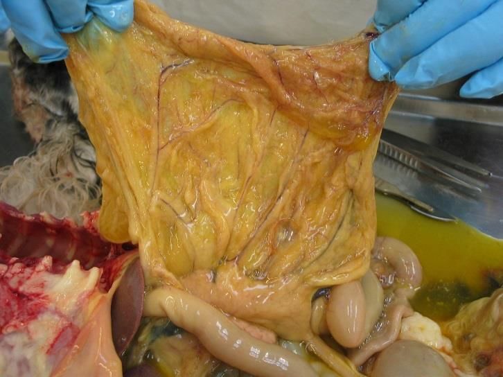
What are coronaviruses?
Coronaviruses are large, enveloped, positive-sense single-stranded RNA viruses with non-segmented genomes
of around 30,000 nucleotides in length{Siddell, 1995 #371}. Feline coronavirus (FCoV) is a sub-species of the
alphacoronavirus I species, along with canine coronavirus (CCoV). Coronaviruses exhibit a high rate of mutation
during RNA replication and therefore exist as clusters of genetically diverse populations, known as
quasispecies{Denison, 2011 #372;Desmarets, 2016 #347}. This genetic diversity, along with a readiness to
recombine with other coronavirus strains, is associated with their pathogenicity and cross-species
transmission. Interestingly, FCoVs appear to have emerged in the 1950s, possibly due to cross-species
transmission{Addie, 2012 #373}. Cats worldwide have been found to be infected with FCoV with the exception
of those residing in a few areas, such as the Galápagos Islands{Levy, 2008 #387} and Falkland Islands{Addie,
2012 #259}, likely due to quarantine of those cats by their isolated island habitat.

Two FCoV so-called serotypes are recognised: Type 1, which represents the vast majority of field strains found
in naturally infected cats, although geographical variation exists{Benetka, 2004 #2;Hohdatsu, 1992 #52}, and
Type 2, which arose following recombination events between Type 1 FCoV and CCoV{Herrewegh, 1998 #377}.
The two FCoV serotypes are distinguished primarily by the genetic and serological differences in their
transmembrane spike (S) gene and protein, respectively. The S protein (Figure 1) is important as it is the part
of the FCoV that binds to the host (feline) receptor, mediating host cell entry{Jaimes, 2018 #409}. The Type 2
FCoV S protein resembles that of CCoV, and, like CCoV, Type 2 FCoVs use aminopeptidase N as their feline host
cell receptor. The receptor for Type 1 FCoVs is unknown{Dye, 2007 #323}.

The complex relationship between feline coronavirus and feline infectious peritonitis (FIP)
Feline coronavirus infection is very common in cats. Around 40% of the domestic cat population has been
infected with FCoV, and this figure increases to 90% in multi-cat households{Addie, 2000 #209;Addie, 1992 #111}.
Modern changes in feline husbandry (e.g. more cats kept indoors and in multi-cat households) are likely to have
led to the increase in FCoV-related disease{Addie, 2012 #373}.

Natural infections with FCoV are transient in ~70% of cats, whereas persistent infections occur in ~13% of
cats{Addie, 2012 #373}. These persistently infected cats are sometimes referred to as ‘carrier’ or ‘chronically
shedding’ cats. In most cases, FCoV infection is asymptomatic or results in only mild gastrointestinal clinical
signs (e.g. inappetence, diarrhoea, vomiting), although occasionally more severe gastrointestinal disease is
seen. Interestingly, around 5-10% of cats are believed to be resistant to FCoV infection{Addie, 2012 #373}.
However, in a small percentage of cases, FCoV infection results in the severe disease of FIP{Pedersen, 2009
#204;Addie, 1995 #40}. Occasionally, and possibly with increasing frequency recently, outbreaks of FIP (when a
larger percentage of cats are affected) in multi-cat households or shelters are reported{Wang, 2013
#269;Barker, 2013 #280}. No curative treatments for FIP are currently available, although some novel
treatments, including protease inhibitors, show promise{Kim, 2016 #360;Legendre, 2017 #374;Pedersen, 2017
#403}.

Asymptomatic FCoV infection was previously believed to be confined to the intestinal tract, but we now know
that healthy FCoV-infected cats can have systemic FCoV infection, albeit with lower FCoV viral loads than cats
with FIP{Kipar, 2006 #130;Kipar, 2010 #227;Meli, 2004 #76;Desmarets, 2016 #347}.

What factors contribute to the development of FIP following FCoV infection?
Viral factors are important in the pathogenesis of FIP. As outlined above, the S protein of FCoV mediates host
cell entry. The S protein contains a putative fusion peptide that enables fusion of the FCoV envelope with the
host cell membrane{Bosch, 2003 #351}. Mutations in the S gene can result in amino acid substitutions in the
transcribed S protein that influence the tropism of FCoV{Belouzard, 2012 #352}. Studies have identified
mutations in the fusion peptide sequence of the FCoV S gene that were thought to be markers of FIP{Chang,
2012 #255;Bank-Wolf, 2014 #395} as well as changes in the closely-related furin cleavage site that were also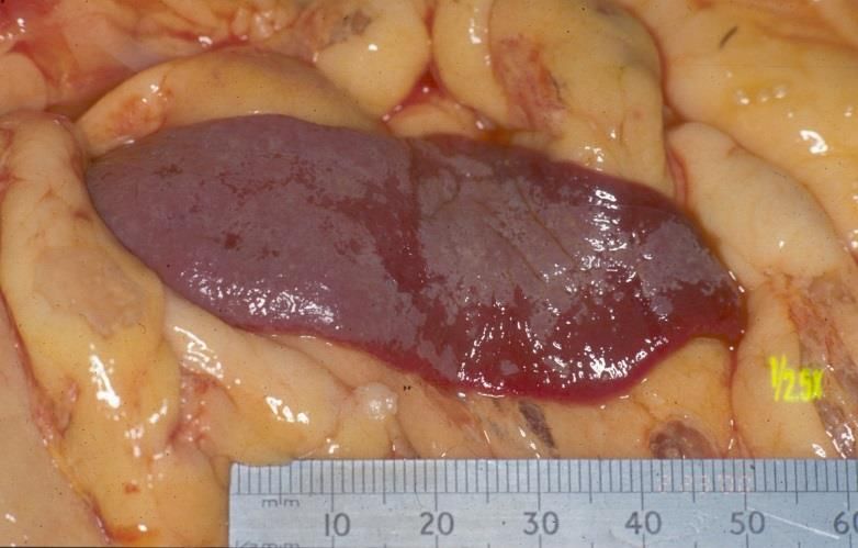
thought to be correlated with FIP{Licitra, 2013 #277}. Recently it has been found that the fusion peptide
mutations are likely to be markers of systemic FCoV infection, which can occur in both FIP and non-FIP cats,
rather than FIP per se{Porter, 2014 #321}. However, these mutations are still important as it is probably via
these, and/or other mutations, that the FCoV acquires its monocyte/macrophage tropism to allow it to spread
systemically, outside of the intestinal tract, and contribute to the development of FIP. Other viral factors then
mediating effective and sustained replication in monocytes, and activation of infected monocytes, are also
likely to be important for the subsequent development of FIP following systemic FCoV infection{Kipar, 2014
#309}.
Host factors are also very likely to play an important part in FIP development. These include the host immune
response (e.g. T-lymphocyte depletion occurs in cats that develop FIP), the ability of monocytes to sustain FCoV
replication, breed and genetics{de Groot-Mijnes, 2005 #314;Golovko, 2013 #294;Pedersen, 2016
#356;Dewerchin, 2005 #394}.
Environmental factors, such as the level of stress and overcrowding in a household, may also play a role. These
may feed into increasing the rate of viral replication in an individual cat{Battilani, 2003 #81}, which could
increase the generation of viral mutants and support FIP development.

Approaching a diagnosis of FIP
Making a definitive diagnosis of FIP
A definitive diagnosis of FIP traditionally relies on histopathological examination of tissues, usually with
detection of the virus within lesions by immunohistochemistry for FCoV antigen. Immunostaining of FCoV
antigen in effusion samples is also an option for definitive diagnosis of FIP in cases with effusions showing
biochemical and cytological features consistent with FIP.

Histopathological examination, effusion analysis and FCoV antigen immunostaining are discussed in more detail
later.

Obtaining a high index of suspicion of a diagnosis of FIP
In the absence of a definitive diagnosis, a high index of suspicion of FIP may be obtained from background
information, clinical signs and routine clinicopathological results. With experience, this can be used as a basis
to discuss with the owner whether additional, more invasive, diagnostic tests (e.g. biopsy of affected tissues)
are warranted. In such cases it may be that euthanasia is discussed as an alternative to pursuing a definitive
diagnosis ante-mortem, and this may be preferable in, for example, shelter cats, where there are financial
limitations, or when cats are very sick and concerns exist over a patient’s ability to tolerate diagnostic
procedures (e.g. surgical biopsy). If euthanasia is performed without a definitive diagnosis, post-mortem
examination is strongly advised and relatively simple to perform{Tasker, 2017 #380}. This permits both
assessment for gross changes consistent with FIP (Figure 2), and sampling for histopathological examination. If
financial limitations preclude the latter, it is worth contacting researchers with an interest in FIP (e.g. the
University of Bristol Feline Coronavirus Research Group & Bristol-Zurich FIP Consortium) to see if samples are
being sought for research studies that could allow for analysis at a reduced cost or free of charge.

Many important differential diagnoses should be considered in cats suspected of having FIP such as
toxoplasmosis, mycobacterial infection and lymphocytic cholangitis. These, and others, are described in Table
1, together with consideration of features distinguishing those diseases from FIP.

Signalment and background evidence for FIP
It should be remembered that FIP is most common in young cats (those less than three years of age, and
especially those less than two years of age{Riemer, 2016 #358}) but a smaller peak of cases is seen in cats older
than 10 years of age. Male cats are also at a slightly higher risk{Riemer, 2016 #358}. Some breeds in some
countries may be predisposed to FIP{Pesteanu-Somogyi, 2006 #121;Worthing, 2012 #252}, but this is likely due
to the presence of unknown specific genetic risk factors in those breeds in those countries, and generalised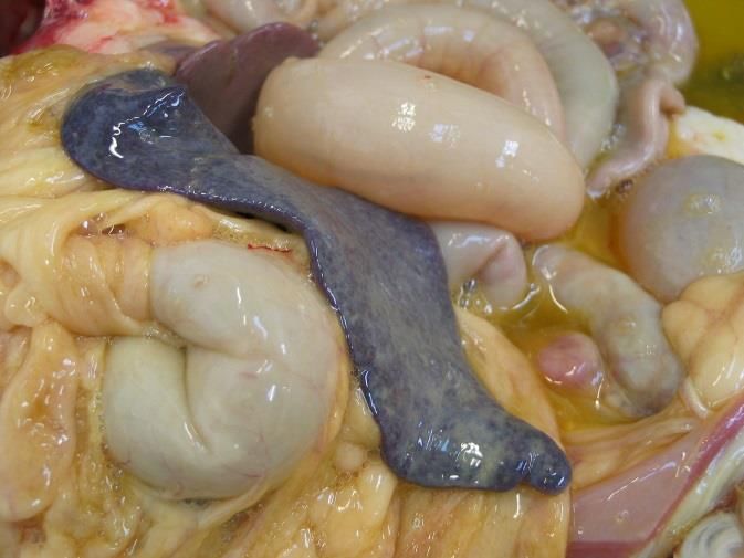
breed predispositions may not exist{Riemer, 2016 #358}. A recent history of stress (e.g, adoption, being in a
shelter, neutering, upper respiratory tract disease, vaccination etc.) may be apparent{Riemer, 2016 #358} and
may play a part in triggering the development of FIP in a FCoV-infected cat. Although living in a multi-cat
household increases the likelihood of being FCoV seropositive, a recent large study{Riemer, 2016 #358} found
that the majority of cats presented to a university hospital with FIP were from households containing a small
number of cats at time of diagnosis.

Clinical signs of FIP
Disease manifestations of FIP typically comprise a vasculopathy resulting in (‘wet’) effusions (up to 80% of FIP
cases have effusions), granuloma formation only resulting in (‘dry’) mass lesions, or a combination of the two;
indeed most FIP cases with effusions also have granulomatous lesions visible at post-mortem examination.
Clinical signs (Figure 3) seen in both effusive and non-effusive FIP include lethargy, anorexia, weight loss (or
failure to gain weight/stunted growth in younger cats), a fluctuating pyrexia that is usually non-responsive to
drugs such as antibiotics or non-steroidal anti-inflammatories, and sometimes jaundice (more common in
effusive FIP). A recent study evaluating referral cat cases with a history of pyrexia found that, at 20.8% (22 of
106 cats), FIP was the most common diagnosis made, showing the importance of FIP as a differential diagnosis
in pyrexic cats in this population of referred cats{Spencer, 2017 #378}. Another study{Riemer, 2016 #358}
describing FIP cases only reported that body temperature exceeded 39.5°C in 81% of cats and 40°C in 39% of
cats. Pyrexia was far more common in cats with effusive FIP than those with neurological non-effusive
FIP{Riemer, 2016 #358}. Lymphadenomegaly can also be present in both the effusive and non-effusive forms of
FIP.

Effusive ‘wet’ FIP is associated with abdominal, pleural and/or pericardial effusions (occasionally in the scrotum
too of entire male cats), and is often quite acute in nature, progressing within a few days or weeks and severely
limiting survival{Ritz, 2007 #117}. These cats can present with dyspnoea, tachypnoea and/or abdominal
distension. Non-effusive ‘dry’ FIP is typically associated with neurological (can be focal, multifocal or diffuse in
nature, often with central vestibular signs, occasionally as a T3-L3 myelopathy{Crawford, 2017 #397}; Figure 3)
and/or ocular (anterior and/or posterior uveitis; Figure 3) signs and is more chronic, progressing over a few
weeks to months. Dermatological signs (manifested typically as small multiple non-pruritic papules or nodules)
signs{Bauer, 2013 #295;Cannon, 2005 #127} have also been reported in dry FIP. Renomegaly may occur in non-
effusive FIP with renal involvement. Occasionally a diffuse pyogranulomatous pneumonia is reported.

It is important to remember that clinical signs of FIP can change over time so repeated clinical examinations
are important to detect newly apparent signs (e.g. development of a small volume of effusion, ocular changes
visible on retinal examination).

Focal non-effusive FIP occasionally occurs, presenting typically as a palpable abdominal mass, and can be
particularly challenging to diagnose as the lesions can be hard to initially differentiate from neoplasia and
mycobacterial infection. Focal FIP case reports have comprised cats presenting with mesenteric lymph node
enlargement due to necrogranulomatous lymphadenitis{Kipar, 1999 #23}, or solitary mural intestinal lesions of
the colon or ileocecocolic junction with associated regional lymphadenopathy {Harvey, 1996 #36}. The cats
with focal intestinal FIP had a history of vomiting and diarrhoea.

Possible routine laboratory test findings in FIP
Routine haematology
Haematological changes in FIP are non-specific but there are a number of abnormalities that can be looked for
to support a diagnosis. Lymphopenia is particularly common (55-77% of cases; although a recent study found
only 49.5% of FIP cases to be lymphopenic{Riemer, 2016 #358}), with neutrophilia (39-57%), a left shift, and
mild-moderate normocytic, normochromic anaemia (37-54%) also reported{Tsai, 2011 #245;Sparkes, 1991
#57;Norris, 2005 #246;Riemer, 2016 #358}. An association between FIP and microcytosis (with or without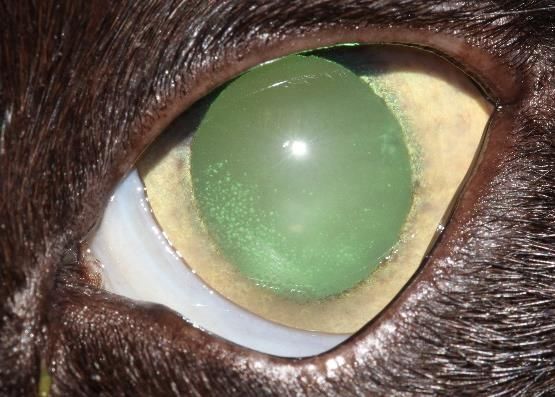
anaemia) was recently reported{Riemer, 2016 #358}. Severe immune-mediated haemolytic anaemia (IMHA),
with an associated regenerative anaemia, can occur with FIP{Norris, 2005 #246}, but is uncommon.
Serum biochemistry
The changes in serum biochemistry in FIP cases are varied and often non-specific but there are a number of
important abnormalities that should be looked for to support a diagnosis of FIP.
Hyperglobulinaemia is reported in 89% of cases, often with hypoalbuminaemia or low-normal serum albumin
(seen in 64.5% of cases){Riemer, 2016 #358}. The presence of hypoalbuminaemia alongside
hyperglobulinaemia means that hyperproteinaemia may not always occur; past reports documented
hyperproteinaemia in up to 60% of cases, especially in dry FIP cases, but lower prevalences of 17.5% have been
reported recently{Riemer, 2016 #358}. The combination of hyperglobulinaemia and hypoalbuminaemia or low-
normal albumin concentration also means that the albumin: globulin (A:G) ratio is low, and this parameter can
be useful to evaluate how likely FIP is in an individual case. Reports of useful cut-off values for A:G ratios in the
diagnosis of FIP vary, but it has been suggested that an A:G ratio of 0.8 makes FIP very unlikely {Tsai, 2011 #245;Sparkes, 1991 #57;Norris, 2005 #246}. Although these
cut-off values are useful to consider, the author does not use a specific value but looks at the A:G ratio in
conjunction with other diagnostic test results; the lower the value, the bigger the suspicion for FIP becomes,
especially if other findings are consistent with a diagnosis of FIP. Interestingly, a study{Jeffery, 2012 #264} in a
population of cats with a low prevalence of FIP (akin to the situation that is usually encountered in veterinary
practice) found that an A:G ratio of >0.6 was useful in ruling out FIP, but that lower ratios were not helpful in
ruling in FIP. Additionally, the frequency and extent of hypoalbuminaemia, hyperglobulinaemia, low A:G ratio
and serum protein electrophoresis (SPE) abnormalities reported in FIP cases have decreased recently{Riemer,
2016 #358;Stranieri, 2017 #379}. With respect to SPE changes, in one study{Stranieri, 2017 #379} cases
diagnosed with FIP from 2013 to 2014 tended to have elevated α2-globulins rather than the elevated γ-
globulins seen in cases from 2004 to 2009; this is possibly due to veterinarians diagnosing FIP earlier, meaning
that cases have not progressed to show elevated γ-globulins. Polyclonal and monoclonal elevated γ-globulins
have been reported with FIP{Taylor, 2010 #388}, although polyclonal elevations are far more common.
Hyperbilirubinaemia occurs in 21-63% of FIP cases, and is especially seen with effusive FIP, often without
marked elevations in alanine aminotransferase (ALT), alkaline phosphatase (ALP) or gamma-
glutamyltransferase (GGT) enzyme activity (although these can be moderately elevated in FIP cases).
Hyperbilirubinaemia due to IMHA is uncommonly reported with FIP{Norris, 2005 #246}, and cats are often not
severely anaemic. Thus the presence of hyperbilirubinaemia in the absence of elevated hepatic enzyme
activities or severe anaemia should raise the index of suspicion of FIP (NB: sepsis and pancreatitis can also
cause hyperbilirubinaemia in the absence of elevated hepatic enzyme activities [Table 1]). Hyperbilirubinaemia
is more commonly identified in FIP cases as the FIP disease progesses; additionally any hyperbilirubinaemia
present can worsen as the FIP disease progresses{Tsai, 2011 #245}.
Acute phase proteins (APPs) are made in the liver in response to cytokines released from macrophages and
monocytes (especially IL-1, IL-6 & TNF-) in many inflammatory and non-inflammatory diseases. 1-acid
glycoprotein (AGP) is an APP, and its measurement can be helpful in the diagnosis of FIP. Although AGP
elevations (>0.48 mg/ml) per se are not specific for FIP, markedly elevated AGP levels (>1.5 mg/ml) are often
seen in FIP cases, so the magnitude of the AGP increase may be helpful in aiding the diagnosis of FIP, with
higher concentrations being more useful in raising the index of suspicion for FIP{Giori, 2011 #242;Duthie, 1997
#244;Paltrinieri, 2007 #260;Hazuchova, 2016 #367}. Indeed, a study found that when the pretest probability of
FIP was high (i.e. history and clinical findings being supportive of FIP), moderate serum AGP levels (1.5-2
mg/ml) could discriminate cats with FIP from cats without FIP, but only higher serum AGP levels (>3 mg/ml)
could support a diagnosis of FIP in cats with a low pretest probability of disease (i.e. history and clinical findings
not supportive of FIP){Paltrinieri, 2007 #260}. However, another, albeit very small, study of unusual cases of FIP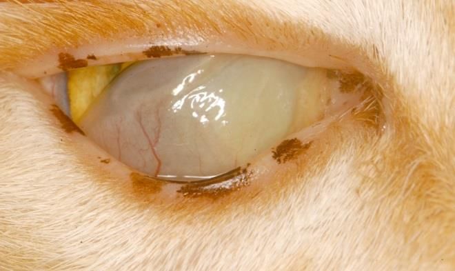
actually found that modest AGP concentrations (>1.5 mg/ml) were still able to discriminate between FIP and
non-FIP cases{Giori, 2011 #242}.

FCoV serology in FIP
Serum FCoV antibody tests are usually enzyme-linked immunosorbent assays (ELISAs), indirect
immunofluorescence antibody (IFA) tests or rapid immunomigration tests{Addie, 2015 #331}. Most tests use
CoV-infected swine or feline cells as a substrate and titres are read in distinct multiples of serum dilutions. A
positive FCoV antibody test indicates that the cat has been infected with FCoV and has seroconverted (this
takes 2-3 weeks from initial infection). Breed-related differences in median FCoV antibody titre have been
detected, and may reflect differences in breed response to FCoV infection{Bell, 2006 #125;Bell, 2006 #126}.
Although FIP cats tend to have higher FCoV antibody titres than non-FIP cats, there is much overlap, with no
difference between median FCoV antibody titres in healthy and suspected FIP cats, so the value in an individual
cat to distinguish cats with FIP is very limited{Bell, 2006 #126}. Many clinically healthy cats (especially those in
multi-cat households) have positive, often very high, FCoV antibody titres, whilst ~10% of cats with FIP are
seronegative (this could be due to the presence of virus in the sample binding antibody and rendering it
unavailable to the serological test{Meli, 2013 #271}), highlighting difficulties in interpretation. It may be that a
negative FCoV antibody result in a suspected dry FIP case is more useful to rule out a diagnosis of FIP{Addie,
2009 #206}; however, negative results have been reported in cases of neurological FIP{Negrin, 2007 #392}.
Clinicians vary as to whether they perform serology or not in suspected cases due to this issue, although a
positive result certainly indicates exposure to FCoV.

Analysis of effusion samples
Analysis of any effusion sample in a suspected case of FIP is extremely helpful for diagnosis, so obtaining
samples of effusions should always be prioritised in investigations of suspected cases. Ultrasonography is
generally regarded as being more sensitive than radiography for the detection of small volumes of fluid in the
thorax and abdomen, but this may depend on where pockets of fluid reside. Repeated ultrasonography to
identify any small volume effusion is recommended and, similarly, ultrasonography can be used to guide
sampling of small pockets of fluid.

FIP effusions (Figure 4) are usually clear, viscous/sticky, straw-yellow and protein-rich (thick eosinophilic
proteinaceous backgrounds are often described on cytology), with a total protein concentration of >35 g/l
(>50% globulins). Very occasionally chylous effusions are described. Typical FIP effusions have similar low A:G
ratios (see above) and raised AGP concentrations to those in serum. A recent study found that effusion AGP
concentrations (of >1.55 mg/ml) were most useful (sensitivity and specificity of 93%) in differentiating FIP and
non-FIP cases compared with AGP levels in the serum or other APPs{Hazuchova, 2016 #367}; however the
‘diagnosis’ of FIP in the cats in this study was not always confirmed by histopathology and immunostaining. FIP
effusions are poorly cellular (usually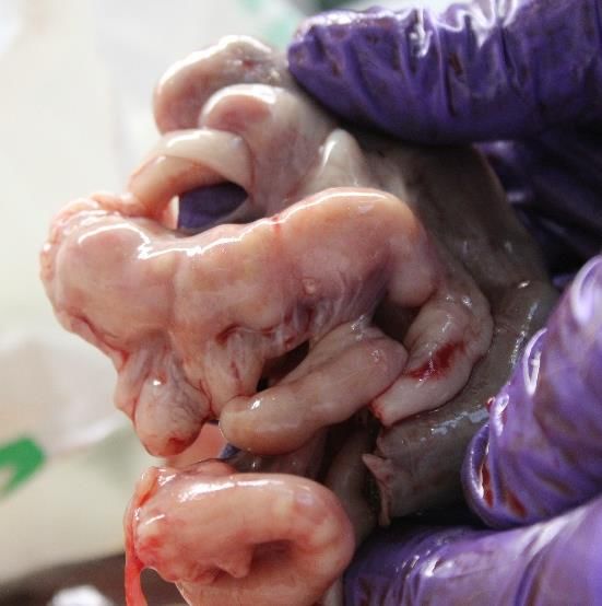
is indicated by the drop disappearing and the solution remaining clear. Interpretation of results can be
problematic due to subjectivity and difficulties in deciding whether a result is truly positive or negative{Fischer,
2013 #382}.

One recent study measured FCoV antibody titres in effusion samples{Meli, 2013 #271} and found an inverse
correlation between FCoV load and FCoV antibodies in some samples, possibly due to antibody being bound by
FCoV and thus not available as a ligand in the serological test; thus making serology unreliable

Serology for FCoV antibodies can also be performed on effusion samples, with very varied results{Meli, 2013
#271;Lorusso, 2017 #401}, so the author does not perform this test in suspected cases of FIP.

Immunostaining for FCoV antigen and reverse-transcriptase polymerase chain reaction (RT-PCR) for FCoV RNA
on effusion samples can also be performed (see later).

Miscellaneous diagnostic tests
In cases with neurological clinical signs, imaging of the brain by magnetic resonance imaging may be useful to
demonstrate changes. For example, obstructive hydrocephalus, syringomyelia, foramen magnum herniation
and marked contrast enhancement of the meninges, third ventricle, mesencephalic aqueduct and brainstem
has been reported with FIP{Foley, 1998 #24;Penderis, 2009 #393;Crawford, 2017 #397}. Cerebrospinal fluid
(CSF) can be collected from neurological cases although care should be taken as the risk of brain herniation is
significant. CSF may show elevated protein concentrations (>30 mg/dl [>0.3 g/l] cisternal samples, >46 mg/dl
[>0.46 g/l] lumbar samples; occasionally FIP cats show marked elevations of >200 mg/dl [>2 g/l]) and an
increased cell count (>8 cells/µl [>8 x 10⁶/l] lumbar and cisternal samples; FIP cats can have cell counts of
>1000 cells/µl [>1000 x 106/l]), with the cell type being predominantly neutrophilic, mononuclear or
mixed{Singh, 2005 #104;Crawford, 2017 #397}. Some neurological cases of FIP have unremarkable CSF analysis
results. Samples of CSF can also be submitted for RT-PCR for FCoV RNA and immunostaining for FCoV antigen
(see below). Add notes on Ab measurement in CSF from {Soma, 2018 #408}{Foley, 1998 #24}

Reverse-transcriptase polymerase chain reaction for FCoV
Background information on FCoV RT-PCR
Reverse transcriptase-PCR assays are available to detect FCoV; however, they are not specific for FIP-associated
FCoVs. FCoV RT-PCR assays amplify both cell-associated subgenomic mRNA (short lengths of transcriptional
RNA produced when the FCoVs replicate), as well as cell-associated or virion-associated genomic RNA, with the
relative abundance of each determined by the positioning of primers (i.e. where along the FCoV sequence the
primers bind during PCR){Barker, 2017 #383}. As viral transcription starts at the 3’ end of the FCoV genome
(Figure 6) there are more subgenomic mRNAs containing viral 3’ sequence than those containing viral 5’
sequence, hence quantitative assays (i.e. RT-qPCR) directed at the 5’ end of the genome (e.g. viral replicase
complex genes) are less susceptible to viral load overestimation than those directed at the 3’ end of the
genome (e.g. 7a/b non-structural protein genes).

Laboratories should be able to report the sensitivity and specificity of the RT-PCRs they are using to detect
FCoV RNA, and the binding site of the primers they use can give some indication as to whether the assay will be
prone to viral load overestimation (see above). As an RNA virus, FCoV shows a high rate of errors during
replication and any viral mutations at the site of primer and/or probe binding can result in loss of PCR assay
efficiency, and ultimately sensitivity. PCR conditions may be altered to tolerate such mutations, but this can
result in a loss of specificity{Barker, 2017 #386}.

Reporting of results for FCoV RNA RT-PCR can be rapid if the laboratory used has a fast turnaround time,
although, once time taken to submit the sample to the laboratory is factored in, reporting of results can still
take a few days. This is usually quicker than immunostaining on tissue samples and may be quicker than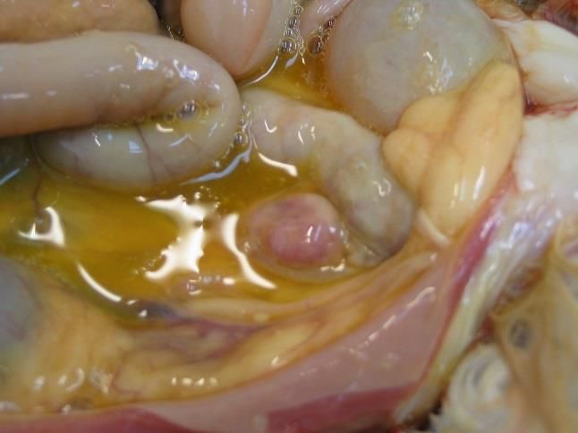
immunostaining on effusion samples. However, it should be noted that immunostaining can provide a
definitive diagnosis whereas RT-PCR does not. Recently a rapid molecular technique (loop mediated isothermal
amplification) for detecting FCoV RNA in-house has been described{Stranieri, 2017 #370}, although it suffered
from poor sensitivity.

Which samples can be tested for FCoV by RT-PCR?
FCoV RT-PCR can be used to detect FCoV RNA in blood, effusion, tissue, CSF, or aqueous humour samples from
suspected cases of FIP. Tissue samples should not be formalin fixed, as formalin degrades the target RNA and
can decrease PCR sensitivity; indeed RNA is very sensitive to degradation and samples for research purposes
are often collected into RNA preservation fluids. However, the need for special collection conditions of samples
outlined above destined for routine diagnostic purposes by RT-PCR is unproven. The presence (particularly of
high levels) of FCoV RNA in blood, effusion, tissue, CSF and/or aqueous humour samples can be highly
supportive of a diagnosis of FIP, as outlined below, but cannot be regarded, in the author’s opinion, as
delivering a definitive diagnosis.

FCoV RT-PCR can also be performed on faecal samples, but this is primarily used to identify cats that are
shedding FCoV for the management of infection in a multi-cat household. Faecal FCoV RT-PCR is not used to
aid in the diagnosis of FIP, but interestingly recent studies have found that cats with FIP are more likely to be
shedding FCoV{Barker, 2017 #383}, and have higher amounts of FCoV RNA as determined by RT-qPCR{Porter,
2014 #321}, in their faeces than cats without FIP.

FCoV RT-PCR on tissue samples
Tissue samples from cats with FIP are significantly more likely to be FCoV RT-PCR positive{Barker, 2017 #383},
and have significantly higher FCoV loads by RT-qPCR{Porter, 2014 #321}, than tissues samples from non-FIP
cats, although cats without FIP can still be positive for FCoV by RT-PCR. For example in a recent extensive study
evaluating FCoV RT-PCR{Barker, 2017 #383}, 90.4% of tissue samples from FIP cats were FCoV RT-qPCR positive
compared to only 7.8% of tissue samples from non-FIP cats. Not surprisingly, in FIP cats, FCoV loads tend to
correlate with histopathological findings suggestive of FIP{Pedersen, 2015 #326;Barker, 2017 #383}. Thus, the
presence of high (i.e. low threshold cycle values of around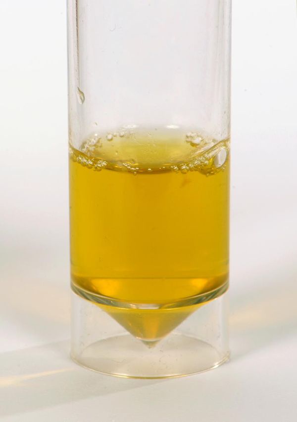
When FCoV RT-PCR was performed on plasma or serum samples from FIP and non-FIP cats{Doenges, 2017
#354;Felten, 2017 #350}, none of the non-FIP cases and very few (9-15.4%) of the FIP cases gave positive
results for FCoV RNA. A recent experimental study{Desmarets, 2016 #347} also failed to show any FCoV RNA in
the plasma of three FCoV-infected cats over the first 12 weeks of infection, and FCoV RNA was rarely detected
in the blood of 20 FIP cases{Pedersen, 2015 #326}. Thus, use of FCoV RT-PCR on blood, plasma or serum
samples is not helpful in the diagnosis of FIP due to low sensitivity. Peripheral blood mononuclear cells (PBMCs)
may be a better target for PCR than serum, as shown in one study{Doenges, 2017 #354}, but sensitivity was still
very poor at 28.6%. Similarly the experimental study{Desmarets, 2016 #347} on three FCoV-infected cats only
infrequently detected cell-associated FCoV in the blood over the first 12 weeks of infection; in this study the
cells used to determine cell-associated viraemia were not stipulated as being PBMCs.

FCoV RT-PCR on cerebrospinal fluid (CSF) samples
A recent paper{Doenges, 2016 #345} described the use of FCoV RT-PCR on CSF samples and found it to have
100% specificity for FIP but a sensitivity of only 41.2%. Our study{Barker, 2017 #383} gave similar results.
However, not all cats included in these studies had neurological signs as CSF was collected at post-mortem
examination independent of presenting signs, such that the population tested may not reflect those that would
have had CSF samples collected for diagnostic purposes. In one study, the sensitivity of RT-PCR rose to
85.7%{Doenges, 2016 #345} when only cats with neurological and ophthalmological signs were considered.
Thus, FCoV RT-PCR on CSF appears to be a useful additional test in cats with neurological signs, as a positive
result highly supports a diagnosis of FIP.

FCoV RT-PCR on aqueous humour samples
Robust studies have yet to be performed evaluating FCoV RT-PCR on aqueous humour samples, although
positive results have been reported in cats with FIP{Barker, 2017 #383}.

Molecular techniques characterizing FCoV S mutations in FCoV-containing samples
How are mutations identified?
Following the detection of FCoV RNA in a sample by RT-PCR, it may be possible to then characterise targeted
sections of FCoV genomic sequences present in that sample using molecular techniques such as
pyrosequencing, Sanger sequencing or PCR with sequence-specific hydrolysis probes. Such techniques are not
always successful in samples positive for FCoV by RT-PCR if, for example, only low levels of FCoV are present
(this can preclude sequence analysis) or if FCoV sequence variability means that targeted sequencing
techniques cannot generate sequence results. Characterisation of FCoV genomic sequences would be most
useful if FIP-specific mutations existed, as the detection of these mutations would be diagnostic for FIP.

Is mutation analysis useful?
Recent research has described amino acid differences in the fusion peptide encoded by the FCoV S gene as
being markers of FCoVs associated with FIP{Chang, 2012 #255}, raising the possibility that detection of the
underlying S gene mutations could be used to definitively diagnose FIP. Similarly, amino acid differences in the
furin cleavage motif, also encoded by the S gene, have been correlated with FIP disease{Licitra, 2013 #277}.
However, these S gene markers were identified by comparing the sequences of FCoVs found in the tissues of
FIP cats with those found in the faeces of healthy non-FIP cats.

Researchers in our group hypothesized that the fusion peptide sequence mutations could reflect the cellular
tropism of the FCoV (i.e. being systemic monocyte- / macrophage-associated FCoV compared to intestinal
epithelium associated FCoV) rather than being specific for FIP, knowing that non-FIP cats can have systemic
FCoV infection. Thus we compared the S gene sequences, from the region of the previously described fusion
peptide mutations, of FCoV detected in the tissues of FIP cats with those detected in the tissues of non-FIP
cats{Porter, 2014 #321}. This allowed us to evaluate the S gene sequences of FCoVs associated with systemic
FCoV infection in both non-FIP and FIP cases. We found that the S gene mutations present in most of the FIP
tissues were also present in most of the tissues of non-FIP cats that had systemic FCoV infection. A recent more
extensive study confirmed the same findings{Barker, 2017 #383}, and calculated that if the identification of S
gene mutated FCoVs was included as an additional confirmatory step to the detection of FCoV alone by RT-
PCR, this only slightly increased specificity for the diagnosis of FIP in tissue samples (from 92.6% to 94.6%) but
moderately decreased sensitivity (from 89.8% to 80.9%, as non-mutated FCoVs were sometimes identified
and mutation analysis was not possible in all tissue samples e.g. due to low FCoV copy numbers or the
presence of Type 2 FCoVs). These results question the value of S gene mutation analysis over and above the
detection of FCoV by RT-PCR, particularly in view of the extra financial expense and time required to perform
this additional analysis.
Analysis for S gene mutations has also been performed on effusions in recently published studies{Felten, 2017
#350;Longstaff, 2015 #349}. The majority of FCoVs in the effusions of FIP cats do indeed have the mutations
described{Chang, 2012 #255}. In one study{Longstaff, 2015 #349}, 12/17 FCoV-positive FIP effusion samples
had S gene mutations, whilst one did not have a mutation and four could not be sequenced due to the low
levels of FCoV present. In another study{Felten, 2017 #350}, 32/36 FCoV-positive FIP effusion samples had S
gene mutations, whilst three did not have mutations and one could not be sequenced. Our recent extensive
study{Barker, 2017 #383} calculated that the identification of S gene mutated FCoVs as an additional step to
the detection of FCoV alone by RT-PCR did not increase specificity for the diagnosis of FIP in fluid (primarily
effusions but also included CSF and aqueous humour) samples (specificity stayed at 97.9%) but markedly
decreased sensitivity (from 78.4% to 60%, for the same reasons as described for the tissue samples above). In
this study{Barker, 2017 #383} all FCoV-positive samples from cats with FIP had S gene mutations in CSF
samples, whilst no non-FIP samples were positive for FCoV. Therefore, S gene mutation analysis in FCoVs does
not substantially improve the ability to diagnose FIP in effusion or fluid samples as compared with detection of
FCoV RNA alone by RT-PCR.
Histopathological examination of tissues
Samples of affected tissues, e.g. liver, kidney or mesenteric lymph nodes, can be collected ante-mortem by
ultrasound-guided percutaneous needle-core biopsy, laparoscopy or laparotomy, although the invasive nature
of collection may preclude carrying this out in sick cats. Often samples are collected following euthanasia due
to a high index of suspicion of FIP at post-mortem examination. Samples are evaluated for characteristic
histopathological changes of FIP, which when present are generally regarded as being reliable for diagnosis,
although immunostaining for FCoV antigen (see below) is usually also recommended to confirm the diagnosis.
However, a lack of histopathological lesions is more difficult to interpret, especially in cases with a high index of
suspicion of FIP, as absence of gross lesions to guide biopsy could lead to sampling of non-affected organs or
tissue{Giordano, 2005 #243}. A small study{Giori, 2011 #242} recently documented that 5/8 FIP cases did not
have histopathology changes typically consistent with FIP, even though large representative biopsies were
taken, and diagnosis in these cases was based on positive FCoV antigen immunostaining.
Immunostaining of FCoV antigen
Immunostaining is performed on formalin-fixed tissues using immunohistochemistry (IHC) or on cytological
(typically effusion) samples using immunocytochemistry (ICC) or immunofluorescence (IF). These techniques
exploit the binding of antibodies to host cell-associated FCoV antigens, which are subsequently visualised by
enzymatic reactions producing a colour change (immunochemistry) or by fluorescence (immunofluorescence).
Positive FCoV antigen immunostaining of tissues is said to confirm a diagnosis of FIP (i.e. it is very specific) but a
negative result does not exclude FIP as a diagnosis as FCoV antigens may be variably distributed within
lesions{Giordano, 2005 #243} and thus are not detected in all histopathological sections prepared from lesions
from FIP cases{Kipar, 2014 #309}. This somewhat contradicts the suggestion by some that immunostaining is
mandatory to confirm/exclude FIP in doubtful cases{Giori, 2011 #242}, but may be overcome by taking multipleand/or large samples with confirmed pathology, as well as possibly requesting additional sections of biopsies
with pathology to be cut and stained.
Immunostaining of effusion samples has shown variable sensitivity (ranging from 57 to 100%){Felten, 2017
#355;Hartmann, 2003 #4;Hirschberger, 1995 #42;Litster, 2013 #274;Paltrinieri, 1999 #109;Parodi, 1993 #342}.
Since this technique relies on staining FCoV within macrophages in the effusion, and the effusion is often cell-
poor and/or the FCoV antigen is masked by FCoV antibodies in the effusion, a false negative result may be
obtained. Immunostaining was thought to be very specific, although two (heart failure and cholangiocarcinoma
cases) of seven non-FIP effusions were positive by IF in one study{Litster, 2013 #274}, and eight (including two
cats with heart failure and two cats with neoplasia) of 29 non-FIP effusions were positive by ICC in
another{Felten, 2017 #355}, questioning the specificity of ICC. However, the reported poorer specificity may be
due to the methodology used in one study (i.e. double staining for both FCoV antigen and macrophages [via
MHC II staining] was used), and the suboptimal storage of slides in the other, which could cause non-specific
staining and false positive results. Some have suggested that using cell pellets prepared from centrifuged
effusion samples to prepare formalin-fixed, paraffin embedded samples that can then be treated like a tissue
specimen for IHC{Kipar, 2014 #309}, can improve the reliability of detection of FCoV antigen{Kipar, 2014 #309},
although the processing time required for this would be longer than for ICC.
FCoV antigen ICC staining has been reported as being successful in detecting FCoV in the CSF of a cat with
neurological FIP{Ives, 2013 #291}. A recent study evaluated ICC in the CSF of cats with and without FIP, that
presented with and without neurological signs, collected at post-mortem examination{Gruendl, 2016 #361};
this study found that 17 of 20 cats with FIP gave positive results but of 18 cats without FIP, three gave positive
results, limiting the test’s specificity, although methodology may again be an issue, as described above. These
analyses excluded those cases in which cellularity was inadequate for ICC to be performed. Application of ICC
to CSF samples collected ante-mortem from a larger number of cats with neurological signs due to FIP and non-
FIP causes would be desirable to further evaluate the usefulness of this technique.
The use of FCoV antigen immunostaining has recently been described in aqueous humour samples collected at
post-mortem examination from cats with and without FIP{Felten, 2017 #384}. Being able to use aqueous
humour for reliable diagnostic investigations in cases of FIP would be especially valuable as it would be possible
to collect this in non-effusive cases, although the sample collection technique used in the study would need to
be modified (e.g. smaller gauge needle) for use ante-mortem. The study evaluating FCoV ICC in aqueous
humour samples from 25 cats with FIP (interestingly the majority were effusive FIP cases, and did not present
with uveitis) and 11 non-FIP cats showed a sensitivity of 64% and specificity of 81.8%; positive results were
obtained in two of the 11 control cats, one with lymphoma and one with pulmonary adenocarcinoma and both
did not have aqueous humour cytological features consistent with FIP (pyogranulomatous inflammation).
Further evaluation of ICC on aqueous humour samples collected ante-mortem from cats with uveitis due to FIP
and non-FIP causes would be useful to further evaluate the usefulness of this technique.
It is possible that fine needle aspirates could also be used as samples FCoV antigen immunostaining but
sensitivity may be poor due to difficulties in targeting lesions; further studies would be necessary to evaluate
their utility in the diagnosis of FIP and no evidence to support this currently exists.
Key Points
• Look for features that could be suggestive of FIP in the history and on clinical examination: young cat,
originally from a multi-cat household (including shelters or catteries), fluctuating non-responsive pyrexia,
evidence of an effusion, ocular or neurological signs
• Look for a lymphopenia on haematology• Look for hyperglobulinaemia, hyperbilirubinaemia (in the absence of moderate to severe increases in ALT and ALP enzyme activity, or anaemia), a reduced albumin:globulin ratio (A:G ratio) of 1.5 mg/ml) on serum biochemistry • Prioritise finding and sampling any effusion, whether pleural, peritoneal or pericardial in type. Effusions due to FIP are typically clear, viscous, straw-yellow and sticky with a total protein concentration of >35 g/l and a low A:G ratio of
Table 1: Diseases to consider in the differential diagnoses of FIP.
Modified from Tasker S and Dowgray N (in production) with permission from BSAVA publications,
Gloucester{Tasker, 2017 #380}.
 Disease/condition         Possible distinguishing features from FIP and course of action
 Toxoplasmosis             Transmission/epidemiology: Acquired via vertical transmission (in young cats) or by
                           hunting or eating raw meat.
                           Clinical signs: Cats may have hepatic, pulmonary, neurological, muscle and/or
                           pancreatic involvement. Signs can include lethargy, anorexia, dyspnoea (pneumonia
                           and/or pleural effusions can occur), jaundice, abdominal effusions, uveitis
                           (especially posterior) and/or neurological signs.
                           Diagnostic testing: Clinical toxoplasmosis is less common than FIP and is not usually
                           associated with the severe hyperglobulinaemia or reduced albumin:globulin ratio
                           often seen with FIP. Hyperbilirubinaemia may occur. Serology (high IgM titre or
                           rising IgG titre) may be helpful for diagnosis. Organisms may be found on sampling
                           and microscopic examination of e.g. lung, lymph node. PCR can also be performed
                           on such samples to demonstrate the presence of Toxoplasma gondii DNA.
                           Cerebrospinal fluid (CSF) PCR can also be performed in cases with neurological signs.
                           Treatment: If toxoplasmosis is suspected, trial treatment with clindamycin can be
                           instigated to see whether there is a positive response.
 Lymphocytic               Epidemiology: Can also occur in young cats. Persians may be predisposed.
 cholangitis (LC)          Clinical signs: Often associated with jaundice. Some cats with LC also have an
                           abdominal (unicavitary) effusion.
                           Diagnostic testing: The nature of the effusion is similar to that seen with FIP in terms
                           of protein concentration (i.e. high), although cell counts in LC are usually higher than
                           those seen with FIP. A marked hyperglobulinaemia can also be seen with LC. Both
                           LC and FIP cases can be hyperbilirubinaemic. Unlike FIP, however, LC is also usually
                           associated with marked increases in liver enzymes, especially cholestatic markers
                           (i.e. ALP and GGT), compared to the more mild or modest increases that occur in FIP
                           cats. Additionally, cats with LC are not usually as sick as those with FIP, e.g. they can
                           be polyphagic rather than inappetent.
 Neoplasia (e.g.           Epidemiology: Lymphoma can affect young cats, but is seen in cats of all ages. Other
 lymphoma, abdominal neoplasias tend to be seen in older cats. Focal FIP lesions in the intestine or
 carcinoma)                (especially mesenteric) lymph nodes can present very similar to cases with
                           apparently solitary neoplasms of these organs.
                           Clinical signs: Lymphoma can involve multiple body organs and, like FIP, it can result
                           in lymphadenopathy and/or bicavity effusions. Cats are often systemically ill.
                           Diagnostic testing: Sampling of affected tissues or effusions followed by cytology
                           may yield a diagnosis of lymphoma rather than the mixed inflammatory cells
                           typically seen on cytological sampling of FIP-affected tissues. Other neoplastic
                           lesions, e.g. carcinomas, may be diagnosed on cytology of effusions.
 Pancreatitis              Clinical signs: Cats may present with anorexia, jaundice and weight loss. Marked
                           pyrexia is not usually a feature, although pyrexia can occur in acute pancreatitis
                           cases that are associated with severe pain and/or sepsis.
                           Diagnostic testing: Hyperbilirubinaemia may occur. A small amount of abdominal
                           fluid (typically with a high protein concentration and high cell count [non-
                           degenerate neutrophils], in contrast to the high protein low cell count effusions with
                           FIP) is sometimes present in acute cases. Pancreatitis can be diagnosed by
                           ultrasonographic examination of the pancreas and measurement of feline
                           pancreatic lipase immunoreactivity.
                           Treatment: Trial treatment with antiemetics and analgesics may be warranted.
Retroviral infection Epidemiology: Feline leukaemia virus (FeLV) and feline immunodeficiency (FIV)
infections are both found more commonly in adults compared to juveniles, but age-
related immunity plays a role in FeLV infection with younger cats being more prone
to infection. Both viruses more likely to occur in outdoor cats. FeLV affects both
males and females whereas males are at increased risk of FIV infection.
Clinical signs: Both FeLV and FIV can be associated with pyrexia, lethargy,
lymphadenopathy and/or uveitis.
Diagnostic testing: FIV infection can be associated with a marked
hyperglobulinaemia.
NB: Retrovirus-positive status may act as a risk factor for the development of FIP.
Mycobacterial Epidemiology: There is a geographical variation in prevalence and infection is usually
infection including associated with a history of outdoor access and hunting.
tuberculosis (TB) Clinical signs: Lymphadenopathy, respiratory signs and/or uveitis may be seen, as
well as draining non-healing wounds. Affected cats may be relatively well despite
the disease, and pyrexia and inappetence are not common features although acute
presentations of e.g. dyspnoea can occur. Mycobacterial infections that involve the
lungs typically affect the lung parenchyma rather than presenting with pleural
effusions as in FIP.
Diagnostic testing: Mycobacterial infection is not usually associated with the severe
hyperglobulinaemia or reduced albumin:globulin ratio seen with FIP.
Hypercalcaemia may be present. Cytology of affected lymph nodes or organs shows
inflammatory changes (macrophages prominent but inflammation can be similar to
FIP with pyogranulomatous changes). Ziehl-Neelsen staining on cytology or biopsy
samples may be positive, and samples can be submitted for culture (although
positive culture results can take weeks to obtain with slower growing organisms,
and may be impossible with some mycobacterial species). An interferon gamma
test, performed on blood samples, is now available to assist in the diagnosis of
suspected feline TB cases.
Pyothorax Clinical signs: Can be associated with pyrexia. A pleural effusion is seen.
Diagnostic testing: Thoracic effusion analysis reveals very high cell counts due to
marked neutrophilic inflammation, with degenerate changes and possibly
intracellular bacteria, although any previous antibiotic treatment may mean that
bacteria are not seen. Unicavity effusion.
Sepsis Can be asscoiated with many conditions e.g. septic peritonitis, pyothorax,
pneumonia, pyelonephritis.
Clinical signs: Can be associated with pyrexia (although cats can also present with
low temperatures), tachycardia or bradycardia, tachypnoea, and other signs
associated with source of sepsis.
Diagnostic testing: Leucocytosis, band neutrophilia, hyperbilirubinaemia (in absence
of raised hepatic enzyme activity) may be present.
Septic peritonitis Clinical signs: Can be associated with pyrexia. An abdominal effusion is seen.
Diagnostic testing: Abdominal effusion analysis reveals very high cell counts due to
marked neutrophilic inflammation, with degenerate changes and possibly
intracellular bacteria, although any previous antibiotic treatment may mean that
bacteria are not seen. Unicavity effusion. Glucose concentration in the abdominal
effusion is lower than that in the blood (by > 1.1 mmol/l).
Congestive heart Clinical signs: Bicavity effusions in the pleural and peritoneal spaces are possible,
failure (CHF) although pleural effusions are far more common with feline CHF than abdominal
effusions, and abdominal effusions alone are very rarely seen with feline CHF. The
presence of a gallop sound, arrhythmia, and possibly heart murmur, may increasethe index of suspicion for CHF. Jugular vein distension may be present with right-
sided CHF. Pyrexia is not a feature.
Diagnostic testing: The fluid is a modified transudate with little protein, in contrast
to the fluid seen with FIP. Echocardiography will confirm cardiac disease and CHF.
Rabies In countries where rabies is endemic, this must be considered as a differential
diagnosis in unvaccinated cats presenting with neurological signs, especially acute
behavioural changes and progressive paralysis.Acknowledgements The author would like to acknowledge the many contributions made by the University of Bristol Feline Coronavirus Research Group & Bristol-Zurich FIP Consortium to the viewpoints and discussions described in this review. Special thanks go to Emi Barker and Samantha Saunders for their helpful comments on this manuscript. Andrew Davidson, Anja Kipar and Stuart Siddell are also thanks for their valued contributions to past and current FCoV research. Additional thanks are made to the veterinary practices, cat breeders and rescue centres that helped in the acquisition of samples used in our research studies and we also thank our colleagues, current and past, at the Feline Centre and Veterinary Pathology Unit, Langford Vets, University of Bristol, who have assisted in obtaining samples.
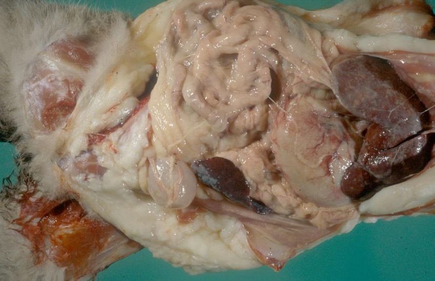
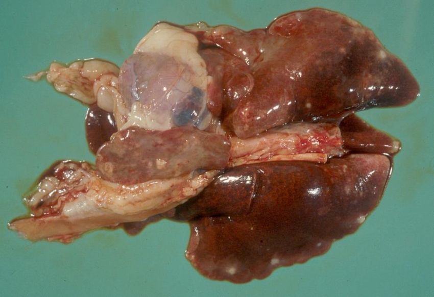
Figure 1: Schematic diagram of a feline coronavirus particle.

Modified with kind permission from Dr Emi Barker.

The spike (S) protein binds to the feline ‘receptor’ mediating host cell entry. The feline receptor is known to be
aminopeptidase N for Type 2 FCoVs, but is, as yet, unknown for Type 1 FCoVs.

Figure 2: Typical gross post-mortem examination findings in cases of FIP.

Granulomatous lesions in organs or fibrinous plaques on the serosa of organs may be visible in the abdominal
or thoracic cavity; tissues that are good to examine for these are the mesenteric lymph nodes, liver, spleen,
kidneys and intestinal surfaces, as well as the peritoneal lining of the abdominal wall and diaphragm. In effusive
cases, yellow sticky fluid can be visible in the pleural and/or peritoneal cavities, but the pericardium can also be
checked for fluid.

Figure 3: Examples of clinical signs seen in cases of FIP.

Clinical signs seen with FIP are typically assigned to wet (blue boxes) or dry (red boxes) FIP presentations, but
much overlap is seen between the two presentations and many signs are seen in both forms (purple boxes).

Figure 4: Typical appearance of effusion seen in cases of FIP.

Effusions in FIP cases are typically clear, viscous and straw-yellow in colour.
Figure 5: Positive Rivalta’s test.

The Rivalta’s test is performed by adding a drop of effusion to the surface of a mixture of 8 mls of distilled
water and 1 drop of 98% acetic acid (vinegar). A positive Rivalta’s test merely indicates that the effusion being
tested is an exudate. It is cheap and quick to do in-house but is not specific for FIP. This is a positive result as
the drop has retained its shape with a connection to the surface of the liquid.
Figure 6: Schematic diagram of the feline coronavirus genome.

Modified with kind permission from Dr Emi Barker.

FCoV RT-PCR assays detect FCoV RNA. The section of the genome amplified by different RT-PCRs varies
depending on the position of the primers used in the assays. As viral transcription starts at the 3’ end of the
FCoV genome, with the production of multiple subgenomic RNAs at this 3’ end, PCR assays with primers
located at the 3’ end of the genome (e.g. in the M or N regions) will be susceptible to viral load overestimation
as these will amplify these subgenomic RNAs, as well as the genomic RNA present in the FCoV. Conversely, PCR
assays with primers located at the 5’ end of the genome (e.g. the RNA polymerase) will amplify primarily
genomic RNA and will be less prone to viral load overestimation. Assays directed at the 3’ end of the FCoV
genome will tend to be more sensitive in detecting the presence of FCoV, due to their ability to amplify both
subgenomic and genomic RNA.
Coronaviruses, including FCoV, frequently undergo mutations and recombinations, meaning that PCRs
designed to be specific for specific sequences may not amplify all FCoVs. PCRs can be designed to target
conserved regions of the genome to minimise this, but elimination of FCoV sequence variability as a cause of
non-amplification is impossible.
Spike protein

Nucleocapsid protein

        RNA

 Envelope protein

       Membrane protein
Granulomas evident on surface of kidneys
Fibrinous plaques visible on the surface of the spleen
Yellow sticky effusion visible in the abdomen
Granulomas visible on surface of intestines
Granulomas visible on surface of the lungs
Fibrinous
plaques visible
on the
omentum
together with
enlargement of
the mesenteric
lymph nodesYou can also read