Expanding the Clinical and Mutational Spectrum of Recessive AEBP1-Related Classical-Like Ehlers-Danlos Syndrome - MDPI
←
→
Page content transcription
If your browser does not render page correctly, please read the page content below
G C A T
T A C G
G C A T
           genes
Article
Expanding the Clinical and Mutational Spectrum of
Recessive AEBP1-Related Classical-Like
Ehlers-Danlos Syndrome
Marco Ritelli 1 , Valeria Cinquina 1 , Marina Venturini 2 , Letizia Pezzaioli 1,3 ,
Anna Maria Formenti 4 , Nicola Chiarelli 1 and Marina Colombi 1, *
 1   Division of Biology and Genetics, Department of Molecular and Translational Medicine,
     University of Brescia, 25123 Brescia, Italy; marco.ritelli@unibs.it (M.R.); v.cinquina@studenti.unibs.it (V.C.);
     l.pezzaioli001@studenti.unibs.it (L.P.); nicola.chiarelli@unibs.it (N.C.)
 2   Division of Dermatology, Department of Clinical and Experimental Sciences,
     Spedali Civili University Hospital, 25123 Brescia, Italy; marina.venturini@unibs.it
 3   Spedali Civili of Brescia, 25123 Brescia, Italy
 4   IRCCS Istituto Ortopedico Galeazzi, 20161 Milano, Italy; annaformenti@live.it
 *   Correspondence: marina.colombi@unibs.it; Tel.: +39-030-3717-240; Fax +39-030-371-7241

 Received: 10 January 2019; Accepted: 8 February 2019; Published: 12 February 2019

 Abstract: Ehlers-Danlos syndrome (EDS) comprises clinically heterogeneous connective tissue
 disorders with diverse molecular etiologies. The 2017 International Classification for EDS recognized
 13 distinct subtypes caused by pathogenic variants in 19 genes mainly encoding fibrillar collagens
 and collagen-modifying or processing proteins. Recently, a new EDS subtype, i.e., classical-like EDS
 type 2, was defined after the identification, in six patients with clinical findings reminiscent of EDS,
 of recessive alterations in AEBP1, which encodes the aortic carboxypeptidase–like protein associating
 with collagens in the extracellular matrix. Herein, we report on a 53-year-old patient, born from
 healthy second-cousins, who fitted the diagnostic criteria for classical EDS (cEDS) for the presence of
 hyperextensible skin with multiple atrophic scars, generalized joint hypermobility, and other minor
 criteria. Molecular analyses of cEDS genes did not identify any causal variant. Therefore, AEBP1
 sequencing was performed that revealed homozygosity for the rare c.1925T>C p.(Leu642Pro) variant
 classified as likely pathogenetic (class 4) according to the American College of Medical Genetics
 and Genomics (ACMG) guidelines. The comparison of the patient’s features with those of the other
 patients reported up to now and the identification of the first missense variant likely associated with
 the condition offer future perspectives for EDS nosology and research in this field.

 Keywords: classical Ehlers-Danlos syndrome; classical-like Ehlers-Danlos syndrome type 2;
 AEBP1; aortic carboxypeptidase-like protein; differential diagnosis; high-frequency ultrasonography;
 reflectance confocal microscopy

1. Introduction
      Ehlers-Danlos syndrome (EDS), with an estimated prevalence of 1/5000, comprises a group
of clinically heterogeneous heritable connective tissue disorders (HCTDs) with diverse molecular
etiologies. The 2017 revised EDS classification recognized 13 distinct subtypes caused by pathogenic
variants in 19 genes mainly encoding fibrillar collagens, collagen-modifying proteins, or processing
enzymes [1]. Classical EDS (cEDS) (MIM #130000), with an estimated prevalence of 1/20,000,
is an autosomal dominant disorder primarily characterized by cutaneous and articular involvement.
Indeed, cEDS is suggested by skin hyperextensibility plus atrophic scarring that must be present
together with the other major criterion, i.e., generalized joint hypermobility (gJHM) evaluated

Genes 2019, 10, 135; doi:10.3390/genes10020135                                         www.mdpi.com/journal/genes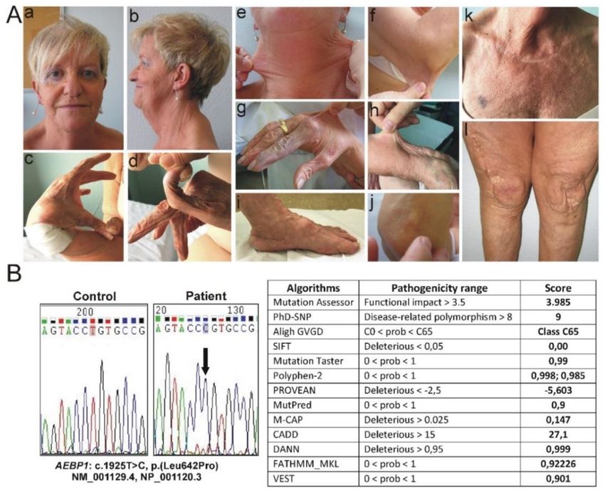
Genes 2019, 10, 135                                                                                  2 of 14

according to the Beighton score (BS ≥5/9), and/or with at least three of the minor criteria
among easy bruising, soft, doughy skin, skin fragility, molluscoid pseudotumors, subcutaneous
spheroids, hernia (or a history of thereof), epicanthal folds, JHM complications (e.g., sprains,
luxation/subluxation, pain, flexible flatfoot), and family history of a first-degree relative who meets
clinical criteria [1–3]. Furthermore, cEDS patients may present distinctive facial features, premature
rupture of fetal membranes, scoliosis, osteoporosis, gastroesophageal reflux, and cardiac and blood
vessel fragility [2,4–8]. Skin is hyperextensible if it can be stretched over a standardized cut off in the
following areas: 1.5 cm for the distal part of the forearms and the dorsum of the hands; 3 cm for neck,
elbow and knees; 1 cm on the volar surface of the hand (palm) [1,2,5]. Atrophic scarring can range
in severity; however, most cEDS patients have wide atrophic scars in different body areas that can
variably assume a cigarette paper, papyraceous, or hemosiderotic appearance [1,2,5].
      Point mutations or intragenic rearrangements of the COL5A1 and COL5A2 genes encoding type V
collagen are recognized in over 90% of patients [4,9], the recurrent heterozygous COL1A1 c.934C>T
(p.Arg312Cys) substitution is rarely found [4,10,11]. Negative molecular testing does not exclude the
diagnosis, as specific types of mutations (e.g., deep intronic variants) may go undetected by standard
diagnostic molecular techniques. Nevertheless, alternative diagnoses should be taken into account in
the absence of a COL5A1, COL5A2, and COL1A1 mutation [1].
      Recognition of cEDS is straightforward in the patient with the typical cutaneous signs and BS ≥5.
However, intra- and interfamilial variability tells a much broader clinical presentation and significant
overlap with other EDS types and HCTDs [1,2,4–6,12]. Differential diagnosis of cEDS should include
the hypermobile EDS (hEDS), particularly in patients without a striking cutaneous involvement [1,12].
Indeed, hEDS shares with cEDS gJHM and many mucocutaneous signs, but generally a lower
grade of skin hyperextensibility and only few small atrophic or post-surgical enlarged scars are
observed [13,14]. In case of a family history compatible with autosomal recessive transmission,
differential diagnosis comprises the rare classical-like EDS type 1 (MIM #606408) due to biallelic
TNXB mutations. These patients show marked skin hyperextensibility, easy bruising, and joint laxity,
but unlike cEDS patients, they do not have atrophic scarring or poor wound healing. Furthermore,
minor criteria such as foot deformities, edema in the legs, mild proximal and distal muscle weakness,
axonal polyneuropathy, and atrophy of muscles in hands and feet facilitates the differential [1,11].
Severe progressive cardiac-valvular problems distinguish the cardiac-valvular EDS type (COL1A2)
from cEDS, severe skin fragility and unusual craniofacial features discriminates the dermatosparaxis
EDS (ADAMTS2), whereas (congenital) kyphoscoliosis and muscle hypotonia differentiates the
kyphoscoliotic EDS (PLOD1, FKBP14), which are other rare recessive EDS types. Bilateral congenital
hip dislocation differentiates the autosomal dominant arthrochalasia EDS (COL1A1, COL1A2) [1,11,12].
      Recently, in six individuals from four unrelated families who presented with a constellation
of clinical findings reminiscent of cEDS such as gJHM, redundant and hyperextensible skin with
poor wound healing and abnormal scarring [15–17], and recessive alterations in the AEBP1 gene,
which encodes the aortic carboxypeptidase-like protein (ACLP) associating with collagens in the
extracellular matrix, were recognized, thus defining a new EDS form labelled as classical-like EDS
type 2 (MIM #618000).
      Herein, we describe an additional patient with a homozygous missense AEBP1 causative variant
and compare her clinical features with those of the other patients reported so far, offering future
perspectives for EDS nosology and research in this field.

2. Patient and Methods

2.1. Molecular Analyses
     The patient was evaluated at the specialized outpatient clinic for the diagnosis of EDS and
related connective tissue disorders, i.e., the Ehlers-Danlos Syndrome and Inherited Connective
Tissue Disorders Clinic (CESED), at the University Hospital Spedali Civili of Brescia. Molecular
Genes 2019, 10, 135 3 of 14
analysis was achieved in compliance with the Italian legislation on genetic diagnostic tests and
the patient provided written informed consent for publication of clinical data and photographs
according to the Italian bioethics laws. Since this report is based on data obtained through routine
clinical care and is not considered research at the involved institutions; formal ethics review was
not obtained. Genomic DNA was extracted from peripheral blood leukocytes using standard
procedures; the exons and intron-flanking regions of COL5A1, COL5A2, and exon 14 of COL1A1
(c.934C>T (p.Arg312Cys) were amplified by PCR and directly sequenced using an ABI PRISM®
3130XL Genetic Analyzer (Life Technologies, Carlsbad, CA, USA), as previously reported [4]. For the
multiplex ligation-dependent probe amplification (MLPA), the commercially available SALSA MLPA
kits P331 and P332 for COL5A1 gene were used, according to the manufacturer’s recommendations
(MRC-Holland, Amsterdam, The Netherlands), as previously described [4]. The primers for AEBP1
Sanger sequencing (Supplementary Table 1) were designed for all coding exons, including the
intron-exon boundaries, and primer sequences were analyzed for the absence of known variants
using the GnomAD database [18]. The sequences were analyzed with the Sequencher 5.0 software and
variants were annotated according to the Human Genome Variation Society (HGVS) nomenclature by
using the Alamut Visual software version 2.11. To evaluate the putative pathogenicity of the AEBP1
missense variant, which was submitted to the LOVD Ehlers–Danlos Syndrome Variant Database [19],
we used the following mutation prediction programs: Mutation Assessor [20], PhD-SNP [21],
Align GVD [22], SIFT [23], Mutation Taster [24], PolyPhen2 [25], PROVEAN [26], MutPred [27],
M-CAP [28], CADD [29], DANN [30], Fathmm-MKL [31], and VEST [32]. The nucleotide and protein
accession numbers correspond to the AEBP1 (NM_001129.4, NP_001120.3) reference sequences.
2.2. High-Frequency Ultrasonography and In Vivo Reflectance Confocal Microscopy
To investigate patient’s skin by a non-invasive approach, we performed high-frequency ultrasonography
(HF-USG) and in vivo reflectance confocal microscopy (RCM) as previously described [33–35].
Briefly, HF-USG was performed on the dorsal and volar side of the forearm of the patient and
10 age- and gender-matched healthy individuals with the same skin phototype and similar sun
exposure history by digital 50-MHz ultrasonography B mode scanning (DUB-USB Skin Scanner,
Taberna Pro Medicum Company, Lueneburg, Germany). For ultrasound transmission, water was
employed as a coupling medium between the transducer and the skin surface. The usable depth of
signal penetration was 4 mm, and the gain was 40 dB. Ultrasonography images were collected under
standard conditions (environmental temperature was 20–23 ◦ C and the patient remained in a lying
position for at least 10 min before examination). Acquired images were exported into a dedicated
database and were evaluated using specific image-analysis software to assess epidermal and dermal
thickness (µM) and lesional echogenicity.
RCM investigation on the same sides of the forearm was achieved with a Vivascope 1500®
microscope (MAVIG GmbH, Lucid Technologies, Henrietta, NY, USA) to visualize in vivo the
horizontal optical skin sectioning at cellular-level resolution (lateral resolution = 0.5–1 µM,
axial resolution = 3–5 µM) from the epidermis to the papillary dermis (200–250 µM in depth).
The system uses a laser source with a wavelength of 830 nm and a powerGenes 2019, 10, 135 4 of 14
3. Results
3.1. Clinical Findings
The proband (LOVD ID AN_006205) was an Italian 53-year-old woman, born from healthy
second-cousins parents, and had two healthy brothers. Clinical history was remarkable for premature
birth at 30 weeks (height 44 cm, weight 1.2 kg) associated with perinatal respiratory distress. Neonatal
severe hypotonia and delayed motor development, i.e., delays in walking (she took her first steps at
four years of age) and acquisition of fine motor skills, were also reported. Medical history further
included propensity to develop ecchymoses either spontaneously or upon minimal trauma often
occurring for motor clumsiness, surgically treated umbilical hernia in infancy, myopia and astigmatism
since childhood, and complete dental loss due to unspecified periodontitis at 14 years old. At age 18,
a clinical diagnosis of unspecified EDS was given for gJHM, skin hyperextensibility, delayed wound
healing, and easy bruising; genetic analyses were not performed. The patient suffered from recurrent
dislocations of knees and occasionally of shoulders and elbows since the age of 10; the objective
patellar instability was surgically treated by capsuloplasty and transposition of the insertion of the
common patellar tendon by tibial tuberosity transplantation followed by skin plastic surgery at the
age of 29 leading to a wide atrophic post-surgical scar (Figure 1A). At 21 and 23 years old, respectively,
she underwent bilateral saphenectomy for symptomatic varicosities with pain, fatigability, heaviness,
and recurrent superficial thrombophlebitis and surgical removal of nodules on vocal cords. At age
41, the patient was subjected to operative treatment of rotator cuff disease in the setting of weakness
and substantial functional disability. Since age 42, she suffered from Achilles tendinopathy with pain
and stiffness, especially at the back of the ankle, treated with on-demand NSAIDs use, conservative
physical therapy, and orthotic insoles for severe pes planus. At 43 years of age, metatarsal osteotomy
on the 3rd toe of the right foot for metatarsalgia and aggravating Achilles tendinopathy was performed.
In the same period, she developed disabling bilateral gonarthrosis, treated with arthroscopic abrasion,
epitrochleitis, and subacromial shoulder impingement associated with night pain. Hypotrophy of the
scapular girdle and weak osteotendinous reflexes were observed at age 50, when she also experienced
the dislocation of the left ankle with soft tissue effusion without reabsorption.
On examination, at 52 years of age, she presented with a height of 150 cm (genetic target 157 cm,
arm span/height ratio 1.03, normal valueGenes 2019, 10, 135 5 of 14
Figure 1. Clinical and molecular findings of the patient. (A) Old-aging appearance of face and
androgenetic alopecia (a,b); laxity of the thumb (c), laxity of the fifth finger (d); hyperextensible skin in
different body areas: neck (e), elbow (f), dorsum of the hand (g) and forearm (h); flat feet and piezogenic
papules (i); subcutaneous spheroid on elbow (j), diffuse PoC-like dermatitis and easy bruising (k);
skin redundancy, atrophic papyraceous scars on knees, postsurgical enlarged scar after right knee
capsuloplasty and skin plastic surgery (l). (B) Sequence chromatograms showing the position of the
c.1925T>C p.(Leu642Pro) variant (arrow) identified in homozygosity in exon 16 of the AEBP1 gene
(seq. Ref.: NM_001129.4, NP_001120.3) and in silico prediction of the pathogenicity of the p.(Leu642Pro)
missense substitution by using 13 different algorithms [20–32].
3.2. Molecular Findings
The patient’s phenotype was suggestive for cEDS, since she fulfilled both major (skin
hyperextensibility plus atrophic scarring and gJHM) and 6 minor criteria according to the 2017 EDS
nosology, i.e., easy bruising, soft, doughy skin, skin fragility, subcutaneous spheroids, a history of
hernia, and JHM complications. Therefore, after written informed consent was obtained, we performed
Sanger sequencing of COL5A1, COL5A2, and of exon 14 of COL1A1 (p.Arg312Cys), integrated by
MLPA analysis of COL5A1, which did not identify any pathogenic variant. Although negative
molecular testing, a clinical diagnosis of cEDS was maintained, since the other EDS types in differential
diagnosis with cEDS (including periodontal EDS) were excluded clinically. Following the discovery
of AEBP1 biallelic variants [15,16], Sanger sequencing of this gene was achieved, which revealed
the homozygosity for the rare c.1925T>C p.(Leu642Pro) variant in exon 16 (Figure 1B), leading to
the substitution of a highly conserved leucine residue with a proline at position 642 within the
metallocarboxypeptidase-like domain of the protein. This variant has been observed in 3 individuals
in GnomAD (rs753531562, 3/282140, no homozygotes, total MAF: C = 0.00001063). Its putative
pathogenicity was estimated through an array of 13 different in silico prediction algorithms that agreed
to define p.(Leu642Pro) as high impacting variant. Given that the variant is located in a critical and
well-established functional domain without benign variation, the extremely low frequency in publicly
available population databases, the multiple lines of computational evidence supporting a deleteriousGenes 2019, 10, 135 6 of 14
effect on the gene product, and the patient’s phenotype highly suggestive for a disease with a single
genetic etiology, the p.(Leu642Pro) missense variant is classified as likely pathogenic (class 4) according
to the guidelines of the ACMG. Samples of the healthy parents or brother were not available for
molecular analyses.
3.3. Instrumental Findings on Patient’s Skin
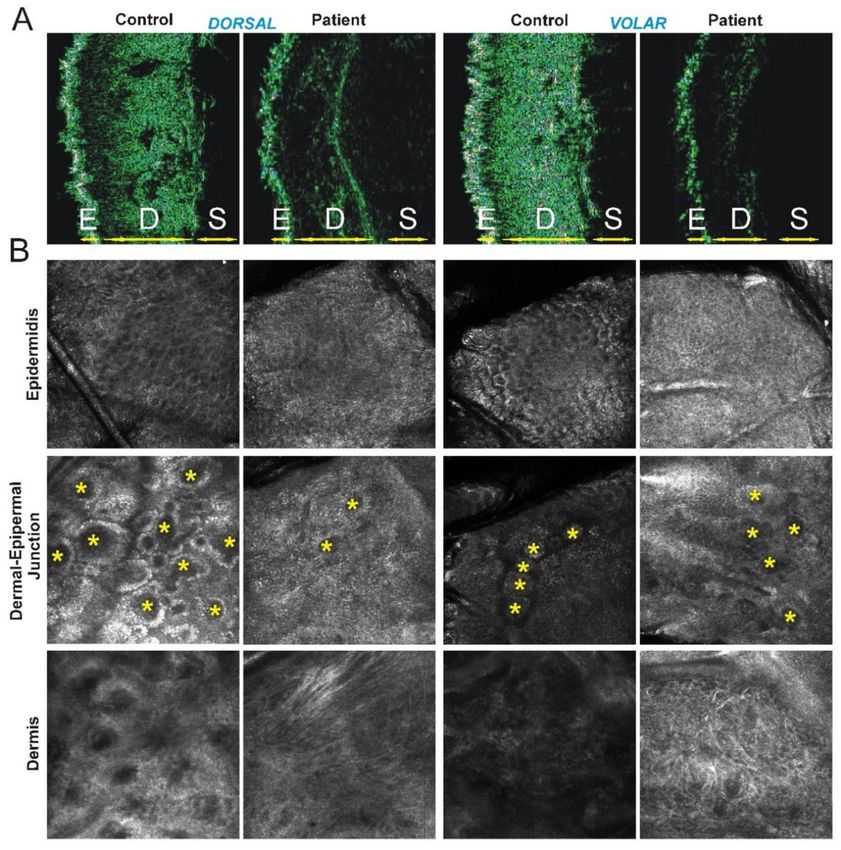
In order to investigate the skin by a non-invasive approach, HF-USG and in vivo RCM were
performed on selected skin areas, i.e., dorsal and volar side of the forearm, showing clinically significant
differences between our patient and 10 healthy individuals (Table 1 and Figure 2). Digital 50-MHz
ultrasonography scanning demonstrated an increase in epidermal entrance echo (highly echogenic
band produced by the differences of the acoustic impedance between gel and skin) corresponding to
increased epidermal thickness, but a decrease in dermal thickness compared to control skin of age- and
gender-matched healthy individuals with the same skin phototype II and similar sun exposure history.
The patient’s epidermis (dorsal thickness = 172 µM; volar thickness = 141 µM) was thicker than that of
healthy controls (dorsal thickness (mean ± standard deviation, SD) = 121 ± 22 µM; volar thickness
(mean ± SD) = 102 ±12 µM), likely due to the multiple and diffuse papules (Table 1). The increased
thickness was more evident on the dorsal side of the forearm that is chronically more photoexposed
compared to the volar side. Contrariwise, the patient’s dermis appeared thinner (dorsal dermal
thickness = 570 µM; volar epidermal thickness = 289 µM) compared to healthy controls (dorsal dermal
thickness (mean ± SD) = 1108 ± 320 µM; volar epidermal thickness (mean ± SD) = 983 ± 205 µM)
(Table 1). Moreover, the considerable hypoechogenicity of the dermal layer suggests disruption of
collagen fibers and accumulation of elastotic material that is typical of chronological and photoinduced
skin aging (Figure 2A). This ultrastructural pattern is known as subepidermal low echogenic band
(SLEB) and derives from skin elastosis and accumulation of glycosaminoglycans that have increased
water-binding capacity [33]. In vivo RCM investigation demonstrated loss of the typical honey-comb
pattern (corresponding to alteration of epidermal thickness), irregularity of the dermal-epidermal
junction and the disarray of the dermis, which was characterized by coarse and fragmented collagen
fibers both on the dorsal and volar side of patient’s forearm (Figure 2B). These alterations are
independent of sun exposure, given that they are present both on dorsal and volar side of the forearm,
suggesting a pronounced and diffuse skin aging due to AEBP1-defect.
Table 1. Epidermal and dermal thickness of patient’ s forearm evaluated by high-frequency ultrasonography
(HF-USG) compared to 10 healthy individuals.
Dorsal Forearm Volar Forearm
Controls Controls
Patient Patient
(mean ± SD) (mean ± SD)
Epidermal thickness (µM) 172 121 ± 22 141 102 ± 12
Dermal thickness (µM) 570 1108 ± 320 289 983 ± 205
4. Discussion
Recently, taking advantage from NGS, a new, autosomal recessive type of EDS has been discovered
due to variants in the AEBP1 gene. This EDS type is very rare and, so far, found in only seven
individuals (including the present patient) from four unrelated families (Table 2). The International
Consortium on EDS and Related Disorders has not yet classified and named this type, but in OMIM
it is labeled as classical-like EDS type 2 (MIM #618000). Indeed, the few patients reported hitherto
(Table 2) share many similarities representative of the classical type as much as they all fulfill the
cEDS diagnostic criteria of the 2017 nosology [1,2] for the presence of the pathognomonic cutaneous
involvement, i.e., soft, doughy and very hyperextensible skin, delayed wound healing with abnormal
atrophic scarring, JHM, and other minor criteria such as easy bruising, subcutaneous spheroids
(observed only in our patient), and JHM complications such as dislocations/subluxations (shoulders,Genes 2019, 10, 135 7 of 14
knees, hips, ankles, elbows, clavicula, wrist, mandibular and distal radioulnar joints, in some cases
requiring surgical treatment), sprain, pain, and flexible flatfoot (Table 2)
Figure 2. Instrumental findings on patient’s skin. (A) Ultrasonography (50 MHz) images of the forearm
skin from the patient and a representative age- and gender-matched healthy individual (control).
E, epidermis, D, dermis, S, subcutaneous adipose tissue (depth of imaging: 4 mm). Disorganization of
collagen fibers and elastosis in patient’s skin appears as a significant thinning and hypoechogenicity
of the dermal layer both on dorsal (left) and volar side (right) compared to control (B) Reflectance
confocal microscopy images of the forearm skin from patient and control (magnification: 500 × 500 µm).
Epidermis: typical honey-comb pattern on dorsal (left) and volar (right) side in healthy skin are
not detectable in patient’s skin. Dermal-epidermal junction: regular edge papillae [rings of basal
keratinocytes surrounding dark circular structures corresponding to dermal papillae (*)] on dorsal
and volar side of control skin are reduced both in number and definition in patient’s skin. Dermis:
Irregular and fragmented collagen fibers that appear bright and coarse on both dorsal and volar side of
patient’s skin compared to control. An increased brightness of all skin structures, corresponding to
chronological and photoinduced skin aging, is present on the dorsal side of healthy skin but not on
the volar side that usually is not photoexposed, whereas in the patient this pronounced skin aging is
present at both sides.Genes 2019, 10, 135 8 of 14
Table 2. Summary of clinical features of individuals with autosomal recessive variants in AEBP1
Citation Present patient P1* P2* P3* P4* P5* P6*
Sex female male male female male female male
Ethnicity white white white Middle Eastern Middle Eastern white white
Age at evaluation 53y 35y 33y 12y 24y 39y 38y
c.1630+1G>A c.1630+1G>A
AEBP1 variant(s) c.1925T>C c.1470del, c.1743C>A c.917dup c.917dup
c.1320_1326del homozygous (r.1609_1630del) (r.1609_1630del)
(NM_001129.4) homozygous compound heterozygous homozygous homozygous
homozygous homozygous
Protein change p.(Asn490_Met495delins40),
p.(Leu642Pro) p.(Arg440Serfs*3) p.(Val537Leufs*31) p.(Val537Leufs*31) p.(Tyr306*) p.(Tyr306*)
(NP_001120.3) p.(Cys581*)
+ + + + + + +
Joint hypermobility (BS)
(5/9) (8/9) (8/9) (8/9) (NA) (6/9) (2/9)
Dislocations/ left ankle, knees, hip, right distal radioulnar hip, knees, ankles shoulders, wrist, mandibular and
hip (congenital), shoulders hips, knees and ankles ankles, knees, clavicula
Subluxations shoulders, elbows joint interphalangeal joints distal radioulnar joints
pes planus, hallux valgus, pes planus, hallux valgus, pes planus, hallux valgus, pes planus, hallux pes planus, hallux valgus, hindfoot deformity,
Foot deformities pes planus, hallux valgus
hammer toes hammer toes hammer toes valgus, toe deformities sandal gap sandal gap
Extensive skin
+ + + + + + +
hyperextensibility
Delayed would healing + + + + + + +
(abnormal scarring) (widened atrophic scars) (widened atrophic scars) (widened atrophic scars, keloids) (widened atrophic scars, keloids) (widened atrophic scars) (widened atrophic scars) (widened atrophic scars)
Redundant skin + old-aging appearance + old-aging appearance + + + + old-aging appearance + old-aging appearance
Easy bruising + + + + NA + +
Prominent chest
- NA + NA NA + +
superficial veins
umbilical
Hernia - large ventral surgical hernia umbilical, ventral, inguinal NA + -
surgically treated
Genitourinary cryptorchidism cryptorchidism
- - - - -
abnormalities surgically corrected surgically corrected
Gastrointestinal
- motility issues bowel rupture - - NA NA
abnormalities
MVP, mildly dilated aortic root,
peripheral artery disease, MVP, circular
Vascular abnormalities MVP bilateral carotids stenosis, aortic - - varicose veins
varicose veins pericardial effusion
dilation requiring surgery
Pyorrhea, complete dental retention of a single abnormal dental
Dentition - abnormal dental alignment - -
loss at age 14 baby tooth alignment
Citation Present patient P1* P2* P3* P4* P5* P6*
Sex female male male female male female male
Ethnicity white white white Middle Eastern Middle Eastern white white
Age at evaluation 53y 35y 33y 12y 24y 39y 38yGenes 2019, 10, 135 9 of 14
Table 2. Cont.
Citation Present patient P1* P2* P3* P4* P5* P6*
bilateral ptosis webbed neck, bilateral ptosis webbed
high palate,
Facial dysmorphisms - micrognathia sagged cheeks large ears, neck, sagged cheeks - -
elongated uvula
narrow palate large ears, narrow palate
skull with ‘copper beaten’
femoral osteopenia, T10 progressive kyphosis,
hip replacement for severe appearance, severe osteopenia,
vertebral deformity, scoliosis, arachnodactyly, kyphoscoliosis,
severe osteopenia of hips osteopenia, upper thoracic narrowing of the interpedicular
Skeletal anomalies scoliosis, lumbar spine positive wrist and thumb arachnodactyly, positive
(mild disc bulging at the scoliosis with degenerative distance of the lumbar spine severe osteopenia
(MRI findings) rectilinization with signs, degeneration of the wrist and thumb signs,
C4-5 and C7-T1 levels) disease and facet arthrosis of distally, short and squared iliac
marked discus ulnaris mild pectus excavatum
spine (empty sella) bones, remodeled long bones of
degenerative arthritis orthopedically treated
the lower extremities
hypotonia, delayed motor
development, multiple
papules (diffuse PoC-like
dermatitis, alopecia,
patellar instability
delays in walking and
surgically treated, rotator
acquisition of fine motor
cuff disease surgically elbow bursitis, piezogenic strabismus surgically
skills, impaired temperature hypotonia, diabetes
Other treated, epitrochleitis, papules, sacral dimple, NA alopecia, skin striae treated,
sensation, mellitus, cellulitis
subacromial shoulder hypertriglyceridemia myopia, astigmatism
keratoconjunctivitis sicca,
impingement, hypotrophy
piezogenic papules
of the scapular girdle,
gonarthrosis, chronic
fatigue, spheroids,
piezogenic
papules, myopia
*Patients reported by Alazami et al., 2016 [15], Blackburn et al., 2018 [16], and Hebebrand et al., 2018 [17]. P1: A-II:1;P2: B-II:1; P3:C-IV:6; P4: C-IV:4 according to [16]; P5: D-II:1; P6: D-II-2
according to [17]. Abbreviations: + present, - absent, NA not available, MVP mitral valve prolapseGenes 2019, 10, 135 10 of 14
Consistent with the multisystemic presentation of EDS in general, there are also variable features
including congenital hip dislocation, hypotonia, delayed motor development, acrogeria, prominent
superficial veins in the chest region, hernias, dental anomalies, gastrointestinal (bowel rupture) and
vascular complications (mitral valve prolapse, aortic root dilation needing surgery), early-onset
varicose veins, and several skeletal anomalies (Table 2). In particular, bone involvement seems
a common feature of classical-like EDS type 2 with osteopenia/osteoporosis affecting hips, femurs,
and spine that are present, at variable degree, in all of the patients reported so far, with the exception
of the two siblings, reported by Hebebrand and coworkers [17], who were not tested for osteopenia.
In addition, degenerative arthritis, (kypho)scoliosis, arachnodactyly, positive wrist and thumb signs,
mild pectus excavatum, T10 vertebral deformity (our patient), narrowing of the interpedicular distance
of the lumbar spine, shortened and squared iliac bones, and remodeling of long bones of the lower
extremities are also encountered (Table 2). In addition, all subjects have severe foot deformities
including bilateral pes planus, hammertoes, hallux valgus, hindfoot deformity, and sandal gap,
which are observed in more than a few other EDS subtypes as well [1]. Although in cEDS patients
a variable degree of low bone mineral density and a high prevalence of radiological vertebral fractures
were reported [7,36], AEBP1-related EDS seems to display a more severe bone involvement that
could potentially facilitate the differential with cEDS. Nevertheless, considering the limited number
of individuals with AEBP1 defect known so far, a larger cohort of patients is needed to confirm this
preliminary observation.
The adipocyte enhancer binding protein 1 gene (AEBP1) encodes a 1158-amino acid secreted
aortic carboxypeptidase-like protein (ACLP) composed of an N-terminal signal sequence, a charged
lysine, proline, and glutamic acid-rich domain, a collagen-binding discoidin domain and
a metallocarboxypeptidase (MCP)-like domain [37,38]. This latter domain is inactive toward standard
MCP substrates, as it lacks several critical active sites and substrate-binding residues that are necessary
for activity [37,38]. Indeed, ACLP acts as an extracellular matrix (ECM)-binding protein rather than as
active MCPs that shows similar embryonic expression pattern as other ECM proteins and is found at
high levels particularly in collagen-rich tissues comprising the dermal layer of the skin, the medial layer
of blood vessels, the basement membrane of the lung, and the periosteum. Consistently, ACLP plays
fundamental roles in both embryonic development and adult tissue homeostasis, particularly in repair
processes [38–43]. Indeed, AEBP1 knock-out mice show ventral wall defects, develop spontaneous
skin ulcerations, and have significantly delayed healing of dermal punch wounds [38]. This cutaneous
phenotype is consistent with the defective wound healing and abnormal scar formation observed
in individuals with AEBP1 defects and suggest that ACLP has a crucial role in damage sensing and
ECM remodeling following injury by regulating fibroblast proliferation and mesenchymal stem cell
differentiation into collagen-producing cells [42,43]. Blackburne and coworkers demonstrated that
ACLP also binds collagens type I, III, and V and is able to promote the polymerization of collagen type
I in vitro [16]. In line with these findings, the ultrastructural study performed by the same authors
on a patient’s skin biopsy revealed reduced dermal collagen and irregular disrupted collagen fibers,
as well as our HF-USG and RCM investigations that disclosed abnormal collagen fibers deposition
together with a reduced dermal thickness. Moreover, we recognized an increase of the epidermal
thickness likely correlating with the diffuse PoC-like dermatitis, which is probably not related to
classical-like EDS type 2. The use of these non-invasive diagnostic techniques may be promising
for the investigation of the qualitative and quantitative cutaneous alterations, but further studies
including electron microscopy on skin as golden standard of reference on large cohorts of patients
are warranted. In our case, we did not perform skin biopsy because of the patient’s will due to
psychological reluctance for her important skin fragility with delayed wound healing.
The AEBP1 variants discovered before our patient’s characterization were all loss-of-function
(LOF) mutations (Table 2) and included compound heterozygous variants [(c.1470del;
p.Asn490_Met495delins40) and (c.1743C>A; p.Cys581*] in the first individual (P1); a homozygous
variant (c.1320_1326del; p.Arg440Serfs*3) in the second individual (P2); a homozygous spliceGenes 2019, 10, 135 11 of 14
variant leading to skipping of the last 22 bp of exon 13 (c.1630+1G>A) in the two siblings from
the third family (P3, P4), and a homozygous nonsense variant (c.917dup; p.Tyr306*) in the two
siblings from the fourth family (P5, P6). Hebebrand and coworkers performed the analysis of all
AEBP1 LOF variants reported in multiple databases showing that these are distributed throughout
the protein and by using conservative criteria for pathogenic LOF variants (nonsense, frameshift,
canonical splice sites, or initiation-codon) these authors estimated a carrier frequency of 1/829 for the
gnomAD database. The analysis of CADD scores for all possible missense variants showed a higher
predicted deleteriousness for positions close to the discoidin and the MCP-like domains, whereas the
unstructured N-/C-terminal parts showed lower scores. The high deleteriousness scores observed for
missense variants within these domains cite evidence in support of additional mutational mechanisms
leading to aberrant function and the authors thus argued that the relatively low estimated carrier
frequencies could be significantly higher if missense variants contribute to a comparable fraction of
disease variants [17]. The present c.1925T>C; p.(Leu642Pro) homozygous variant disclosed within
the MCP-like domain of the protein corroborates this hypothesis, since it represents the first likely
pathogenic AEBP1 missense substitution (ACMG class 4) associated with classical-like EDS type 2.
The variant is predicted in silico to affect the tertiary structure of the protein by disrupting an α-helix
located in a highly conserved domain, thus likely interfering with its function in terms of impaired
partner binding capability. Nevertheless, a definite proof of variant’ s causality is lacking, since the
effective functional consequences on the ECM organization, particularly of collagens, and on the
other not yet well-defined roles of the ACLP protein were not studied, because the patient refused
skin biopsy.
5. Conclusions
Our findings expand the knowledge of the clinical phenotype of this recently defined autosomal-
recessive EDS subtype, provide the first evidence that missense variants contribute to the allelic
repertoire of AEBP1, and suggest that in the diagnostic process of a cEDS patient this gene should
be investigated when a recessive inheritance is compatible and no causal variant is identified in the
other cEDS genes. Further reports are needed to better characterize the AEBP1-related phenotype,
define specific clinical criteria that might facilitate the differential with the other EDS forms, delineate
genotype-phenotype correlations, and collect natural history data for prognostication. Finally, ACLP
function needs to be explored more in-depth to provide insights into molecular mechanisms involved
in the pathophysiology of AEBP1-related EDS that may represent a starting point for identifying
potential therapeutic options.
Supplementary Materials: The following are available online at http://www.mdpi.com/2073-4425/10/2/135/s1,
Table S1. Primers.
Author Contributions: M.C. and M.R. conceived the study. M.V. and M.C. performed the clinical diagnosis of the
patient, genetic counselling and follow-up; M.V. performed skin evaluations; L.P. and A.M.F. investigated bone
health parameters; M.R. and V.C. carried out the molecular analyses; M.R. and N.C. researched the literature; M.R.,
A.M.F., and M.V. prepared the manuscript; M.C. edited and coordinated the manuscript. All authors discussed,
read, and approved the manuscript.
Funding: No funding was active on this project.
Acknowledgments: The authors want to thank the patient for her cooperation during the diagnostic process and
the Fazzo Cusan family for its generous support.
Conflicts of Interest: All authors declare that there is no conflict of interest concerning this work.Genes 2019, 10, 135 12 of 14
References
1. Malfait, F.; Francomano, C.; Byers, P.; Belmont, J.; Berglund, B.; Black, J.; Bloom, L.; Bowen, J.M.;
Brady, A.F.; Burrows, N.P.; et al. The 2017 international classification of the Ehlers-Danlos syndromes.
Am. J. Med. Genet. C 2017, 175, 8–26. [CrossRef] [PubMed]
2. Bowen, J.M.; Sobey, G.J.; Burrows, N.P.; Colombi, M.; Lavallee, M.E.; Malfait, F.; Francomano, C.A.
Ehlers-Danlos syndrome, classical type. Am. J. Med. Genet. Part C 2017, 75, 27–39. [CrossRef] [PubMed]
3. Beighton, P.; De Paepe, A.; Steinmann, B.; Tsipouras, P.; Wenstrup, R.J. Ehlers-Danlos syndromes: Revised
nosology, Villefranche, 1997. Ehlers-Danlos National Foundation (USA) and Ehlers-Danlos Support Group
(UK). Am. J. Med. Genet. 1998, 77, 31–37. [CrossRef]
4. Ritelli, M.; Dordoni, C.; Venturini, M.; Chiarelli, N.; Quinzani, S.; Traversa, M.; Zoppi, N.; Vascellaro, A.;
Wischmeijer, A.; Manfredini, E.; et al. Clinical and molecular characterization of 40 patients with classic
Ehlers-Danlos syndrome: Identification of 18 COL5A1 and 2 COL5A2 novel mutations. Orphanet J. Rare Dis.
2013, 12, 8–58. [CrossRef] [PubMed]
5. Colombi, M.; Dordoni, C.; Venturini, M.; Ciaccio, C.; Morlino, S.; Chiarelli, N.; Zanca, A.; Calzavara-Pinton, P.;
Zoppi, N.; Castori, M.; et al. Spectrum of mucocutaneous, ocular and facial features and delineation of novel
presentations in 62 classical Ehlers-Danlos syndrome patients. Clin. Genet. 2017, 92, 624–631. [CrossRef]
[PubMed]
6. Colombi, M.; Dordoni, C.; Cinquina, V.; Venturini, M.; Ritelli, M. A classical Ehlers-Danlos syndrome family
with incomplete presentation diagnosed by molecular testing. Eur. J. Med. Genet. 2018, 61, 17–20. [CrossRef]
[PubMed]
7. Mazziotti, G.; Dordoni, C.; Doga, M.; Galderisi, F.; Venturini, M.; Calzavara-Pinton, P.; Maroldi, R.;
Giustina, A.; Colombi, M. High prevalence of radiological vertebral fractures in adult patients with
Ehlers-Danlos syndrome. Bone 2016, 84, 88–92. [CrossRef] [PubMed]
8. Borck, G.; Beighton, P.; Wilhelm, C.; Kohlhase, J.; Kubisch, C. Arterial rupture in classic Ehlers-Danlos
syndrome with COL5A1 mutation. Am. J. Med. Genet. Part A 2010, 152, 2090–2093. [CrossRef]
9. Symoens, S.; Syx, D.; Malfait, F.; Callewaert, B.; De Backer, J.; Vanakker, O.; Coucke, P.; De Paepe, A.
Comprehensive molecular analysis demonstrates type V collagen mutations in over 90% of patients with
classic EDS and allows to refine diagnostic criteria. Hum. Mutat. 2012, 33, 1485–1493. [CrossRef] [PubMed]
10. Colombi, M.; Dordoni, C.; Venturini, M.; Zanca, A.; Calzavara-Pinton, P.; Ritelli, M. Delineation of
Ehlers-Danlos syndrome phenotype due to the c.934C>T, p.(Arg312Cys) mutation in COL1A1: Report
on a three-generation family without cardiovascular events, and literature review. Am. J. Med. Genet. Part A
2017, 173, 524–530. [CrossRef] [PubMed]
11. Brady, A.F.; Demirdas, S.; Fournel-Gigleux, S.; Ghali, N.; Giunta, C.; Kapferer-Seebacher, I.; Kosho, T.;
Mendoza-Londono, R.; Pope, M.F.; Rohrbach, M.; et al. The Ehlers-Danlos syndromes, rare types.
Am. J. Med. Genet. C 2017, 175, 70–115. [CrossRef] [PubMed]
12. Colombi, M.; Dordoni, C.; Chiarelli, N.; Ritelli, M. Differential diagnosis and diagnostic flow chart of
joint hypermobility syndrome/Ehlers-Danlos syndrome hypermobility type compared to other heritable
connective tissue disorders. Am. J. Med. Genet. Part C 2015, 169, 6–22. [CrossRef] [PubMed]
13. Tinkle, B.; Castori, M.; Berglund, B.; Cohen, H.; Grahame, R.; Kazkaz, H.; Levy, H. Hypermobile
Ehlers-Danlos syndrome (a.k.a. Ehlers-Danlos syndrome Type III and Ehlers-Danlos syndrome
hypermobility type): Clinical description and natural history. Am. J. Med. Genet. Part C 2017, 175, 48–69.
[CrossRef] [PubMed]
14. Castori, M.; Dordoni, C.; Morlino, S.; Sperduti, I.; Ritelli, M.; Valiante, M.; Chiarelli, N.; Zanca, A.; Celletti, C.;
Venturini, M.; et al. Spectrum of mucocutaneous manifestations in 277 patients with joint hypermobility
syndrome/Ehlers-Danlos syndrome, hypermobility type. Am. J. Med. Genet. Part C 2015, 169, 43–53.
[CrossRef] [PubMed]
15. Alazami, A.M.; Al-Qattan, S.M.; Faqeih, E.; Alhashem, A.; Alshammari, M.; Alzahrani, F.; Al-Dosari, M.S.;
Patel, N.; Alsagheir, A.; Binabbas, B.; et al. Expanding the clinical and genetic heterogeneity disorders of
connective tissue. Hum. Genet. 2016, 135, 525–540. [CrossRef] [PubMed]Genes 2019, 10, 135 13 of 14
16. Blackburn, P.R.; Xu, Z.; Tumelty, K.E.; Zhao, R.W.; Monis, W.J.; Harris, K.G.; Gass, J.M.; Cousin, M.A.;
Boczek, N.J.; Mitkov, M.V.; et al. Bi-allelic alterations in AEBP1 lead to defective collagen assembly and
connective tissue structure resulting in a variant of Ehlers-Danlos syndrome. Am. J. Hum. Genet. 2018, 102,
696–705. [CrossRef] [PubMed]
17. Hebebrand, M.; Vasileiou, G.; Krumbiegel, M.; Kraus, C.; Uebe, S.; Ekici, A.B.; Thiel, C.T.; Reis, A.; Popp, B.
A biallelic truncating AEBP1 variant causes connective tissue disorder in two siblings. Am. J. Med. Genet. A
2018. [CrossRef]
18. GnomAD Database. Available online: http://gnomad.broadinstitute.org/ (accessed on 7 January 2019).
19. Dalgleish, R. The human collagen mutation database 1998. Nucleic Acids Res. 1998, 26, 253–255. [CrossRef]
20. Reva, B.; Antipin, Y.; Sander, C. Determinants of protein function revealed by combinatorial entropy
optimization. Genome Biol. 2007, 8, R232. [CrossRef]
21. PhD-SNP Web Server. Available online: http://snps.biofold.org/phd-snp/phd-snp.html (accessed on
21 August 2018).
22. Tavtigian, S.V.; Deffenbaugh, A.M.; Yin, L.; Judkins, T.; Scholl, T.; Samollow, P.B.; de Silva, D.; Zharkikh, A.;
Thomas, A. Comprehensive statistical study of 452 BRCA1 missense substitutions with classification of eight
recurrent substitutions as neutral. J. Med. Genet. 2006, 43, 295–305. [CrossRef]
23. Sim, N.L.; Kumar, P.; Hu, J.; Henikoff, S.; Schneider, G.; Ng, P.C. SIFT web server: Predicting effects of amino
acid substitutions on proteins. Nucleic Acids Res. 2012, 40, W452–W457. [CrossRef] [PubMed]
24. Schwarz, J.M.; Cooper, D.N.; Schuelke, M.; Seelow, D. MutationTaster2: Mutation prediction for the
deep-sequencing age. Nat. Methods 2014, 11, 361–362. [CrossRef] [PubMed]
25. Adzhubei, I.A.; Schmidt, S.; Peshkin, L.; Ramensky, V.E.; Gerasimova, A.; Bork, P.; Kondrashov, A.S.;
Sunyaev, S.R. A method and server for predicting damaging missense mutations. Nat. Methods. 2010, 7,
248–249. [CrossRef] [PubMed]
26. Choi, Y.; Chan, A.P. PROVEAN web server: A tool to predict the functional effect of amino acid substitutions
and indels. Bioinformatics 2015, 31, 2745–2747. [CrossRef] [PubMed]
27. Pejaver, V.; Urresti, J.; Lugo-Martinez, J.; Pagel, K.A.; Ning Lin, G.; Nam, H.J.; Mort, M.; Cooper, D.N.;
Sebat, J.; Iakoucheva, L.M.; et al. MutPred2: Inferring the molecular and phenotypic impact of amino acid
variants. bioRxiv 2017, 134981. [CrossRef]
28. Jagadeesh, K.A.; Wenger, A.M.; Berger, M.J.; Guturu, H.; Stenson, P.D.; Cooper, D.N.; Bernstein, J.A.;
Bejerano, G. M-CAP eliminates a majority of variants of uncertain significance in clinical exomes at high
sensitivity. Nat. Genet. 2016, 48, 1581–1586. [CrossRef] [PubMed]
29. Rentzsch, P.; Witten, D.; Cooper, G.M.; Shendure, J.; Kircher, M. CADD: Predicting the deleteriousness of
variants throughout the human genome. Nucleic Acids Res. 2018, 47, D886–D894. [CrossRef] [PubMed]
30. Quang, D.; Chen, Y.; Xie, X. DANN: A deep learning approach for annotating the pathogenicity of genetic
variants. Bioinformatics 2015, 31, 761–763. [CrossRef] [PubMed]
31. Shihab, H.A.; Rogers, M.F.; Gough, J.; Mort, M.; Cooper, D.N.; Day, I.N.; Gaunt, T.R.; Campbell, C.
An integrative approach to predicting the functional effects of non-coding and coding sequence variation.
Bioinformatics 2015, 31, 1536–1543. [CrossRef]
32. Carter, H.; Douville, C.; Stenson, P.D.; Cooper, D.N.; Karchin, R. Identifying mendelian disease genes with
the variant effect scoring tool. BMC Genom. 2013, 14 (Suppl. 3), S3. [CrossRef]
33. Polańska, A.; Dańczak-Pazdrowska, A.; Jałowska, M.; Żaba, R.; Adamski, Z. Current applications of
high-frequency ultrasonography in dermatology. Postepy Dermatol. Alergol. 2017, 34, 535–542. [CrossRef]
[PubMed]
34. Calzavara-Pinton, P.; Longo, C.; Venturini, M.; Sala, R.; Pellacani, G. Reflectance confocal microscopy for
in vivo skin imaging. Photochem. Photobiol. 2008, 84, 1421–1430. [CrossRef] [PubMed]
35. Rajadhyaksha, M.; González, S.; Zavislan, J.M.; Anderson, R.R.; Webb, R.H. In vivo confocal scanning
laser microscopy of human skin II: Advances in instrumentation and comparison with histology.
J. Invest. Dermatol. 1999, 113, 293–303. [CrossRef] [PubMed]
36. Theodorou, S.J.; Theodorou, D.J.; Kakitsubata, Y.; Adams, J.E. Low bone mass in Ehlers–Danlos syndrome.
Intern Med. 2012, 51, 3225–3226. [CrossRef] [PubMed]
37. Reznik, S.E.; Fricker, L.D. Carboxypeptidases from A to Z: Implications in embryonic development and Wnt
binding. Cell Mol. Life Sci. 2001, 58, 1790–1804. [CrossRef] [PubMed]Genes 2019, 10, 135 14 of 14
38. Layne, M.D.; Yet, S.F.; Maemura, K.; Hsieh, C.M.; Bernfield, M.; Perrella, M.A.; Lee, M.E. Impaired
abdominal wall development and deficient wound healing in mice lacking aortic carboxypeptidase-like
protein. Mol. Cell Biol. 2001, 21, 5256–5261. [CrossRef] [PubMed]
39. Ith, B.; Wei, J.; Yet, S.F.; Perrella, M.A.; Layne, M.D. Aortic carboxypeptidase-like protein is expressed in
collagen-rich tissues during mouse embryonic development. Gene Expr. Patterns 2005, 5, 533–537. [CrossRef]
[PubMed]
40. Layne, M.D.; Endege, W.O.; Jain, M.K.; Yet, S.F.; Hsieh, C.M.; Chin, M.T.; Perrella, M.A.; Blanar, M.A.; Haber, E.;
Lee, M.E. Aortic carboxypeptidase-like protein, a novel protein with discoidin and carboxypeptidase-like domains,
is up-regulated during vascular smooth muscle cell differentiation. J. Biol. Chem. 1998, 273, 15654–15660. [CrossRef]
[PubMed]
41. Layne, M.D.; Yet, S.F.; Maemura, K.; Hsieh, C.M.; Liu, X.; Ith, B.; Lee, M.E.; Perrella, M.A. Characterization
of the mouse aortic carboxypeptidase-like protein promoter reveals activity in differentiated and
dedifferentiated vascular smooth muscle cells. Circ. Res. 2002, 90, 728–736. [CrossRef]
42. Schissel, S.L.; Dunsmore, S.E.; Liu, X.; Shine, R.W.; Perrella, M.A.; Layne, M.D. Aortic carboxypeptidase-like
protein is expressed in fibrotic human lung and its absence protects against bleomycin-induced lung fibrosis.
Am. J. Pathol. 2009, 174, 818–828. [CrossRef]
43. Tumelty, K.E.; Smith, B.D.; Nugent, M.A.; Layne, M.D. Aortic carboxypeptidase-like protein (ACLP)
enhances lung myofibroblast differentiation through transforming growth factor β receptor-dependent
and -independent pathways. J. Biol. Chem. 2014, 289, 2526–2536. [CrossRef] [PubMed]
© 2019 by the authors. Licensee MDPI, Basel, Switzerland. This article is an open access
article distributed under the terms and conditions of the Creative Commons Attribution
(CC BY) license (http://creativecommons.org/licenses/by/4.0/).You can also read