Shell disease syndrome in the edible crab, Cancer pagurus - isolation, characterization and pathogenicity of chitinolytic bacteria
←
→
Page content transcription
If your browser does not render page correctly, please read the page content below
Microbiology (2002), 148, 743–754                                                                                                                                                           Printed in Great Britain

                                                                                Shell disease syndrome in the edible crab,
                                                                                Cancer pagurus – isolation, characterization
                                                                                and pathogenicity of chitinolytic bacteria
                                                                                Claire L. Vogan, Carolina Costa-Ramos and Andrew F. Rowley

                                                                                Author for correspondence : Andrew F. Rowley. Tel : j44 1792 295455. Fax : j44 1792 295447.
                                                                                e-mail : a.f.rowley!swansea.ac.uk

School of Biological                                                            Chitinolytic bacteria are believed to be the primary aetiological agents of shell
Sciences, University of Wales                                                   disease syndrome in marine crustaceans. The disease principally results from
Swansea, Singleton Park,
Swansea SA2 8PP, UK                                                             the breakdown of their chitinous exoskeletons by the shell disease pathogens,
                                                                                but pathogenicity may also manifest internally should a breach of the carapace
                                                                                occur. The current study looks at the pathogenicity of a number of bacteria
                                                                                (predominantly from the genus Vibrio) isolated from the edible crab, Cancer
                                                                                pagurus. All chitinase-producing bacteria investigated were capable of growth
                                                                                in a minimal medium consisting of chitin powder from crab shells, but differed
                                                                                in their speed of growth and nature of chitinolytic activity, suggesting that
                                                                                they have different roles within the lesion community. Two isolates
                                                                                (designated I4 and I7) were chosen for studies on internal pathogenicity, which
                                                                                included the effect of the pathogen on crab tissues, the ability of the host to
                                                                                remove the bacteria from circulation and the antibacterial activity of crab
                                                                                blood. Initially, I4 was rapidly removed from circulation, but began to reappear
                                                                                in the blood after 24 h. By 100 h, 100 % of crabs were moribund. The
                                                                                septicaemic effects of the isolate were reflected in the low levels of its killing
                                                                                by blood-cell lysate and serum. By contrast, I7 was only slowly removed from
                                                                                circulation and caused the rapid mortality of all crabs in T 3 h. A large decline
                                                                                in the number of circulating blood cells following injection of I7 was mirrored
                                                                                by an accumulation of these cells in the gills. Initial experiments suggest that
                                                                                the death of the crabs following injection with I7 may be caused by toxic
                                                                                extracellular bacterial products that exert their effects on the blood cells and
                                                                                nervous system of the crabs.

                                                                                       Keywords : enzymic activity, histopathology, bacterial extracellular products

INTRODUCTION                                                                                                                                        disease have frequently been reported amongst crus-
                                                                                                                                                    taceans living in degraded environmental conditions,
Chitin is an abundant polymer within the marine                                                                                                     such as aquaculture systems (e.g. Delves-Broughton &
environment, thus chitinolytic bacteria are both com-                                                                                               Poupard, 1976 ; Prince et al., 1995) or polluted environ-
mon and vital to nutrient recycling (reviewed by Keyhani                                                                                            ments (e.g. Young & Pearce, 1975, Sawyer, 1991).
& Roseman, 1999). Chitin breakdown is largely re-
stricted to the moulted shells of marine crustaceans. In                                                                                            The disease commences with the removal of the non-
situations where chitin degradation occurs on the                                                                                                   chitin-containing outer layer of the exoskeleton, called
exoskeletons (cuticle) of living crustaceans the condition                                                                                          the epicuticle, and may occur by proteolytic and lipolytic
is known as shell disease or black-spot. In natural                                                                                                 microbial activities (Cipriani et al., 1980), predatory or
populations the disease is rarely reported in more than                                                                                             cannibalistic attacks (Dyrynda, 1998), chemical attack
10 % of individuals (e.g. Ayres & Edwards, 1982 ; Baross                                                                                            (Schlotfeldt, 1972) or the abrasive action of sediment
& Tester, 1978). However, elevated levels of shell                                                                                                  and\or articulated body parts (Young, 1991 ; Vogan et
                                                                                                                                                    al., 1999). Once the underlying chitin-containing procu-
.................................................................................................................................................   ticle is exposed, shell degradation is largely attributed to
Abbreviations : DMAB, p-dimethylaminobenzaldehyde ; ECP, extra-                                                                                     the chitinolytic members of the lesion microbial com-
cellular product ; pNP, p-nitrophenol ; SEM, scanning electron microscopy.                                                                          munity (for reviews see Getchell, 1989 ; Stewart, 1993).

0002-5109 # 2002 SGM                                                                                                                                                                                           743
C. L. Vogan, C. Costa-Ramos and A. F. Rowley
Bacteria belonging to the genera Vibrio, Aeromonas, V. alginolyticus (NCIMB 1339 and a further strain courtesy of
Pseudomonas, Alteromonas, Flavobacterium, Spirillum, DRIM Laboratories, Montpellier, France) and V. splendidus
Moraxella, Pasteurella and Photobacterium are all (courtesy of DRIM Laboratories) were run in parallel. Isolates
reported as probable agents involved in the disease were taken from slopes and subcultured twice on MA plates
syndrome (Getchell, 1989). Although the disease is not before determining each test characteristic.
believed to be fatal in its initial stages, mortality is Temperature tolerance, the level and requirement of sodium
thought to result in the later stages of the disease either ions for growth, susceptibility towards the vibriostatic agent
from (i) unsuccessful moulting (e.g. Smolowitz et al., V0129 (10 and 150 µg), motility and catalase activity were all
investigated using standard techniques. API 20E and 20NE
1992) or (ii) as a result of septicaemic infections by isolate profiles were obtained according to the instructions of
pathogenic bacteria originating by entry through the the manufacturer (bioMe! rieux) with the following recom-
lesion sites (Baross & Tester, 1978 ; Vogan et al., 2001). mended modifications for use with marine bacteria. Sus-
Previous studies have found high prevalences of shell pension medium (AUX, supplied with the bioMe! rieux API
disease in the commercially fished edible crab, Cancer 20NE kit) used for assimilation tests in API 20NE was
pagurus, from the Swansea Bay region of South Wales supplemented with 200 µl saturated NaCl solution. All other
(Vogan et al., 1999). The current study investigates the bacterial suspensions for strip inoculations were made using
3n2 % NaCl. Biolog GN Microplates (Don Whitely Scientific)
pathogenic effects of a number of exoskeletal chitinolytic were used to assess utilization of 95 different carbon sources
isolates both in terms of their abilities to degrade by the isolates. The plating protocol of the supplier for use
cuticular chitin (i.e. external pathogenicity) and their with marine bacteria was followed using a marine cation
effect on the host following penetration into blood (i.e. supplement (2n5 % NaCl, 0n8 % MgCl , 0n05 % KCl) as the
internal pathogenicity). #
suspension medium. All plates were incubated at 25 mC for
72 h and positive reaction wells were recorded.
METHODS Bacteria were also tested for their ability to produce caseinase,
lipase, phospholipase and gelatinase on agar plates as de-
Animals. Edible crabs, Cancer pagurus, were obtained from
scribed by Zhang & Austin (2000). Two selective agars were
pots anchored between Oxwich Bay and Pwlldu Head, or used for the partial differentiation of Vibrio spp. Cellobiose–
Langland Bay, Gower, UK. All were intermoult males colistin agar (CC agar) and Vibrio vulnificus medium (VVM
(100–150 mm carapace width). Initial bacterial isolation was agar) were prepared and isolate-inoculated plates were incu-
performed immediately after return to the laboratory on crabs bated as described by Høi & Dalsgaard (2000) and Cerda' -
with variable levels of shell disease on their external body Cue! llar et al. (2000), respectively. Presumptive V. vulnificus
surfaces. Crabs used in bacterial pathogenicity experiments isolates produce yellow colonies on CC agar and bright yellow
were maintained in aerated tanks in a circulating sea-water colonies with yellow diffusion haloes on VVM agar. According
aquarium (15 mC and 35 = salinity) for about 24 h prior to their to Cerda' -Cue! llar et al. (2000), on VVM agar Pseudomonas
use and had 1 % of their external surfaces covered with aeruginosa, Vibrio (Listonella) anguillarum, Vibrio cholerae,
black-spot lesions. Vibrio mediterranei, Vibrio mimicus and Vibrio proteolyticus
Bacteria. Vibrio alginolyticus (NCIMB 1339), Vibrio splen- appear as green colonies, whereas Vibrio campbellii, Vibrio
didus (NCIMB 2251), Vibrio vulnificus (NCIMB 2139) and carchariae (harveyi) and Vibrio navarrensis produce yellow
Vibrio pelagius (Listonella pelagia ; NCIMB 1900) were all colonies.
routinely maintained on tryptic soy agar containing 1 % NaCl
Extracellular chitinase activity of bacterial isolates. The
(TSAj1 % NaCl) or Difco Marine Agar 2216 (MA).
patterns of chitinase production by bacterial isolates were
Bacterial isolation. Bacteria were cultured from the blood determined in a minimal chitin medium which consisted of
(haemolymph) and exoskeleton of healthy and shell-diseased 4 % (w\v) Sigma sea salts, 5 % (v\v) 1 M Tris\HCl, pH 7n5,
specimens of C. pagurus. Bacteria from the blood of crabs and 1 % (w\v) chitin powder (from crab shells ; Sigma).
were isolated on MA as described by Vogan et al. (2001). Isolates used in chitinase production experiments were ini-
Exoskeletal bacterial isolates were obtained by abrasion of tially grown for 24 h in tryptic soy broth (TSB) supplemented
shell-disease lesion and non-lesion areas of both dorsal and with 1 % NaCl and washed twice in sterile Tris-buffered
ventral surfaces (see Fig. 1) using a sterile scalpel. Each sea-salt solution (4 %, w\v, Sigma sea salts, 5 %, v\v, 1 M
scraping was transferred into 600 µl sterile 3n2 % NaCl Tris\HCl, pH 7n5) prior to inoculation into the minimal
solution, homogenized briefly and spread-plated in triplicate chitinase medium. Bacterial counts were adjusted to give a
onto MA and chitin agar (comprising an underlay containing final concentration of 2i10' bacteria ml−" in inoculated
20 g Difco agar l−", 1 g NH Cl l−", 75 mg K HPO .3H O l−", minimal medium. Both test and control flasks (containing
25 mg FeSO .7H O l−", 50%ml 1 M Tris\HCl, # % ml
500 # sea
sterile chitinase medium) were incubated in triplicate at 25 mC
% #
water and 450 ml H O, pH 7n5, plus an overlay containing
# and aliquots were removed aseptically at a number of time
10 g Difco Noble agar, 75 mg K HPO .3H O l−", 25 mg points. Initial time series chitinase profiles (using I3 ; see Table
FeSO .7H O l−", 50 ml 1 M Tris\HCl, # 500 %ml colloidal
# chitin
% # 1) were determined from 50 ml cultures with aliquots (6 ml)
and 450 ml H O, pH 7n5). Plates were incubated at 10, 20 or taken at 0, 48 and 96 h, and thereafter at every 24 h until the
25 mC for up to # 10 days. Pure cultures from randomly chosen
bacteria had been growing for 408 h. All other chitinase
colonies were placed on MA slopes and stored at 4 mC until incubation experiments were measured in cultures (25 ml)
use. with 4 ml aliquots taken after 0, 192 and 408 h. Each aliquot
Characterization of chitinolytic bacterial isolates. Based on was enumerated on a Thoma bacterial counting chamber and
preliminary characterization (colony morphology, chitinase the remainder was centrifuged (1800 g, 5 min, room tem-
production on chitin agar, bacterial morphology and Gram- perature) before filter-sterilization. Aliquots of each crude
staining), nine chitinolytic isolates were chosen for initial enzyme supernatant were heat-inactivated (100 mC, 5 min). All
phenotypic profiling. Each of the chosen isolates, designated enzyme and heat-inactivated supernatant samples were stored
I1–I9, were distinctive in terms of these tests. Known strains of at k20 mC until analysis of their chitinolytic abilities.
744Shell disease in crabs
(a)
(b) (c)
(d)
c
d
.................................................................................................................................................................................................................................................................................................................
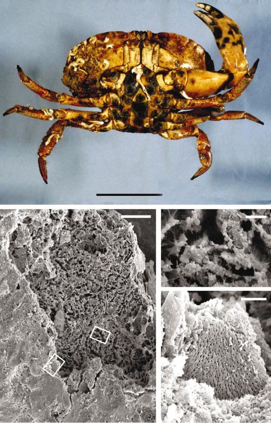
Fig. 1. (a) The ventral surfaces of C. pagurus displaying the characteristic black-spot lesions of shell disease. (b) Low
power scanning electron micrograph of a ventral carapace lesion. Boxed regions are enlarged in (c) and (d). Note the
columnar pattern of degradation in the central regions of the lesion (c) which contrasts with the lamellar cleavage planes
at lesion peripheries (d). Bars : 5 cm, (a) ; 100 µm (b) ; 10 µm (c, d).
Total chitinase activity was assessed by the production of N- 125 µl supernatant was added to 25 µl 0n8 M boric acid
acetylglucosamine when the crude enzyme supernatants were (adjusted to pH 10n2 using KOH) and the mixture was heated
incubated with colloidal chitin using a method adapted from in a boiling water bath for 3 min. The sample was cooled (30
Yanai et al. (1992). Briefly, enzyme supernatant samples min, 4 mC) and 750 µl p-dimethylaminobenzaldehyde (DMAB)
(250 µl) were incubated with 125 µl colloidal chitin and 125 µl solution [1 %, w\v, p-dimethylaminobenzaldehyde (Sigma),
0n1 M Tris\HCl, pH 7n5, containing 0n08 % sodium azide, for 99 %, v\v, glacial acetic acid, 1 %, v\v, concentrated HCl]
46 h at 25 mC. After centrifugation (21 000 g, 2 min, 25 mC), was added. After incubation at 37 mC for 20 min, each sample
745C. L. Vogan, C. Costa-Ramos and A. F. Rowley
Table 1. Summary of the principal test characteristics for chitinolytic bacterial isolates 1–9 (I1–I9)
.................................................................................................................................................................................................................................................................................................................
All isolates were found to be Gram-negative rods which were motile in liquid medium. Cluster groups are based on the isolates
displaying 85 % simple matching coefficients to each other (see Fig. 2). j, Positive activity ; k, negative enzyme activity ; , strain
did not grow on medium.
Test Cluster group
A D E F G
I1 I2 I6 I3 I4 I8 I5 I7 I9
Na+ requirement j j j j k k j j j
Max. Na+ where growth observed (%) 8 8 8 10 3 3 3 3 10
Oxidase j j j j k k j j j
Catalase j j j j j j j k j
V0129 sensitivity j j j j k k j j k
Water-soluble pigment k k k k k j (black) k k k
-Mannitol utilization j j j j k k j j j
Caseinase k j k k j j k j k
Gelatinase* k(j) (j) k(j) (j) j j (k) (j) k
Phospholipase j j j j j j k k
Lipase k k k k j j j j
Colony colour on VVM agar Green\yellow
Colony colour on CC agar Green\yellow Green Yellow Yellow
Closest genus† ( % S ) Vibrio Xanthomonas Vibrio
SM
(100) (100) (100) (100) (80) (90) (88) (82) (82)
Closest species† (% S ) V. splendidus b1 V. anguillarum X. maltophilia V. splendidus V. pelagius V. splendidus
SM
b2 b1\V. vulnificus b2
(93) (87) (93) (82) (86) (86) (80) (71) (80)
* Values in parentheses indicate differing results detected using API commercial test kits.
† Compared to those defined in Holt et al. (1994).
was plated in triplicate onto a 96-well microtitre plate and the blanks (10 µl heat-inactivated supernatants) and negative
OD was measured. Sample blanks (containing 250 µl heat- controls (sterile control culture supernatants) were run in
&&!
inactivated supernatants) and negative controls (consisting of parallel. The pNP substrate microplates were sealed and
250 µl supernatant from sterile control culture flasks) were run incubated at 25 mC for 46 h. Reactions were terminated by the
in parallel. A standard curve, for the linear range of the assay, addition of 10 µl 1 M NaOH to each well and the OD was
was produced using serial dilutions of N-acetylglucosamine measured. One unit of enzyme activity was defined %!& as the
(Sigma) and DMAB detection. One unit of enzyme activity amount of enzyme activity that liberated 1 µmol pNP min−"
was defined as the amount of enzyme required to produce under the above conditions. According to dal Soglio et al.
sugars equivalent to 1 µmol N-acetylglucosamine min−" under (1998), 1 mM pNP is equivalent to an OD increase of 6n367.
the above conditions. %!&
Clearance of chitinolytic isolates in crabs. I4 and I7 (see Table
‘ Endo-acting ’ chitinase activity (i.e. that principally acting on 1) were grown overnight in TSBj1 % NaCl at 25 mC with
the central portions of the chitin polymer) of each supernatant shaking. The bacteria were washed twice in sterile marine
was detected in a turbidometric microplate assay using saline [MS ; 0n58 M NaCl, 20 mM CaCl , 12 mM KCl,
colloidal chitin (adapted from Tronsmo & Harman, 1993). #
0n56 mM disodium phosphate, 0n05 M Tris(hydroxymethyl)-
Aliquots (150 µl) of crude enzyme supernatants (test samples) methylamine, pH 7n4] and adjusted to 2i10* bacteria ml−".
and heat-inactivated supernatants (sample blanks) were added All injections into crabs were made through the surface-
in triplicate to wells that contained 50 µl colloidal chitin. sterilized soft membrane where the fifth pereiopod (fourth
Negative controls that contained 150 µl of supernatants from walking limb) joins the body. Blood samples were removed for
sterile control culture flasks were also included on each plate. blood-cell (haemocyte) and bacterial counts from the soft
After a 10 s shake, a time zero OD reading was taken on a membrane on the cheliped (fighting limb) as described in
microplate reader. The plates were %*#sealed and incubated at Vogan et al. (2001).
25 mC for 14 days. Endochitinase activity for each bacterial Total blood volume in groups of C. pagurus was estimated
supernatant was defined as a reduction in the sample OD
between time zero and 14 days compared with its sample %*# using the Amaranth dye dilution technique (Smith & Ratcliffe,
1980) and the volume of bacterial suspension (2i10* bacteria
blank. ml−") to be injected was calculated so as to give a circulating
The chitinolytic activities of ‘ exo-acting ’ chitinases (acting on blood dose of 2i10' bacteria (ml blood)−". Prior to bacterial
terminal locations on the chitin polymer) were quantified in injection into the crabs, a blood sample was removed from
two separate assays by the release of p-nitrophenol (pNP) each animal for blood-cell counts and bacterial sterility checks.
from p-nitrophenyl-N-acetyl-β--glucosaminide and p-nitro- Control crabs received a dose of sterile MS in place of the
phenyl-β--N,Nh-diacetylchitobiose (Sigma), respectively. bacterial suspension. After 1, 6, 24 and 48 h, a 200 µl blood
Stock solutions of each substrate were prepared at a con- sample was removed from each crab and serially diluted in
centration of 0n275 mM in 1 M Tris\HCl (0n02 % sodium sterile sea salts solution (Sigma). A 100 µl aliquot was spread
azide) and frozen at k20 mC until use in the microtitre assay. in triplicate onto TSAj1 % NaCl plates and incubated for
Crude enzyme supernatant (10 µl) was added in triplicate to about 24 h to allow for bacterial growth. The original bacterial
wells containing 90 µl pNP substrate stock solutions. Sample suspension (2i10* cell ml−") was also plated. Total blood-cell
746Shell disease in crabs
counts were conducted between 5 min and 48 h post-injection UPGMA (unweighted pair group method using arithmetic
using a haemocytometer. averages) clustering method in the Genstat software package.
Experiments involving injection of extracellular products Bacterial diversity frequencies were assessed using con-
(ECPs) from I7 (see Table 1) followed the same protocol, tingency tables followed by either χ# test (for tables with
except for the substitution of filter-sterilized (0n22 µm) aliquots two rows or columns) or Fisher’s exact test (for tables
from 48 h TSBj1 % NaCl cultures in test animals and filter- with two rows or columns). For mean valuesp the
sterilized un-inoculated TSBj1 % NaCl for control animals.
appropriate parametric test (t-test or ANOVA) was
Crabs were considered dead when no ventilatory or sensory employed. For ANOVA, the Tukey’s, Bonferroni and
movements were detected. Crabs that died during the ex- Dunnett post-tests were used as recommended by Instat
perimental period were immediately fixed for histology. (GraphPad).
Control crabs were sacrificed for histology at the end of the
experimental period (48 h).
Histopathology and scanning electron microscopy (SEM). All RESULTS AND DISCUSSION
crabs that died during the experimental period were im- Bacterial diversity in shell disease lesions and
mediately injected with about 50 ml Bouin’s sea water fixative diseased crabs
and dissected to remove the main organs. Tissues were stored
and processed as detailed in Vogan et al. (2001). Wax sections Crabs with shell disease syndrome show characteristic
(about 10 µm thick) were cut and stained using Cole’s darkened lesions through the cuticle (Fig. 1a). Such
haematoxylin and eosin. Photographs were taken using a lesions have previously been shown to occur on all
Zeiss Photomicroscope II. For SEM, small pieces (about regions of the exoskeleton (Vogan et al., 1999). SEM
2 mm#) of lesion and non-lesion crab cuticle were fixed for
about 12 h in ethanol and transferred into acetone (30 min)
examination of these lesions revealed the extensive
prior to air-drying and coating with gold. All samples were nature of the cuticular erosion into the chitinous
examined with a JEOL scanning electron microscope. procuticle (Fig. 1b–d). In all regions of the lesion the
epicuticle was absent as indicated by the appearance of
Antibacterial activity assay. The in vitro antibacterial activity small pore canals (about 1 µm in diameter) that permeate
of crab blood towards I4 and I7 was assessed using a method through the underlying procuticle. Exoskeletal degra-
described by Chisholm & Smith (1992) adapted for use in a
turbidometric growth microplate assay. Test samples con- dation was most severe towards the centre of the lesion,
taining 225 µl of either clotted whole blood (serum ; 6n4i which tended to display sever pitting with a columnar
10%p0n8i10% µg protein ml−" ; p1, n l 6) or a lysate of pattern of degradation (Fig. 1c). Towards the peripheries
the blood cells (1n0i10#p0n2i10# µg protein ml−" ; p1, of the lesion the degree of pitting decreased and
n l 6), prepared as described by Vogan & Rowley (2002), displayed areas of exposed procuticle which had broken
were incubated with 25 µl I4 or I7 in MS (4i10' bacteria ml−"). along the lamellar cleavage planes that run parallel to
For controls, test samples were substituted with equal volumes the epidermis (Fig. 1d).
(225 µl) of sterile MS. Sample blanks contained 225 µl of each
particular test sample and 25 µl sterile MS. All samples, for a The majority of bacteria cultured from the blood (n l
given bacterial isolate, were incubated at room temperature 19) and exoskeleton (n l 67) of shell-diseased C. pag-
with constant shaking for 30 min. They were then rapidly urus were Gram-negative rods (57n9 and 89n6 % re-
flooded with 2n25 ml TSBj1 % NaCl and 200 µl aliquots were spectively). The proportion of Gram-positive eubacteria
plated in triplicate into a 96-well microtitre plate. The OD was significantly higher amongst the blood-derived
of each plate was measured on a microplate reader, after &&!
isolates (33 %) than those found in the exoskeleton
incubation at 25 mC for differing lengths of time so as to give (7n7 % ; χ# test, P l 0n0009). Chitinase-producing bac-
an OD of control wells that lies in the middle of the
&&! growth phase for each bacterial isolate.
exponential teria were isolated from the blood (21n1 % of total
culturable isolates) and from both lesioned and non-
Protein concentrations in serum and blood-cell lysate were lesioned portions of the exoskeleton (26n9 % of total
determined using a BCA (bicinchoninic acid) assay kit in culturable isolates). No significant differences were
accordance with the manufacturer’s instructions for use in a found in the percentages of culturable chitinolytic
microtitre plate (Pierce & Warriner). All samples were
replicated in triplicate and calibrated against a bovine serum
bacteria residing in lesion (31 %) and non-lesion (15 %)
albumin (BSA) standard curve (100–1000 µg ml−") run on the areas (Fisher’s exact test, P l 0n2289).
same plate. From the 22 bacterial colony types that were positive for
Data analyses. All data used for numerical taxonomic chitinolytic activity, 9 isolates (designated I1–I9) were
comparisons were converted into binary figures, scoring ‘ 1 ’ selected for further phenotypic analysis. Selection was
for positive reactions and ‘ 0 ’ for negative reactions. In- based on their distinct behaviour in several preliminary
conclusive results were omitted from all comparisons. A tests, including Gram staining, colony and bacterial
positive test result (1) was recorded when 75 % of the strains morphology. I1 and I8 were from blood of shell-diseased
tested were positive, a negative test result (0) was recorded individuals. I2, I4, I5, I6, I7 and I9 were from exoskeletal
when 25 % of the strains tested were positive and tests with lesions, while I3 was from a non-lesion area. All were
results between 25 and 75 % were taken as being inconclusive
(omitted from comparison), as described by Holt et al. (1994). motile, Gram-negative rods that generally varied in
The isolate test results were then compared to each other and length between 0n6 and 2 µm. However, the rods of I4
to data published in Holt et al. (1994) using the simple were elongate (1n5–5 µm) and formed long filamentous
matching coefficient of similarity. The resultant similarities chains, whereas those of I9 were almost spherical in
(0–100 %) were then plotted onto dendrograms using the appearance. These isolates were clustered based on 157
747C. L. Vogan, C. Costa-Ramos and A. F. Rowley
formic acid, glucose-1-phosphate). I5, I7 and I9 were
85 % similar to all others and hence were contained
within individual clusters.
According to Holt et al. (1994) members of the family
Vibrionaceae (Bergey’s Group 5.2) are Gram-negative,
oxidase-positive, motile rods, capable of growth at
37 mC as long as there is a source of sodium ions.
Although growth at 37 mC was not tested, I1, I2, I5, I6, I7
and I9 all grew at 35 mC and satisfy the other require-
ments for classification into Vibrionaceae (Table 1).
Comparisons of the test isolates to genera known to
contain chitinolytic strains were made using data in
Holt et al. (1994) (Table 1). It was found that I1, I2, I3
and I6 were most likely to be Vibrio species (100 %
simple matching similarity). I5, I7 and I9 were also most
likely to belong within the genus Vibrio (88, 82 and 82 %
simple matching similarities, respectively). Like the
majority of members of this genus, I5 and I7 were
sensitive to the vibriostatic agent V0129 (150 µg) and
utilized -mannitol as carbon source. I9, however, was
.................................................................................................................................................
resistant to V0129 and did not utilize -mannitol,
Fig. 2. UPGMA clustering of bacterial isolates based on 157 suggesting that it may not be a Vibrio species. However,
individual test results, using a simple matching coefficient of
similarity. I1–I9 are the chitinolytic isolates. V1–V3 are V. Holt et al. (1994) recorded that some strains of V.
alginolyticus (NCIMB 1339), V. alginolyticus (‘ French ’ strain) and alginolyticus, Vibrio cincinnatiensis and Vibrio para-
V. splendidus, respectively. Cluster groups A–G are based on haemolyticus are resistant to V0129. In the current
isolates displaying 85 % simple matching coefficients. study, a lack of sensitivity to V0129 was also displayed
by both V. alginolyticus and V. splendidus.
Urakawa et al. (1999) found that V. splendidus strains
1010 70 were the most abundant culturable vibrios from sea
Total chitinase activity (mU ml–1)
water. The current study found five of the nine strains
Bacterial count (c.f.u. ml–1)
60 isolated from C. pagurus to be most closely related to V.
109 50 splendidus (2 biovars listed in Holt et al., 1994). I7
displayed joint highest similarities to V. pelagius (71 %)
40 and V. vulnificus. It was concluded that this isolate was
108 unlikely to be a strain of V. vulnificus, since it did not
30
produce the characteristic yellow diffusion haloes on
20 VVM agar (Table 1).
107
10 The isolates of cluster group D (I4 and I8) were distinct
0
from the other isolates in that they were oxidase-
106 negative and capable of growth in the absence of sodium
0 24 72 120 168 216 264 312 360 408
Time (h)
ions. Chitinolytic Gram-negative rods that are oxidase-
negative either belong to the family Enterobacteriaceae
................................................................................................................................................. (Bergey’s Group 5.1) or within Bergey’s Group 4.4A
Fig. 3. Total extracellular chitinase activity (– – –) in relation to (Holt et al., 1994). Although the similarity comparison
growth of cultures (–––) of I3. The bacterial isolate was grown calculations were based only upon seven test charac-
in a minimal chitin powder medium at 25 mC with shaking for
408 h. Mean valuesp1 SE are shown (n l 3). teristics, I4 and I8 both seem to be most closely related
to Xanthomonas maltophilia (as listed in Holt et al.,
1994). These results are in agreement with those found
when comparing data to the API published profile (data
not shown) and the genus identification (Table 1).
test characteristics (Fig. 2 ; Table 1). Bacteria in cluster
group A (I1, I2, I3 and I6) differed mainly in their ability It is generally accepted that a large proportion of
to produce β-galactosidase, ferment amygdalin and bacteria in the marine environment are not detected by
arabinose, and in several carbon utilization tests. Cluster culturing onto high nutrient media. Over the last 15
D contained two isolates, I4 and I8, that were distinct years, the existence and nature of a viable but non-
from all others tested and were less likely to belong culturable state has been much debated (e.g. McDougald
within the genus Vibrio. Within the cluster the two et al., 1998). Roszak & Colwell (1987) described the
strains differed in pigment production, cell form and a dynamic state of the microbial cell in its natural state as
small number of carbon utilization tests (Tween 40, being able to readily adapt and survive fluctuations in
N-acetyl--glucosamine, acetic acid, cis-aconitic acid, environmental parameters such as nutrient source and
748Shell disease in crabs
* *
8
10–9 × bacterial count (ml–1)
(a)
7
6
5
4
3
2
1
0
I3 I4 I7 I8
*
14 (b) 0·4
(mU per 1 × 10⁹ bacteria)
(c)
Total chitinase activity
12
(Reduction in OD492nm
Endochitinase activity
per 1 × 10⁹ bacteria)
10 a 0·3
8
0·2
6
a
4
0·1
2
0 0·0
I3 I4 I7 I8 I3 I4 I7 I8
* * *
1·0 1·0
(d) (e)
(mU per 1 × 10⁹ bacteria)
(mU per 1 × 10⁹ bacteria)
a ab
Exochitinase activity
Exochitinase activity
0·8 0·8
0·6 0·6
a b
a
0·4 0·4
0·2 0·2
0·0 0·0
I3 I4 I7 I8 I3 I4 I7 I8
.................................................................................................................................................................................................................................................................................................................
Fig. 4. Chitinase activity from culture supernatants of I3, I4, I7 and I8 grown in a minimal chitin powder medium when
sampled after 192 ( ) and 408 h ( ) of growth. (a) Number of bacteria (ml culture)−1. (b) Total chitinase activity, (c)
endo-acting chitinase activity and (d, e) exo-acting chitinase against the pNP-monomer (d) and dimer (e) in culture
supernatants standardized for 1i109 bacteria. Mean values p1 SE are shown (n l 3). Significant differences (paired
t-test, P 0n05) for individual isolates between 192 and 408 h are indicated by asterisks. Significant interisolate
differences (ANOVA, P 0n05 ; Tukey’s post-test, P 0n05) at either 192 or 408 h are indicated by roman or italicized
letters, respectively.
availability, temperature and light. Thus, culturing onto perhaps, be considered analogous to the primary lesion
standard laboratory media provides non-comparable colonizers, i.e. having the ability to rapidly detect and
conditions to those of oligotrophy that are found in the adapt to newly exposed chitin.
natural environment. Hence, only those organisms that
are most able to adapt to the specific culture conditions Pathogenicity of chitinolytic isolates
will be able to grow and divide. The current study was
not intended to provide an exhaustive list of all micro- In this study, the pathogenicity of the chitinolytic (shell
organisms involved in or capable of exoskeletal degra- disease) isolates was considered on two levels : (i) their
dation. Compared to the vast majority of the marine ability to contribute to exoskeletal breakdown by the
environment, shell disease lesions provide a high nu- expression of chitinase activity (external pathogenicity),
trient niche and thus, culturing onto a chitin-containing and (ii) should they penetrate the cuticle, their ability to
medium may not be as extreme as, for instance, culturing cause damage to host tissues and to overwhelm the
directly from sediments. Thus, the types of chitinolytic cellular and humoral defences of the host (internal
micro-organisms cultured on chitin agar plates might, pathogenicity).
749C. L. Vogan, C. Costa-Ramos and A. F. Rowley
100 (a) Tris-buffered sea salts alone, did not induce chitinase
production (data not shown). The crude chitinase found
10
in supernatant samples was stable at k20 mC without a
Bacterial survival (%)
1 significant loss in activity for at least 6 months (data not
shown).
0·1
For I3, chitinase activity in the culture supernatant was
0·01 not detected before 96 h of bacterial growth (Fig. 3).
Activity levels were not significantly higher than control
0·001
flasks until the isolate had undergone 144 h of growth
0·0001 (ANOVA, P 0n01 ; Bonferroni post-test, P 0n001
1 2 3 4 5 6 24 48 from 144 to 408 h). The maximal rate of chitinase
synthesis was found to occur during the late exponential
120 (b)
and early stationary phases of growth (120–192 h). This
result is consistent with patterns of chitinase production
100 for Bacillus spp. (Priest, 1977 ; Frandberg & Schnurer,
1994) and the fungus Metarhizium anisopliae (Kang et
80
al., 1999). The highest rates of chitinase production
60 between 120 and 192 h were found when the culture
Total blood-cell count (percentage of t0)
supernatant from I3 was tested for endo- and exo-acting
40 activity (data not shown).
Death All the isolates tested (I3, I4, I7, I8) were able to grow
20
using 1 % chitin powder as their nutrient source (Fig.
4a). I3 and I4 were found to have similar numbers of
1 2 3 4 5 6 24 48 bacteria at both 192 and 408 h (paired t-tests for
individual isolates, P 0n05), indicating that these
120 (c)
cultures had reached, or were close to reaching the
stationary phase of growth (Fig. 4a). This was further
100 reflected when total chitinase activity was standardized
80
for 1i10* bacteria with no significant differences found
in the amount of extracellular chitinase activity for I3
60 and I4 between the two sample times (Fig. 4b). When
chitinase activity was divided in terms of its activity
40 towards the chitin polymer (i.e. endo- and exo-acting) it
Death was found that I3 and I4 displayed low to moderate
20 levels of both endo- and exo-activity that did not change
significantly over the culture period (Fig. 4c–e ; paired
t-tests, P 0n05).
1 2 3 4 5 6 24 48
Time (h) In contrast to I3 and I4, the culture flasks for I7 and I8
were found to have significantly higher bacterial num-
.................................................................................................................................................
bers at 408 h compared to 192 h, suggesting that both
Fig. 5. Response of C. pagurus to injection with I4, I7 and ECPs
from I7. (a) Recovery of viable bacteria from blood samples
cultures were still in exponential growth phase. Signifi-
taken over a 48 h post-bacterial injection period. (b) Total cant increases in total chitinase activity between the two
blood-cell counts from crabs after injection with bacteria incubation times were only found for I7 (Fig. 4b ; paired
(ANOVA, P 0n05 ; Dunnett’s post-test, *P 0n05, **P 0n01, t-test, P l 0n0316). Thus, although cultures of I8 were
compared to the control for the individual time periods). (c) still actively dividing, a higher rate of chitinase synthesis
Total blood-cell counts from crabs after injection with 0n5 ml
ECPs (unpaired t-test, *P 0n05, ***P 0n001). Mean values was reached earlier in the growth period. Indeed, I8
p1 SE are shown (n l 4–6). =, I4 ; , I7 ; $, control. cultures showed the highest total chitinase activity per
standard number of bacteria at both time periods. I8
produced the highest quantities of endo-acting chitinase
at 192 h and the highest amount of exo-type activity by
Chitinase activity of bacterial isolates (external 408 h (Fig. 4d, e).
pathogenicity)
Overall within the culture system, I3 and I4, with their
Preliminary experiments investigating the media re- faster growth rates and production of moderate levels of
quired to induce chitinase production in liquid culture enzymes capable of degrading all areas of the chitin
found chitinase activity to be present only within polymer, are presumed to represent primary colonizers
inoculated flasks that contained Tris-buffered sea salts of chitin powder. By comparison, I7 and I8 grew more
and 1 % chitin powder (data not shown). Inoculated slowly, producing greater amounts of chitinases later in
flasks, run in parallel, that contained either Tris-buffered the culture period and thus may be more likely to
sea salts, 1 % chitin powder plus 0n01 % tryptone or represent secondary colonizers. However, it should be
750Shell disease in crabs
(a) noted that I8 initially (192 h) produced high levels of
endo-acting enzyme and subsequently (408 h) released
large quantities of extracellular enzymes capable of
degrading the smaller chitin oligomer. Thus, I8 is
potentially best adapted for the degradation of the chitin
powder.
The present investigation examined the production of
chitinases in bacteria in culture systems and thus is
unlikely to reflect their behaviour within the natural
lesion community. Pure cultures fail to consider such
factors as the interactive effects of the various lesion-
BS dwelling bacteria and predation rates (Wardell, 1988).
Chitin powder used as a substrate is both deproteinized
and decalcified compared to its native form within the
cuticle. Both microbial attachment (Dietrich et al., 1984)
and chitinase production (Hood & Meyers, 1977) have
been shown to be enhanced when microbial populations
are exposed to native chitin. In the current study,
preliminary experiments using shell pieces derived from
(b) C. pagurus as the growth substrate within the culture
system have indicated that levels of extracellular chi-
tinase activity increase for I3 (data not shown). This
may be an approach for future studies.
C. pagurus has been shown to have a chitin : protein
ratio (calculated from the percentage organic frac-
tion ; dry wt) of approximately 7 : 1 within the exo-
skeleton (Brimacombe & Webber, 1964). The outermost
waterproof cuticle layer, the epicuticle, is ultrathin and
composed of lipid and protein alone. Thus, although
chitinolytic activity is fundamental to lesion progres-
sion, microbial proteases and lipases may also be
involved in exoskeletal breakdown, particularly in the
initial stages of shell disease (Cipriani et al., 1980).
Although not widely investigated, protease activity
[specifically gelatinase (collagenase)] was found in all
but one of the isolates (I9) while lipase activity amongst
the exoskeletal isolates was restricted to cluster groups
D, E and F (I4, I5, I7, I8) (Table 1).
(c)
Internal pathogenicity
The internal effects of only two of the isolates (I4 and I7)
on the crab host were studied. These were chosen
because both isolates displayed distinct morphological
characteristics that would enable their identity to be
rapidly ascertained on reisolation and growth following
intrahaemocoelic injections (i.e. I4 had an elongate cell
morphology while I7 was negative for catalase).
I4 was rapidly cleared from the blood of crabs, with
99 % of c.f.u. removed by 15 min post-injection (Fig.
5a). This rapid reduction in bacterial number coincided
Fig. 6. Changes observed within C. pagurus gills after injection
with (a) marine saline (control), (b) 2i106 I7 ml−1 and (c) ECPs
(0n5 ml) from I7 cultures after 48 h of growth. Arrows indicate
the accumulation of blood cells at the base of the gills near the
branchial space (BS). Bar, 200 µm.
751C. L. Vogan, C. Costa-Ramos and A. F. Rowley with a marked reduction in the number of circulating major determinants of pathogenicity. There is surpris- blood cells (haemocytes ; Fig. 5b). By 24 h, c.f.u. numbers ingly little known about the mechanisms of patho- on spread plates increased and at 100 h mortality was genicity for vibrios in marine invertebrates. A number of seen in 80 % of individuals. All control (saline-injected) heat-sensitive proteases produced by V. parahaemolyt- crabs survived and only showed transient changes in the icus have been shown to be toxic for tiger prawns total numbers of circulating blood cells (Fig. 5b). (Penaeus monodon), but the nature of these enzymes is The dynamics of clearance and pathology of I7 was unknown (Sudheesh & Xu, 2001) while alkaline serine markedly different than for I4. Injection of I7 (9i10$p proteases produced by V. alginolyticus have been 6n7i10$ c.f.u. ml−") resulted in a more modest reduction reported to be toxic for Kurama prawns (Penaeus in the number of viable bacteria from circulation. For japonicus) (Liu et al., 1997). Although it is premature to example, after 60 min bacterial numbers had only speculate on the activities of the toxic ECPs from I7, the decreased to 17n8 % of the original. The number of symptoms of the disease, including limb tremors and eye circulating blood cells showed a rapid decline, such that stalk withdrawal, suggest a neurotoxic component. after 60 min only 4n7p1n1 % (mean value p1 , n l 5) Both serum and blood-cell lysates displayed variable of the cells remained in circulation. Of importance was levels of antibacterial activity against I4 and I7. In the the finding that by 3 h post-injection all crabs were case of I4, 91n3p3n7 % bacterial survival was found after moribund. Indeed, after only 15 min post-injection, 30 min incubation with the crab blood-cell lysate, while crabs showed early changes in their behaviour, including 61n7p10n2 % bacterial survival was observed after retraction of eye stalks and reduced or increased gill incubation with neat whole serum (mean valuesp, n ventilation movements. Subsequently, they all exhibited l 3). I7 was more effectively killed by both blood-cell loss of limb movement by 60 min and some also showed lysates and serum preparations (0n26p0n17 % bacterial tremors in their walking limbs. Because such changes survival compared with 4n3p2n5 % bacterial survival indicated that mortality may have been caused by respectively ; mean valuesp, n l 3). Notably, with I7, toxins (i.e. ECPs), crabs were injected with filter- and to some extent I4, the blood-cell lysate preparations sterilized culture filtrates taken from 48 h cultures of I7. were particularly active in bacterial killing, taking into All crabs injected were moribund 60 min after injection, account the difference in total protein content between while culture filtrate from un-inoculated cultures had no this lysate and serum (the concentration of protein in the effect. The decline in the number of circulating blood blood-cell lysate was about 1000-fold less concentrated cells seen after injection of I7 was also observed with the than in serum). The limited killing of I4 by both serum crude ECP preparation (Fig. 5c). Furthermore, the and blood-cell lysates partially explains why it is able to behaviour of crabs injected with ECPs was the same as multiply in vivo and cause limited mortality. The nature that seen following injection of I7 bacteria. Histological of this antibacterial activity was not investigated further. examination of crabs injected with I7 bacteria or ECPs However, in a closely related species of crab, the shore revealed a massive accumulation of blood cells in the crab (Carcinus maenas), Schnapp et al. (1996) identified gills (Fig. 6). Such accumulations effectively blocked the four proteins with antimicrobial activity that reside in central vascular region of the gills (branchial spaces). the blood cells. In the shrimp, Pennaeus vannamei, a There was also evidence in other organs that blood cells new family of antimicrobial peptides, termed penaei- dropped out of circulation and attached to such surfaces dins, have also been reported to be located in certain following exposure to ECPs. blood-cell types (Destoumieux et al., 1997). Although Much is known of the cellular defence responses in there have been reports of serum antibacterial activity in crustaceans to micro-organisms. Injection of heat- crabs (e.g. Noga et al., 1994) the extreme fragility of killed Bacillus cereus into shore crabs (Carcinus their blood cells suggests that some or all of the serum maenas) results in their rapid clearance as a result of the killing factors may be derived from the haemocytes by formation of clumps of blood cells and bacteria, termed degranulation following bleeding. nodules, in the gills (Smith & Ratcliffe, 1981). These In conclusion, shell disease is not a disease caused by a observations largely accounted for the rapid reduction single pathogen and should not be considered a disease in the number of circulating blood cells and bacteria. In solely restricted to the exoskeleton. Numerous bacteria the current study with I4, following an initial rapid within the marine environment are capable of degra- clearance of the bacteria, presumably by nodule forma- dation of the chitin component of the crustacean cuticle tion, there was some evidence that bacterial replication and it is likely that the collective effects of the lesion occurred in vivo, leading to limited septicaemia and community lead to further exoskeletal degradation. If death. It must be remembered, however, that the dose breach of the cuticle occurs, infection of the body cavity injected (2i10' bacteria ml−" ; Thoma count) and the of the crustacean may result, with the internal symptoms method of delivery suggest that this bacterium has differing depending on the nature of the penetrating limited virulence in edible crabs. bacteria and this may ultimately lead to the death of the The mechanism by which I7 kills the crabs is clearly animal. different to that employed by I4. In particular, injection of culture filtrates (containing ECPs) resulted in the ACKNOWLEDGEMENTS same symptoms observed after the injection of washed We are grateful to Messrs Paul Llewellyn, Paul Jenkins and I7 bacteria. Hence, in the case of I7, ECPs appear to be Wyn Morris for their assistance with crab collection and to the 752
Shell disease in crabs
DRIM laboratories, Montpellier, France, for stains of V. chitin catabolism in marine bacteria. Biochim Biophys Acta 1473,
alginolyticus and V. splendidus. We also thank Dr M. R. 108–122.
Fordy for assistance with the SEM studies. C. L. V. was Liu, P. C., Lee, K. K., Tu, C. C. & Chen, S. N. (1997). Alkaline serine
supported by a University of Wales Swansea studentship. protease is an exotoxin of Vibrio alginolyticus in Kurama prawn,
Penaeus japonicus. Curr Microbiol 34, 110–117.
REFERENCES McDougald, D., Rice, S. A., Weichart, D. & Kjelleberg, S. (1998).
Nonculturability : adaptation or debilitation? FEMS Microbiol
Ayres, P. A. & Edwards, E. (1982). Notes on the distribution of Ecol 25, 1–9.
‘ black spot ’ shell disease in crustacean fisheries. Chem Ecol 1, Noga, E. J., Engel, D. P., Arroll, T. W., McKenna, S. & Davidian, M.
125–130. (1994). Low serum antibacterial activity coincides with increased
Baross, J. A. & Tester, P. A. (1978). Incidence, microscopy, and prevalence of shell disease in blue crabs Callinectes sapidus. Dis
etiology of exoskeletal lesions in the tanner crab, Chionoecetes Aquat Org 19, 121–128.
tanneri. J Fish Res Board Can 35, 1141–1149. Priest, F. G. (1977). Extracellular enzyme synthesis in the genus
Brimacombe, J. S. & Webber, J. M. (1964). Mucopolysaccharides. Bacillus. Bacteriol Rev 41, 711–753.
Amsterdam : Elsevier. Prince, D. L., Bayer, R. C., Gallagher, M. L. & Subramanyam, M.
Cerda' -Cue! llar, M., Jofre, J. & Blanch, A. R. (2000). A selective (1995). Reduction of shell disease with an experimental diet in a
medium and a specific probe for detection of Vibrio vulnificus. Nova-Scotian lobster pound. J Shellfish Res 14, 205–207.
Appl Environ Microbiol 66, 855–859. Roszak, D. B. & Colwell, R. R. (1987). Survival strategies of bacteria
Chisholm, J. R. S. & Smith, V. J. (1992). Antibacterial activity in in the natural environment. Microbiol Rev 51, 365–379.
the haemocytes of the shore crab, Carcinus maenas. J Mar Biol Sawyer, T. K. (1991). Shell disease in the Atlantic rock crab,
Assoc UK 72, 529–542. Cancer irroratus say, 1817, from the northeastern United States.
Cipriani, G. R., Wheeler, R. S. & Sizemore, R. K. (1980). Charac- J Shellfish Res 10, 495–497.
terization of brown spot disease of Gulf Coast shrimp. J Invertebr Schlotfeldt, H.-J. (1972). Jahreszeitliche abhangigkeit der
Pathol 36, 255–263. ‘ schwarzfleckenkrankheit ’ bei der garnele, Crangon crangon (L).
dal Soglio, F. K., Bertagnolli, B. L., Sinclair, J. B., Yu, G. Y. & Ber Wissen Komm Meeresfors 22, 397–399.
Eastburn, D. M. (1998). Production of chitinolytic enzymes and Schnapp, D., Kemp, G. D. & Smith, V. J. (1996). Purification and
endoglucanase in the soybean rhizosphere in the presence of characterization of a proline-rich antibacterial peptide, with
Trichoderma harzianum and Rhizoctonia solani. Biol Control 12, sequence similarity to bactenecin-7, from the haemocytes of the
111–117. shore crab, Carcinus maenas. Eur J Biochem 240, 532–539.
Delves-Broughton, J. & Poupard, C. W. (1976). Disease problems Smith, V. J. & Ratcliffe, N. A. (1980). Host defence reactions in the
of prawns in recirculation systems in the UK. Aquaculture 7, shore crab, Carcinus maenus (L.) : clearance and distribution of
201–217. test particles. J Mar Biol Assoc U K 60, 89–102.
Destoumieux, D., Bulet, P., Loew, D., Van Dorsselaer, A., Smith, V. J. & Ratcliffe, N. A. (1981). Pathological changes in the
Rodriguez, J. & Bache' re, E. (1997). Penaeidins, a new family of nephrocytes of the shore crab, Carcinus maenas, following
antimicrobial peptides isolated from the shrimp Penaeus van- injection of bacteria. J Invertebr Pathol 38, 113–121.
namei (Decapoda). J Biol Chem 272, 28398–28406.
Smolowitz, R. M., Bullis, R. A. & Abt, D. A. (1992). Pathological
Dietrich, M. A., Hackney, C. R. & Grodner, R. M. (1984). Factors cuticular changes of winter impoundment shell disease preceding
affecting the adherence of Vibrio cholerae to blue crab (Callinectes and during intermolt in the American lobster, Homarus ameri-
sapidus) shell. In Vibrios in the Environment, pp. 601–611. Edited canus. Biol Bull 183, 99–112.
by R. R. Colwell. New York : Wiley.
Stewart, J. E. (1993). Infectious diseases of marine crustaceans. In
Dyrynda, E. A. (1998). Shell disease in the common shrimp Pathobiology of Marine and Estuarine Organisms, pp. 319–342.
Crangon crangon : variations within an enclosed estuarine system. Edited by J. A. Couch & J. W. Fournie. Boca Raton : CRC Press.
Mar Biol 132, 445–452.
Sudheesh, P. S. & Xu, H. S. (2001). Pathogenicity of Vibrio
Frandberg, E. & Schnurer, J. (1994). Chitinolytic properties of parahaemolyticus in tiger prawn Penaeus monodon fabricius :
Bacillus pabuli K1. J Appl Bacteriol 76, 361–367. possible role of extracellular proteases. Aquaculture 196, 37–46.
Getchell, R. G. (1989). Bacterial shell disease in crustaceans : a Tronsmo, A. & Harman, G. E. (1993). Detection and quantification
review. J Shellfish Res 8, 1–6. of N-acetyl-β--glucosaminidase, chitobiosidase, and endochit-
Høi, L. & Dalsgaard, A. (2000). Evaluation of a simplified semi- inase in solutions and on gels. Anal Biochem 208, 74–79.
quantitative protocol for the estimation of Vibrio vulnificus in Urakawa, H., Kita-Tsukamoto, K. & Ohwada, K. (1999). Re-
bathing water using cellobiose-colistin agar : a collaborative study striction fragment length polymorphism analysis of psychrophilic
with 13 municipal food controlling units in Denmark. J Microbiol and psychrotrophic Vibrio and Photobacterium from the north-
Methods 41, 53–57. western Pacific Ocean and Otsuchi Bay, Japan. Can J Microbiol
Holt, J. G., Krieg, N. R., Sneath, P. H. A., Staley, J. T. & Williams, 45, 67–76.
S. T. (1994). Bergey’s Manual of Determinative Bacteriology, 9th Vogan, C. L. & Rowley, A. F. (2002). Effects of shell disease
edn. Baltimore : Williams & Wilkins. syndrome on the haemocytes and humoral defences of the edible
Hood, M. A. & Meyers, S. P. (1977). Rates of chitin degradation in crab, Cancer pagurus. Aquaculture (in press).
an estuarine environment. J Oceanogr Soc Jpn 33, 328–334. Vogan, C. L., Llewellyn, P. J. & Rowley, A. F. (1999). Epidemiology
Kang, S. C., Park, S. & Lee, D. G. (1999). Purification and and dynamics of shell disease in the edible crab Cancer pagurus :
characterization of a novel chitinase from the entomopathogenic a preliminary study of Langland Bay, Swansea, UK. Dis Aquat
fungus, Metarhizium anisopliae. J Invertebr Pathol 73, 276–281. Org 35, 81–87.
Keyhani, N. O. & Roseman, S. (1999). Physiological aspects of Vogan, C. L., Costa-Ramos, C. & Rowley, A. F. (2001). A his-
753C. L. Vogan, C. Costa-Ramos and A. F. Rowley tological study of shell disease syndrome in the edible crab, deep-sea red crabs (Chaceon quinquedens, Smith 1879) in relation Cancer pagurus. Dis Aquat Org 47, 209–217. to ocean dumping of sewage sludge. J Shellfish Res 10, 499–503. Wardell, J. N. (1988). Methods for the study of bacterial at- Young, J. S. & Pearce, J. B. (1975). Shell disease from crabs and tachment. In Methods in Aquatic Bacteriology, pp. 389–415. lobsters from New York Bight. Mar Pollut Bull 6, 101–105. Edited by B. Austin. Chichester : Wiley. Zhang, X. H. & Austin, B. (2000). Pathogenicity of Vibrio harveyi Yanai, K., Takaya, N., Kojima, N., Horiuchi, H., Ohta, A. & Takagi, to salmonids. J Fish Dis 23, 93–102. M. (1992). Purification of 2 chitinases from Rhizopus oligosporus and isolation and sequencing of the encoding genes. J Bacteriol ................................................................................................................................................. 174, 7398–7406. Received 16 July 2001 ; revised 24 October 2001 ; accepted 12 November Young, J. S. (1991). Prevalence and severity of shell disease among 2001. 754
You can also read