AGAR PLATE-BASED SCREENING METHODS FOR THE IDENTIFICATION OF POLYESTER HYDROLYSIS BY PSEUDOMONAS SPECIES - JUSER
←
→
Page content transcription
If your browser does not render page correctly, please read the page content below
bs_bs_banner

Brief Report

Agar plate-based screening methods for the
identification of polyester hydrolysis by Pseudomonas
species
Rebecka Molitor,1 Alexander Bollinger,1                               pre-screening with short-chain and middle-chain-
Sonja Kubicki,1            Anita Loeschcke,1                          length triglycerides as substrates to identify enzymes
                       1,2
Karl-Erich Jaeger               and Stephan Thies1*                   with lipolytic activity to be further tested for polyester-
1
  Institute of Molecular Enzyme Technology, Heinrich-                 ase activity. We applied these assays to experimen-
Heine-University Du    € sseldorf, Forschungszentrum Ju  € lich,      tally demonstrate polyesterase activity in bacteria
D-52425, Ju   € lich, Germany.                                        from the P. pertucinogena lineage originating from
2
  Institute of Bio- and Geosciences IBG-1: Biotechnology,             contaminated soils and diverse marine habitats.
Forschungszentrum Ju       € lich GmbH, D-52425, Ju
                                                  € lich,
Germany.
                                                                      Introduction
                                                                      Recent attention of both the scientific community and the
Summary
                                                                      public was drawn to microorganisms with enzymatic
Hydrolases acting on polyesters like cutin, polycapro-                capabilities to degrade the plastic polymer polyethylene
lactone or polyethylene terephthalate (PET) are of                    terephthalate (PET) (Wei et al., 2016; Wierckx et al.,
interest for several biotechnological applications like               2018) that was assumed to be biologically inert for a
waste treatment, biocatalysis and sustainable poly-                   long time (Moharir and Kumar, 2019). Probably the most
mer modifications. Recent studies suggest that a                       prominent example is the b-proteobacterium Ideonella
large variety of such enzymes are still to be identified               sakaiensis isolated from a plastic-polluted site (Yoshida
and explored in a variety of microorganisms, includ-                  et al., 2016) which produces an enzyme named IsPE-
ing bacteria of the genus Pseudomonas. For activity-                  Tase (Austin et al., 2018; Gong et al., 2018; Joo et al.,
based screening, methods have been established                        2018) that was shown to be responsible for the
using agar plates which contain nanoparticles of poly-                biodegradation of PET. Crystallographic studies revealed
caprolactone or PET prepared by solvent precipitation                 that this enzyme shows a cutinase-like structure (Joo
and evaporation. In this protocol article, we describe a              et al., 2018) which is in line with other studies on enzy-
straightforward agar plate-based method using emul-                   matic degradation of PET by enzymes that were initially
sifiable artificial polyesters as substrates, namely                    described as cutinases (Nikolaivits et al., 2018).
Impranil DLN and liquid polycaprolactone diol (PLD).                    Cutinases are lipolytic enzymes and thus primarily active
Thereby, the currently quite narrow set of screening                  on carboxylic ester bonds (EC 3.1.1) but defined by activity
substrates is expanded. We also suggest optional                      on polyesters like the plant surface material cutin (Nikolaiv-
                                                                      its et al., 2018) and, as a consequence, were assigned to a
                                                                      distinct enzyme subclass (EC 3.1.1.74). Cutinases are now
Received 23 January, 2019; revised 5 April, 2019; accepted 8 April,   spotlighted in the development of new strategies to deal
2019.                                                                 with man-made plastic pollution: Most studies attempting to
*For correspondence. E-mail s.thies@fz-juelich.de; Tel. +49 2461
613790; Fax + 49 2461 612461.                                         hydrolyze artificial polyesters are conducted applying such
Microbial Biotechnology (2020) 13(1), 274–284                         cutinase-like enzymes (Korpecka et al., 2010; Austin et al.,
doi:10.1111/1751-7915.13418                                           2018; Nikolaivits et al., 2018). However, lipolytic enzymes
Funding Information
The authors received funding from the European Union’s Horizon        clustering within other families (Arpigny and Jaeger, 1999),
2020 research and innovation programme (Blue Growth: Unlocking        e.g. family VIII (b-lactamase like), were likewise associated
the potential of Seas and Oceans) through the Project ‘INMARE’        with polyesterase activity very recently (Biundo et al., 2017;
under grant agreement No. 634486. ST, SK and AL are financially
supported by the ministry of Culture and Science of the German        Mu €ller et al., 2017; Hajighasemi et al., 2018). Besides
State of North Rhine-Westphalia within in the framework of the        biodegradation of artificial polyesters like PET, cutinases/
NRW Strategieprojekt BioSC (No. 313/323-400-00213).

ª 2019 The Authors. Microbial Biotechnology published by John Wiley & Sons Ltd and Society for Applied Microbiology.
This is an open access article under the terms of the Creative Commons Attribution-NonCommercial License, which permits use,
distribution and reproduction in any medium, provided the original work is properly cited and is not used for commercial purposes.
Assays for functional polyesterase screenings 275
polyesterases are discussed for different biotechnological solvent precipitation and evaporation techniques (Jarrett
applications, e.g. sustainable polymerization and polymer et al., 1984; Nishida and Tokiwa, 1993; Wei et al., 2014).
modification processes or biocatalytic transesterification Notably, these assays imply safety hazards and the pro-
and ester synthesis reactions (Nikolaivits et al., 2018). Most duction of organic solvent waste. In this protocol article,
polyesterases known today are secreted into the extracellu- we describe water-emulsifiable polyesters (Fig. S1) as
lar medium, potentially facilitating industrial production with substrates for rapid and straightforward agar plate-based
either wild-type or suitable recombinant host strains. screening assays as an alternative or at least complemen-
Currently, high-throughput activity-based screening tary strategy to identify polyesterase activity in bacterial
assays are a frequently applied method to identify novel clones, here exemplified by the identification of such enzy-
biocatalysts within environmental isolates or metagenomic matic activities exhibited by yet unexplored Pseudomonas
libraries (Popovic et al., 2015; Martin et al., 2016; Pen ~a- species. These assays generally allow for high-throughput
Garcıa et al., 2016; Thies et al., 2016). These assays are identification of relevant clones, e.g. in metagenomic or
of key importance for reducing the experimental workload genomic libraries (Fig. 1).
to allow assigning of an activity of interest to an individual
clone which can then be further characterized. To this
Step-by-step protocols for agar plate preparation
end, agar plate-based activity assays are typically applied.
and polyesterase activity screening
Here, clear or coloured zones which are formed around
the bacterial colonies indicate the production of a Polyesterases are lipolytic enzymes and are thus detected
catalytically active enzyme. Suchlike approaches to iden- by non-specific esterase assays like an agar plate-based
tify organisms or clones with polyesterase activity cur- screening with the substrate tributyrin. The use of this uni-
rently mostly rely on clearance of media containing versal substrate with a short-chain fatty acid triglyceride
polycaprolactone (PCL) or PET nanoparticles prepared by will also detect activities of esterases, true lipases,
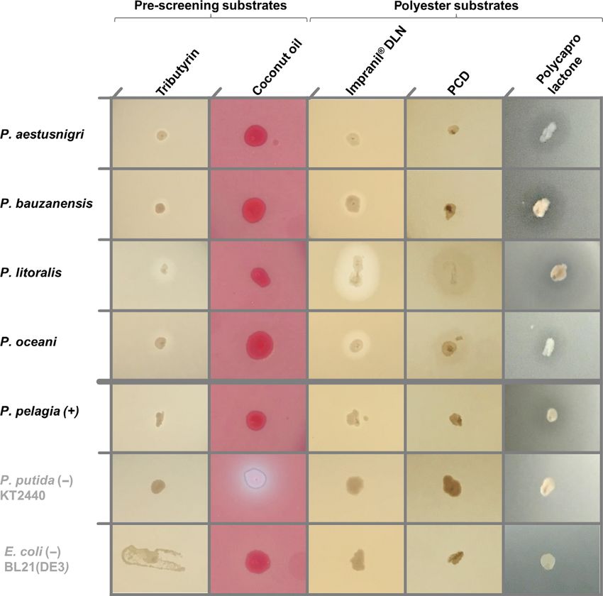
Fig. 1. Workflow for agar plate-based screening for polyesterase active clones.
A. Steps of plate preparation and screening: 1. Prepare an emulsion/suspension with the respective substrate (if necessary). 2. Combine sub-
strate emulsion/suspension and molten agar-containing nutrient medium. 3. Pour the warm medium into suitable Petri dishes and let the agar
solidify. Suitable supplements for induction of gene expression or selection may be included as well. 4. Plate bacteria either by transfer of single
colonies using autoclaved toothpicks, 96 pin replicators or a robotic colony picker, or spread appropriate cell suspensions with glass beads or a
Drigalski spatula. Incubate for at least 16 h at a temperature optimal for the applied organism. 5. Document the appearance of halos and/or flu-
orescence if applicable.
B. Overview on the described substrates (including the chain lengths of the dominant fatty acid for the triacylglycerides) and the enzymatic
activities that can be identified with the respective screening plates.
ª 2019 The Authors. Microbial Biotechnology published by John Wiley & Sons Ltd and Society for Applied Microbiology., Microbial
Biotechnology, 13, 274–284276 R. Molitor et al.
phospholipases or even peptidases and acyl transferases. autoclaving. An Ultra Turrax T25 basic (IKA Labortechnik,
The use of triglycerides with long-chain fatty acids (FA) Staufen, Germany), previously rinsed with 70% (v/v)
like olive oil instead is more selective for lipases because ethanol, was applied with 16 000 rpm for both the
activity towards substrates with fatty acid chains > C10 is preparation of substrate emulsions in sterile deionized water
a characteristic of these enzymes (Kouker and Jaeger, (if applicable) and their homogeneous emulsification into
1987). However, cutinases have been categorized molten LB agar (cooled to a temperature of about 60–70°C).
between esterases and true lipases because they are Emulsification using an ultrasonic emulsifier according to
reported to have higher affinities for short-chain to middle- manufacturer’s instructions is also possible. Here, it was in
chain FA ester substrates with chain lengths up to C8 or particular applied for smaller volumes (≤ 1 ml). The
C12 (Nikolaivits et al., 2018); as a result of this substrate emulsion of the substrates in hot agar is recommended to
specificity, established lipase-specific screenings with maintain sterility of the plates.
long-chain plant oils like olive oil (Kouker and Jaeger,
1987) may miss lipolytic enzymes with additional polyes- Bacterial clones. In the presented examples, single
terase activity. The application of coconut oil that contains, colonies of Pseudomonas sp. and E. coli BL21(DE3),
in contrast, a large portion of C6-C14 FA esters respectively, are transferred from a master plate to the
(Sankararaman and Sferra, 2018), may bridge the gap indicator plates using sterile toothpicks. In general, it
between too universal and too lipase-specific substrates should also be applicable to directly plate (meta-)genomic
used for screening (tributyrin and olive oil respectively). libraries prepared in E. coli (Katzke et al., 2017) using
Here, we suggest using the substrates tributyrin and coco- commercially available kits for TopoTA-cloning (Thermo
nut oil for an optional pre-screening to identify lipolytic Scientific, Waltham, MA, USA) or CopyControlTM Fosmid
activity because both substrates are inexpensive and Library Production (epicentre, Madison, WI, USA), or
easily available. As a second step, we describe the utiliza- mutagenesis libraries of specific genes in expression
tion of easy-to-emulsify polyesters which can serve as vectors prepared by standard molecular cloning methods.
appropriate substrates, i.e. Impranil DLN, an anionic ali- Agar plate-based screening assays are typically suitable
phatic polyester polyurethane, and polycaprolactone diol for that application. As an example, tributyrin plates are an
PCDMn530 as a polycaprolactone derivative of lower established tool for metagenomic library screenings (Pen ~a-
molecular weight. Impranil DLN emulsion was already Garcıa et al., 2016). Plating of dilutions of environmental
described as a substrate in agar plates for polyurethanase samples to isolate species of interest might also be tested.
screening (Howard et al., 2001). PCDMn530 constitutes a However, it has to be kept in mind that the applied growth
viscous liquid which can be emulsified in liquid media in medium and incubation conditions will in general select for
contrast to amorphous or crystalline solids like the com- a subpopulation of the plated microbial strains. Hence, a
monly applied polycaprolactone. good part of the natural diversity may be lost.
In the here presented example, plates were incubated
General remarks at the optimal growth temperature of the used bacterial
strains to allow colony formation and afterwards incu-
Media preparation. For the plate assays, we used
bated at 4°C. At low temperatures, halo formation pro-
autoclaved (121°C, 20 min) LB medium (Carl Roth,
ceeds, whereas bacterial growth is slowed down.
Karlsruhe, Germany), consisting of 10 g l1 tryptone
Thereby, the perception of activity is often facilitated
(peptone from casein), 5 g l1 yeast extract and 10 g l1
without the danger of overgrowing (see below, section
NaCl solubilized in deionized water supplemented with
‘example’). Hence, prolonged incubation of screening
15 g l1 agar–agar (Carl Roth) as the growth medium
plates at a lower temperature is a common strategy to
because it proved suitable for growth of the selected
detect poor activities in activity-based metagenomic
Pseudomonas strains despite their partial marine origin and
library screenings (Popovic et al., 2015, 2017; Thies
Escherichia coli that was included as negative control.
et al., 2016). Incubation temperature, incubation time to
However, other growth media or agar plates supplemented
establish growth and the necessity for prolonged incuba-
with antibiotics or expression inductors may also be tested,
tion at 4°C depend of course on the investigated organ-
if required. As examples, polyesterase screening plates
isms and enzymes.
supplemented with the here introduced polyesterase
substrates polycaprolactone diol and Impranil DLN based
Universal screening for lipolytic enzymes
on MME minimal medium (Vogel and Bonner, 1956) as well
as artificial seawater medium (Passeri et al., 1992) with Tributyrin assay.
regard to the sea-born strains of the P. pertucinogena
lineage (Fig. S2). The agar was melted just before plate (i) Prepare a 50% (v/v) tributyrin (Applichem, Darmstadt,
preparation or, alternatively, applied immediately after Germany) emulsion in sterile distilled water and add
ª 2019 The Authors. Microbial Biotechnology published by John Wiley & Sons Ltd and Society for Applied Microbiology., Microbial
Biotechnology, 13, 274–284Assays for functional polyesterase screenings 277
1
50 g l gum arabic (Carl Roth) (Jaeger and Kovacic, (vi) Positive clones are identified by a fluorescent halo after
2014). Gum arabic powder is used as an emulsifying overnight growth or after prolonged (2–7 days) incuba-
agent for the triglyceride. Homogenize the mixture for tion at 4°C for clones expressing low amounts or less
at least 1 min to yield a stable emulsion, e.g. using active enzymes. Because of the low solubility of mid-
an Ultra Turrax (see general remarks). dle- and long-chain fatty acids in aqueous media,
Note: Add the gum arabic powder to the respective clearing halos are barely formed. Hence, esterase/li-
volume of water. Filling water into a tube or a bottle with pase activity is detected by fluorescent complexes that
a layer of the powder at the bottom should be avoided are formed between the cationic rhodamine B and free
because it will result in a hard-to-dissolve clot of gum. fatty acids released from the substrate lead to yellow
fluorescing colonies and/or halos around active colo-
(ii) Add 30 ml of tributyrin emulsion per 1 l of molten LB
nies. These can be visualized by irradiation of the plate
agar (see general remarks) and mix thoroughly, e.g.
with UV light, e.g. at 254 nm, for example on a UV
using an Ultra Turrax (see general remarks).
table for visualization of ethidium bromide-labelled
(iii) Pour 25 ml medium portions into appropriate Petri
DNA after gel electrophoresis. Photodocument agar
dishes and allow the agar to solidify for at least
plates (e.g. with a digital camera) and further proceed
15 min.
with selected clones, which were identified as lipolyti-
(iv) Plate bacterial clones (see general remarks) and
cally active, as appropriate.
incubate at optimal growth temperature for the speci-
Note: If the propagation of the colonies in further
fic organism for at least 16 h.
experiments is planned, apply UV radiation only for a
(v) Positive clones are identified by a clearing halo after
short period of time (a few seconds) to prevent damag-
overnight growth or after prolonged (2–7 days) incu-
ing effects of the UV light. Alternatively, blue light can be
bation at 4°C for clones expressing low amounts or
used for excitation, e.g. by NGFG15-FastGene Blue/
less active enzymes. Photodocument agar plates
Green LED Gel TransIlluminator XL (460–530 nm). How-
(e.g. with a digital camera) and further proceed with
ever, background fluorescence of rhodamine B (excita-
selected clones, which were identified as lipolytically
tion maximum 580 nm) increases (Fig. S3).
active, as appropriate.
Note: Agar plates containing oils and rhodamine B con-
stitute a frequently applied robust assay to detect lipase
Coconut oil assay.
activities in different bacteria (Kouker and Jaeger, 1987;
Jaeger and Kovacic, 2014). However, the production of
(i) Melt coconut oil (Biozentrale Naturprodukte, Wit-
fluorescent pigments may interfere with the rhodamine B
tibreut – Ulbering, Germany) by incubation at 30–
fluorescence. The fluorescent siderophore pyoverdine
37°C. Pre-heat sterile distilled water to 60°C. Heating
leads to a bright fluorescence of many Pseudomonas
the water in advance should avoid a drop of tempera-
strains under UV light exposure. Production of fluorescent
ture below 30°C during the preparation of the emul-
siderophores should not be an issue for libraries within
sion in the next step and therefore prevent a partial
E. coli or the strains of the P. pertucinogena lineage
hardening of the coconut oil which will hamper suc-
investigated here because of the absence of respective
cessful emulsification.
gene clusters (Bollinger et al., 2018). However, if pyover-
(ii) Prepare a 50% (v/v) coconut oil emulsion in the pre-
dine producers are the strains of interest, supplementing
heated water containing 50 g l1 gum arabic (Carl
additional iron to the medium decreases the siderophore
Roth) and 0.35 g l1 rhodamine B (Sigma-Aldrich/
production and therefore the autofluorescence (Fig. S4).
Merck, Darmstadt, Germany). Homogenize the mix-
The necessary amount is probably dependent on the
ture for at least 1 min to yield a stable emulsion.
physiology of the investigated strain and the applied
Note: Add the gum arabic powder to the respective
growth medium. Concentrations described in protocols for
volume of water. Filling water into a tube or a bottle with
appropriate mineral media for the respective strain may
a layer of the powder at the bottom should be avoided
offer a suitable starting point.
because it will result in a hard-to-dissolve clot of gum.
(iii) Add 20 ml of coconut oil emulsion per 1 l of molten
Polyesterase screenings
LB agar (see general remarks) and mix thoroughly,
e.g. using an Ultra Turrax (see general remarks). Impranil DLN assay.
(iv) Pour 25 ml medium portions into appropriate Petri
dishes and allow the agar to solidify for at least 15 min. (i) Add 4 ml of Impranil DLN-SD emulsion (COVES-
(v) Plate bacterial clones (see general remarks) and TRO, Leverkusen, Germany) per 1 l of sterile molten
incubate at optimal growth temperature for the speci- LB agar (see general remarks) and mix thoroughly,
fic organism for at least 16 h. e.g. using an Ultra Turrax (see general remarks).
ª 2019 The Authors. Microbial Biotechnology published by John Wiley & Sons Ltd and Society for Applied Microbiology., Microbial
Biotechnology, 13, 274–284278 R. Molitor et al.
(ii) Pour 25 ml medium portions into appropriate Petri clearing halos by enzymatic activity on PCD agar
dishes and allow the agar to solidify for at least plates is often accompanied by a grainy accumula-
15 min. tion of apparent hydrolysis or transesterification
(iii) Plate bacterial clones (see general remarks) and products at the edges of the halo, which is not
incubate at optimal growth temperature for the observed on plates supplemented with tributyrin or
specific organism for at least 16 h. Impranil DLN. However, this even enhances the
(iv) Positive clones are identified by a clearing halo after perceptibility of the halo (Fig. S4). Photodocument
overnight growth or after prolonged (2–7 days) incu- agar plates (e.g. with a digital camera) and further
bation at 4°C for clones expressing low amounts or proceed with selected clones, which were identified
less active enzymes. Photodocument agar plates as active on polyesters, as appropriate.
(e.g. with a digital camera) and further proceed with
selected clones, which were identified as active, as Assay with polycaprolactone (PCL) nanoparticle plates
appropriate. (common polyesterase assay, protocol derived from
Note: Impranil DLN-SD emulsion contains isothia- Jarrett et al. (1984)).
zolones as biocidal supplements to prevent spoilage. In
the concentrations used here, we observed no impaired (i) Prepare a 5 g l1 PCL solution by completely solving
growth of the investigated bacteria. PCL (average Mn ~10 000 by GPC, density 1.146
Note: The anionic Impranil DLN-SD may become dif- g ml1, Sigma-Aldrich/Merck) in pre-heated acetone at
ficult to emulsify into the agar in our experience when a 50°C under continuous stirring. Pre-heat an appropriate
salt-rich growth medium is applied. volume of sterile water likewise to 50°C for the next step.
Note: Impranil DLN is also described as a useful (ii) Prepare a PCL particle suspension by slowly pouring
substrate to uncover polyurethanase activities. Although the PCL solution drop by drop under continuous stir-
this activity, like polyester hydrolysis, is not widespread, ring into the water until a final acetone percentage of
the verification of hits from an Impranil DLN-based ca. 10–15% is reached.
screening by determination of sequence homology or Note: A turbid dispersion should be formed. Pour care-
additional esterase activity assays is suggested (not fully, because too fast supplementation of PCL solution
described in this article). easily leads to the formation of tiny globular plastic parti-
cles instead of a homogenous suspension.
Polycaprolactone diol (PCDMn530) assay.
(iii) Add 100 ml of the warm PCL suspension per 1 l of
LB agar (see general remarks) and mix thoroughly,
(i) Prepare a 50% (v/v) PCD (average Mn 530 Da) emul-
e.g. using an Ultra Turrax (see general remarks).
sion: Mix the PCDMn530 (Sigma-Aldrich/Merck) and
(iv) Pour 25 ml medium into appropriate Petri dishes
50 g l1 gum arabic with sterile distilled water.
and allow the agar to solidify for at least 15 min.
Homogenize the mixture for at least 1 min to yield a
(v) Plate bacterial clones (see general remarks) and
stable emulsion, e.g. using an Ultra Turrax (see gen-
incubate at optimal growth temperature for the
eral remarks).
specific organism for at least 16 h.
Note: Add the gum arabic powder to the respective
(vi) Positive clones are identified by a clearing halo on
volume of water. Filling water into a tube or a bottle with
slightly turbid plates after overnight growth or after
a layer of the powder at the bottom should be avoided
prolonged (2–7 days) incubation at 4°C for clones
because it will result in a hard-to-dissolve clot of gum.
expressing low amounts or less active enzymes.
(ii) Add 30 ml of PCDMn530 emulsion per 1 l of LB agar Photodocument agar plates (e.g. with a digital
and mix thoroughly, e.g. using an Ultra Turrax (see camera) and further proceed with selected clones,
general remarks). which were identified as active on polyesters, as
(iii) Pour 25 ml medium portions into appropriate Petri appropriate.
dishes and allow the agar to solidify for at least
15 min.
Example: Identification of polyesterase activity
(iv) Plate bacterial clones (see general remarks) and
among members of the P. pertucinogena group
incubate at optimal growth temperature for the
specific organism for at least 16 h. The recently established Pseudomonas pertucinogena
(v) Positive clones are identified by a distinct halo after lineage (Peix et al., 2018) consists of several species
overnight growth or after prolonged (2–7 days) incu- barely explored until today. The group appears espe-
bation at 4°C for clones expressing low amounts or cially interesting for its distinct characteristics with
less active enzymes. Notably, the formation of respect to metabolism, genome size and, not least,
ª 2019 The Authors. Microbial Biotechnology published by John Wiley & Sons Ltd and Society for Applied Microbiology., Microbial
Biotechnology, 13, 274–284Assays for functional polyesterase screenings 279
habitats with very specific conditions including cold, (Loeschcke and Thies, 2015) with a versatile metabo-
high-salt and chemically contaminated environments lism, but without previously described polyesterase activ-
(Bollinger et al., 2018). Remarkably for the predomi- ity. In addition, we comprised E. coli BL21(DE3) (Studier
nantly terrestrial genus Pseudomonas, most of the spe- and Moffatt, 1986) as a negative control because E. coli
cies within this lineage were isolated from marine or is applied as a standard host for metagenomic library
saline habitats. Unlike other Pseudomonas species, screenings and recombinant esterase production,
which are well known for their versatile metabolism, bac- respectively, with negligible background activity.
teria of the P. pertucinogena lineage seem to have a All strains were streaked on LB agar plates and incu-
more niche-adapted metabolism in common (Bollinger bated at 30°C for 24 h. Distinct colonies of each strain
et al., 2018). This is indicated by a comparably small were transferred to the indicator plates using sterile
genome and a limited spectrum of utilizable carbon toothpicks and grown for 24 h at 30°C. Plates were
sources. However, the current knowledge about the photodocumented (Fig. 2), incubated further 24 h at
specific ecological and physiological properties of these 30°C and afterwards stored at 4°C for 4 days before the
species is very limited. An in silico search for cutinase final photodocumentation (Fig. S5). All strains showed
homologous proteins uncovered a lipase of Pseu- activity on the indicator plates, except E. coli BL21(DE3)
domonas pelagia (PpelaLip) which was recombinantly and P. putida KT2440 which appeared as polyesterase-
expressed in E. coli and proven to hydrolyze different negative. The production of the fluorescent siderophore
artificial aromatic polyesters, among them poly(oxyethy- pyoverdine leads to bright fluorescence of the latter
lene terephthalate) (Haernvall et al., 2017a; Haernvall strain under UV light exposure. As halos were formed
et al., 2018). The strain itself exhibited likewise activity not only on Impranil DLN, which may also result from
on the polyesters (Haernvall et al., 2017a). The occur- other enzymatic activities (Fig. 1), but also on the two
rence of genes encoding closely related proteins to Ppe- other polyester substrates, the tested strains of the
laLip appeared to be a common feature of this lineage of P. pertucinogena lineage can be assumed to produce
Pseudomonas sp. (Bollinger et al., 2018). Therefore, we lipolytic and/or polyester hydrolyzing enzymes. Hydroly-
investigated one terrestrial and four species from differ- sis of the applied substrates by polyesterases was fur-
ent marine habitats differing in temperature, type of con- thermore confirmed by clearing halos exhibited by E. coli
tamination and water depth for polyester hydrolyzing with pEBP18_Cut (Troeschel et al., 2012). This strain is
properties (Table 1). All strains were purchased from able to express the cutinase gene from Fusarium solani
DSMZ (Deutsche Sammlung von Mikroorganismen und f.sp. pisi which constitutes a well-characterized enzyme
Zellkulturen, Braunschweig) and included P. pelagia CL- known for its polyesterase activity (Wei et al., 2016;
AP6T as described producer of PpelaLip as a positive Wierckx et al., 2018), from a shuttle vector applicable to
control. We further included P. bauzanensis BZ93T iso- metagenomic library screenings (Thies et al., 2016)
lated from contaminated soil and the marine species (Fig. S6).
P. litoralis 2SM5T, P. aestusnigri VGXO14T and In conclusion, polyesterase activity that was sug-
P. oceani KX 20T. P. putida KT2440 (Belda et al., 2016) gested by the previous identification of respective genes
was also included as a well-established and frequently by sequence homology searches (Bollinger et al., 2018)
applied member of the fluorescent Pseudomonads could be experimentally confirmed for these strains. The
Table 1. Pseudomonas strains analysed for polyesterase activity.
Species DSMZ No. Habitata Origina Referencesb
P. aestusnigri VGXO14T 103 065 Crude oil-contaminated Spain nchez et al. (2014);
Sa
intertidal sand samples 42°460 29.27″ N 9°70 27.08″ W Gomila et al. (2017)
P. bauzanensis BZ93T 22 558 Soil from an industrial site Bozen, South Tyrol, Italy Zhang et al. (2011)
P. litoralis 2SM5T 26 168 Seawater of the Spain Pascual et al. (2012)
Mediterranean coast 40° 270 24″ N 0° 310 36″ E
P. oceani KX 20T 100 277 Deep-sea (1350 m) Okinawa Trough, Pacific Ocean Wang and Sun (2016);
s et al. (2018)
Garcıa-Valde
T
P. pelagia CL-AP6 25 163 Antarctic green Antarctic Ocean Hwang et al. (2009); Koh et al. (2013)
algae co-culture
P. putida KT2440c 6125 Plasmid free derivative of P. putida mt-2, isolated from soil Nakazawa (2002); Belda et al. (2016)
in Japan
a. Environment from which the species was isolated (habitat) and geographical origin of the sample (origin) as stated in the type strain descrip-
tion.
b. References for original descriptions and, if applicable, genome announcements.
c. P. putida was included as an established representative of the fluorescent Pseudomonads.
ª 2019 The Authors. Microbial Biotechnology published by John Wiley & Sons Ltd and Society for Applied Microbiology., Microbial
Biotechnology, 13, 274–284280 R. Molitor et al.
comparison of the halo sizes as an indicator for the
Discussion
enzymatic activity revealed remarkable differences: (i)
Closely related species exhibited very different strengths Lipolytic enzymes with activities on polyesters are
of activity. P. litoralis and P. oceani showed large hydrol- already highly interesting for a variety of industrial appli-
ysis halos already after one night of incubation, whereas cations and may become even more important in the
the activity of P. pelagia became clearly visible only after future for plastic waste and microplastic removal (Wei
growth for 48 h and several further days at 4°C and Zimmermann, 2017; Urbanek et al., 2018). Cur-
(Fig. S5). (ii) The activity on polyester substrates rently, many scientific studies on the detection of bacte-
appeared more prominent than on tributyrin and coconut rial polyesterase enzyme production rely on a first
oil while a polyesterase itself should be able to hydrolyze screening step utilizing the clearance of media from
the triglyceride substrates likewise very well (A. Bollin- polymer nanoparticles, often polycaprolactone as a sim-
ger, unpublished). Both observations may be caused by ple aliphatic polyester or PET as prominent industrially
variances in the specific activities of the different applied polyester (Jarrett et al., 1984; Nishida and
enzymes or attributable to differentially regulated polyes- Tokiwa, 1993; Wei et al., 2014). These procedures are
terase production and secretion by the different bacteria associated with certain disadvantages as they imply
in reaction to the substrates. Further studies are neces- solving of the solid polymer in hazardous organic sol-
sary to assess whether the enzyme biochemistry or the vents like acetone, dichloromethane or 1,1,1,3,3,3-hexa-
bacterial physiology is the dominant factor behind the fluoro-2-propanol and subsequent precipitation by careful
apparently massive differences in polyesterase activity. addition to heated water or immediately to molten agar.
Fig. 2. Polyesterase activities exhibited by Pseudomonas species. The colonies were grown for 24 h at 30°C on LB agar plates supplemented
with different substrates: Tributyrin (esterase activity); coconut oil + rhodamine B (mid-chain-length hydrolyzing esterase); Impranil DLN (syn-
thetic polyester polyurethane, polyesterase activity); PCDMn530, polycaprolactone diol (synthetic polyester, polyesterase activity); and polycapro-
lactone nanoparticles (current standard for polyesterase screens, polyesterase activity). P. putida as an example for a fluorescent
Pseudomonad and E. coli as a negative control are indicated by grey letters. The white halo around P. putida relies on the fluorescence of the
siderophore pyoverdine and does not indicate clearance of the substrate. All plates were photodocumented under white light, except coconut oil
+ rhodamine B-supplemented plates which were exposed to UV light (k = 254 nm). Shown are exemplary colonies of a set of at least three
colonies for each combination on independent plates. Halo formation of the depicted colony is representative for all replicates.
ª 2019 The Authors. Microbial Biotechnology published by John Wiley & Sons Ltd and Society for Applied Microbiology., Microbial
Biotechnology, 13, 274–284Assays for functional polyesterase screenings 281
In both cases, a temperature just below the boiling point of novel types of polyesterases within a set of hydro-
of the applied solvents is necessary. Hence, the boiling lases from metagenomic libraries that were identified
temperature may easily be exceeded and the risk of a by their lipolytic activity in previous studies (Hajigha-
sudden evaporation of hot solvents is immanent. Con- semi et al., 2018).
nected to this is the reassembly of larger globular poly- The halo formation on the opaque white or yellowish
mer particles because of the sudden exposition of the Impranil DLN agar (in dependence of the used med-
insoluble polymer to water. Finally, the solvent has to be ium) and the dark framed halos on PCD plates appeared
evaporated by heat or ultrasonication to avoid detrimen- to facilitate visual recognition of poor activities in
tal effects to the cells which are to be investigated. In comparison with semi-transparent nanoparticle plates.
addition, this step is necessary for the exchange of sol- This straightforward readout might also be useful in
vent shells around the plastic nanoparticles against applications using cutinases as reporter proteins in
water which makes them accessible for hydrolases. In high-throughput approaches. Examples include transcrip-
our experience, the named procedures not only bear tional fusions confirming the successful transcription of
safety hazards but also require considerable handling target operons to identify promising expression strains
practice to obtain reproducible results. The application of (Domro €se et al., 2017), or as a model protein for studies
emulsifiable polyesters like Impranil DLN or lower on protein secretion, e.g. using signal peptide libraries
molecular weight polycaprolactone derivatives instead (Knapp et al., 2017). In addition, both substrates gener-
appeared in our hands to be a more rapid and straight- ally expand the set of polymers applicable to screenings.
forward procedure. While PCD–agar was to our knowl- They may be used in combination with nanoparticle-
edge not described to prepare screening plates before, based screenings to increase hit rates and to detect a
Impranil DLN-supplemented agar has previously been broad variety of enzymatic activities in mixed samples as
described and applied to identify and characterize poly- it can be assumed that different enzymes are differen-
urethanases, e.g. in biofilms that degrade coatings in tially active on diverse unnatural substrates. Certainly,
military aircrafts (Howard et al., 2001; Biffinger et al., the aim of the screening was an important determinant
2015, 2018; Hung et al., 2016). In our experiences, this for the selection of the substrate. Both assays described
substrate is also perfectly suited to identify polyes- here apply aliphatic polyesters (Fig. S1), whereas many
terases. This observation is in line with studies using this of the widely used polyesters like PET contain aromatic
substrate to assess cutinase activities in turbidometric building blocks. However, previous studies showed that
experiments (Schmidt et al., 2017). It is further sup- a large portion of enzymes is active on both types of
ported by the fact that F. solani f.sp. pisi cutinase substrates (Wei et al., 2014; Danso et al., 2018). This
producing recombinant E. coli that are able to hydrolyze suggests that aliphatic polyesters might still serve as a
the polyester polyurethane (Fig. S6). However, useful substrate to pre-select candidates for further
Impranil DLN screening may yield false positive hits investigation even if aromatic polyester hydrolysis is the
constituting protease- or amidase-like enzymes rather activity of interest. However, evolutive development of
than esterases. Hence, hits from these screenings respective specificities towards a separation of both
should be verified using esterase activity assays based activities is discussed (Austin et al., 2018); the pre-
on the hydrolysis of, e.g., triglycerides or p-nitrophenol sented assays are probably not suitable to indicate activ-
esters (Jaeger and Kovacic, 2014). Generally, applica- ity of such enzymes that are selective for aromatic
tion of inexpensive and easily available triglyceride sub- polyesters.
strates like tributyrin and coconut oil for screening may In conclusion, the presented assays are suitable for
be highly useful to pre-select esterolytic organisms or high-throughput screening applications and may not
clones in a library in advance to specific polyesterase completely replace but functionally complement the
assays (Fig. S7). PCDMn530 is near twice as expensive existing nanoparticle-based activity assays to exploit
as the applied polycaprolactone (source: Sigma-Aldrich); novel organisms and biocatalysts with polyesterase
however, the small amounts necessary to prepare one activity. For optimal results, these methods need to be
litre medium for screening approaches render this sub- interlinked with appropriate in silico strategies to exploit
strate also affordable according to our experience. the available DNA sequence information. By using a hid-
Impranil DLN-SD emulsion is conventionally purchased den Markov Model-based search strategy to screen
as a bulk product for industrial coating applications. sequence data sets, Danso and co-authors showed that
Hence, conditions to obtain small scaled product sam- a surprisingly large variety of potential polyesterases is
ples for the laboratory application have to be enquired still to be discovered, in particular in bacteria which are
on an individual basis. The applicability of a two-step currently not considered as a prime source for cutinases
strategy combining pre-selection and subsequent poly- (Danso et al., 2018). Pseudomonas species may consti-
esterase activity assay was shown by the identification tute an example; in the context of polymer hydrolysis,
ª 2019 The Authors. Microbial Biotechnology published by John Wiley & Sons Ltd and Society for Applied Microbiology., Microbial
Biotechnology, 13, 274–284282 R. Molitor et al.
they appeared as a source for enzymes hydrolyzing and engineering of a plastic-degrading aromatic polyester-
polyurethane (Wilkes and Aristilde, 2017) for a long time, ase. Proc Natl Acad Sci USA 115: E4350–E4357.
but some very recent reports by the Guebitz group indi- Belda, E., van Heck, R.G.A., Jose Lopez-Sanchez, M., Cru-
veiller, S., Barbe, V., Fraser, C., et al. (2016) The revis-
cated also polyesterase activity in Pseudomonads
ited genome of Pseudomonas putida KT2440 enlightens
(Haernvall et al., 2017a,b; Wallace et al., 2017). The its value as a robust metabolic chassis. Environ Microbiol
here reported confirmation of polyesterase activity of 18: 3403–3424.
bacteria from the P. pertucinogena lineage, that was Biffinger, J.C., Barlow, D.E., Cockrell, A.L., Cusick, K.D.,
already suggested by sequence homology searches Hervey, W.J., Fitzgerald, L.A., et al. (2015) The applicabil-
(Bollinger et al., 2018), underlines the biotechnological ity of ImpranilDLN for gauging the biodegradation of
potential of this group of bacteria. The predominantly polyurethanes. Polym Degrad Stab 120: 178–185.
Biffinger, J.C., Crookes-Goodson, W.J. and Barlow, D.E.
marine Pseudomonas lineage, which includes psy-
(2018) Assignment of direct vs. indirect mechanisms used
chrophilic, halophilic, as well as hydrocarbonoclastic, by fungi for polyurethane coating degradation. SERDP
and heavy metal-tolerant species, may harbour many Final Report for SEED WP-2745. [WWW document] URL:
more intriguing biocatalysts with extraordinary properties. https://www.serdp-estcp.org/content/download/47939/
Agar plate-based assays are a frequently applied tool 456696/file/WP-2745%20Final%20Report.pdf.
for the activity-based screenings of metagenomic Biundo, A., Ribitsch, D., Steinkellner, G., Gruber, K., and
libraries, in particular for lipolytic enzymes (Popovic Guebitz, G.M. (2017) Polyester hydrolysis is enhanced by
~a-Garcıa et al., 2016; Thies et al., a truncated esterase: less is more. Biotechnol J 12: 8.
et al., 2015, 2017; Pen
Bollinger, A., Thies, S., Katzke, N. and Jaeger, K.-E. (2018)
2016), also with special emphasis on pollutant degrading The biotechnological potential of marine bacteria in the novel
enzymes (Ufarte et al., 2015). The functionality of this lineage of Pseudomonas pertucinogena. Microb Biotechnol.
assay with the typical host for metagenomic libraries, https://doi.org/10.1111/1751-7915.13288. [Epub ahead of
E. coli, expressing a cutinase encoding gene was indi- print].
cated here (Fig. S6). In this light, the here presented Danso, D., Schmeisser, C., Chow, J., Zimmermann, W.,
assays may also prove useful to identify polyester-hydro- Wei, R., Leggewie, C., et al. (2018) New insights into the
function and global distribution of polyethylene terephtha-
lytic biocatalysts within metagenomic libraries containing
late (PET)-degrading bacteria and enzymes in marine and
DNA, e.g. from microplastic-polluted habitats. In the terrestrial metagenomes. Appl Environ Microbiol 84:
future, this may contribute to the exploitation of novel AEM.02773-17.
biocatalysts for biotechnological and environmental appli- Domro €se, A., Weihmann, R., Thies, S., Jaeger, K.-E., Drep-
cations and shed light on natural plastic degradation pro- per, T., and Loeschcke, A. (2017) Rapid generation of
cesses in microbial communities. recombinant Pseudomonas putida secondary metabolite
producers using yTREX. Synth Syst Biotechnol 2: 310–
319.
Acknowledgements Garcıa-Valde s, E., Gomila, M., Mulet, M., and Lalucat, J.
(2018) Draft genome sequence of Pseudomonas oceani
The authors received funding from the European dsm 100277T, a Deep-sea bacterium. Genome Announc
Union’s Horizon 2020 research and innovation pro- 6: e00254-18.
gramme (Blue Growth: Unlocking the potential of Seas Gomila, M., Mulet, M., Lalucat, J., and Garcıa-Valde s, E.
and Oceans) through the Project ‘INMARE’ under grant (2017) Draft genome sequence of the marine bacterium
agreement No. 634486. ST, SK and AL are financially Pseudomonas aestusnigri VGXO14T. Genome Announc
5: e00765-17.
supported by the ministry of Culture and Science of the
Gong, J., Kong, T., Li, Y., Li, Q., Li, Z., Zhang, J., et al.
German State of North Rhine-Westphalia within in the (2018) Biodegradation of microplastic derived from poly
framework of the NRW Strategieprojekt BioSC (No. (ethylene terephthalate) with bacterial whole-cell biocata-
313/323-400-00213). lysts. Polymers (Basel) 10: 1326.
Haernvall, K., Zitzenbacher, S., Wallig, K., Yamamoto, M.,
Schick, M.B., Ribitsch, D., and Guebitz, G.M. (2017a)
Conflict of interest Hydrolysis of ionic phthalic acid based polyesters by
wastewater microorganisms and their enzymes. Environ
None declared.
Sci Technol 51: 4596–4605.
Haernvall, K., Zitzenbacher, S., Yamamoto, M., Schick, M.B.,
References Ribitsch, D., and Guebitz, G.M. (2017b) A new arylester-
ase from Pseudomonas pseudoalcaligenes can hydrolyze
Arpigny, J.L., and Jaeger, K.-E. (1999) Bacterial lipolytic ionic phthalic polyesters. J Biotechnol 257: 70–77.
enzymes: classification and properties. Biochem J 343(Pt Haernvall, K., Zitzenbacher, S., Biundo, A., Yamamoto, M.,
1): 177–183. Schick, M.B., Ribitsch, D., and Guebitz, G.M. (2018)
Austin, H.P., Allen, M.D., Donohoe, B.S., Rorrer, N.A., Enzymes as enhancers for the biodegradation of synthetic
Kearns, F.L., Silveira, R.L., et al. (2018) Characterization polymers in wastewater. ChemBioChem 19: 317–325.
ª 2019 The Authors. Microbial Biotechnology published by John Wiley & Sons Ltd and Society for Applied Microbiology., Microbial
Biotechnology, 13, 274–284Assays for functional polyesterase screenings 283
Hajighasemi, M., Tchigvintsev, A., Nocek, B., Flick, R., from moss-associated microorganisms. Appl Environ
Popovic, A., Hai, T., et al. (2018) Screening and charac- Microbiol 83: e02641-16.
terization of novel polyesterases from environmental Nakazawa, T. (2002) Travels of a Pseudomonas, from
metagenomes with high hydrolytic activity against syn- Japan around the world. Environ Microbiol 4: 782–786.
thetic polyesters. Environ Sci Technol 52: 12388–12401. Nikolaivits, E., Kanelli, M., Dimarogona, M., Topakas, E.,
Howard, G.T., Vicknair, J., and MacKie, R.I. (2001) Sensi- Nikolaivits, E., Kanelli, M., et al. (2018) A middle-aged
tive plate assay for screening and detection of bacterial enzyme still in its prime: recent advances in the field of
polyurethanase activity. Lett Appl Microbiol 32: 211–214. cutinases. Catalysts 8: 612.
Hung, C.-S., Zingarelli, S., Nadeau, L.J., Biffinger, J.C., Nishida, H., and Tokiwa, Y. (1993) Distribution of poly(b-
Drake, C.A., Crouch, A.L., et al. (2016) Carbon catabolite hydroxybutyrate) and poly(e-caprolactone) aerobic
repression and impranil polyurethane degradation in degrading microorganisms in different environments. J
Pseudomonas protegens strain Pf-5. Appl Environ Micro- Environ Polym Degrad 1: 227–233.
biol 82: 6080–6090. Pascual, J., Lucena, T., Ruvira, M.A., Giordano, A., Gamba-
Hwang, C.Y., Zhang, G.I., Kang, S.-H., Kim, H.J., and Cho, corta, A., Garay, E., et al. (2012) Pseudomonas litoralis
B.C. (2009) Pseudomonas pelagia sp. nov., isolated from sp. nov., isolated from Mediterranean seawater. Int J Syst
a culture of the Antarctic green alga Pyramimonas gelidi- Evol Microbiol 62: 438–444.
cola. Int J Syst Evol Microbiol 59: 3019–3024. Passeri, A., Schmidt, M., Haffner, T., Wray, V., Lang, S.,
Jaeger, K.-E., and Kovacic, F. (2014) Determination of lipoly- and Wagner, F. (1992) Marine biosurfactants. IV. Produc-
tic enzyme activities. Methods Mol Biol 1149: 111–134. tion, characterization and biosynthesis of an anionic glu-
Jarrett, P., Benedict, C.V., Bell, J.P., Cameron, J.A., and cose lipid from the marine bacterial strain MM1. Appl
Huang, S.J. (1984) Mechanism of the Biodegradation of Microbiol Biotechnol 37: 281–286.
polycaprolactone. In Polymers as Biomaterials. Boston, Peix, A., Ramırez-Bahena, M.-H., and Vela zquez, E. (2018)
MA: Springer US, pp. 181–192. The current status on the taxonomy of Pseudomonas
Joo, S., Cho, I.J., Seo, H., Son, H.F., Sagong, H.-Y., Shin, revisited: an update. Infect Genet Evol 57: 106–116.
T.J., et al. (2018) Structural insight into molecular mecha- Pen~a-Garcıa, C., Martınez-Martınez, M., Reyes-Duarte, D.,
nism of poly(ethylene terephthalate) degradation. Nat and Ferrer, M. (2016) High throughput screening of
Commun 9: 382. esterases, lipases and phospholipases in mutant and
Katzke, N., Knapp, A., Loeschcke, A., Drepper, T., and Jae- metagenomic libraries: a review. Comb Chem High
ger, K.-E. (2017) Novel tools for the functional expression Throughput Screen 19: 605–615.
of metagenomic DNA. In Metagenomics-Methods and Popovic, A., Tchigvintsev, A., Tran, H., Chernikova, T.N.,
Protocols. Streit, W.R., and Daniel, R. (eds). New York: Golyshina, O.V., Yakimov, M.M., et al. (2015) Metage-
Springer, New York, pp. 159–196. nomics as a tool for enzyme discovery: hydrolytic
Knapp, A., Ripphahn, M., Volkenborn, K., Skoczinski, P., enzymes from marine-related metagenomes. Adv Exp
and Jaeger, K.-E. (2017) Activity-independent screening Med Biol 883: 1–20.
of secreted proteins using split GFP. J Biotechnol 258: Popovic, A., Hai, T., Tchigvintsev, A., Hajighasemi, M.,
110–116. Nocek, B., Khusnutdinova, A.N., et al. (2017) Activity
Koh, H.Y., Jung, W., Do, H., Lee, S.G., Lee, J.H., and Kim, screening of environmental metagenomic libraries reveals
H.J. (2013) Draft genome sequence of Pseudomonas novel carboxylesterase families. Sci Rep 7: 44103.
pelagia CL-AP6, a psychrotolerant bacterium isolated nchez, D., Mulet, M., Rodrıguez, A.C., David, Z., Lalucat,
Sa
from culture of Antarctic green alga Pyramimonas gelidi- J., and Garcıa-Valde s, E. (2014) Pseudomonas aestusni-
cola. Genome Announc 1: e00699-13. gri sp. nov., isolated from crude oil-contaminated intertidal
Korpecka, J., Heumann, S., Billig, S., Zimmermann, W., sand samples after the Prestige oil spill. Syst Appl Micro-
Zinn, M., Ihssen, J., et al. (2010) Hydrolysis of cutin by biol 37: 89–94.
PET-hydrolases. Macromol Symp 296: 342–346. Sankararaman, S., and Sferra, T.J. (2018) Are we going
Kouker, G., and Jaeger, K.-E. (1987) Specific and sensitive nuts on coconut oil? Curr Nutr Rep 7: 107–115.
plate assay for bacterial lipases. Appl Env Microbiol 53: Schmidt, J., Wei, R., Oeser, T., Dedavid e Silva, L., Breite,
211–213. D., Schulze, A. and Zimmermann, W. (2017) Degradation
Loeschcke, A., and Thies, S. (2015) Pseudomonas putida-a of polyester polyurethane by bacterial polyester hydro-
versatile host for the production of natural products. Appl lases. Polymers (Basel) 9: 65.
Microbiol Biotechnol 99: 6197–6214. Studier, F.W., and Moffatt, B.A. (1986) Use of bacterio-
Martin, M., Vandermies, M., Joyeux, C., Martin, R., Barbey- phage T7 RNA polymerase to direct selective high-level
ron, T., Michel, G., and Vandenbol, M. (2016) Discovering expression of cloned genes. J Mol Biol 189: 113–130.
novel enzymes by functional screening of plurigenomic Thies, S., Rausch, S.C., Kovacic, F., Schmidt-Thaler, A.,
libraries from alga-associated Flavobacteriia and Wilhelm, S., Rosenau, F., et al. (2016) Metagenomic
Gammaproteobacteria. Microbiol Res 186–187: 52–61. discovery of novel enzymes and biosurfactants in a
Moharir, R.V., and Kumar, S. (2019) Challenges associated slaughterhouse biofilm microbial community. Sci Rep 6:
with plastic waste disposal and allied microbial routes for 27035.
its effective degradation: a comprehensive review. J Troeschel, S.C., Thies, S., Link, O., Real, C.I., Knops, K.,
Clean Prod 208: 65–76. Wilhelm, S., et al. (2012) Novel broad host range shuttle
Mu€ller, C.A., Perz, V., Provasnek, C., Quartinello, F., Gueb- vectors for expression in Escherichia coli, Bacillus subtilis
itz, G.M., and Berg, G. (2017) Discovery of polyesterases and Pseudomonas putida. J Biotechnol 161: 71–79.
ª 2019 The Authors. Microbial Biotechnology published by John Wiley & Sons Ltd and Society for Applied Microbiology., Microbial
Biotechnology, 13, 274–284284 R. Molitor et al.
Ufarte Duquesne, S., and Potocki-Veronese,
, L., Laville, E., Hydrocarbon and Lipid Microbiology. Steffan, R. (ed).
G. (2015) Metagenomics for the discovery of pollutant Cham: Springer International Publishing, pp. 1–29.
degrading enzymes. Biotechnol Adv 33: 1845–1854. Wilkes, R.A., and Aristilde, L. (2017) Degradation and meta-
Urbanek, A.K., Rymowicz, W., and Miron czuk, A.M. (2018) bolism of synthetic plastics and associated products by
Degradation of plastics and plastic-degrading bacteria in Pseudomonas sp.: capabilities and challenges. J Appl
cold marine habitats. Appl Microbiol Biotechnol 102: Microbiol 123: 582–593.
7669–7678. Yoshida, S., Hiraga, K., Takehana, T., Taniguchi, I., Yamaji,
Vogel, H.J., and Bonner, D.M. (1956) Acetylornithinase of H., Maeda, Y., et al. (2016) A bacterium that degrades
Escherichia coli: partial purification and some properties. and assimilates poly(ethylene terephthalate). Science
J Biol Chem 218: 97–106. 351: 1196–1199.
Wallace, P.W., Haernvall, K., Ribitsch, D., Zitzenbacher, S., Zhang, D.-C., Liu, H.-C., Zhou, Y.-G., Schinner, F., and Mar-
Schittmayer, M., Steinkellner, G., et al. (2017) PpEst is a gesin, R. (2011) Pseudomonas bauzanensis sp. nov., iso-
novel PBAT degrading polyesterase identified by pro- lated from soil. Int J Syst Evol Microbiol 61: 2333–2337.
teomic screening of Pseudomonas pseudoalcaligenes.
Appl Microbiol Biotechnol 101: 2291–2303.
Wang, M., and Sun, L. (2016) Pseudomonas oceani sp. Supporting information
nov., isolated from deep seawater. Int J Syst Evol Micro-
biol 66: 4250–4255. Additional supporting information may be found online in
Wei, R., and Zimmermann, W. (2017) Microbial enzymes for the Supporting Information section at the end of the article.
the recycling of recalcitrant petroleum-based plastics: how
far are we? Microb Biotechnol 10: 1308–1322. Fig. S1. Molecular structures of the discussed substrates.
Wei, R., Oeser, T., Then, J., Ku €hn, N., Barth, M., Schmidt, Fig. S2. Growth and polyesterase activities exhibited by
J., and Zimmermann, W. (2014) Functional characteriza- Pseudomonas species on agar plates based on artificial
tion and structural modeling of synthetic polyester-degrad- sea medium and MME minimal medium.
ing hydrolases from Thermomonospora curvata. AMB Fig. S3. Coconut oil/rhodamine B agar plates exposed to
Express 4: 44. different light conditions.
Wei, R., Oeser, T., Schmidt, J., Meier, R., Barth, M., Then, Fig. S4. Effect of the additional supplementation of Fe2+ on
J., and Zimmermann, W. (2016) Engineered bacterial the autofluorescence of P. putida on coconut oil +rhodamine
polyester hydrolases efficiently degrade polyethylene B agar plates.
terephthalate due to relieved product inhibition. Biotechnol Fig. S5. Polyesterase activities exhibited by Pseudomonas
Bioeng 113: 1658–1665. species after prolonged incubation at 4°C.
Wierckx, N., Narancic, T., Eberlein, C., Wei, R., Drzyzga, Fig. S6. Polyesterase exhibited by E. coli BL21(DE3)
O., Magnin, A., et al. (2018) Plastic biodegradation: Chal- expressing the F. solani f.sp pisi cutinase gene.
lenges and opportunities. In Consequences of Microbial Fig. S7. Schematic workflow for agar plate-based screening
Interactions with Hydrocarbons, Oils, and Lipids: for polyesterase active clones within a (meta-)genomic
Biodegradation and Bioremediation. Handbook of library.
ª 2019 The Authors. Microbial Biotechnology published by John Wiley & Sons Ltd and Society for Applied Microbiology., Microbial
Biotechnology, 13, 274–284You can also read