Carp Edema Virus Infection Is Associated With Severe Metabolic Disturbance in Fish
←
→
Page content transcription
If your browser does not render page correctly, please read the page content below
BRIEF RESEARCH REPORT
                                                                                                                                                published: 19 May 2021
                                                                                                                                        doi: 10.3389/fvets.2021.679970

                                             Carp Edema Virus Infection Is
                                             Associated With Severe Metabolic
                                             Disturbance in Fish
                                             Jiri Pikula 1,2*† , Lubomir Pojezdal 3 , Ivana Papezikova 1,2 , Hana Minarova 1,3 ,
                                             Ivana Mikulikova 1 , Hana Bandouchova 1,2 , Jana Blahova 4 , Małgorzata Bednarska 5 ,
                                             Jan Mares 2 and Miroslava Palikova 1,2†
                                             1
                                               Department of Ecology and Diseases of Zoo Animals, Game, Fish and Bees, Faculty of Veterinary Hygiene and Ecology,
                                             University of Veterinary Sciences Brno, Brno, Czechia, 2 Department of Zoology, Fisheries, Hydrobiology and Apiculture,
                                             Mendel University in Brno, Brno, Czechia, 3 Department of Infectious Diseases and Preventive Medicine, Veterinary Research
                          Edited by:         Institute, Brno, Czechia, 4 Department of Animal Protection and Welfare and Veterinary Public Health, Faculty of Veterinary
                     Subhash Verma,          Hygiene and Ecology, University of Veterinary Sciences Brno, Brno, Czechia, 5 Department of Epizootiology and Clinic of Bird
 Chaudhary Sarwan Kumar Himachal             and Exotic Animals, Faculty of Veterinary Medicine, Wrocław University of Environmental and Life Sciences, Wrocław, Poland
 Pradesh Krishi Vishvavidyalaya, India

                         Reviewed by:
                                             Significant mortalities associated with emerging viral diseases are challenging the
                     Dieter Steinhagen,
      University of Veterinary Medicine      economy of common carp aquaculture. As such, there is an increased need to
                    Hannover, Germany        disentangle how infected fish cope with progressive disease pathology and lose the
                     Bozidar Raskovic,
        University of Belgrade, Serbia       ability for homeostatic maintenance of key physiological parameters. A natural carp
                   *Correspondence:          edema virus (CEV) infection outbreak at a carp fish farm provided an opportunity to
                             Jiri Pikula     examine diseased and healthy carp in the same storage pond, thereby contributing
                        pikulaj@vfu.cz
                                             to our better understanding of CEV disease pathophysiology. The disease status
     † These   authors have contributed      of fish was determined using PCR-based virus identification combined with analysis
                    equally to this work
                                             of gill pathology. Compared with healthy control carp, the blood chemistry profile
                    Specialty section:
                                             of CEV-infected fish revealed major disruptions in electrolyte and acid-base balance
         This article was submitted to       (i.e., hyponatraemia, hypochloraemia, hyperphosphatemia, elevated pH, base excess,
        Veterinary Infectious Diseases,
                                             and anion gap and decreased partial dissolved carbon dioxide). In addition, we
                a section of the journal
        Frontiers in Veterinary Science      recorded hyperproteinaemia, hyperalbuminaemia, hypotonic dehydration, endogenous
           Received: 12 March 2021           hyperammonaemia, and decreased lactate along with increased creatinine, alkaline
            Accepted: 21 April 2021          phosphatase, alanine aminotransferase, and aspartate aminotransferase. Red blood
            Published: 19 May 2021
                                             cell associated hematology variables were also elevated. The multivariate pattern of
                              Citation:
    Pikula J, Pojezdal L, Papezikova I,
                                             responses for blood chemistry variables (driven by sodium, pH, partial dissolved carbon
              Minarova H, Mikulikova I,      dioxide, ammonia, and albumin in the principal component analysis) clearly discriminated
          Bandouchova H, Blahova J,
                                             between CEV-infected and control carp. To conclude, we show that CEV infection in carp
Bednarska M, Mares J and Palikova M
 (2021) Carp Edema Virus Infection Is        exerts complex adverse effects and results in severe metabolic disturbance due to the
   Associated With Severe Metabolic          impaired gill respiratory and excretory functioning.
                   Disturbance in Fish.
            Front. Vet. Sci. 8:679970.       Keywords: emerging viral diseases, fish, pathophysiology, electrolyte and acid-base imbalance, hypotonic
     doi: 10.3389/fvets.2021.679970          dehydration, endogenous hyperammonaemia

Frontiers in Veterinary Science | www.frontiersin.org                               1                                              May 2021 | Volume 8 | Article 679970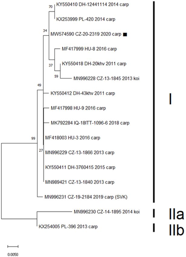
Pikula et al.                                                                                             Carp Edema Virus Infection Pathophysiology

INTRODUCTION                                                                neurological abnormalities associated with loss of electrolytes,
                                                                            and (4) dehydration. An improved understanding of how CEV
Mass mortalities associated with viral diseases challenge the               infection alters the homeostatic maintenance of key physiological
economy of common carp (Cyprinus carpio) aquaculture (1–                    parameters will have positive ramifications for fish medicine as
3). Over the last decade, carp edema virus (CEV) infection,                 regards other infections causing gill pathology, such as the koi
also termed koi sleepy disease, has emerged as a serious disease            herpes virus (CyHV-3).
threatening the European common carp aquaculture and koi
trade (1, 4, 5). CEV infections, both in common and koi carps,
                                                                            MATERIALS AND METHODS
are currently known from many European countries (1, 4–9).
Outbreaks of CEV in the Czech Republic date back to 2013.                   Outbreak Description
Since then, CEV genogroups I and II have been detected in                   In mid-December 2020, we investigated a case of high mortality
this Central European country based on sequencing the 357-bp                in market-size common carp (2,200–2,500 g) kept at a storage
nucleotide encoding the P4a protein (10). Interestingly, common             pond at the lowland Napajedla fish farm (Czech Republic). Based
carps purchased in the Czech Republic for restocking a pond                 on available records, it became clear that the first clinical signs
played probably a role in the CEV outbreak in Austria in 2014               of CEV were recognized about 1 month after translocation of
(5). While rapid and accurate diagnostic assays are imperative for          about 1,500 kg of fish from a highland pond. No disease had been
effective surveillance and control of such infectious diseases (11),        reported from either site at the time of fish movement. According
advances in recognition of the pathophysiological mechanisms                to the respective fish farm managers, while water temperatures
underlying clinical infection signs and mortality improve our               ranged from 10 to 12◦ C at the highland pond on the date of
understanding of disease progression.                                       translocation (16 October 2020), temperatures at the lowland
    CEV is characterized by lethargic behavior, fish lying on the           storage pond were around 17◦ C. Interestingly, the fish farmers
bottom of ponds, skin lesions around the mouth and at the base              also stressed that only translocated fish were showing signs of the
of fins, inflammation of the anus, enophthalmos, and swollen                disease, including extreme lethargy, shoaling close to the storage
and necrotic gills that induce hypoxia and respiratory distress.            pond water inflow, enophthalmos, and gill necrotic lesions.
Mortality may be as high as 100% and adverse effects are believed           At that point, the same storage pond contained specimens of
to increase with the extent of gill pathology. As CEV requires              clinically diseased carp and those showing no clinical signs of
permissive temperatures of between 15 and 25◦ C, outbreaks tend             the disease. Suspecting a viral infection, we captured 22 carp
to occur seasonally in spring and autumn, often affecting new               and took blood samples, following which they were euthanised
fish following the stress of transportation and restocking (1, 4, 5).       and subjected to dissection and virological examination. Aquatic
Outbreaks of infection may, however, occur also at lower water              environmental quality parameters were measured in situ at the
temperatures (6–10◦ C), resulting in a protracted course of the             time of carp capture (15 December 2020) using an HQ40D
disease (9, 12).                                                            portable multimeter (Hach, Loveland, Colorado, United States).
    Little is known about the blood profile responses of CEV-               The results obtained indicated a water temperature of 4.2◦ C,
infected carp as, up to now, only single specimens have                     oxygen content at 9.46 mg/L and a pH of 7.9. Considering the
been blood-sampled for examination (5) and/or only a few                    outcome of CEV infection at the respective farm, while diseased
blood profile parameters have been reported (13). Moreover,                 carps were sampled in mid-December 2020, farmers reported
comparison of blood profile parameters with normal ranges for               100% mortality of translocated fish by the end of January 2021.
the species (14) will be of limited value as such parameters
are strongly influenced by variations in environmental physical             Virus Identification, PCR, and Sequence
and chemical properties. Finally, where CEV-induced mortality               Analysis
approaches 100%, it can be difficult to obtain comparative data             Gill samples were processed for virus identification using
from healthy control fish (i.e., CEV-negative) sampled under the            standardized polymerase chain reaction (PCR) methods. DNA
same conditions at the same site.                                           was extracted from the gill tissue using the QIAamp DNA
    Recently, we had an unparalleled opportunity to examine                 Mini kit (Qiagen, Germany), as described in the manufacturer’s
CEV-infected and healthy carp in the same fish farm storage                 instructions. DNA extracts were tested (i) for presence of
pond under conditions of a natural CEV infection outbreak.                  koi herpes virus (CyHV-3), using nested PCR according to
To better understand CEV disease pathophysiology, we report                 Bercovier et al. (15), and (ii) for presence and viral load of
on blood profile parameters sampled during this outbreak                    CEV (measured as copies of the P4a gene per 250 ng of
event. It seems reasonable to hypothesize that manifestations of            extracted CEV DNA) using nested PCR and real-time PCR,
CEV infection in carp, including extreme lethargy, decreased                respectively, as described by Matras et al. (7). Primers used
locomotor activity, and enophthalmos, can best be explained                 in the present study were as follows: (1) CEV conventional
by the pathophysiological consequences of impaired respiratory              PCR: CEV-ForB: 5′ -ATG GAG TAT CCA AAG TAC TTA
and excretory functioning in the gills, leading to osmotic, ionic,          G-3′ ; CEV-RevJ: 5′ -CTC TTC ACT ATT GTG ACT TTG-3′ ;
and acid-base imbalance. Alongside extensive necrotic branchial             (2) CEV nested PCR: CEV-ForB Internal: 5′ -GTT ATC AAT
lesions in CEV-infected carp, we predict (1) insufficient oxygen            GAA ATT TGT GTA TTG-3′ ; CEV-RevJ Internal: 5′ -TAG CAA
delivery for tissue metabolism, (2) sedative/narcotic effects of            AGT ACT ACC TCA TCC-3′ ; (3) CEV qPCR: CEV-qFor1:
metabolic carbon dioxide elevation and respiratory acidosis, (3)            5′ -AGTTTTGTAKATTGTAGCATTTCC-3′ ;                 CEV-qRev1:

Frontiers in Veterinary Science | www.frontiersin.org                   2                                       May 2021 | Volume 8 | Article 679970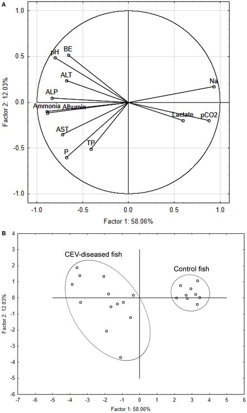
Pikula et al.                                                                                          Carp Edema Virus Infection Pathophysiology

5′ -GATTCCTCAAGGAGTTDCAGTAAA-3′ ;               Cev-qProbe1:             were undertaken using Statistica for Windows R 10 (StatSoft,
5′ -FAM-AGAGTTTGTTTCTTGCCATACAAACT-3′ ; (4) KHV                          Inc., USA).
qPCR: KHV-86f: 5′ -GACGCCGGAGACCTTGTG-3′ ; KHV-
163r: 5′ -CGGGTTCTTATTTTTGTCCTTGTT-3′ ; KHV-109p:
5′ -FAM-CTTCCTCTGCTCGGCGAGCACG-3′ . PCR products                         RESULTS
were visualized using the Fast Gene R GelPic Imaging System
                                                                         In total, 13 of the 22 gill samples (59%) proved positive for
(GmbH, Germany) following electrophoresis in 2% agarose gel
                                                                         CEV. Viral loads ranged from 442,228 to 3,036,744 copies of
stained by 1× Gel Red (Biotium, USA). PCR products were
                                                                         viral DNA per 250 ng of extracted DNA. Sanger sequencing of
sequenced by Sanger direct sequencing at a commercial company
                                                                         the product from nested PCR resulted in a 433-bp nucleotide
(SEQme, Czech Republic). The CEV sequences obtained were
                                                                         sequence identical for all 13 positive samples. This sequence was
compared with other 357-bp nucleotide sequences encoding
                                                                         uploaded to the GenBank database under the accession number
the P4a protein found in GenBank analyzed in a maximum-
                                                                         MW574590. Analysis of a 357-bp segment of the sequence
likelihood phylogenic tree using the Jukes–Cantor model in
                                                                         assigned the isolate to CEV genogroup I, with five published
MEGA v. 6 Software (16), with the robustness of the tree being
                                                                         sequences (originating from Czechia, Germany, and Poland)
tested using 1,000 bootstrap replicates.
                                                                         showing only a single nucleotide difference (Figure 1). None of
                                                                         the samples proved positive for koi herpes virus.
Blood Sampling and Measurement                                              Based on the above virological findings, along with presence
The caudalis vessels of 22 carp were punctured with an
                                                                         of branchial lesions ranging from excessive mucus and gill tissue
18G needle and blood drawn into a 5 mL heparinised
                                                                         swelling to extensive necroses with secondary fungal overgrowth
polypropylene syringe. Immediately after collection, an i-STAT
                                                                         (Figure 2), enophthalmos, skin erosions, and/or increased mucus
portable clinical analyser for veterinary use (EC8+ diagnostic
                                                                         production on body surface on gross examination, blood profile
cartridge based on electrochemical sensing technologies; Abaxis,
                                                                         parameters for individual carp were assigned to either CEV-
USA) was used to measure the following blood profile
                                                                         infected or the uninfected control group to allow comparative
parameters: sodium (Na, mmol/L), potassium (K, mmol/L),
                                                                         analysis. Of the 30 blood profile variables measured, 19
chloride (Cl, mmol/L), total dissolved carbon dioxide (tCO2 ,
                                                                         showed clear differences between CEV-infected and healthy carp
mmol/L), blood urea nitrogen (mmol/L), glucose (mmol/L),
                                                                         (Table 1). Likewise, there were clear differences in red blood
pH, partial dissolved carbon dioxide (pCO2 , kPa), bicarbonate
                                                                         cell associated hematology variables between the two groups.
(HCO3 , mmol/L), base excess (BE, mmol/L), and anion gap
                                                                         Both sodium and chloride electrolyte values were drastically
(AnGap, mmol/L). The remaining portion of each blood
                                                                         reduced in CEV-infected carp, while phosphorus levels had
sample was transported to the laboratories of the University
                                                                         increased. Concerning acid-base balance variables, pH, partial
of Veterinary Sciences Brno (Czech Republic), where it was
                                                                         dissolved carbon dioxide, base excess and anion gap were all
used to determine red blood cell count (T/L), hemoglobin
                                                                         significantly altered in CEV-infected carp, while both protein
(g/L), and hematocrit (L/L), to calculate hematological indices
                                                                         catabolism products ammonia and creatinine were elevated in
mean corpuscular volume (fL), mean corpuscular hemoglobin
                                                                         CEV-infected fish. Moreover, CEV infection was associated with
(pg) and mean corpuscular hemoglobin concentration (L/L),
                                                                         elevated levels of total protein, albumin, alkaline phosphatase,
and, following centrifugation to obtain plasma, Ca (mmol/L),
                                                                         alanine aminotransferase, and aspartate aminotransferase, and a
P (mmol/L), Mg (mmol/L), creatinine (µmol/L), ammonia
                                                                         decrease in lactate.
(µmol/L), triglycerides (mmol/L), lactate (mmol/L), albumin
                                                                            The multivariate response pattern for blood chemistry
(g/L), total protein (g/L), alkaline phosphatase (µkat/L), alanine
                                                                         variables showed a clear discrimination between CEV-infected
aminotransferase (µkat/L), aspartate aminotransferase (µkat/L),
                                                                         and control carp, with variable component weights revealing
and lactate dehydrogenase (µkat/L), spectrophotometrically
                                                                         separation between the two groups driven mainly by sodium,
using a Konelab 20i biochemical analyser and commercial test
                                                                         pH, partial dissolved carbon dioxide, ammonia and albumin
kits (Biovendor, Czech Republic) as described elsewhere (17).
                                                                         (Figure 3A). Specimen samples plotted onto a two-dimensional
                                                                         space according to principal components 1 and 2 (Figure 3B)
Data Analysis                                                            separated infected and healthy carp along the axis of the first
Using findings of virology and pathology to assign data to both
                                                                         principal component, which explained 58.06% of variation. In
groups, hematology and blood chemistry parameters measured
                                                                         comparison, the second principal component only explained
in CEV-infected and healthy control carp were compared using
                                                                         12.03% of variation.
the Kolmogorov–Smirnov and Shapiro–Wilks tests, one-way
analysis of variance (ANOVA) and the non-parametric Kruskal
Wallis, Tukey’s multiple comparison and Mann–Whitney U                   DISCUSSION
tests. Levels of significance were set at either p < 0.05 or
p < 0.01. Multivariate analysis of blood profile parameters              In the present study, we provide the first insight into the
was performed using principal components analysis (PCA) to               interaction between carp hosts and their pathogenic poxvirus
differentiate between CEV-infected fish and healthy control              under conditions of a natural CEV infection outbreak. We show
specimens. Original measured blood profile data used in this             that CEV infection resulted in severe metabolic disturbance, with
study are available as Supplementary Table 1. All analyses               significant differences from control healthy fish revealed by both

Frontiers in Veterinary Science | www.frontiersin.org                3                                       May 2021 | Volume 8 | Article 679970
Pikula et al. Carp Edema Virus Infection Pathophysiology FIGURE 1 | Phylogenetic analysis of a 357-bp nucleotide sequence encoding the CEV P4a core protein. Included was the sequence obtained in this study (MW574590, marked), previously published Czech (CZ) and Slovak (SVK) isolates, and CEV sequences obtained from GenBank with highest identity to the presented case from Germany (DH), Hungary (HU), Poland (PL), and Iraq (IQ). Three proposed CEV genogroups are marked as I, IIa, and IIb. The Maximum Likelihood Tree was constructed using a Jukes-Cantor model and the robustness of the tree was tested using 1,000 bootstrap replicates. The branch length is proportional to the number of substitutions per site. univariate and multivariate analyses. The blood profile of infected controls and were otherwise within normal ranges for common fish was characterized by increased red blood cell associated carp (14, 17). hematology variables, electrolyte and acid-base imbalance, Mortality amounted to 100% within
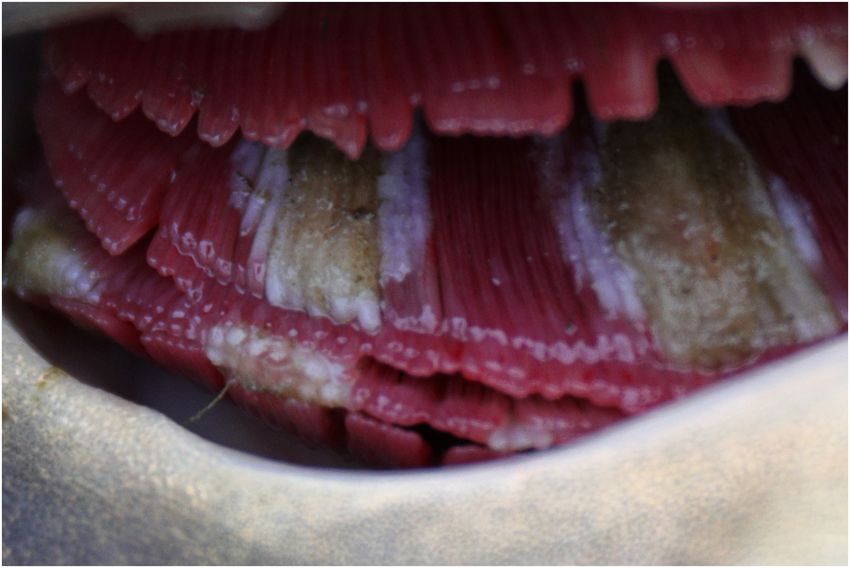
Pikula et al.                                                                                                        Carp Edema Virus Infection Pathophysiology

  FIGURE 2 | Typical gill pathology observed in carp assigned to the CEV-infected group.

numbers of CEV-specific DNA in the branchial tissue were wide                         decreased consciousness, and/or coma as a sequel to cerebral
ranging, they corresponded with published data (1, 18). Adamek                        edema. A decreased ability of fish to control extracellular ions fits
et al. (1) mentioned some discrepancy between clinical signs                          well with the observed pathology of gills (the target organ of CEV
and gill pathology based on experimental infections. Contrary                         replication), which included gill swelling, hyperplasia, clubbing
to this, the present study suggests that the viral load may be                        of lamellae and necrosis (1), as well as the corresponding clinical
a suitable proxy for estimation of health deterioration in CEV-                       signs (4, 5). The observed skin lesions may also contribute
diseased carps. There is no quarantine pond at the farm under                         to a loss of electrolytes from the body (24). Enophthalmos, a
study. However, placing translocated carps to quarantine would                        pathological sign observed in CEV-infected fish (4, 5), can be
not prevent the outbreak because it was most probably the                             attributed to hypotonic dehydration associated with a severe
stress of translocation plus the change in water temperature that                     loss of electrolytes (Na+ and Cl− ) from the body, combined
promoted the outbreak among fish already carrying the agent (5).                      with increased hematocrit, hemoglobin, red blood cell count,
   Mechanisms controlling fluid and acid-base balance,                                hyperproteinaemia, and hyperalbuminaemia (24). Given the
electrolyte concentration, glucose level, and disposal of                             reported absence of albumin in the common carp (25), the
metabolic waste products are of vital importance for ensuring                         finding of hyperalbuminaemia may seem to be controversial and
stable biochemical reactions and cellular processes (19). Thus,                       should be further pursued. Interestingly, elevation of high density
any loss in a fish’s ability for homeostatic maintenance of such                      lipoproteins might have been detected instead of carp albumin
key physiological parameters will be damaging. The gills of fishes                    (25). On the other hand, serum chemistry reference ranges of
combine respiratory and excretory functions with osmotic, ionic,                      albumin in Cyprinus carpio were published (14, 17, 26, 27).
and acid–base regulation (20–23). While the gills and intestines                          We observed considerable alterations in the blood acid-
are the principal organs for osmoregulation in fishes, the kidneys                    base balance in CEV-infected fish (24). Contrary to our
also contribute to fluid volume and electrolyte concentration,                        prediction of respiratory acidosis, higher pH and decreased
though to a lesser extent. Active uptake of ions across the                           pCO2 levels are indicative of respiratory alkalosis, probably due
intestines and gills is necessary to maintain hyperosmotic ionic                      to hyperventilation and/or suppressed metabolism and cellular
homeostasis in all freshwater bony fishes (24), while chloride                        respiration. Hyperammonaemia, as a combined metabolic
cells in the gills, rich in mitochondria, regulate the ionic and                      component of the acid-base balance, is probably responsible for
acid-base balance through exchange of Na+ /H+ /NH+          4 and                     the increase in base excess value observed in CEV-infected carp.
Cl− /HCO−   3 . Regarding   the  blood  electrolyte concentrations                    Elevation of the anion gap, a measure of electrical neutrality,
measured in our CEV-infected carp, severe hyponatremia                                corresponds well with electrolyte imbalance, as well as marked
and hypochloraemia indicate gill function disruption, while                           alterations in albumin and lactate (24).
hyperphosphatemia suggests alterations in kidney function (24).                           Ammonia (NH3 ), the major catabolite of proteins in
In general, manifestations of hyponatremia and hypochloraemia                         freshwater fishes (21, 28), is dissolved in body fluids and
will include lethargy, fatigue, loss of appetite, muscle weakness,                    the milieu of blood and/or is ionized to ammonium ions

Frontiers in Veterinary Science | www.frontiersin.org                             5                                        May 2021 | Volume 8 | Article 679970
Pikula et al.                                                                                                               Carp Edema Virus Infection Pathophysiology

TABLE 1 | Haematology and blood chemistry values measured in carp edema                       at both the cellular and organism levels. Furthermore, ionized
virus (CEV)-infected and control fish (Cyprinus carpio).                                      ammonium ions can replace potassium ions (K+ ) in ion
                                            CEV-infected fish          Control fish
                                                                                              transporters and disrupt central nervous system electrochemical
                                                                                              gradients. Indeed, passage of ammonia across the blood-brain
Variable                                           N = 13                  N=9                barrier is why the brain is the principal target of ammonia
                                                                                              toxicity in fish. According to Randall and Tsui (29), fish may
Haematocrit (L/L)                              0.36 ± 0.09**           0.24 ± 0.05
                                                                                              be threatened by both environmental exposure and endogenous
Hemoglobin (g/L)                               94.64 ± 7.93**         72.62 ± 15.56
                                                                                              ammonia. In this study, low blood ammonia levels in healthy
Red blood cell count (T/L)                      1.67 ± 0.49*           1.13 ± 0.31
                                                                                              control fish from the same storage pond excluded the possibility
Mean corpuscular volume (fL)                  208.39 ± 23.89         209.95 ± 31.35
                                                                                              of environmental ammonia contamination. Endogenous sources
Mean corpuscular hemoglobin (pg)               56.59 ± 5.81           64.03 ± 15.25
                                                                                              of ammonia in fish are associated with protein metabolism in
Mean corpuscular hemoglobin                    0.27 ± 0.003**          0.31 ± 0.03
                                                                                              the liver (amounting to 50–70% of total production), intestinal
concentration (L/L)
                                                                                              epithelial cells, kidneys, and skeletal muscles (28). Factors
Na (mmol/L)                                   103.32 ± 6.71**         143.46 ± 4.35
                                                                                              responsible for increased ammonia production may include
K (mmol/L)                                      4.37 ± 1.23            3.46 ± 0.74
                                                                                              postprandial metabolism, burst, and/or exhaustive exercise,
Cl (mmol/L)                                   68.15 ± 17.82**         111.06 ± 5.47
                                                                                              starvation and stress (28, 30). Considering the lethargic behavior
Ca (mmol/L)                                     2.71 ± 0.66            2.29 ± 0.19
                                                                                              of our CEV-infected carp, which were fasted over the autumn
P (mmol/L)                                     2.98 ± 0.73**           1.50 ± 0.22
                                                                                              period, starvation- and stress-induced catabolism, combined
Mg (mmol/L)                                     1.19 ± 0.10            1.23 ± 0.05
                                                                                              with the compromised excretory function of gills, would appear
Blood urea nitrogen (mmol/L)                    1.19 ± 0.23            0.99 ± 0.00
                                                                                              to be implicated in the elevated ammonia levels observed in this
Creatinine (µmol/L)                           30.00 ± 11.28**          13.71 ± 1.92
                                                                                              study. Moreover, ammonia toxicity can further deteriorate the
Ammonia (µmol/L)                            592.24 ± 177.84**        231.60 ± 65.37
                                                                                              condition of gills as both CEV and ammonia autointoxication
Glucose (mmol/L)                                1.56 ± 1.54            2.53 ± 0.74
                                                                                              result in gill necrosis (4, 5, 9, 28). Ammonia accumulation in fish
Triglycerides (mmol/L)                          1.11 ± 0.51            1.08 ± 0.18            blood, muscles, and brain, along with subsequent neuromotor
Lactate (mmol/L)                               5.76 ± 4.12**           11.01 ± 2.58           dysfunction, are known to manifest as impaired locomotor
pH                                             7.43 ± 0.25**           7.13 ± 0.05            performance (30), which corresponds with the clinical signs of
pCO2 (kPa)                                     3.58 ± 0.90**           5.78 ± 0.82            lethargy and inactivity observed in CEV-infected carp (4, 5).
tCO2 (mmol/L)                                  20.34 ± 8.17            15.66 ± 1.58           Importantly, ammonia can also be considered a respiratory gas
HCO3 (mmol/L)                                  19.42 ± 8.15            14.28 ± 1.31           produced in ammoniotelic fishes at a rate equalling 10 to 20%
Base Excess (mmol/L)                          −4.84 ± 11.96*            −15 ± 1.65            of CO2 production or O2 uptake (22), stimulating ventilation by
Anion Gap (mmol/L)                             15.25 ± 2.62*           8.66 ± 3.35            an increase in the ventilatory stroke volume, rather than the rate,
Albumin (g/L)                                  15.18 ± 1.98**          7.68 ± 2.39            in order to allow for ammonia excretion through the branchial
Total protein (g/L)                           40.89 ± 10.99*           31.54 ± 4.08           surface (31).
Alkaline phosphatase (µkat/L)                  1.07 ± 0.43**           0.30 ± 0.18                Levels of blood urea nitrogen, another catabolite excreted
Alanine aminotransferase (µkat/L)              0.92 ± 0.73**           0.10 ± 0.05            through fish gills and an ammonia detoxification product, were
Aspartate aminotransferase (µkat/L)            3.89 ± 1.85**           1.30 ± 0.91            comparably low in both CEV-infected and healthy control carp,
Lactate dehydrogenase (µkat/L)                 10.41 ± 6.21            10.54 ± 2.27           again suggesting starvation. On the other hand, elevated levels of
                                                                                              creatinine, which is mainly excreted through the kidney, suggests
Values represent mean ± standard deviation, * p< 0.05, ** p< 0.01 when comparing CEV-
infected fish against the healthy control. While non-parametric testing was performed
                                                                                              alterations in renal functioning in CEV-infected carp. Elevated
for analysis of anion gap, alkaline phosphatase, aspartate aminotransferase and lactate       levels for the enzymes ALP, ALT, and AST supports the finding
dehydrogenase, parametric tests were used in the rest of variables.                           of systemic responses seen in multiple organs, including the
                                                                                              hepatopancreas, spleen, heart, and kidney, in addition to the
                                                                                              gills (5).
                                                                                                  Considering energy metabolism, no stress response was
(NH+ 4 ). In addition to temperature, the ratio of NH3 /NH4
                                                              +
                                                                                              observable as there was only a non-significant reduction observed
will depend on pH, with a higher pH favoring higher NH3                                       in glucose levels (32, 33). On the other hand, the triglyceride
concentrations and increased toxicity due to the ease with                                    levels observed suggest no alteration to the lipid metabolism.
which it can cross biological membranes. In line with this,                                   Glycolysis, a key metabolic pathway supplying organisms with
the hyperammonaemia combined with alkalosis documented                                        energy, may function both aerobically and anaerobically. In the
in our CEV-infected carp is likely to exert increased adverse                                 absence of oxygen, it stops at pyruvate, which is reduced to
effects due to the higher proportion of toxic unionized ammonia                               lactate by lactate dehydrogenase (34). Elevation of lactate in
circulating in the blood. As reviewed in Ip and Chew (21),                                    blood indicates that the oxygen supply to tissues (e.g., white
ammonia toxicity affects many cellular processes. It is known                                 muscles in fish) is lower than that required for aerobic glycolysis
for its stimulatory effects on glycolysis through activation of                               (32). However, the decreased lactate levels observed in our
phosphofructokinase I in cytosolic fluid, for interference with                               study indicate that CEV-infected fish were not exposed to
energy metabolism related to impairment of the tricarboxylic                                  hypoxic stress (33), suggesting that, aside from gill pathology,
acid cycle in mitochondria and for disruption of ionic balance                                oxygen demand was significantly lowered due to the carp’s

Frontiers in Veterinary Science | www.frontiersin.org                                     6                                       May 2021 | Volume 8 | Article 679970
Pikula et al. Carp Edema Virus Infection Pathophysiology FIGURE 3 | Principal component analysis (PCA) discriminating between carp edema virus (CEV)-infected and control fish (Cyprinus carpio). Component weight (A) and component score (B) plots are based on multiple blood chemistry variables. ALP, alkaline phosphatase; ALT, alanine aminotransferase; AST, aspartate aminotransferase; BE, base excess; Na, sodium; P, phosphorus; pCO2 , partial dissolved carbon dioxide; TP, total protein. Frontiers in Veterinary Science | www.frontiersin.org 7 May 2021 | Volume 8 | Article 679970
Pikula et al.                                                                                                                 Carp Edema Virus Infection Pathophysiology

lethargic inactivity and disrupted hypometabolic condition.                              ETHICS STATEMENT
Unfortunately, blood oxygen saturation was not measured in the
present study. In contrast to our findings indicating an absence                         The animal study was reviewed and approved by The Ministry of
of hypoxic stress, Lewisch et al. (5) reported clinical signs of                         Agriculture of the Czech Republic (permission No. MZe 1842).
dyspnoea and hypoxia in association with CEV, meaning that                               Written informed consent was obtained from the owners for the
carp clustered at the inflow and utilized air-breathing at the                           participation of their animals in this study.
water’s surface.
    While infectious agents are known to elicit disruption of blood                      AUTHOR CONTRIBUTIONS
homeostasis, multiple physical, chemical and biological stressors
may often be combined (35–39), making it difficult to disentangle                        JP and MP conceived the study and analyzed the data and wrote
the link between infection-associated pathophysiological                                 the first draft of the manuscript. JM and MP acquired funding. LP,
mechanisms of morbidity and mortality (40, 41). Interestingly,                           IP, HM, IM, HB, JB, and MB performed field and/or laboratory
loss of osmoregulatory functioning in the gills, gut and kidneys                         investigations. All authors contributed critical comments on
are thought to contribute to mortality in carp affected with                             manuscript drafts and gave final approval for its publication.
another viral agent, the koi herpes virus (34), which none of
our samples was positive for. However, blood chemistry data for                          FUNDING
carp infected with koi herpes virus are limited (42), preventing
detailed comparison.                                                                     This work was supported by the Ministry of Education, Youth,
    To conclude, our blood profile study increases our                                   and Sports through the projects CR PROFISH (Sustainable
understanding of CEV disease in carp, explaining how impaired                            production of healthy fish under various aquaculture systems)
respiratory and excretory functioning of gills, together with                            and by the Ministry of Agriculture of the Czech Republic through
osmotic, ionic and acid-base disruption, exert complex adverse                           the project (New viral diseases of common carp—diagnosis and
effects on the ability of fish to maintain homeostasis and support                       prevention) (No. NAZV QK1710114).
essential bodily functions. Further research should aim to
quantify levels of infection intensity, the extent of gill damage                        ACKNOWLEDGMENTS
and neurological abnormalities associated with neurotoxicity of
ammonia as well as factors of fish susceptibility and conditions                         We are grateful to Dr. Kevin Roche for his correction and
promoting disease manifestation.                                                         improvement of the English text.

DATA AVAILABILITY STATEMENT                                                              SUPPLEMENTARY MATERIAL
The original contributions generated for the study are included                          The Supplementary Material for this article can be found
in the article/Supplementary Material, further inquiries can be                          online at: https://www.frontiersin.org/articles/10.3389/fvets.
directed to the corresponding author/s.                                                  2021.679970/full#supplementary-material

REFERENCES                                                                                7. Matras M, Borzym E, Stone D, Way K, Stachnik M, Maj-Paluch J, et al.
                                                                                             Carp edema virus in Polish aquaculture - evidence of significant sequence
 1. Adamek M, Oschilewski A, Wohlsein P, Jung-Schroers V, Dawson A, Gela D,                  divergence and a new lineage in common carp Cyprinus carpio (L.). J Fish
    et al. Experimental infections of different carp strains with the carp edema             Dis. (2017) 40:319–25. doi: 10.1111/jfd.12518
    virus (CEV) give insights into the infection biology of the virus and indicate        8. Radosavljevic V, Adamek M, Milicevic V, Maksimovic-Zoric J, Steinhagen
    possible solutions to problems caused by koi sleepy disease (KSD) in carp                D. Occurrence of two novel viral pathogens (CEV and CyHV-2) affecting
    aquaculture. Vet Res. (2017) 48:12. doi: 10.1186/s13567-017-0416-7                       Serbian cyprinid aquaculture and ichthyofauna. J Fish Dis. (2018) 41:851–4.
 2. OIE - World Organisation for Animal Health. Manual of Diagnostic Tests for               doi: 10.1111/jfd.12789
    Aquatic Animals. (2019). Available online at: https://www.oie.int/standard-           9. Way K, Haenen O, Stone D, Adamek M Bergmann SM, Bigarre L, et al.
    setting/aquatic-manual/access-online/ (accessed February 21, 2021).                      Emergence of carp edema virus (CEV) and its significance to European
 3. Gilad O, Yun S, Zagmutt-Vergara FJ, Leutenegger CM, Bercovier H, Hedrick                 common carp and koi Cyprinus carpio. Dis Aquat Org. (2017) 126:155–66.
    RP. Concentrations of a Koi herpesvirus (KHV) in tissues of experimentally-              doi: 10.3354/dao03164
    infected Cyprinus carpio koi as assessed by real-time TaqMan PCR. Dis Aquat          10. Matějíčková K, Pojezdal L, Pokorová D, Reschová S, Piačková V, Palíková M,
    Org. (2004) 60:179–87. doi: 10.3354/dao060179                                            et al. Carp oedema virus disease outbreaks in Czech and Slovak aquaculture. J
 4. Jung-Schroers V, Adamek M, Teitge F, Hellmann J, Bergmann SM, Schutze H,                 Fish Dis. (2020) 43:971–8. doi: 10.1111/jfd.13179
    et al. Another potential carp killer?: Carp Edema Virus disease in Germany.          11. Adams A, Thompson KD. Recent applications of biotechnology to novel
    BMC Vet Res. (2015) 11:114. doi: 10.1186/s12917-015-0424-7                               diagnostics for aquatic animals. Rev Sci Tech Off Int Epizoot. (2008) 27:197–
 5. Lewisch E, Gorgoglione B, Way K, El-Matbouli M. Carp Edema Virus/Koi                     209. doi: 10.20506/rst.27.1.1792
    Sleepy Disease: an emerging disease in Central-East Europe. Transbound               12. Pragyan D, Bajpai V, Suman K, Mohanty J, Sahoo PK. A review of current
    Emerg Dis. (2015) 62:6–12. doi: 10.1111/tbed.12293                                       understanding on carp edema virus (CEV): a threatful entity in disguise. Int J
 6. Adamek M, Baska F, Vincze B, Steinhagen D. Carp edema virus from three                   Fish Aquat Stud. (2019) 7:87–93.
    genogroups is present in common carp in Hungary. J Fish Dis. (2018) 41:463–          13. Seno R, Hata N, Oyamatsu T, Fukuda H. Curative effect of 0.5% salt
    8. doi: 10.1111/jfd.12744                                                                water treatment on carp, Cyprinus carpio, infected with Carp Edema Virus

Frontiers in Veterinary Science | www.frontiersin.org                                8                                               May 2021 | Volume 8 | Article 679970
Pikula et al. Carp Edema Virus Infection Pathophysiology
(CEV) results mainly from reviving the physiological condition of the host. 30. McKenzie DJ, Shingles A, Claireaux G, Domenici P. Sublethal
Suisanzoshoku. (2003) 51:123–4. doi: 10.11233/aquaculturesci1953.51.123 concentrations of ammonia impair performance of the teleost fast-start
14. Groff JM, Zinkl JG. Hematology and clinical chemistry of cyprinid fish: escape response. Physiol Biochem Zool. (2009) 82:353–62. doi: 10.1086/
common carp and goldfish. Vet Clin North Am Exot Anim Pract. (1999) 590218
2:741–76. doi: 10.1016/S1094-9194(17)30120-2 31. Wright PA, Wood CM. Seven things fish know about ammonia and we
15. Bercovier H, Fishman Y, Nahary R, Sinai S, Zlotkin A, Eyngor M, et al. don’t. Respir Physiol Neurobiol. (2012) 184:231–40. doi: 10.1016/j.resp.2012.
Cloning of the koi herpesvirus (KHV) gene encoding thymidine kinase and 07.003
its use for a highly sensitive PCR based diagnosis. BMC Microbiol. (2005) 5:13. 32. Pottinger TG. Changes in blood cortisol, glucose and lactate in carp retained
doi: 10.1186/1471-2180-5-13 in anglers’ keepnets. J Fish Biol. (1998) 53:728–42. doi: 10.1006/jfbi.1998.0737
16. Tamura K, Stecher G, Peterson D, Filipsky A, Kumar S. MEGA6: molecular 33. Wells RMG, Pankhurst NW. Evaluation of simple instruments for
evolutionary genetics analysis version 6.0. Mol Biol Evol. (2013) 30:2725–9. the measurement of blood glucose and lactate, and plasma protein
doi: 10.1093/molbev/mst197 as stress indicators in fish. J World Aquacult Soc. (1999) 30:276–84.
17. Svobodová Z, Vykusová B, Modrá H, Jarkovský J, Smutná M. Haematological doi: 10.1111/j.1749-7345.1999.tb00876.x
and biochemical profile of harvest-size carp during harvest and post-harvest 34. Dando P. Lactate metabolism in fish. J Mar Biol Assoc UK. (1969) 49:209–23.
storage. Aquac Res. (2006) 37:959–65. doi: 10.1111/j.1365-2109.2006.01511.x doi: 10.1017/S002531540004652X
18. Adamek M, Jung-Schroers V, Hellmann J, Teitge F, Bergmann SM, Runge 35. Rodrigues RA, Silva ALD, Siqueira MS, Pilarski F, Leal CRB, Kuibida KV,
M, et al. Concentration of carp edema virus (CEV) DNA in koi tissues et al. Hematological, biochemical, and histopathological responses in sorubim
affected by koi sleepy disease (KSD). Dis Aquat Org. (2016) 119:245–51. Pseudoplatystoma spp. experimentally infected with Lactococcus garvieae.
doi: 10.3354/dao02994 Aquacult Int. (2020) 28:1907–23. doi: 10.1007/s10499-020-00566-5
19. Woods HA, Wilson JK. An information hypothesis for the 36. Rehulka J. Haematological analyses in rainbow trout Oncorhynchus mykiss
evolution of homeostasis. Trends Ecol Evol. (2013) 28:283–9. affected by viral haemorrhagic septicaemia (VHS). Dis Aquat Organ. (2003)
doi: 10.1016/j.tree.2012.10.021 56:185–93. doi: 10.3354/dao056185
20. Gilmour KM. New insights into the many functions of carbonic 37. Rehulka J, Minarík B. Blood parameters in brook trout Salvelinus fontinalis
anhydrase in fish gills. Respir Physiol Neurobiol. (2012) 184:223–30. (Mitchill, 1815), affected by columnaris disease. Aquac Res. (2007) 38:1182–
doi: 10.1016/j.resp.2012.06.001 97. doi: 10.1111/j.1365-2109.2007.01786.x
21. Ip YA, Chew SF. Ammonia production, excretion, toxicity, and defense in fish: 38. Shah SL. Impairment in the haematological parameters of tench (Tinca
a review. Front Physiol. (2010) 1:134. doi: 10.3389/fphys.2010.00134 tinca) infected by Saprolegnia spp. Turk J Vet Anim Sci. (2010) 34:313–8.
22. Randall DJ, Ip YK. Ammonia as a respiratory gas in water and doi: 10.3906/vet-0706-4
air-breathing fishes. Respir Physiol Neuro. (2006) 154:216–25. 39. Witeska M. Anemia in teleost fishes. Bull Eur Assoc Fish Pathol. (2015)
doi: 10.1016/j.resp.2006.04.003 35:148–60.
23. Sardella BA, Brauner CJ. The osmo-respiratory compromise in fish: the effects 40. Bass D, Stentiford GD, Wang HC., Koskella B, Tyler CR. The pathobiome
of physiological state and the environment. In: Fernandes MN, Rantin FT, in animal and plant diseases. Trends Ecol Evol. (2019) 34:996–1008.
Glass ML, Kapoor BG, editors. Fish Respiration and Environment. Enfield, NH: doi: 10.1016/j.tree.2019.07.012
Science Publishers (2007). p. 147–65. 41. Gorgoglione B, Bailey C, Fast MD, Bass D, Saraiva M, Adamek M, et al. Co-
24. Campbell TW. Clinical chemistry of fish and amphibians. In: Thrall MA, infections and multiple stressors in fish: EAFP 19th International Conference
Weiser G, Allison RW, Campbell TW, editors. Veterinary Hematology and on Diseases of Fish and Shellfish special edition workshop report. Bull Eur
Clinical Chemistry, 2nd edn. Hoboken, NJ: John Wiley & Sons (2012). p. 607– Assoc Fish Pathol. (2020) 40:4–19.
14. 42. Negenborn J, van der Marel MC, Ganter M, Steinhagen D. Cyprinid
25. De Smet H, Blust R, Moens L. Absence of albumin in the plasma herpesvirus-3 (CyHV-3) disturbs osmotic balance in carp (Cyprinus carpio
of the common carp Cyprinus carpio: binding of fatty acids to L.) - a potential cause of mortality. Vet Microbiol. (2015) 177:280–8.
high density lipoprotein. Fish Physiol Biochem. (1998) 19:71–81. doi: 10.1016/j.vetmic.2015.03.018
doi: 10.1023/A:1007734127146
26. Nagano H, Hosaka K, Shukuya R. Comparative biochemistry of Conflict of Interest: The authors declare that the research was conducted in the
serum albumin. A serum albumin-like protein from carp, Cyprinus absence of any commercial or financial relationships that could be construed as a
carpio. Comp Biochem Physiol B-Biochem Mol Biol. (1975) 50:573–8. potential conflict of interest.
doi: 10.1016/0305-0491(75)90092-9
27. Tripathi N, Latimer K, Lewis T, Burnley VV. Biochemical reference Copyright © 2021 Pikula, Pojezdal, Papezikova, Minarova, Mikulikova,
intervals for koi (Cyprinus carpio). Comp Clin Path. (2003) 12:160–5. Bandouchova, Blahova, Bednarska, Mares and Palikova. This is an open-access
doi: 10.1007/s00580-003-0495-x article distributed under the terms of the Creative Commons Attribution License (CC
28. Smutná M, Vorlová L, Svobodová Z. Pathobiochemistry of ammonia in the BY). The use, distribution or reproduction in other forums is permitted, provided
internal environment of fish (Review). Acta Vet BRNO. (2002) 71:169–81. the original author(s) and the copyright owner(s) are credited and that the original
doi: 10.2754/avb200271020169 publication in this journal is cited, in accordance with accepted academic practice.
29. Randall DJ, Tsui TKN. Ammonia toxicity in fish. Mar Pollut Bull. (2002) No use, distribution or reproduction is permitted which does not comply with these
45:17–23. doi: 10.1016/S0025-326X(02)00227-8 terms.
Frontiers in Veterinary Science | www.frontiersin.org 9 May 2021 | Volume 8 | Article 679970You can also read