Brain Reconstruction Across the Fish-Tetrapod Transition; Insights From Modern Amphibians
←
→
Page content transcription
If your browser does not render page correctly, please read the page content below
ORIGINAL RESEARCH
                                                                                                                                              published: 19 March 2021
                                                                                                                                        doi: 10.3389/fevo.2021.640345

                                            Brain Reconstruction Across the
                                            Fish-Tetrapod Transition; Insights
                                            From Modern Amphibians
                                            Alice M. Clement 1* , Corinne L. Mensforth 1 , T. J. Challands 2 , Shaun P. Collin 3 and
                                            John A. Long 1
                                            1
                                             College of Science and Engineering, Flinders University, Adelaide, SA, Australia, 2 School of Geosciences, Grant Institute,
                                            University of Edinburgh, Edinburgh, United Kingdom, 3 School of Life Sciences, La Trobe University, Bundoora, VIC, Australia

                                            The fish-tetrapod transition (which incorporates the related fin-limb and water-land
                                            transitions) is celebrated as one of the most important junctions in vertebrate
                                            evolution. Sarcopterygian fishes (the “lobe-fins”) are today represented by lungfishes
                                            and coelacanths, but during the Paleozoic they were much more diverse. It was some
                                            of these sarcopterygians, a lineage of the tetrapodomorph fishes, that gave rise to
                                            tetrapods (terrestrial vertebrates with limbs bearing digits). This spectacular leap took
                          Edited by:        place during the Devonian Period. Due to the nature of preservation, it is the hard parts
                   Ingmar Werneburg,
     University of Tübingen, Germany
                                            of an animal’s body that are most likely to fossilize, while soft tissues such as muscular
                         Reviewed by:
                                            and brain tissues, typically fail to do so. Thus, our understanding of the adaptations
                               Min Zhu,     of the hard skeletal structures of vertebrates is considerably greater than that of the
   Institute of Vertebrate Paleontology     soft tissue systems. Fortunately, the braincases of early vertebrates are often ossified
      and Paleoanthropology, Chinese
          Academy of Sciences, China        and thereby have the potential to provide detailed morphological information. However,
                                Jing Lu,    the correspondence between brain and endocast (an internal mold of the cavity)
   Institute of Vertebrate Paleontology
      and Paleoanthropology, Chinese
                                            has historically been considered poor in most “lower” vertebrates and consequently
          Academy of Sciences, China        neglected in such studies of brain evolution. Despite this, recent work documenting the
                  *Correspondence:          spatial relationship in extant basal sarcopterygians (coelacanth, lungfish, axolotl, and
                    Alice M. Clement        salamander) has highlighted that this is not uniformly the case. Herein, we quantify
        alice.clement@flinders.edu.au
                                            and illustrate the brain-endocast relationship in four additional extant basal tetrapod
                   Specialty section:       exemplars: neobatrachian anurans (frogs) Breviceps poweri and Ceratophrys ornata;
         This article was submitted to
                                            and gymnophionans (caecilians) Gegeneophis ramaswamii and Rhinatrema bivittatum.
                         Paleontology,
                a section of the journal    We show that anurans and caecilians appear to have brains that fill their endocasts to a
    Frontiers in Ecology and Evolution      similar degree to that of lungfishes and salamanders, but not coelacanth. Ceratophrys
       Received: 11 December 2020           has considerably lower correspondence between the brain and endocast in the olfactory
        Accepted: 25 February 2021
         Published: 19 March 2021
                                            tract and mesencephalic regions, while Breviceps has low correspondence along its
                               Citation:
                                            ventral endocranial margin. The brains of caecilians reflect their endocasts most closely
          Clement AM, Mensforth CL,         (vol. ∼70%). The telencephalon is tightly fitted within the endocast in all four taxa. Our
 Challands TJ, Collin SP and Long JA
                                            findings highlight the need to adequately assess the brain-endocast relationship in a
  (2021) Brain Reconstruction Across
 the Fish-Tetrapod Transition; Insights     broad range of vertebrates, in order to inform neural reconstructions of fossil taxa using
            From Modern Amphibians.         the Extant Phylogenetic Bracket approach and future studies of brain evolution.
           Front. Ecol. Evol. 9:640345.
      doi: 10.3389/fevo.2021.640345         Keywords: Sarcopterygii, Anura, Gymnophiona, neurobiology, brain, endocast, diceCT

Frontiers in Ecology and Evolution | www.frontiersin.org                           1                                            March 2021 | Volume 9 | Article 640345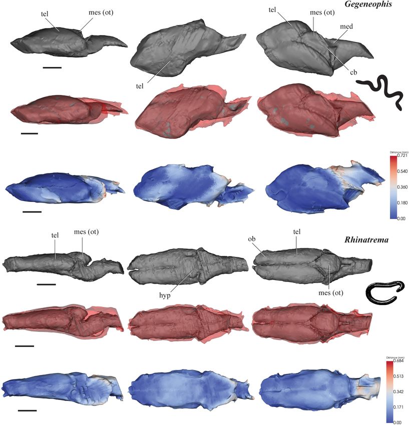
Clement et al.                                                                                      Fish-Tetrapod Transition Brain Reconstructions

INTRODUCTION                                                             (Frasnian) of East Greenland, have had only their ethmosphenoid
                                                                         regions modeled (Snitting, 2008; Lu et al., 2012). Work
Plants and invertebrates had already ventured onto land in               currently underway (Clement, pers. comm.) will soon add
the Silurian Period (419–443 million years ago), well before             a complete endocast of Cladarosymblema, a megalichthyinid
the transition onto land by the first vertebrates (back-boned            from the Carboniferous of Australia, to the list of those stem-
animals) during the Devonian Period (359–419 million years               tetrapods where the morphology of the endocast has been
ago). Nevertheless, the move from water to land by these animals,        examined.
known as the “fish-tetrapod transition,” is widely celebrated                The record of endocasts for basal tetrapods is similarly
as a highly significant evolutionary leap which eventually               depauperate, and includes the following: a partial endocast
gave rise to roughly half of all today’s vertebrate diversity            from the well-known Devonian tetrapod Ichthyostega (Clack
(including humans).                                                      et al., 2003); a cranial endocast from the early Carboniferous
    Due to the nature of fossilization, changes over time in             tetrapod Lethiscus (Pardo et al., 2017b); an otic capsule
the skeleton are much better documented and understood                   endocast from the Carboniferous temnospondyl, Dendrerpeton
(e.g., Cloutier et al., 2020) than the related soft tissues such         (Robinson et al., 2005); two Permian temnospondyls, Eryops
as muscle and brain. In particular, preserved brains are                 and Edops, had their endocasts described in good detail
exceedingly rare in the fossil record (although see Pradel               but were made using destructive techniques (Dempster, 1935;
et al., 2009), and where they are preserved they unlikely                Romer and Edinger, 1942); the Permian recumbirostran
reflect brain morphology during life due to desiccation. As              Brachydectes examined using non-destructive tomographic
such, the internal part of the skull that houses the brain, or           methods (Pardo and Anderson, 2016); and the Triassic
“endocast” has instead been used as a proxy for visualizing              stegocephalians Deinosuchus, Lyrocephalus, and Aphaneramma
the brain, the shape of its component parts and quantitatively           (Stensiö, 1963). This is, in part, due to many basal crown
assessing their size, in a field of study known as palaeoneurology       tetrapods possessing neurocrania that are poorly ossified
(Edinger, 1921).                                                         compared to many tetrapodomorph fishes.
    By analyzing cranial endocasts (the internal space within the            However, despite this pioneering work on these early
cranial cavity) of fossil taxa, certain inferences about behavior        vertebrates, most comparative neurological or palaeoneurological
can be drawn, guided by the principle of proper mass, whereby            studies continue to focus on birds and mammals, due to
“the mass of neural tissue controlling a particular function             their brains largely filling the cranial vault with a tight
is appropriate (proportionate) to the amount of information              correspondence with their braincases (Jerison, 1973). The
processing involved in performing the function” (Jerison, 1973,          brains of “lower” (anamniote) vertebrates generally incompletely
pg. 8). That is to say, the more important or acute a sense              fills the internal braincase space, and therefore have been
or behavior is to an animal, the more likely that brain region           considered of limited use for interpreting brain morphology.
will be relatively larger than would otherwise be expected,              Jerison (1973, pg. 121) claims that the brain of lungfishes
and consequently be reflected in the shape of the braincase              is only 10% as big as the endocast, and that it would
internally. Applying this principal to endocasts of fossils can be       thus be “difficult, although not impossible, to understand
problematic in those taxa which brains incompletely fill their           the brain’s anatomy from an endocranial cast.” Although a
endocasts, and the internal spatial relationship between brain and       large disparity was recorded between the lungfish brain and
endocast is not known.                                                   endocast, the disparity is even greater still in the coelacanth
    Despite the significance afforded to the fish-tetrapod               (Latimeria) with a brain just 1% of its endocranial volume
transition, there remain relatively few known cranial endocasts          (Millot and Anthony, 1965).
of tetrapodomorph fishes (stem-tetrapods). The best-known                    Recent work based on CT and magnetic resonance imaging
example is that of Eusthenopteron foordi, a tristichopterid              (MRI) scans has interrogated these assertions. Clement et al.
fish from the Late Devonian (mid-Frasnian) Escuminac                     (2015) revealed that the brain of a sub-adult Australian
Formation in Canada (Whiteaves, 1883). Using Sollas’ grinding            lungfish (Neoceratodus) fills the majority of its endocast
method common in embryology, palaeoichthyologists from                   (∼80%), while the adult coelacanth (Latimeria) was confirmed
the “Stockholm School” and elsewhere, serially ground several            to indeed have a brain one hundredth of the volume of
Paleozoic 3D-preserved early vertebrates to reconstruct their            its capacious cavity (Dutel et al., 2019). While it is well
endocasts, including that of Eusthenopteron foordi (Jarvik, 1980).       understood that ontogeny can have an effect on relative
Romer (1937) followed the same method to reconstruct the                 brain size, adult specimens from both extant lungfish families
endocast of the Permian megalichthyid Ectosteorhachis from               (Neoceratodontidae, Lepidosirenidae) were later found to have
the United States.                                                       a brain filling upward of 40% of their endocasts, even without
    More recently, advances in computed tomography (CT)                  correction for ostensible shrinkage (Challands et al., 2020), a
scanning technology have enabled several more endocasts                  proportion that is significantly higher than the reported 10%
to be investigated non-destructively. Gogonasus, from the                from Jerison (1973). In fact, more recent data (Yopak et al.,
Late Devonian (Frasnian) Gogo Formation in Australia is                  2010; Iwaniuk, 2017; Striedter and Northcutt, 2020) continues
known from complete, exceptionally-preserved 3D material                 to show a more nuanced picture with greater degree of overlap
(Holland, 2014), while Tungsenia from the Early Devonian                 in brain volume to body mass between the “lower” and “higher”
(Pragian) of China, and Spodichthys from the Late Devonian               vertebrates, and thus we consider that it can be reasonably

Frontiers in Ecology and Evolution | www.frontiersin.org             2                                    March 2021 | Volume 9 | Article 640345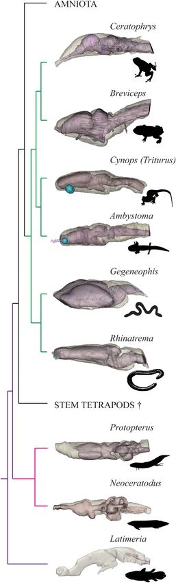
Clement et al.                                                                                              Fish-Tetrapod Transition Brain Reconstructions

expected that the relationship between brain and endocast is               the first tetrapods, we hereby aim to quantify the brain-endocast
similarly complicated.                                                     spatial relationship in four more extant lissamphibian basal
    The “heat-map” surface-surface distance measurement                    tetrapod exemplars: neobatrachian anurans Breviceps poweri,
method of quantifying the brain-endocast spatial relationship              and Ceratophrys ornata; and gymnophionans Gegeneophis
in the Australian lungfish developed by Clement et al. (2015),             ramaswamii and Rhinatrema bivittatum. We hypothesize that
was recently applied to six basal sarcopterygian taxa as a proxy           these taxa will have brains that fill their endocasts to a
to elucidate the changes that occurred over the fish-tetrapod              similar degree as that of lungfishes and salamanders, and
transition (Challands et al., 2020). Taxa investigated included            the region of closest fit will be around the forebrain. As
the coelacanth (Latimeria chalumnae), Australian lungfish                  more 3D morphological data such as these are created, brain
(Neoceratodus forsteri), African lungfish (Protopterus aethiopicus,        reconstruction models, such as that of Clement et al. (2016),
P. dolloi), the axolotl (Ambystoma mexicanum) and fire newt                can be further refined by incorporating data from the full extant
[Triturus (Cynops) pyrrhogaster].                                          phylogenetic bracket for any given fossil taxon.
    Challands et al. (2020) confirmed earlier work that showed the
coelacanth to be an outlier, with a miniscule brain lying within a
cavernous endocast (Dutel et al., 2019), which is in stark contrast        MATERIALS AND METHODS
to the brain of lungfishes and salamanders that occupy ∼40–
50% of the endocast. Those authors found that the Australian               Tomographic Data
and African lungfishes have quite a good fit between brain and             Diffusible     iodine-based     contrast-enhanced    computed
endocast internally, although the olfactory tract morphology in            tomography (diceCT) scan data of B. poweri (Power’s Rain
Protopterus spp. is not faithfully reflected in its endocast. In the       Frog), Media number: M13270-22958, was obtained under CC
salamanders, Triturus (Cynops) and Ambystoma, there is also                BY-NC-SA copyright from MorphoSource1 . The pixel size was
quite a tight fit except for the region where the trigeminal nerve         21 microns (µm).
complex exits the braincase. Overall, these findings suggest that             The C. ornata (Argentine Horned Frog) diceCT scan data
certain inferences can be made about the size of the brain from            was originally generated by Kleinteich and Gorb (2015), but later
its endocast in basal sarcopterygians, in particular with respect to       downloaded for this study from Dryad Digital Repository2 under
some brain regions, such as the telencephalon (forebrain).                 a CC0 1.0 Universal Public Domain Dedication license. The pixel
    Challands et al. (2020) suggested that the closeness of fit of         size was 27 µm.
different regions of the brain to the brain cavity may be influenced          The G. ramaswamii (tenmalai blind caecilian) and
by the mandibular musculature and so, to a certain extent,                 R. bivittatum (two-lined caecilian) diceCT scan data were
could be predicted from the skull morphology where musculature             downloaded from Duke University (see text footnote 1), with
can be reconstructed. Regions of lowest brain-endocast disparity           Media numbers: M71659-133946 and M33276-61864, and
occurred where bony reinforcement of the neurocranium lies                 obtained under -CC BY-NC-SA- attribution. Florida Museum
adjacent to mandibular musculature, whereas regions of highest             of Natural History, University of Florida provided access to
disparity were found in regions with lower reinforcement (e.g.,            these data, the collection of which was funded by oVert TCN;
where mandibular masticatory musculature mass was lower                    NSF DBI-1701714. The Gegeneophis dataset was subsequently
or absent). Such regions included where the trigeminal nerve               subsampled so the resultant pixel size was 25 µm, whereas the
complex exits the braincase and where endolymphatic sacs are               Rhinatrema data were subsampled to 32 µm.
present within the braincase.                                                 All data were segmented and rendered, and stereolithographs
    There are three living genera of lungfishes and one genus              (STL’s) produced, in MIMICS v.18–19 (Materialise, 1992–2020).
of coelacanth, which united represent the only extant piscine
sarcopterygians, and are therefore of particular relevance to
the fish-tetrapod transition. Conversely, on the other side                Surface Distance Analysis and
of the extant phylogenetic bracket are lissamphibians, such                Visualization
as frogs, salamanders and caecilians, which can also likely                The “closeness of fit” between 3D surface mesh STL’s of the brain
provide equally valuable insights into brain evolution of the              and the braincase’s internal “endocast” were analyzed using the
first tetrapods. For the purposes outlined in this paper, we               “Surface-to-Surface Distance Measurement Script” of Clement
have adopted the phylogeny of extant amphibians from Pyron                 et al. (2015) using Python, with script updates from Challands
and Wiens (2011) and do not herein attempt to comment                      et al. (2020). This custom script is publicly available from3 . The
of the controversy surrounding the origins of Lissamphibia                 resulting images visualize the surface-to-surface distance between
(Marjanovic and Laurin, 2007; Ruta and Coates, 2007). We                   STL’s using a “heat map” whereby cooler colors (blue) indicate
accept that the first tetrapods appeared during the Devonian, the          smaller distances (a closer fit), and warmer colors (red) indicate
tetrapod crown node arose during the Carboniferous, and the                greater distance.
extant amphibian orders most likely have their origins dating
back throughout the Permo-Triassic (Anderson et al., 2008;                 1
                                                                             www.morphosource.org
Clack, 2012; Pardo et al., 2017a).                                         2
                                                                             https://doi.org/10.5061/dryad.066mr
    Since we cannot assume a priori that salamanders are                   3
                                                                             https://www.researchgate.net/publication/345917984_Surface-to-surface_
necessarily the best representatives to interpret the condition of         distance_measurement_script

Frontiers in Ecology and Evolution | www.frontiersin.org               3                                           March 2021 | Volume 9 | Article 640345
Clement et al. Fish-Tetrapod Transition Brain Reconstructions
Comparison of volumes were calculated from 3D model The olfactory tracts (olf) extend a relatively long way into
volume values obtained from MIMICS. Figures were the cranial endocast (1.2 mm, 23% of the length of the
prepared and assembled in Adobe Photoshop and Illustrator, endocast). The olfactory tracts run parallel to each other and
GIMP and Inkscape. lie 0.3 mm apart. The olfactory bulbs (ob) are recognizable as a
separate bulge distinct from the telencephalon (tel) anteriorly,
which extends anteroventrally from the olfactory tracts as
RESULTS triangular-shaped structures. The telencephalic hemispheres
are small, paired, oval prominences. The brain narrows
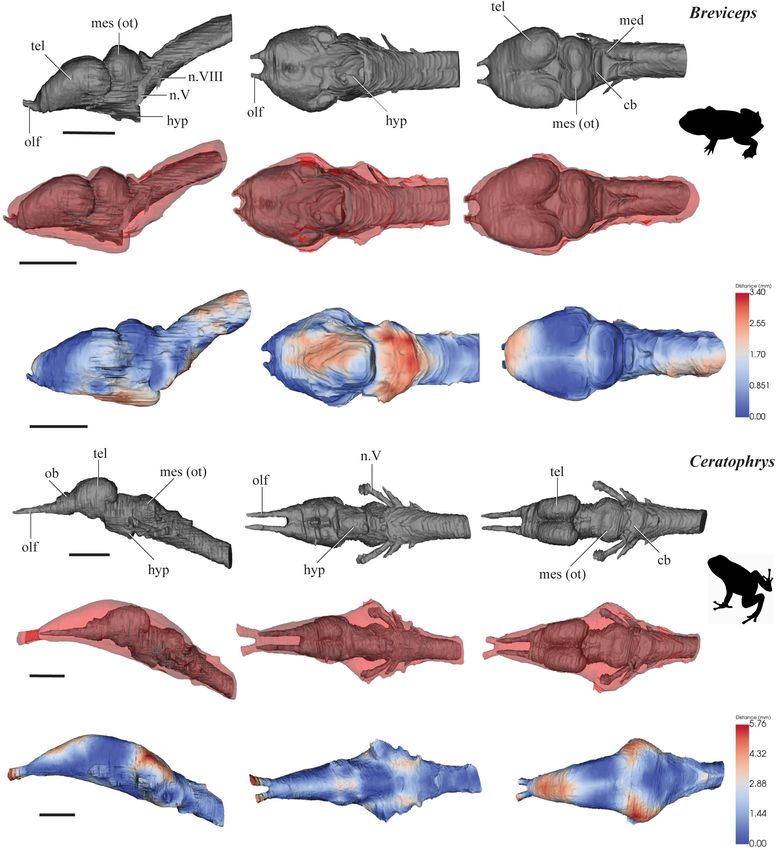
The segmented brains (gray) and the brain-endocast overlays considerably in the diencephalic region and the mid- and
(gray and red), and surface-surface distance “heat maps” are hindbrain angles ventrally downward some 45◦ from the
shown in Figures 1, 2. Brain volume as a percentage of endocast preceding forebrain. The hypophyseal region (hyp) is short
volume in mm3 is given in Table 1. and angled posteroventrally. The brain expands only slightly
in the midbrain (mes) to accommodate the optic tecta. The
Breviceps poweri brain then narrows toward the hindbrain (med) and the
The brain of B. poweri measures 3.0 mm long from the anterior anterior edge of the spinal cord. The trigeminal (n.V), facial
edge of the olfactory bulbs to the spinal cord, 1.3 mm across (n.VII), auditory (n.VIII), glossopharyngeal (n.IX) and vagus
the widest point of the brain, and 1.4 mm at the highest nerves (n.X) are visible from the scan data in the hindbrain
point of the brain (Figure 1A). The forebrain (consisting of region (Figure 1D).
the telencephalon and diencephalon) comprises 48% of the In contrast to the bulbous brain, the cranial endocast
total brain length, the midbrain (mesencephalon) 21%, and of C. ornata is smooth and lacking obvious protuberances
the hindbrain (metencephalon and myelencephalon) 31%. The (Figure 1E). It slowly widens from the olfactory epithelia to
cranial endocast of B. poweri measures 3.4 mm in length, 1.8 mm the midbrain. Following the course of the brain, the mid- and
at its widest point, and 1.6 mm dorsoventrally. hindbrain endocast regions also angles down ventrally to the
The forebrain of B. poweri is dominated by the large, anterior end of the spinal cord. Dorsally, the widest region
bulbous, oval-shaped telencephalic (tel) hemispheres which are accommodates the midbrain and the nerves emerging from the
slightly wider than they are long (Figure 1A). The boundary hindbrain (n.V-X), and then tapers toward the hindbrain ventral
between the olfactory bulbs and the telencephalon is difficult edge. The surface-surface distances between the cranial endocast
to discern. There is 0.1 mm of olfactory nerve visible between and brain of C. ornata are 0.2 mm between the brain and the endocast on the dorsal
the midbrain. The spinal cord is angled upward dorsally at surface, where the olfactory nerves extend into the endocast, and
about 45◦ from the rest of the brain. The trigeminal (n.V) and on the lateral surfaces near the trigeminal nerve’s entry to the
auditory (n.VIII) nerves are visible emerging from the hindbrain brain (Figure 1F).
from the scan data.
The cranial endocast of B. poweri, when viewed laterally, Gegeneophis ramaswamii
steadily increases in both dorsoventral and lateral diameter from The brain of G. ramaswamii measures 6.0 mm from the anterior
the olfactory bulbs through to the hindbrain and then rapidly edge of the olfactory bulbs to the spinal cord, is 3.4 mm wide
constricts near the spinal cord (Figures 1B,C). When viewing across the broadest point of the brain, and is 1.2 mm in height
the cranial endocast dorsally, the shape of forebrain is almost (Figure 2A). The forebrain contributes 61% of the total brain
circular, with an abrupt widening from the olfactory bulbs and length, while the midbrain and hindbrain contribute 17 and 22%,
then constricting behind the optic tecta. The endocast is close to respectively. The cranial endocast measures 6.2 mm in length,
the surface of the brain (0.17 mm over the on the scans and anatomical features are still recognizable
majority of the area (Figure 1C). and measurements have been approximated along the
compressed edge.
Ceratophrys ornata The forebrain of G. ramaswamii consists of large telencephalic
The brain of C. ornata (Figure 1D) measures 4.2 mm from the hemispheres (tel) that are much longer than they are wide
anterior edge of the olfactory bulbs to the spinal cord and is (Figure 2A). When viewed laterally, the height of the brain slowly
1.1 mm at its widest point and 1.2 mm at the highest point of increases from the anterior edge of the olfactory bulbs (ob) to
the brain. The forebrain comprises 43% of the total brain length, the midpoint of the telencephalon, and then slightly decreases
the midbrain 31%, and the hindbrain 26%. The cranial endocast in height toward the posterior edge of the hindbrain. The brain
of C. ornata measures 5.2 mm long, 2.1 mm at the widest point, narrows behind the midbrain (mesencephalon) before it expands
and 1.7 mm in height. as a conical structure in the hindbrain toward the spinal cord.
Frontiers in Ecology and Evolution | www.frontiersin.org 4 March 2021 | Volume 9 | Article 640345Clement et al. Fish-Tetrapod Transition Brain Reconstructions FIGURE 1 | 3D models of brains (gray), endocasts (red), and surface-surface distance heatmaps of two anurans in left lateral, ventral and dorsal views. (A–C) Breviceps; (D–F) Ceratophrys. Anterior to the left. cb, cerebellum; hyp, hypophysis; med, medulla oblongata; mes (ot), mesencephalon/optic tectum; tel, telencephalon; ob, olfactory bulb; olf, olfactory tract; n.V, trigeminal nerve complex; n.VIII, auditory nerve. Scale 10 mm. The optic tectum (mes) appears as a single protuberance rather slightly different degrees in the mid- and hindbrain region, than a paired structure. From the lateral view, the brain appears leaving distances of >0.3 mm in these regions. approximately linear. The cranial endocast of G. ramaswamii fits very tightly around Rhinatrema bivittatum the forebrain (Figures 2B,C), with
Clement et al.                                                                                                          Fish-Tetrapod Transition Brain Reconstructions

  FIGURE 2 | 3D models of brains (gray), endocasts (red), and surface-surface distance heatmaps of two caecilians in left lateral, ventral and dorsal views. (A–C)
  Gegeneophis; and (D–F) Rhinatrema. Anterior to the left. cb, cerebellum; med, medulla oblongata; mes (ot), mesencephalon/optic tectum; tel, telencephalon; ob,
  olfactory bulb. Scale 1 mm.

across the broadest point at the midbrain, and is 1.7 mm in                           separated by a small groove. The midbrain increases in diameter
height at the highest part of the brain (Figure 2D). The forebrain                    along all axes around the optic tecta, and the hindbrain tapers
contributes 63% of the total brain length, with the midbrain                          toward the spinal cord. From the lateral view, the brain appears
and hindbrain contributing 24 and 13%, respectively. The cranial                      approximately linear except for some dorsal expansion of the
endocast measures 5.7 mm in length, 2.3 mm in width and                               mesencephalic region (mes).
2.0 mm in height.                                                                        The cranial endocast of R. bivittatum closely mirrors the shape
   When observing the brain of R. bivittatum dorsally, the                            of the forebrain, with
Clement et al.                                                                                                       Fish-Tetrapod Transition Brain Reconstructions

TABLE 1 | Brain volume as a percentage of endocast volume in mm3 in extant piscine sarcopterygians and select Lissamphibian taxa.

Taxon                                  Source                               Brain vol. (mm3 )              Endocast vol. (mm3 )               Percentage filled%

Latimeria (adult)                      Challands et al., 2020                     1,973                          201,276                               1
Neoceratodus (juv.)                    Clement et al., 2015                        5.3                             6.8                                 78
Neoceratodus (adult)                   Challands et al., 2020                      630                            1,403                                45
Protopterus aethiopicus                Challands et al., 2020                      457                            1,097                                42
Protopterus dolloi                     Challands et al., 2020                      407                             857                                 47
Rhinatrema                             This study                                   9                              14                                  64
Gegeneophis                            This study                                   14                             18                                  78
Ambystoma                              Challands et al., 2020                       28                             74                                  38
Triturus (Cynops)                      Challands et al., 2020                       14                             34                                  41
Breviceps                              This study                                   26                             41                                  63
Ceratophrys                            This study                                   38                             77                                  49

Additional comparative data from Clement et al. (2015) and Challands et al. (2020) also included.

leaving distances >0.3 mm between the majority of these areas                           other taxa (e.g., still possessing a tail), and as one of the
(Figures 2E,F).                                                                         most basal caecilians it may thus reasonably be expected
                                                                                        to retain a more plesiomorphic condition of the brain and
                                                                                        braincase. Indeed, the telencephalic hemispheres of Rhinatrema
DISCUSSION                                                                              are elongate, remaining narrow along their whole length without
                                                                                        the expansion posterolaterally as in other more derived caecilians
The Extant Phylogenetic Bracket method (EPB), whereby two                               such as Ichthyophis (Nieuwenhuys et al., 1998) and Typhlonectes
or more extant outgroups are used to provide rigorous limits                            (Schmidt et al., 1996; Striedter and Northcutt, 2020). In this
of biological inference to a fossil taxon in question (Witmer,                          aspect the telencephalon actually resembles those of several
1995), is a valued approach to aid in the reconstruction of soft                        other sarcopterygians more so than other caecilians (Challands
tissues in extinct taxa. Although extant tetrapods are separated                        et al., 2020) and perhaps reflects the plesiomorphic condition.
by over 360 million years from the first terrestrial vertebrates,                       The braincase of a stem caecilian from the Jurassic Period,
the anatomy and morphology of their closest piscine relatives                           Eocaecilia, suggests that earliest members of this lineage retained
(sarcopterygian fishes) and basal tetrapods (lissamphibians)                            a more robust skull than is found in more recent, derived taxa
can provide invaluable insight into the condition of their                              (Maddin et al., 2012a).
long extinct kin.                                                                           In contrast, as a member of the Caeciliidae, Gegeneophis
    The brain-endocast relationships of piscine sarcopterygians                         is a more derived caecilian than Rhinatrema (Pyron and
and salamanders have been considered elsewhere (Clement                                 Wiens, 2011), and its brain shape, with posterolaterally-expanded
et al., 2015; Dutel et al., 2019; Challands et al., 2020) but are                       telencephalic hemispheres, is more typical for the group. Being a
complemented by the additions in this study investigating frogs                         blind animal, one might expect the optic tectum to be reduced
and caecilians to fulfill the EPB for Palaeozoic stem tetrapods                         in Gegeneophis but it appears to remain as prominent as in
(Figure 3). By quantifying the spatial relationship between                             other taxa (Figure 1). Nevertheless, the entire morphology of
the brain and internal cavity of the braincase (the endocast)                           caecilians is strongly modified in association with their ecology,
across sarcopterygians ranging from the coelacanth, both lungfish                       and the close correspondence internally between their brain and
families, and all three orders of extant amphibians, we have                            braincase likely reflects morphological constraints related to their
provided further information that can be used to identify a range                       fossorial lifestyle.
of brain volumes for early stem tetrapods (assuming the EPB                                 Compared to caecilians, anurans are a much more diverse
method is robust), and thus have established likely minimum                             and well-studied group. The brains of frogs and toads have
volumes for the brains of these animals.                                                been considered relatively similar across taxa, although there
    Caecilians are enigmatic and generally poorly understood                            is evidence of variation in brain morphology correlated with
animals although recent work has elucidated data on their                               seasonality (Luo et al., 2017), habitat and locomotion (Taylor
crania relating to its musculature (Kleinteich and Haas, 2007),                         et al., 1995; Manzano et al., 2017). The brains of both Breviceps
shape (Müller et al., 2009; Sherratt et al., 2014), and modularity                      and Ceratophrys are both a similar size in proportion to their
(Marshall et al., 2019). Work on braincase morphology (Maddin                           overall body size (brain length 20–22% of snout/vent length),
et al., 2012a,b) and the labyrinth system (Maddin and Anderson,                         but that of Breviceps fills its endocast to a greater degree than
2012; Maddin and Sherratt, 2014) has provided a breadth of new                          Ceratophrys, by almost 15%. While the olfactory canals are
information, but to the best of our knowledge, we are herein the                        short in Breviceps, those in Ceratophrys extend far anteriorly
first to produce cranial endocasts for the lineage.                                     before exiting the braincase, similar to the condition in some
    As the sister-group to all other caecilians (Pyron and                              lungfishes (Protopterus spp., see Challands et al., 2020, Figure 6).
Wiens, 2011), the Rhinatrematidae retain features lacking in                            Breviceps is a burrowing frog (Hofrichter, 2000), thus its close

Frontiers in Ecology and Evolution | www.frontiersin.org                           7                                        March 2021 | Volume 9 | Article 640345
Clement et al. Fish-Tetrapod Transition Brain Reconstructions
relationship between the brain and braincase may be related to
its highly fossorial lifestyle, as we have hypothesized for caecilians
also. However, of the six frog brain morphotypes shown in
Manzano et al. (2017), we note that the brain of Breviceps is
most similar in overall morphology to that of the “hopper-
walker” Rhinella fernandezae, the Bella Vista Toad. On the other
hand, Ceratophrys has a brain more similar to the waxy monkey
tree frog, Phyllomedusa sauvagii, a “climber-walker,” or largely
arboreal frog (Manzano et al., 2017). It is clear that further work
is required to elucidate brain ecomorphology in the hyper diverse
and specialized anurans.
While employing the full Extant Phylogenetic Bracket to
reconstruct soft tissue in fossils is considered a relatively rigorous
approach, the inclusion of particularly specialized or derived
groups must only be done so with caution. One may consider
that the lower limit of brain volume in the first tetrapods may
be similar to the condition found in Latimeria, although we do
not believe this to be the case. Extant coelacanths live in the
deep ocean and retain an intracranial joint, while lungfish and
tetrapods lost this feature early in their history. The enlargement
of the notochord in Latimeria (Dutel et al., 2019) and the
persistence of the intracranial joint both plausibly contribute to
the large disparity between the brain and endocast in this taxon,
where its expansion is most likely limited by both ecological and
biomechanical constraints. Furthermore, it is known that fish
living in the deep-sea and less complex environments tend to
have smaller brains compared to their shallow water counterparts
(Kotrschal, 1998).
Moreover, both caecilians and anurans are considered to be
highly adapted to their respective modes of life as fossorial
animals or specialized jumpers and vocalizers. Consideration
of the ecology and habitat of the first tetrapods (Clack, 2012)
lends support to the use of lungfishes and salamanders as the
best extant representatives for interpreting the brain morphology
in the earliest terrestrial vertebrates. The combination of data
from the coelacanth, lungfish and now all three orders of extant
amphibians herein demonstrates the importance of considering
ecology and habit when using the EPB.
If the coelacanth, caecilians and frogs are used to estimate and
constrain the brain volume of extinct stem and/or early tetrapods
then we might expect a range of brain volumes of between 1–
78% of the internal brain cavity/endocast. Such a range is hardly
informative and using the EPB may perhaps more useful in
constraining character states in extinct taxa. For example, we may
infer the presence of elongate parallel telencephalic hemispheres
in extinct early tetrapod taxa seeing as this condition is seen in
the extant, yet basal, caecilian Rhinatrema.
Eliminating those taxa that possess characters not seen in early
tetrapods (e.g., Latimeria due to the presence of an intracranial
joint) and those taxa that are ecologically highly specialized
FIGURE 3 | Brain-braincase relationship in extant piscine sarcopterygians and
(Gymnophiona and Anura) limits brain volume estimates to
select Lissamphibian taxa. Endocasts (light brown) shown in transparent be restricted to those seen in extant Dipnoi and Caudata
overlay on brains (pink) in left lateral view. Latimeria, Neoceratodus, i.e., between 38–47%. That said, many early tetrapods were
Protopterus, Ambystoma, and Cynops from Challands et al. (2020), phylogeny themselves highly specialized in terms of ecology and habit, for
adapted from Amemiya et al. (2013) and Pyron and Wiens (2011). Images not
example the limbless Lethiscus (Pardo et al., 2017b) and the
to scale. Gegeneophis silhouette drawn by AMC, all other animal silhouettes
from http://phylopic.org, see Acknowledgments for individual image credits.
robust, possibly burrowing recumbirostran Brachydectes (Pardo
and Anderson, 2016), and so it may be expected that in some taxa,
Frontiers in Ecology and Evolution | www.frontiersin.org 8 March 2021 | Volume 9 | Article 640345Clement et al. Fish-Tetrapod Transition Brain Reconstructions
the brain volume in these extreme cases may be more similar to AUTHOR CONTRIBUTIONS
different extant, but ecologically similar taxa. The emphasis here
is on selecting a suitable EPB that reflects both phylogeny and AC conceived and designed the study. AC and CM performed the
ecology so as to obtain informed estimates of features in extinct 3D segmentation and wrote the first draft of the manuscript. TC
taxa rather than using a wholesale approach. analyzed the data. AC and TC created figures and Supplementary
Data. SC, TC, and JL inputted to the writing of the manuscript.
JL provided materials and software. All authors contributed to
CONCLUSION the interpretation of results, editing of the manuscript, and have
approved this final version for re-submission.
In conclusion, we analyzed the correspondence between the brain
and endocast in two anurans, B. poweri and C. ornata, and two
caecilians, R. bivittatum and G. ramaswamii, using diceCT data.
We found that these taxa had brains that filled their endocasts to
FUNDING
a greater extent than that of extant salamanders and lungfishes, AC, SC, CM, and JL are supported by funding from Australian
and infer that such disparity may be a product of their highly Research Council grant DP 200103398. TC was supported by
specialized ecology and habit. The telencephalon (forebrain) was Callidus Services Ltd., United Kingdom.
the region of closest fit across all taxa. Ceratophrys differed
from the other taxa in that it had considerable surface-surface
distance surrounding the olfactory tracts and mesencephalon.
Our findings help to constrain the minimum and maximum ACKNOWLEDGMENTS
expected brain volumes for extinct tetrapods employing an
We would like to thank Thomas Kleinteich for use of the
Extant Phylogenetic Bracket approach. However, we emphasize
Ceratophrys dataset and Hugo Dutel for supplying STL’s of
the need to make an informed choice for the EPB and not to
Latimeria for use in Figure 3. We would also like to thank
assume inferences can be made from the phylogenetic position
Catherine Early and David Blackburn for bringing my attention
of the EPB taxa alone, but also to consider ecology, habit and
to caecilian scan data available on MorphoSource (https://www.
lifestyle. Lastly, we have created the first virtual cranial endocasts
morphosource.org/). Animal silhouettes displayed in figures
for frogs and caecilians and envisage that these will be useful for
are copyright-free images courtesy of PhyloPic (http://www.
future analyses of endocast shape in amphibians.
phylopic.org/): Latimeria, Rhinatrema, Yan Wong; Neoceratodus,
Roberto Díaz Sibaja; Protopterus, Ceratophrys, Steven Traver;
Ambystoma, Jake Warner; Cynops, Neil Kelley; and Breviceps,
DATA AVAILABILITY STATEMENT under CC BY 3.0 from Nobu Tamura at http://phylopic.org/
The original contributions presented in the study are included image/a3b917bc-142d-4314-aa82-2215f5487daf/. We would also
in the article/Supplementary Materials, further inquiries can be like to thank two reviewers and Guest Associate Editor Ingmar
directed to the corresponding author/s. Werneburg for their criticisms that improved an earlier draft of
this manuscript.
ETHICS STATEMENT
SUPPLEMENTARY MATERIAL
Ethical review and approval was not required for the animal
study because data already existed freely available online (we did The Supplementary Material for this article can be found
not deal with the animals scanned ourselves, the data was from online at: https://www.frontiersin.org/articles/10.3389/fevo.2021.
previous studies). 640345/full#supplementary-material
REFERENCES Clack, J. A., Ahlberg, P. E., Finney, S. M., Dominguez Alonso, P., Robinson, J.,
and Ketcham, R. A. (2003). A uniquely specialized ear in a very early tetrapod.
Amemiya, C. T., Alfoldi, J., Lee, A. P., Fan, S. H., Philippe, H., MacCallum, I., Nature 425, 65–69. doi: 10.1038/nature01904
et al. (2013). The African coelacanth genome provides insights into tetrapod Clement, A. M., Nysjö, J., Strand, R., and Ahlberg, P. E. (2015). Brain – endocast
evolution. Nature 496, 311–316. doi: 10.1038/Nature12027 relationship in the Australian lungfish, Neoceratodus forsteri, elucidated from
Anderson, J. S., Reisz, R. R., Scott, D., Frobisch, N. B., and Sumida, S. S. tomographic data (Sarcopterygii: Dipnoi). PLoS One 10:e0141277. doi: 10.1371/
(2008). A stem batrachian from the Early Permian of Texas and the journal.pone.0141277
origin of frogs and salamanders. Nature 453, 515–518. doi: 10.1038/nature0 Clement, A. M., Strand, R., Nysjö, J., Long, J. A., and Ahlberg, P. E. (2016). A new
6865 method for reconstructing brain morphology: applying the brain-neurocranial
Challands, T. J., Pardo, J. D., and Clement, A. M. (2020). Mandibular musculature spatial relationship in an extant lungfish to a fossil endocast. R. Soc. Open Sci.
constrains brain-endocast disparity between sarcopterygians. R. Soc. Open Sci. 3:160307. doi: 10.1098/rsos.160307
7, 1–20. Cloutier, R., Clement, A. M., Lee, M. S. Y., Noël, R., Béchard, I., Roy, V., et al.
Clack, J. A. (2012). Gaining Ground: The Origin and Evolution of Tetrapods. (2020). Elpistostege and the origin of the vertebrate hand. Nature 579, 549–554.
Bloomington, IN: Indiana University Press. doi: 10.1038/s41586-020-2100-8
Frontiers in Ecology and Evolution | www.frontiersin.org 9 March 2021 | Volume 9 | Article 640345Clement et al. Fish-Tetrapod Transition Brain Reconstructions
Dempster, W. T. (1935). The brain case and endocranial cast of Eryops Pardo, J. D., and Anderson, J. S. (2016). Cranial morphology of the Carboniferous-
megacephalus (Cope). J. Comp. Neurol. 62, 171–196. doi: 10.1002/cne. Permian tetrapod Brachydectes newberryi (Lepospondyli, Lysorophia): new data
900620108 from µCT. PLoS One 11:1–34.
Dutel, H., Galland, M., Tafforeau, P., Long, J. A., Fagan, M. J., Janvier, P., et al. Pardo, J. D., Small, B. J., and Huttenlocker, A. K. (2017a). Stem caecilian from from
(2019). Neurocranial development of the coelacanth and the evolution of the the Triassic of Colorado sheds light on the origins of Lissamphibia. Proc. Natl.
sarcopterygian head. Nature doi: 10.1038/s41586-019-1117-3 Acad. Sci. U. S. A. 114, E5389–E5395. doi: 10.1073/pnas.1706752114
Edinger, T. (1921). Über nothosaurus, ein steinkern der schädelhöhle. Pardo, J. D., Szostakiwskyj, M., Ahlberg, P. E., and Anderson, J. S. (2017b). Hidden
Senckenbergiana 3, 121–129. morphological diversity among early tetrapods. Nature 546, 642–645. doi: 10.
Hofrichter, R. (2000). Amphibians: the world of frogs, toads, salamanders and 1038/nature22966
newts. Pradel, A., Langer, M., Maisey, J. G., Geffard-Kuriyama, D., Cloetens, P., Janvier, P.,
Holland, T. (2014). The endocranial anatomy of Gogonasus andrewsae Long, 1985 et al. (2009). Skull and brain of a 300-million-year-old chimaeroid fish revealed
revealed through micro CT-scanning. Earth Environ. Sci. Trans. R. Soc. Edinb. by synchrotron holotomography. Proc. Natl. Acad. Sci. U. S. A. 106, 5224–5228.
105, 9–34. doi: 10.1017/s1755691014000164 doi: 10.1073/pnas.0807047106
Iwaniuk, A. N. (2017). “The evolution of cognitive brains in non-mammals,” in Pyron, R. A., and Wiens, J. J. (2011). A large-scale phylogeny of Amphibia
Evolution of the Brain, Cognition, and Emotion in Vertebrates, eds S. Watanabe, including over 2800 species, and a revised classification of extant frogs,
M. A. Hofman, and T. Shimizu (Berlin: Springer), 101–124. doi: 10.1007/978- salamanders, and caecilians. Mol. Phylogenet. Evol. 61, 543–583. doi: 10.1016/j.
4-431-56559-8_5 ympev.2011.06.012
Jarvik, E. (1980). Basic Structure and Evolution of Vertebrates. London: Academic Robinson, J., Ahlberg, P. E., and Koentges, G. (2005). The braincase and middle ear
Press. region of Dendrerpeton acadianum (Tetrapoda: Temnospondyli). Zool. J. Linn.
Jerison, H. J. (1973). Evolution of the Brain and Intelligence. Cambridge, MA: Soc. 143, 577–597. doi: 10.1111/j.1096-3642.2005.00156.x
Academic Press, Inc. Romer, A. S. (1937). The braincase of the Carboniferous Crossopterygian
Kleinteich, T., and Gorb, S. N. (2015). Frog tongue acts as muscle-powered adhesive Megalichthys nitidus. Bull. Mus. Comp. Zool. 132, 1–73. doi: 10.1007/978-3-
tape. R. Soc. Open Sci. 2:150333. doi: 10.1098/rsos.150333 319-23534-9_1
Kleinteich, T., and Haas, A. (2007). Cranial musculature in the larva of the Romer, A. S., and Edinger, T. (1942). Endocranial casts and brains of living and
caecilian, Ichthyophis kohtaoensis (Lissamphibia: Gymnophiona). J. Morphol. fossil amphibia. J. Comp. Neurol. 77, 355–389. doi: 10.1002/cne.900770203
268, 74–88. doi: 10.1002/jmor.10503 Ruta, M., and Coates, M. I. (2007). Dates, nodes and character conflict: addressing
Kotrschal, K. (1998). Fish brains: evolution and environmental relationships. Rev. the lissamphibian origin problem. J. Syst. Palaeontol. 5, 69–122. doi: 10.1017/
Fish Biol. Fish. 8, 373–408. s1477201906002008
Lu, J., Zhu, M., Long, J. A., Zhao, W., Senden, T. J., Jia, L. T., et al. (2012). Schmidt, A., Wake, D. B., and Wake, M. H. (1996). Motor nuclei of nerves
The earliest known stem-tetrapod from the Lower Devonian of China. Nat. innervating the tongue and hypoglossal musculature in a caecilian (Amphibia:
Commun. 3:1160. doi: 10.1038/ncomms2170 Gymnophiona), as revealed by HRP transport. J. Comp. Neurol. 370, 342–349.
Luo, Y., Zhong, M. J., Huang, Y., Li, F., Liao, W. B., and Kotrschal, A. (2017). doi: 10.1002/(sici)1096-9861(19960701)370:33.0.co;2-4
Seasonality and brain size are negatively associated in frogs: evidence for the Sherratt, E., Gower, D. J., Klingenberg, C. P., and Wilkinson, M. (2014). Evolution
expensive brain framework. Sci. Rep. 7:16629. of cranial shape in Caecilians (Amphibia: Gymnophiona). Evol. Biol. 41, 528–
Maddin, H. C., and Anderson, J. S. (2012). Evolution of the amphibian ear with 545. doi: 10.1007/s11692-014-9287-2
implications for lissamphibian phylogeny: insight gained from the caecilian Snitting, D. (2008). A redescription of the anatomy of the Late Devonian
inner ear. Fieldiana Life Earth Sci. 76, 59–77. doi: 10.3158/2158-5520-5.1.59 Spodichthys buetleri Jarvik, 1985 (Sarcopterygii, Tetrapodomorpha) from East
Maddin, H. C., Jenkins, F. A. J., and Anderson, J. S. (2012a). The braincase Greenland. J. Vertebr. Paleontol. 28, 637–655. doi: 10.1671/0272-4634(2008)
of Eocaecilia micropodia (Lissamphibia, Gymnophiona) and the origin 28[637:arotao]2.0.co;2
of caecilians. PLoS One 7:e50743. doi: 10.1371/journal.pone.005 Stensiö, E. (1963). The Brain and the Cranial Nerves in Fossil, Lower Craniate
0743 Vertebrates. Oslo: Skrifter utgitt av Det Norske Videnskaps-Akademi, 1–120.
Maddin, H. C., Russell, A. P., and Anderson, J. S. (2012b). Phylogenetic Striedter, G. F., and Northcutt, R. G. (2020). Brains Through Time: A Natural
implications of the morphology of the braincase of caecilian amphibians History of Vertebrates. New York, NY: Oxford University Press.
(Gymnophiona). Zool. J. Linn. Soc. 166, 160–201. Taylor, G. M., Nol, E., and Boire, D. (1995). Brain regions and encephalization in
Maddin, H. C., and Sherratt, E. (2014). Influence of fossoriality on inner ear anurans: adaptation or stability? Brain Behav. Evol. 45, 96–109. doi: 10.1159/
morphology: insights from caecilian amphibians. J. Anat. 225, 83–93. doi: 000113543
10.1111/joa.12190 Whiteaves, J. F. (1883). Recent discoveries of fossil fishes in the Devonian rocks of
Manzano, A. S., Herrel, A., Fabre, A. C., and Abdala, V. (2017). Variation in brain Canada. Am. Nat. 17, 158–164. doi: 10.1086/273271
anatomy in frogs and its possible bearing on their locomotor ecology. J. Anat. Witmer, L. M. (1995). “The extant phylogenetic bracket and the importance of
231, 38–58. doi: 10.1111/joa.12613 reconstructing soft tissues in fossils,” in Functional Morphology in Vertebrate
Marjanovic, D., and Laurin, M. (2007). Fossils, molecules, divergence times, Palaeontology, ed. J. Thomason (Cambridge: Cambridge University Press),
and the origin of lissamphibians. Syst. Biol. 56, 369–388. doi: 10.1080/ 19–33.
10635150701397635 Yopak, K. E., Lisney, T. J., Darlington, R. B., Collin, S. P., Montgomery, J. C.,
Marshall, A. F., Bardua, C., Gower, D. J., Wilkinson, M., Sherratt, E., and and Finlay, B. L. (2010). A conserved pattern of brain scaling from sharks to
Goswami, A. (2019). High-density three-dimensional morphometric analyses primates. Proc. Natl. Acad. Sci. U. S. A. 107, 12946–12951. doi: 10.1073/pnas.
support conserved static (intraspecific) modularity in caecilian (Amphibia: 1002195107
Gymnophiona) crania. Biol. J. Linn. Soc. 126, 721–742. doi: 10.1093/biolinnean/
blz001 Conflict of Interest: The authors declare that the research was conducted in the
Materialise (1992–2020). Materialise Mimics. Leuven: Materialise. absence of any commercial or financial relationships that could be construed as a
Millot, J., and Anthony, J. (1965). Anatomie de Latimeria Chalumnae: Système potential conflict of interest.
Nerveux et Organes des sens. Centre National de la Recherche Scientifique. Paris:
Centre National de la Recherche Scientifique. Copyright © 2021 Clement, Mensforth, Challands, Collin and Long. This is an open-
Müller, H., Wilkinson, M., Loader, S. P., Wirkner, C. S., and Gower, D. J. (2009). access article distributed under the terms of the Creative Commons Attribution
Morphology and function of the head in foetal and juvenile Scolecomorphus License (CC BY). The use, distribution or reproduction in other forums is permitted,
kirkii (Amphibia: Gymnophiona: Scolecomorphidae). Biol. J. Linn. Soc. 96, provided the original author(s) and the copyright owner(s) are credited and that the
491–504. doi: 10.1111/j.1095-8312.2008.01152.x original publication in this journal is cited, in accordance with accepted academic
Nieuwenhuys, R., Donkelaar, H. J., and Nicholson, C. (1998). The Central Nervous practice. No use, distribution or reproduction is permitted which does not comply
System of Vertebrates. Berlin: Springer. with these terms.
Frontiers in Ecology and Evolution | www.frontiersin.org 10 March 2021 | Volume 9 | Article 640345You can also read