FMRI reveals neural activity overlap between adult and infant pain
←
→
Page content transcription
If your browser does not render page correctly, please read the page content below
SHORT REPORT
          elifesciences.org

                                   fMRI reveals neural activity overlap
                                   between adult and infant pain
                                   Sezgi Goksan1, Caroline Hartley2, Faith Emery2, Naomi Cockrill2, Ravi Poorun1,
                                   Fiona Moultrie2, Richard Rogers1, Jon Campbell1, Michael Sanders1, Eleri Adams2,
                                   Stuart Clare1, Mark Jenkinson1, Irene Tracey1, Rebeccah Slater1,2*
                                   1
                                    Oxford Centre for Functional Magnetic Resonance Imaging of the Brain, Nuffield
                                   Department of Clinical Neurosciences, University of Oxford, Oxford, United
                                   Kingdom; 2Department of Paediatrics, University of Oxford, Oxford, United Kingdom

                                   Abstract Limited understanding of infant pain has led to its lack of recognition in clinical practice.
                                   While the network of brain regions that encode the affective and sensory aspects of adult pain are
                                   well described, the brain structures involved in infant nociceptive processing are less well known,
                                   meaning little can be inferred about the nature of the infant pain experience. Using fMRI we
                                   identified the network of brain regions that are active following acute noxious stimulation in newborn
                                   infants, and compared the activity to that observed in adults. Significant infant brain activity was
                                   observed in 18 of the 20 active adult brain regions but not in the infant amygdala or orbitofrontal
                                   cortex. Brain regions that encode sensory and affective components of pain are active in infants,
                                   suggesting that the infant pain experience closely resembles that seen in adults. This highlights the
                                   importance of developing effective pain management strategies in this vulnerable population.
                                   DOI: 10.7554/eLife.06356.001

                                   Introduction
                                   The network of brain regions that encode both the affective and sensory aspects of the pain
                                   experience have been well described in the adult (Apkarian et al., 2005; Tracey and Mantyh, 2007).
*For correspondence: rebeccah.
slater@paediatrics.ox.ac.uk        It is not known which cortical and subcortical brain structures are activated following noxious events in
                                   infants. Early evidence demonstrated that infants exhibited reflex responses and concluded that pain
Competing interests: The           was not processed at the level of the cortex (Rodkey and Pillai Riddell, 2013). This, coupled with an
authors declare that no
                                   infant’s inability to describe their pain experience verbally, led to extreme controversy regarding
competing interests exist.
                                   whether an infant has the ability to experience the unpleasant affective components of pain (Rodkey
Funding: See page 11               and Pillai Riddell, 2013). Consequently, infants have received poor pain management, exemplified
Received: 06 January 2015
                                   during the 1980s by surgery being routinely performed using neuromuscular blocks without provision
Accepted: 11 March 2015            of adequate analgesia (Anand and Hickey, 1987). More recent research has primarily focussed on
Published: 21 April 2015           behavioural and physiological measures, which has led to the development of a number of infant pain
                                   assessment tools (Duhn and Medves, 2004). However, the lack of sensitivity and specificity of these
Reviewing editor: Jody C
                                   measures means the trend to undertreat pain remains in clinical practice (Carbajal et al., 2008),
Culham, University of Western
                                   despite concerted efforts to improve the management of pain in this population (Anand and
Ontario, Canada
                                   International Evidence-Based Group for Neonatal Pain, 2001). For example, it is remarkable that
    Copyright Goksan et al. This   current UK NHS guidelines for ankyloglossia (tongue tie) surgery state that ‘in small babies, being
article is distributed under the   cuddled and fed are more important than painkillers’ (NHS Choices, 2015). Indeed, a recent review of
terms of the Creative Commons
                                   neonatal pain management practice in intensive care highlighted that although infants experience an
Attribution License, which
                                   average of 11 painful procedures per day, 60% of the population did not receive any pharmacological
permits unrestricted use and
                                   analgesia (Roofthooft et al., 2014).
redistribution provided that the
original author and source are          Recent studies using EEG and near-infrared spectroscopy have been used to provide
credited.                          reliable evidence that nociceptive information is transmitted to the newborn infant brain

Goksan et al. eLife 2015;4:e06356. DOI: 10.7554/eLife.06356                                                                          1 of 13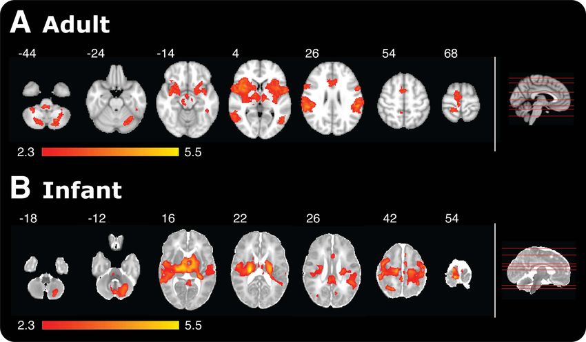
Short report                                                                                                      Neuroscience

                                   eLife digest Doctors long believed that infants do not feel pain the way that older children and
                                   adults do. Instead, they believed that the infants’ responses to discomfort were reflexes. Based on
                                   these beliefs, it was a routine practice to perform surgery on infants without suitable pain relief up
                                   until the late 1980s. Even now, infants may receive less than ideal pain relief. For example, a review
                                   found that although newborns in intensive care units undergo 11 painful procedures per day on
                                   average, more than half of the babies received no pain medications. Some guidelines continue to
                                   emphasize that for infants cuddling and feeding are more important sources of comfort than pain-
                                   relieving drugs.
                                       There is growing support for better pain control for infants. Doctors and nurses now routinely
                                   observe behaviour and physiological responses—such as heart rate—to assess whether infants are
                                   experiencing pain. When an infant shows signs of pain, medical staff may give the infant sugar water
                                   or other interventions aimed at reducing their distress. However, recordings of brain activity suggest
                                   that infants may experience pain without exhibiting physical signs and that sugar water may reduce
                                   the behaviours associated with pain but not the pain itself.
                                       More objective measurements of infant pain would be useful, but to create such measure-
                                   ments scientists must first understand how infants experience pain. So Goksan et al. used
                                   a technique called functional magnetic resonance imaging (fMRI) to compare the brain responses
                                   of adults and newborns to the same stimulus—a sharp poke of the foot. The adults were also
                                   asked about the pain they experienced, and whether the infants pulled their foot away when
                                   poked was documented.
                                       The fMRI results revealed that pain increased activity in 20 regions in the adults’ brains, and 18
                                   of the same regions in the infants’ brains. The brain regions activated in the infants’ brains in
                                   response to a poke on the foot are involved in processing sensations and emotions. The two
                                   regions that did not activate in the infant brains—the amygdala and the orbitofrontal
                                   cortex—help individuals interpret the stimuli. Goksan et al. therefore conclude that infants
                                   experience pain in similar ways to adults, though they may not experience all the emotions that
                                   adults have when they are in pain. It is, therefore, important to give infants suitable pain relief
                                   during potentially painful procedures.
                                   DOI: 10.7554/eLife.06356.002

                                   (Slater et al., 2006, 2010a, 2010b), and have highlighted the limitations of using observational
                                   behavioural measures to quantify pain in infants. For example, nociceptive information can be
                                   processed in the infant brain without a concomitant behavioural response (Slater et al., 2008),
                                   and interventions thought to alleviate pain (i.e., oral sucrose) can reduce clinical pain scores without
                                   reducing evoked nociceptive brain activity (Slater et al., 2010a). While these studies confirm
                                   that the infant central nervous system can process noxious stimulation, they do not elucidate the
                                   nature of the infant experience—in particular, which brain regions are involved, and therefore,
                                   whether the sensory, cognitive, and emotional aspects of pain are present in this population.
                                   Here, we identify the cortical and subcortical structures activated following acute noxious
                                   stimulation in the healthy newborn infant, and compare the activity with that observed in adults.
                                   The feasibility of this approach was demonstrated in a foundational pilot study (Williams et al.,
                                   2015). A case study in a single infant demonstrated that noxious stimulation evoked widespread
                                   brain activity (Williams et al. 2015), which included brain regions previously reported to be
                                   involved in adult pain (Tracey et al., 2007). Using a reverse inference approach to compare
                                   active brain regions in infants with those reported during adult pain, we postulate which aspects
                                   of the pain experience are present (Wager et al., 2013), providing an opportunity to gain insight
                                   into the organisation of nociceptive circuitry in the naı̈ve infant brain.
                                      In this study, acute noxious stimulation (PinPrick Stimulators, MRC Systems) was applied to the foot
                                   in both adults (n = 10; applied force: 32–512 mN) and infants (n = 10; applied force: 32–128 mN;
                                   greater force was not applied due to the potential risk of tissue damage). Using functional magnetic
                                   resonance imaging (fMRI) changes in blood oxygen level dependent (BOLD) activity in the brain were
                                   recorded in response to the stimuli. Adults were asked to verbally report their pain intensity and,
                                   using the McGill Pain questionnaire (Melzack and Torgerson, 1971), to describe the quality of the

Goksan et al. eLife 2015;4:e06356. DOI: 10.7554/eLife.06356                                                                         2 of 13
Short report Neuroscience
pain they experienced. As infants are unable to describe their pain, reflex leg withdrawal from the
stimuli was visually observed during scanning. Parents were present during the studies and no infants
were withdrawn from the study after recruitment.
Results and discussion
Adult participants reported increased pain with increasing stimulus intensity (r = 0.48; p < 0.0001),
and most frequently described the pain as pricking (n = 8 of 10) and sharp (n = 6 of 10). In infants,
application of the stimuli evoked visible withdrawal of the stimulated leg, which could be observed at
all stimulus intensities, whereas in adults, reflex withdrawal of the leg or foot was not observed at any
stimulus intensity. While low threshold stimuli can also evoke reflex withdrawal in infants (Cornelissen
et al., 2013), this observation confirms that the stimuli applied in this study were detected by the
peripheral nervous system and transmitted to the central nervous system. Although noxious
stimulation can elicit reflex limb withdrawal in adults, supraspinal modulation of the input means this
activity is often suppressed in experimental studies.
In adults, noxious stimulation evoked significant increases in BOLD activity in cortical and
subcortical brain regions, including primary somatosensory cortices, anterior cingulate cortex (ACC),
bilateral thalamus, and all divisions of the insular cortices (Figure 1). All brain regions that had
a significant increase in BOLD following noxious stimulation are identified in Table 1, and are
consistent with previous literature (Tracey and Mantyh, 2007). In infants, the increases in BOLD
activity evoked by the noxious stimulation were extremely similar to that seen in adults, and all but two
of the 20 regions that were active in the adults were active in infants (Table 1 and Figure 1). While in
adults the parietal lobe, pallidum, and precuneus cortex were only active in the brain regions
contralateral to the site of stimulation, in infants these brain regions were also active on the ipsilateral
side to the stimulus. Additional brain regions that were only active in the infants included the bilateral
auditory cortices, hippocampus, and caudate (Table 1). The increased bilateral activity and greater
number of active regions in infants are likely due to the immature cortico-cortical and interhemispheric
Figure 1. Comparison of nociceptive-evoked brain activity in selected brain regions that are active in both adults
and infants. Significantly, active voxels across each stimulus intensity level are presented for (A) adult and (B) infant
participants (applied force: adults 32–512 mN; infants 32–128 mN). Each colour represents activity in a different
anatomical brain region. (A) Adult activity is overlaid onto a standard T1 weighted MNI template and (B) infant
activity is overlaid onto a standard T2 weighted neonatal template, corresponding to a 40-week gestation infant.
ACC: anterior cingulate cortex; S1: primary somatosensory cortex: PMC: primary motor cortex; SMA: supplementary
motor area.
DOI: 10.7554/eLife.06356.003
Goksan et al. eLife 2015;4:e06356. DOI: 10.7554/eLife.06356 3 of 13Short report Neuroscience
pathways (Kostovic and Jovanov-Milosevic, 2006). Major reorganisation of the cortical circuitry
occurs after the first postnatal month when there is a striking retraction of exuberant axons in the
corpus callosum and there is a cessation of growth of the long cortico-cortical afferent pathways
(Jovanov-Milosevic et al., 2006; Kostovic and Jovanov-Milosevic, 2006).
Although the infant brain activity was widespread, the specificity of the response was
demonstrated, as it was not present across all brain regions. For example, brain regions not
commonly associated with the cerebral processing of nociceptive stimulation in the adult, such as the
olfactory cortex, cuneus, and fusiform gyrus, were also not active in the infants. 14% of voxels across
the whole brain were active following the application of the 128 mN stimuli in infants compared with
9% of voxels following the 512 mN stimuli in adults (Figure 2). In contrast, the 128 mN stimulus
activated less than 1% of voxels in the adult brain. This demonstrates that the coverage and
distribution of brain activity evoked by the 128 mN stimulus in infants was most similar to that
evoked by the 512 mN stimulus in adults (Figure 2). This suggests that infants have increased
sensitivity to nociceptive stimuli compared with adults, which is supported by previous data that
show spinal nociceptive reflex withdrawal activity has greater amplitude and duration in infants
compared with adults (Andrews and Fitzgerald, 1999; Skljarevski and Ramadan, 2002;
Cornelissen et al., 2013). These data strongly imply that the threshold for evoking widespread
nociceptive brain activity in infants is substantially lower than in adults. It is, however, not known
whether the increased brain activity observed at a lower threshold in the infants is due to increased
peripheral drive, for example due to differences in skin thickness between the adult and infant
populations, or due to differences in transduction or subsequent central processing of the
nociceptive input.
Noxious stimulation in infants did not evoke activity in the amygdala or orbitofrontal cortex (OFC)
(Table 1), and in contrast to the adults, where activity was present across all divisions of bilateral
insular cortices, activity in the anterior division was not present (Figure 1). A recent white matter
tractography study of the adult brain shows that the anterior insula has dominant connections with the
OFC (Wiech et al., 2014). Based on many imaging studies spanning a range of stimuli and tasks, it is
thought that activation in the anterior insula reflects the net evaluation of the affective impact of an
impending situation. Similarly, the OFC is sensitive to stimuli with an emotional valence, however, it
primarily responds to the reward value of the stimulus (including negative value) rather than its
sensory features. Importantly, the OFC also encodes the anticipation of future outcomes, which makes
it well suited for guiding subsequent decisions (Kahnt et al., 2010). It is likely that the infants are too
immature and inexperienced to evaluate and contextualise the nociceptive stimulus into a coordinated
decision and response, which might account for the lack of activity within these regions. Similarly, in
adults the amygdala is thought to attach emotional significance to the nociceptive inputs it receives,
and to play a role in fear and anxiety (Simons et al., 2014), which may reflect affective qualities that
the newborn infant does not yet ascribe to the stimulus.
In light of these observations, it is plausible that infants do not experience the full range of aversive
qualities that adults associate with nociceptive input. Indeed, this hypothesis is supported by evidence
from rat pups, which shows that avoidance behaviour in a fear-conditioning paradigm does not manifest
until postnatal day 10, and is associated with the enhancement of neural activity within the amygdala
(Sullivan et al., 2000; Sullivan, 2001). Nevertheless, the observation that brain structures involved in
affective processing, such as the anterior cingulate cortex, are activated following noxious stimulation
suggests that infants do have the capacity to experience an emotionally relevant context related to
incoming sensory input. Indeed, in adults the modulation of pain-related activity in the anterior cingulate
cortex closely parallels a selective change in perceived unpleasantness (Rainville et al., 1997).
11 brain regions significantly encoded stimulus intensity in adults, whereas none of the active
regions in infants exhibited significant intensity encoding (Table 1). Although the trend for intensity
encoding in infants is clearly evident in some brain regions, these data suggest that infants do not
discriminate stimulus intensity as well as adults (Figure 2—figure supplement 1). As only three
stimulus intensities were applied to the infants it is plausible that if the intensity range were increased,
significant intensity encoding may be observed. Nevertheless, when considering adult brain regions
that did significantly intensity encode, three of the four highest ranked brain regions (ranked
according to the degree of intensity encoding, and identified as the contralateral temporal gyri,
opercular cortex, and all divisions of the insular cortex), were ranked in the same order within the top
three regions in infants, highlighting the remarkable similarity in how the immature infant brain and
Goksan et al. eLife 2015;4:e06356. DOI: 10.7554/eLife.06356 4 of 13Short report Neuroscience
adult brain encode nociceptive information (Table 1). Intensity encoding has been reported following
low intensity von Frey hair stimulation (Williams et al., 2015).
Inferences about the subjective experience of pain are highly speculative, whether based on brain
imaging data, behavioural responses or other autonomic or physiological observations. In most adults,
where the pain experience can be communicated verbally, it is not always necessary to rely on
surrogate measures when attempting to quantify an individual’s pain experience or when assessing
the need for analgesic provision. However, where verbal report is not possible as in the infant
population or in those who are cognitively impaired, reliance on surrogate measures is essential when
making inferences about pain perception. As cortical activation is a fundamental requirement for an
experience to be interpreted as painful, inferences based on patterns of brain activity may provide the
most reliable surrogate measure of pain compared with alternative approaches based on behavioural
and physiological indicators that may not be reliably linked to central sensory or emotional processing
in the brain (Oberlander et al., 2002; Ranger et al., 2007). This does not, however, negate the
importance of taking a multidimensional approach to infant pain assessment by considering
measures of brain activity in the context of other well-characterised behavioural and physiological
indicators. Indeed, some researchers have argued that reverse inference based on brain imaging
results should be used merely as a guide to direct further enquiry rather than a direct means to
interpret results (Poldrack, 2008). Nevertheless, it has been shown using multivariate pattern
analysis that pain-related brain activity can be classified and discriminated from other
psychological states, suggesting a neural state for pain perception that is distinct from other
sensory modalities and affective experiences (Yarkoni et al., 2011; Wager et al., 2013). Although
we cannot necessarily infer an infant’s subjective experience based on a given pattern of brain
activity, these results make certain conclusions more likely. The closer the pattern of brain activity
mimics activity observed in adults—who can report their subjective experience—the stronger the
Figure 2. Noxious-evoked brain activity in response to the maximal presented stimulus in adults (512 mN) and
infants (128 mN). Red-yellow coloured areas represent active brain regions (threshold z ≥ 2.3 with a corrected cluster
significance level of p < 0.05). An image of a midline sagittal brain slice (right panel) identifies the location of each
example slice in the horizontal plane. (A) Adult activity is overlaid onto a standard T1 weighted MNI template and (B)
infant activity is overlaid onto a standard T2 weighted neonatal template, corresponding to a 40-week gestation
infant.
DOI: 10.7554/eLife.06356.005
The following figure supplement is available for figure 2:
Figure supplement 1. Relationship between percentage change in BOLD signal and stimulus intensity (force) in four
example active brain regions in adult and infant participants.
DOI: 10.7554/eLife.06356.006
Goksan et al. eLife 2015;4:e06356. DOI: 10.7554/eLife.06356 5 of 13Table 1. Identification of all active brain regions in adults and infants following acute noxious stimulation at all stimulus intensities (applied force: adults 32–512 mN;
infants 32–128 mN)
Adults Infants
Neonate
template
Peak Z Slope of Peak Z Slope of
MNI coords coords
Anatomical within regression P within regression P
Short report
area Region cluster x y z Rank (*E-03) val* cluster x y z Rank (*E-03) val*
Temporal gyrus Contra 3.92 64 −34 20 1 1.01 0.0002 3.05 32 −32 12 1 2.46 0.0083
Cingulate gyrus Anterior 4.11 6 4 40 2 0.65 0.0005 2.58 −1 1 26 11 1.01 0.3971
Opercular cortex Contra 5.60 40 6 10 3 0.63 0.0001 3.38 32 −13 19 2 2.23 0.0391
Insula Contra 4.18 34 14 6 4 0.61 0.0001 3.04 19 −22 23 3 2.15 0.0207
Supramarginal Contra 4.33 64 −38 20 5 0.60 0.0008 3.29 25 −23 39 9 1.08 0.1749
gyrus
Intensity
Postcentral gyrus Contra 4.28 58 −18 22 6 0.60 0.0012 3.85 15 −22 52 10 1.01 0.2667
encoding regions
(in adults) Visual cortex Contra 3.62 44 −62 4 7 0.59 0.0004 3.25 21 −52 34 6 1.41 0.0814
Putamen Contra 3.68 22 6 6 8 0.55 0.0001 3.30 17 −17 18 8 1.20 0.1656
Goksan et al. eLife 2015;4:e06356. DOI: 10.7554/eLife.06356
Thalamus Contra 3.51 14 −14 0 9 0.50 0.0010 3.58 6 −16 15 4 1.91 0.0592
Insula Ipsi 4.67 −38 −18 14 10 0.49 0.0001 2.59 −26 −14 14 5 1.69 0.1015
Supplementary Contra 3.91 8 4 46 11 0.39 0.0008 3.50 6 −18 48 7 1.23 0.2315
motor area
Cerebellum Ipsi 3.88 −20 −66 −44 0.35 0.0029 3.53 −3 −46 −6 3.57 0.0164
Temporal gyrus Ipsi 3.72 −52 −56 10 0.18 0.5487 3.41 −32 −22 14 2.90 0.0196
Supramarginal Ipsi 4.59 −64 −28 20 0.51 0.0035 3.13 −31 −24 30 2.79 0.0055
gyrus
Cerebellum Contra 3.36 20 −70 −50 0.31 0.0246 3.16 2 −44 −6 2.72 0.1634
Opercular cortex Ipsi 5.23 −50 −28 26 0.50 0.0018 2.69 −27 −12 13 2.23 0.0710
Postcentral gyrus Ipsi 4.71 −62 −18 24 0.44 0.0375 3.52 −31 −15 41 2.12 0.0845
Thalamus Ipsi 3.52 −12 −14 10 0.42 0.0018 3.48 −1 −20 13 1.67 0.1009
Active regions in
Angular gyrus Ipsi 3.59 −58 −50 18 0.53 0.0107 2.98 −23 −39 33 1.56 0.0528
both adults and Non intensity
infants encoding regions Precentral gyrus Ipsi 4.01 −58 0 10 0.43 0.0578 3.46 −23 −17 48 1.53 0.1247
(in adults) Frontal gyrus Contra 3.88 58 12 0 0.56 0.0212 3.11 11 −12 48 1.42 0.0646
Cingulate gyrus Posterior 3.71 −14 −28 38 0.08 0.2480 3.18 −9 −23 35 1.42 0.1101
Angular gyrus Contra 3.71 60 −46 18 0.54 0.0080 3.12 22 −51 35 1.42 0.0407
Precuneous Contra 3.60 16 −68 40 0.38 0.0714 3.70 5 −30 52 1.19 0.1623
cortex
Visual cortex Ipsi 3.82 −52 −70 10 −0.09 0.3758 2.59 −7 −40 11 1.17 0.1657
Brainstem 3.86 10 −26 −8 0.33 0.1710 2.99 −3 −27 −10 1.11 0.4350
Parietal lobule Contra 3.10 20 −44 68 0.61 0.1097 3.10 27 −24 46 1.09 0.1271
Table 1. Continued on next page
6 of 13
NeuroscienceTable 1. Continued
Adults Infants
Neonate
template
Peak Z Slope of Peak Z Slope of
MNI coords coords
Anatomical within regression P within regression P
area Region cluster x y z Rank (*E-03) val* cluster x y z Rank (*E-03) val*
Short report
Putamen Ipsi 3.63 −16 10 −2 0.45 0.0023 3.13 −14 −14 19 0.92 0.2813
Supplementary Ipsi 3.55 −6 4 44 0.40 0.0219 3.16 −4 −10 46 0.91 0.3903
motor area
Precentral Gyrus Contra 4.05 58 4 8 0.44 0.0276 3.76 6 −20 53 0.88 0.2672
Frontal gyrus Ipsi 3.57 −8 22 32 −0.24 0.1954 2.79 −13 −9 50 0.70 0.4820
Pallidum Contra 3.40 16 −4 −4 0.49 0.0071 2.84 13 −13 13 0.64 0.4863
Amygdala Contra 3.49 20 −2 −14 0.69 0.0160
Amygdala Ipsi 4.28 −20 −2 −12 0.43 0.0860
Active regions in Orbitofrontal Ipsi 3.40 −18 4 −16 0.42 0.0157 no activity
adults only cortex
Orbitofrontal Contra 3.57 34 30 0.44 0.0460
Goksan et al. eLife 2015;4:e06356. DOI: 10.7554/eLife.06356
−2
cortex
Precuneous Ipsi 3.80 −1 −26 52 1.26 0.1699
cortex
Pallidum Ipsi 3.16 −8 −5 14 0.59 0.4787
Parietal lobule Ipsi 3.31 −28 −23 33 0.99 0.2711
Auditory cortex Contra 2.89 26 −14 18 3.07 0.0119
Auditory cortex Ipsi 3.34 −17 −29 19 2.56 0.0304
Active regions in
infants only Caudate Contra no activity 3.61 13 −17 22 0.59 0.5822
Caudate Ipsi 3.47 −7 −8 18 1.05 0.3415
Hippocampus Contra 2.61 21 −25 9 1.84 0.1288
Hippocampus Ipsi 2.77 −15 −31 9 1.00 0.3326
Parahippocampus Contra 3.02 11 −23 0 1.53 0.3740
Parahippocampus Ipsi 2.99 −7 −24 −8 0.19 0.9013
Active brain regions were defined as regions with more than one active voxel with significant positive BOLD activity (z = 2.3; corrected cluster significance level of p < 0.05). The intensity
encoding regions are reported with the corresponding p values and slope of the regression that refer to the degree of intensity encoding. The intensity encoding regions (in adults) are ranked
according to the slope of the regression. * Threshold for significant intensity encoding was p < 0.00156 following a Bonferroni correction.
DOI: 10.7554/eLife.06356.004
7 of 13
NeuroscienceShort report Neuroscience
inference. The patterns of brain activity observed in this study make it likely that the infant
experience is similar to that described by adults.
Pain is defined as an unpleasant sensory and emotional experience. This study provides the first
demonstration that many of the brain regions that encode pain in adults are also active in full-term
newborn infants within the first 7 days of life. This strongly supports the hypothesis that infants are
able to experience both sensory and affective aspects of pain, and emphasizes the importance of
effective clinical pain management.
Materials and methods
Participants
10 healthy adults (mean age = 28.3 years; range: 23–36) and 10 healthy term-born infants (mean
gestational age at time of study = 40.6 weeks; range: 38.6–42.7) participated in the study. Adult
participants were members of staff or postgraduate students at The University of Oxford, and
infants were recruited from the Maternity Unit at the John Radcliffe Hospital, Oxford. At the time of
study all infants were less than 7 days old (mean postnatal age = 3 days; range: 1–6). Infant
participants were eligible for inclusion in the study if they were healthy, had no history of
neurological problems, born after 37 weeks gestation, self-ventilating in air and clinically stable at
the time of study.
Recruitment
Informed written consent and consent to publish the results were provided by adult participants or by
the infant’s parent before the study commenced. The study was approved by the National Research
Ethics Service and the University of Oxford Central University Research Ethics Committee. The study
conformed to the standards set by the Declaration of Helsinki and Good Clinical Practice guidelines.
A member of the research team identified infants who were eligible for inclusion in the study
shortly after birth. Prior to obtaining consent for infants to take part, parents were shown the
experimental stimulators and given the opportunity to test the stimulators before they were applied
to the infants. A full description of the MRI scanning environment was also provided. Parents of 113
infants were approached to take part in the study. 44 parents expressed an interest in the proposed
research and 11 infants were recruited to the study. Parents were invited to stay with their infants
during the study and in nearly all cases, one or both parents chose to do so. Parents were also
informed that if their infant became restless while in the scanner, the study would be stopped.
Recruitment success rate was highly dependent on infant availability during the pre-booked MRI
scan slots. One study was stopped due to the baby being restless when placed on the MRI bed. In
adults, 100% of the subjects (10 out of 10) who were approached to take part in the study gave their
consent for the psychophysical and MRI aspects of the study.
Experimental study design
Functional magnetic resonance imaging (fMRI) of the brain was performed on all participants in
response to acute noxious stimulation. On a second test occasion in the adults, the experimental
protocol was repeated outside the scanner and the psychophysical data were recorded. During this
session, participants were asked to verbally rate pain intensity using a numerical scale (0–10) and to
describe the type of pain they experienced using the McGill pain questionnaire (Melzack and
Torgerson, 1971).
Experimental techniques
Noxious stimulation
Acute noxious (non-skin-breaking) stimulation was applied using graded nociceptive stimulators
(PinPrick Stimulators, MRC Systems). In adults, five intensities of stimulation were applied to the
dorsum of the left foot (applied force: 32, 64, 128, 256, and 512 mN). In infants, three intensities of
stimulation were applied to the heel of the left foot (applied force: 32, 64, 128 mN). Greater force was
not applied in infants to avoid the potential risk of tissue damage. Each stimulus was delivered 10
times with a minimum inter-stimulus interval (ISI) of 25 s. In all cases, stimuli were delivered by the
experimenter in one smooth motion and lasted approximately 1 s.
Goksan et al. eLife 2015;4:e06356. DOI: 10.7554/eLife.06356 8 of 13Short report Neuroscience
Recording techniques
MRI study protocol
Preparation
All MRI scans conformed to the FMRIB (Functional Magnetic Resonance Imaging of the Brain) Centre
ethical and safety guidelines. Adult participants were screened for MRI safety by a radiographer.
Ear protection was provided (foam ear plugs, 3M, St. Paul, Minnesota; sound attenuation 28 dB) and
adults were made comfortable while lying on the MRI scanner bed. The head was positioned inside
the head coil and padding was used to restrict head movement.
Infants were accompanied and transported to FMRIB by a member of clinical staff, trained in
neonatal life support, who remained with each infant throughout the study to ensure the infant’s
safety and wellbeing. Infants were screened for metal items (including metal poppers on clothing that
were in direct contact with their skin) and were fed and swaddled before being placed on a vacuum-
positioning mattress on the MRI bed. Ear putty, ear muffs (Minimuffs, Natus Medical Inc., Galway,
Ireland), and ear-defenders (Em’s 4 Bubs Baby Earmuffs, Em’s 4 Kids, Brisbane, Australia) were fitted
(sound attenuation levels: 23 dB, 7 dB, and 22 dB, respectively). Finally, extra padding was placed
around the ear-defenders to restrict head movement. The infant’s temperature was measured before
the scan commenced, and heart rate and oxygen saturation was monitored throughout the scan using
a 3T MRI compatible neonatal monitoring probe placed on the right foot (Fibre Optic Pulse Oximeter;
Nonin Medical, Plymouth, Minnesota). Parents who accompanied their infants were also MRI safety
screened and provided with adequate ear protection, and were asked to sit inside the MRI scan room
throughout the scans.
MR image acquisition
MRI data were acquired using a Siemens 3-Tesla Magnetom Verio system (Erlangen, Germany) with
a 32-channel head coil. Anatomical scans were first acquired and if excessive motion was identified,
a second acquisition was attempted. For adults, a T1-weighted sequence (MPRAGE; TR = 2040 ms; TE
= 4.7 ms; flip angle 8˚; resolution 1 × 1 × 1 mm; axial slices = 192) was acquired and for infants a T2-
weighted sequence (TSE; TR = 13871 ms; TE = 89 ms; flip angle 150˚; resolution 1 × 1 × 1 mm; slices =
80) was used. BOLD images were acquired using a T2* weighted echo-planar imaging (EPI) sequence
with an echo time (TE) optimised for either adults (TR = 3280 ms; TE = 30 ms; flip angle = 90˚; FOV =
192 mm; imaging matrix 64 × 64; resolution 3 × 3 × 3 mm; slices = 50; average total volumes = 96) or
infants (TR = 2500 ms; TE = 40 ms; flip angle = 90˚; FOV = 192 mm; imaging matrix 64 × 64; resolution
3 × 3 × 3 mm; slices = 33; average total volumes = 136). Field map images were obtained for post-
acquisition correction of gradient field effects. Prospective Acquisition Correction for head motion
(PACE) was applied during all EPI scans. PACE is a motion correction technique that tracks the
position of the head during scan acquisition and applies a real-time correction for large head
movements (Thesen et al., 2000). The noxious stimuli were time-locked to the fMRI recording using
Neurobehavioural Systems (Presentation) software that recorded each time the experimenter pressed
a button while simultaneously applying the experimental stimuli to the participant’s foot.
The MR data acquisition protocol was 28 min in infants and 40 min in adults. On average infants
spent 60 min in the scanner room, which allowed time to prepare and settle the infants before and
during scanning.
Sleep state
Infant sleep state could not be controlled during the study as infants fluctuated between being quietly
awake and asleep. Adults were not instructed to stay awake during scanning and three adults
reported that they fell asleep.
Adult psychophysics and pain questionnaire
Participants were asked to lie down on a patient bed. Throughout the experiment, adults were asked
to verbally state a pain score following each individual stimulus using a pain scoring system where 0 is
no pain and 10 is the worst pain imaginable. Once all stimuli had been presented, the participants
were asked to describe the type of pain they experienced by completing the McGill Pain
Questionnaire (Melzack and Torgerson, 1971).
Goksan et al. eLife 2015;4:e06356. DOI: 10.7554/eLife.06356 9 of 13Short report Neuroscience
Data analysis
MR data
All MR data processing was done using the FMRIB Software Library (FSL) (www.fmrib.ox.ac.uk/fsl). FSL
Version 4.9.1 (with no boundary based registration [BBR]) was used in infants and FSL Version 5.0 was
used in adults (Woolrich et al., 2009). Standard preprocessing steps were performed in all fMRI data
sets using the FMRI Expert Analysis Tool (FEAT, version 6.0). The FSL Brain Extraction Tool (BET) was
used to remove non-brain structures from the adult and infant structural images and from the adult
field map images (Smith, 2002). In the infant field maps, brain extraction was achieved using a mask
of the infant’s brain-extracted structural image to guide the field map preparation. For each adult, the
functional data were registered using a two-step registration: (i) the EPI image was registered to the
subject’s T1-weighted structural image, with a rigid body transformation, six DOF and BBR, using
FMRIBs Linear Image Registration Tool (FLIRT) (Jenkinson and Smith, 2001; Jenkinson et al., 2002;
Greve and Fischl, 2009); and (ii) the T1-weighted structural image was registered to a standard MNI
image (http://www.bic.mni.mcgill.ca/ServicesAtlases/ICBM152NLin6) using FMRIBs Non-linear Reg-
istration Tool (FNlRT) with a non-linear transformation and 12 DOF. In each infant, the functional data
were registered using a three step registration: (i) the EPI image was registered to the subjects T2-
weighted structural images using FLIRT, with a rigid body transformation with six DOF and no BBR
(Jenkinson and Smith, 2001; Jenkinson et al., 2002); (ii) the T2-weighted structural images were
registered to a neonatal specific template image, which corresponded to the gestational age of the
infant at the time of the study (Serag et al., 2012); and (iii) the template images were then registered
to a standardized infant template, corresponding to a 40-week gestation infant (Serag et al., 2012).
The final two stages of the infant registration were carried out using a non-linear transformation
(FNIRT) and 12 DOF. The 40-week gestation template was chosen because it most closely matched
the median age of the infants.
Functional data were spatially smoothed (full width half maximum = 5 mm) and temporal filtering
(high pass cut off = 90 s) was also applied. Motion artifacts were minimised using Motion Correction with
MCFLIRT (Jenkinson et al., 2002) and by the addition of motion-derived explanatory variables (EV) in
the models. A single EV was included for each volume that was identified as having a large deviation in
head position (FSL motion outliers were calculated per data set using a framewise displacement),
effectively removing the signals associated with the identified timepoint from the analysis. Probabilistic
independent component analysis was applied using MELODIC (model-free fMRI analysis using
probabilistic independent component analysis) and components resembling movement were removed.
Time-series analysis was performed using a general linear model (GLM) by convolving the
experimental design with either a standard adult hemodynamic response function (HRF) or a neonatal-
specific HRF (Arichi et al., 2012). This approach was used to identify voxels in the brain that have
a significantly increased level of BOLD activity (threshold at z = 2.3 with a corrected cluster
significance level of p < 0.05). Group analyses were performed separately on adults and infants, and
were performed independently for each stimulus intensity.
A voxel-based conjunction analysis was not performed between adult and infant participants
because of extreme differences between infant and adult brain anatomy, which would make such an
analysis unreliable. As the insula is a key region of interest in nociceptive processing the distribution of
activity within the insula was also reported.
Identifying active regions
Anatomical brain regions were classified using the Adult Harvard–Oxford cortical and subcortical
atlases (Desikan et al., 2006) and a neonatal-specific atlas, which uses similar anatomical
nomenclature as the Harvard–Oxford atlases (Shi et al., 2011). As the cerebellum was not identified
in either the adult or neonatal atlas, and the brainstem not identified in the neonatal atlas, therefore
masks (which were available as part of the standard templates in each population) were used to
identify these regions. Comparisons between the infant and adult brain activity was considered on
a gross anatomical scale. For example, the temporal gyrus, visual cortex, and brainstem were each
considered as single structures. Active brain regions were defined as regions with more than one
active voxel with significant positive BOLD activity (thresholded at z = 2.3 with a corrected cluster
significance level of p < 0.05). A conversion from standard to functional space was performed to
calculate the number of active voxels.
Goksan et al. eLife 2015;4:e06356. DOI: 10.7554/eLife.06356 10 of 13Short report Neuroscience
In adults, the FSL function Atlasquery was used to generate a list of active brain regions at each
stimulus intensity, based on the masked clusters in the Harvard–Oxford cortical and subcortical atlases
(Desikan et al., 2006). The use of Atlasquery ensured that all active voxels were identified in each brain
region. The percentage of active voxels in each anatomical mask was then used to threshold regions,
such that only regions with more than one active voxel were identified as active. The FSL function Cluster
was used to identify the peak z statistic and MNI coordinates in each active region (see Table 1).
In infants, the active brain regions were identified using MATLAB. Infant activity at each stimulus
intensity level (thresholded at z = 2.3 with a corrected cluster significance level of p < 0.05) was used
to mask the neonatal atlas (Shi et al., 2011). The masked image was imported into MATLAB so that
each active region could be identified and the number of active voxels within each region calculated in
the neonatal atlas space. A conversion from standard to functional space allowed quantification of the
number of active voxels in the infant functional space and brain regions with more than one active
voxel were identified as active. The FSL function Cluster was used to identify peak z statistics and
coordinates in neonatal template space for each active region (Table 1).
Percentage BOLD increase in active anatomical brain regions
Once the active brain regions were identified at each stimulus intensity, an activity mask was created
for each brain region based on the group analysis of all inputs across all stimulus intensities
(z threshold = 2.3) for both the adults and infants. The activity mask was separated into anatomical
regions of interest (based on brain regions which had been identified as active) and using Featquery
the parameter estimate of the average percentage BOLD increase within each mask for each
participant at each stimulus intensity was calculated.
Statistics
MRI data—intensity analysis
Regression analysis was carried out using the software packages Graphpad Prism & R. Mean
percentage signal change was plotted against stimulus intensity and regression analysis was used to
test the null hypothesis that no intensity encoding was present within the masked activity within each
anatomical brain region. A Bonferroni correction for multiple comparisons was used to determine the
p threshold required in order to reject the null hypothesis. The corrected significance threshold was p
= 0.00156. Parameter estimates for the gradient of the regression were used to rank brain regions
that were active and exhibited significant intensity encoding.
Adult psychophysics
The mean pain score across each train of 10 stimuli at each stimulus intensity was calculated. The
relationship between the mean pain scores and stimulus intensity was quantified using linear
regression.
Acknowledgements
This work was funded by the Wellcome Trust. Sezgi Goksan is a MRC funded DPhil student. We would
like to thank Eugene Duff, Jelena Bozek Mouthuy, Gabriela Schmidt Mellado, Sheula Barlow, Gabrielle
Green, Falk Eippert, David Parker, and Caroline Young for their analytical, clinical and technical
support. We would also like to thank the infants, adults, and parents who took part in this study.
Additional information
Funding
Funder Grant reference Author
Wellcome Trust Wellcome Trust Career Rebeccah
Development Fellowship, Slater
WT095802MA
Medical Research Council Graduate Student Fellowship Sezgi Goksan
(MRC)
The funders had no role in study design, data collection and interpretation, or the
decision to submit the work for publication.
Goksan et al. eLife 2015;4:e06356. DOI: 10.7554/eLife.06356 11 of 13Short report Neuroscience
Author contributions
SG, RS, Conception and design, Acquisition of data, Analysis and interpretation of data, Drafting or
revising the article; CH, RP, Acquisition of data, Analysis and interpretation of data, Drafting or
revising the article; FE, Conception and design, Acquisition of data; NC, FM, Analysis and
interpretation of data, Drafting or revising the article; RR, EA, IT, Conception and design, Analysis
and interpretation of data, Drafting or revising the article; JC, MS, Acquisition of data, Drafting or
revising the article; SC, Conception and design, Acquisition of data, Analysis and interpretation of
data; MJ, Acquisition of data, Analysis and interpretation of data
Author ORCIDs
Caroline Hartley, http://orcid.org/0000-0002-7981-0836
Ethics
Human subjects: Informed written consent and consent to publish was provided by adult participants
or by the infant’s parents. The study was approved by the Oxford and South Central Research Ethics
Committees of the National Research Ethics Service and the University of Oxford Central University
Research Ethics Committee (CUREC) (refs.: Investigating pain in the developing human brain; study
number: 12/SC/0447; Human pain perception; study number: 11/SC/0249; CUREC study number:
MSD/IDREC/C1/2011/143). The study conformed to the standards set by the Declaration of Helsinki
and Good Clinical Practice guidelines.
References
Anand KJ, Hickey PR. 1987. Pain and its effects in the human neonate and fetus. The New England Journal of
Medicine 317:1321–1329. doi: 10.1056/NEJM198711193172105.
Anand KJ, International Evidence-Based Group for Neonatal Pain. 2001. Consensus statement for the prevention
and management of pain in the newborn. Archives of pediatrics & adolescent medicine 155:173–180. doi: 10.
1001/archpedi.155.2.173.
Andrews K, Fitzgerald M. 1999. Cutaneous flexion reflex in human neonates: a quantitative study of threshold and
stimulus-response characteristics after single and repeated stimuli. Developmental Medicine and Child
Neurology 41:696–703. doi: 10.1017/S0012162299001425.
Apkarian AV, Bushnell MC, Treede RD, Zubieta JK. 2005. Human brain mechanisms of pain perception and
regulation in health and disease. European Journal of Pain 9:463–484. doi: 10.1016/j.ejpain.2004.11.001.
Arichi T, Fagiolo G, Varela M, Melendez-Calderon A, Allievi A, Merchant N, Tusor N, Counsell SJ, Burdet E,
Beckmann CF, Edwards AD. 2012. Development of BOLD signal hemodynamic responses in the human brain.
Neuroimage 63:663–673. doi: 10.1016/j.neuroimage.2012.06.054.
Carbajal R, Rousset A, Danan C, Coquery S, Nolent P, Ducrocq S, Saizou C, Lapillonne A, Granier M, Durand P,
Lenclen R, Coursol A, Hubert P, de Saint Blanquat L, Boëlle PY, Annequin D, Cimerman P, Anand KJ, Bréart G.
2008. Epidemiology and treatment of painful procedures in neonates in intensive care units. JAMA 300:60–70.
doi: 10.1001/jama.300.1.60.
Cornelissen L, Fabrizi L, Patten D, Worley A, Meek J, Boyd S, Slater R, Fitzgerald M. 2013. Postnatal temporal,
spatial and modality tuning of nociceptive cutaneous flexion reflexes in human infants. PLOS ONE 8:e76470.
doi: 10.1371/journal.pone.0076470.
Desikan RS, Segonne F, Fischl B, Quinn BT, Dickerson BC, Blacker D, Buckner RL, Dale AM, Maguire RP, Hyman
BT, Albert MS, Killiany RJ. 2006. An automated labeling system for subdividing the human cerebral cortex on MRI
scans into gyral based regions of interest. Neuroimage 31:968–980. doi: 10.1016/j.neuroimage.2006.01.021.
Duhn LJ, Medves JM. 2004. A systematic integrative review of infant pain assessment tools. Advances in Neonatal
Care 4:126–140. doi: 10.1016/j.adnc.2004.04.005.
Greve DN, Fischl B. 2009. Accurate and robust brain image alignment using boundary-based registration.
Neuroimage 48:63–72. doi: 10.1016/j.neuroimage.2009.06.060.
Jenkinson M, Bannister P, Brady M, Smith S. 2002. Improved optimization for the robust and accurate linear
registration and motion correction of brain images. Neuroimage 17:825–841. doi: 10.1006/nimg.2002.1132.
Jenkinson M, Smith S. 2001. A global optimisation method for robust affine registration of brain images. Medical
Image Analysis 5:143–156. doi: 10.1016/S1361-8415(01)00036-6.
Jovanov-Milosević N, Benjak V, Kostovic I. 2006. Transient cellular structures in developing corpus callosum of the
human brain. Collegium Antropologicum 30:375–381.
Kahnt T, Heinzle J, Park SQ, Haynes JD. 2010. The neural code of reward anticipation in human orbitofrontal
cortex. Proceedings of the National Academy of Sciences of USA 107:6010–6015. doi: 10.1073/pnas.
0912838107.
Kostovic I, Jovanov-Milosevic N. 2006. The development of cerebral connections during the first 20-45 weeks’
gestation. Seminars in Fetal & Neonatal Medicine 11:415–422. doi: 10.1016/j.siny.2006.07.001.
Melzack R, Torgerson WS. 1971. On the language of pain. Anesthesiology 34:50–59.
NHS Choices. 2015. Tongue tie. http://wwwnhsuk/conditions/tongue-tie/Pages/Introductionaspx.
Goksan et al. eLife 2015;4:e06356. DOI: 10.7554/eLife.06356 12 of 13Short report Neuroscience
Oberlander TF, Grunau RE, Fitzgerald C, Whitfield MF. 2002. Does parenchymal brain injury affect biobehavioral
pain responses in very low birth weight infants at 32 weeks’ postconceptional age? Pediatrics 110:570–576.
doi: 10.1542/peds.110.3.570.
Poldrack RA. 2008. The role of fMRI in cognitive neuroscience: where do we stand?Current Opinion in
Neurobiology 18:223–227. doi: 10.1016/j.conb.2008.07.006.
Rainville P, Duncan GH, Price DD, Carrier B, Bushnell MC. 1997. Pain affect encoded in human anterior cingulate
but not somatosensory cortex. Science 277:968–971. doi: 10.1126/science.277.5328.968.
Ranger M, Johnston CC, Anand KJ. 2007. Current controversies regarding pain assessment in neonates. Seminars
in Perinatology 31:283–288. doi: 10.1053/j.semperi.2007.07.003.
Rodkey EN, Pillai Riddell R. 2013. The infancy of infant pain research: the experimental origins of infant pain denial.
The Journal of Pain 14:338–350. doi: 10.1016/j.jpain.2012.12.017.
Roofthooft DW, Simons SH, Anand KJ, Tibboel D, van Dijk M. 2014. Eight years later, are we still hurting newborn
infants?Neonatology 105:218–226. doi: 10.1159/000357207.
Serag A, Aljabar P, Ball G, Counsell SJ, Boardman JP, Rutherford MA, Edwards AD, Hajnal JV, Rueckert D. 2012.
Construction of a consistent high-definition spatio-temporal atlas of the developing brain using adaptive kernel
regression. Neuroimage 59:2255–2265. doi: 10.1016/j.neuroimage.2011.09.062.
Shi F, Yap PT, Wu G, Jia H, Gilmore JH, Lin W, Shen D. 2011. Infant brain atlases from neonates to 1- and 2-year-
olds. PLOS ONE 6:e18746. doi: 10.1371/journal.pone.0018746.
Simons LE, Moulton EA, Linnman C, Carpino E, Becerra L, Borsook D. 2014. The human amygdala and pain:
evidence from neuroimaging. Human Brain Mapping 35:527–538. doi: 10.1002/hbm.22199.
Skljarevski V, Ramadan NM. 2002. The nociceptive flexion reflex in humans – review article. Pain 96:3–8. doi: 10.
1016/S0304-3959(02)00018-0.
Slater R, Cantarella A, Franck L, Meek J, Fitzgerald M. 2008. How well do clinical pain assessment tools reflect pain
in infants?PLOS Medicine 5:e129. doi: 10.1371/journal.pmed.0050129.
Slater R, Cantarella A, Gallella S, Worley A, Boyd S, Meek J, Fitzgerald M. 2006. Cortical pain responses in human
infants. The Journal of neuroscience 26:3662–3666. doi: 10.1523/JNEUROSCI.0348-06.2006.
Slater R, Cornelissen L, Fabrizi L, Patten D, Yoxen J, Worley A, Boyd S, Meek J, Fitzgerald M. 2010a. Oral sucrose
as an analgesic drug for procedural pain in newborn infants: a randomised controlled trial. Lancet 376:
1225–1232. doi: 10.1016/S0140-6736(10)61303-7.
Slater R, Worley A, Fabrizi L, Roberts S, Meek J, Boyd S, Fitzgerald M. 2010b. Evoked potentials generated by
noxious stimulation in the human infant brain. European Journal of Pain 14:321–326. doi: 10.1016/j.ejpain.2009.
05.005.
Smith SM. 2002. Fast robust automated brain extraction. Human Brain Mapping 17:143–155. doi: 10.1002/hbm.
10062.
Sullivan RM. 2001. Unique characteristics of neonatal classical conditioning: the role of the amygdala and locus
coeruleus. Integrative Physiological and Behavioral Science 36:293–307. doi: 10.1007/BF02688797.
Sullivan RM, Landers M, Yeaman B, Wilson DA. 2000. Good memories of bad events in infancy. Nature 407:38–39.
doi: 10.1038/35024156.
Thesen S, Heid O, Mueller E, Schad LR. 2000. Prospective acquisition correction for head motion with image-based
tracking for real-time fMRI. Magnetic Resonance in Medicine 44:457–465. doi: 10.1002/1522-2594(200009)44:
33.0.CO;2-R.
Tracey I, Mantyh PW. 2007. The cerebral signature for pain perception and its modulation. Neuron 55:377–391.
doi: 10.1016/j.neuron.2007.07.012.
Wager TD, Atlas LY, Lindquist MA, Roy M, Woo CW, Kross E. 2013. An fMRI-based neurologic signature of
physical pain. The New England Journal of Medicine 368:1388–1397. doi: 10.1056/NEJMoa1204471.
Wiech K, Jbabdi S, Lin CS, Andersson J, Tracey I. 2014. Differential structural and resting state connectivity
between insular subdivisions and other pain-related brain regions. Pain 155:2047–2055. doi: 10.1016/j.pain.
2014.07.009.
Williams G, Fabrizi L, Meek J, Jackson D, Tracey I, Robertson N, Slater R, Fitzgerald M. 2015. Functional magnetic
resonance imaging can be used to explore tactile and nociceptive processing in the infant brain. Acta Paediatrica
104:158–166. doi: 10.1111/apa.12848.
Woolrich MW, Jbabdi S, Patenaude B, Chappell M, Makni S, Behrens T, Beckmann C, Jenkinson M, Smith SM.
2009. Bayesian analysis of neuroimaging data in FSL. Neuroimage 45:1 SupplS173–S186. doi: 10.1016/j.
neuroimage.2008.10.055.
Yarkoni T, Poldrack RA, Nichols TE, Van Essen DC, Wager TD. 2011. Large-scale automated synthesis of human
functional neuroimaging data. Nature Methods 8:665–670. doi: 10.1038/nmeth.1635.
Goksan et al. eLife 2015;4:e06356. DOI: 10.7554/eLife.06356 13 of 13You can also read