Lateral orbitofrontal dysfunction in
←
→
Page content transcription
If your browser does not render page correctly, please read the page content below
Research Paper
Lateral orbitofrontal dysfunction in the
Sapap3 knockout mouse model of
obsessive–compulsive disorder
Huimeng Lei, PhD; Juan Lai, MS; Xiaohong Sun, BS; Qunyuan Xu, MD, PhD;
Guoping Feng, MD, PhD
Background: Obsessive–compulsive disorder (OCD) is a common psychiatric disorder that affects about 2% of the population, but the
underlying neuropathophysiology of OCD is not well understood. Although increasing lines of evidence implicate dysfunction of the orbito-
frontal cortex (OFC) in OCD, a detailed understanding of the functional alterations in different neuronal types in the OFC is still elusive.
Methods: We investigated detailed activity pattern changes in putative pyramidal neurons and interneurons, as well as local field potential
oscillations, in the lateral OFC underlying OCD-relevant phenotypes. We applied in vivo multichannel recording in an awake OCD mouse
model that carried a deletion of the Sapap3 gene, and in wild type littermates. Results: Compared with wild type mice, the lateral OFC of
Sapap3 knockout mice exhibited network dysfunction, demonstrated by decreased power of local field potential oscillations. The activity of
inhibitory and excitatory neurons in the lateral OFC showed distinct perturbations in Sapap3 knockout mice: putative interneurons exhib-
ited increased activity; putative pyramidal neurons exhibited enhanced bursting activity; and both putative pyramidal neurons and inter
neurons exhibited enhanced discharge variability and altered synchronization. Limitations: To exclude motor activity confounders, this
study examined functional alterations in lateral OFC neurons only when the mice were stationary. Conclusion: We provide, to our know
ledge, the first direct in vivo electrophysiological evidence of detailed functional alterations in different neuronal types in the lateral OFC of
an OCD mouse model. These findings may help in understanding the underlying neuropathophysiology and circuitry mechanisms for phe-
notypes relevant to OCD, and may help generate and refine hypotheses about potential biomarkers for further investigation.
Introduction impaired in people with OCD.16,17 For this reason, the OFC is
well suited as a neural substrate for OCD pathogenesis.
Obsessive–compulsive disorder (OCD) is a debilitating Although a large number of functional neuroimaging
neuropsychiatric condition with a lifetime prevalence of 2%.1 studies have shown altered metabolic activity in the OFC of
It is characterized by persistent intrusive thoughts (obses- people with OCD, a detailed understanding of the functional
sions) and repetitive actions (compulsions). Although dys- alterations is still elusive. For example, a majority of studies
function of the cortico–striato–thalamo–cortical circuitry has have reported increased resting metabolic activity in the OFC
been implicated in the pathogenesis of OCD2–5 and is sup- of people with OCD,18–22 which is exacerbated by symptom
ported by neuroimaging studies in patients,6–9 the underlying provocation23,24 and alleviated after successful treatment.25–30
neuropathological changes are still not well understood. The However, these studies can only measure metabolic levels to
orbitofrontal cortex (OFC) may be central to our understand- indirectly reflect general neuronal activity levels. The nonin-
ing of OCD, because it is the most frequently reported region vasive methods used in clinical studies also have limited spa-
of structural, functional and connectivity alterations in tial and temporal resolution. Direct electrophysiological evi-
patients with OCD.10–12 The OFC is thought to update out- dence of detailed activity change of different neuronal types
come expectations when rules linking stimuli to outcomes in the OFC of people with OCD or animal models is still lack-
are changed.13–15 Therefore, the OFC is essential for behaviour ing. Furthermore, there are discrepancies in the directionality
flexibility and goal-directed behaviours, both of which are of findings in clinical neuroimaging studies that may be due
Correspondence to: H. Lei, Department of Neurobiology, Capital Medical University, You An Men Wai, Xi Tou Tiao, No. 10 Beijing, Beijing
1000069, China; leihm@ccmu.edu.cn; G. Feng, McGovern Institute for Brain Research, Department of Brain and Cognitive Sciences,
Massachusetts Institute of Technology, Cambridge, MA 02139; fengg@mit.edu.
Submitted Feb. 28, 2018; Revised May 18, 2018; Accepted Jun. 27, 2018; Published online Nov. 7, 2018
DOI: 10.1503/jpn.180032
© 2019 Joule Inc. or its licensors
120 J Psychiatry Neurosci 2019;44(2)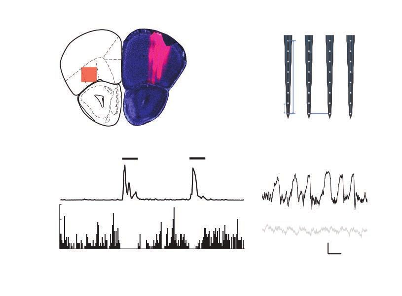
Orbitofrontal dysfunction in an OCD mouse model
to the heterogeneity of the disorder, comorbidities, medica- were anesthetized by intraperitoneal injection of Avertin so-
tion history or different subregions of the OFC analyzed. lution (20 mg/mL, 0.5 mg/g body weight) and then
In the present study, we investigated detailed functional mounted in a stereotactic holder and kept warm (37°C) with
change in different neuronal types in the lateral OFC (lOFC) an electric heating pad (BrainKing Biotech). A small skull re-
that underlie OCD-relevant phenotypes by applying in vivo gion (~1 mm in diameter) located posterior to the lOFC based
multichannel recording in an awake OCD mouse model on stereotactic coordinates (anterior–posterior = 2.3 mm,
that carried a deletion of the Sapap3 gene. These Sapap3 medial–lateral = 1.3 mm) was thinned but not broken with a
knockout (KO) mice demonstrate several OCD-like behav- high-speed drill. A custom-made head plate with a hole
iours, including excessive and pathological self-grooming 2 mm in diameter was placed on the skull, with the hole
and increased anxiety-like behaviours, suggesting potential centred over the thinned region above the lOFC. The head
relevance to OCD. As well, the entire constellation of OCD- plate was affixed to the skull with Meta-bond (Parkell Inc.),
like behaviours in Sapap3 KO mice is alleviated by chronic and the thinned skull and hole in the head plate were then
fluoxetine, a first-line treatment for OCD.31 Human genetics covered with Kwik-sil (World Precision Instruments) for pro-
studies also support a role for Sapap genes in OCD.32–34 tection. Mice were individually housed after surgery and al-
Therefore, a detailed understanding of functional altera- lowed to recover for 3 to 5 days before habituation training.
tions in the OFC of Sapap3 KO mice could help identify po- To minimize potential stress effects, mice were trained to
tential circuit mechanisms for behaviours relevant to OCD. habituate to a head-fixed spherical treadmill for 2 to 4 hours
The OFC consists of lateral and medial subregions. The lat- each day for 4 consecutive days before recording. Mice
eral and medial OFC may perform different functions, such quickly learned to balance and walk on the apparatus and
as processing negative versus positive valence.35,36 A previ- stayed quiet for most of the time during recording, indicating
ous study from our group found that selective stimulation low stress.
of the lOFC suppressed overexpression of both spontan
eous and conditioned repetitive grooming behaviours, Electrophysiological recording
suggesting involvement of the lOFC in these behaviours in
Sapap3 KO mice.37 To investigate functional alterations in the During electrophysiological recording, the mouse’s head was
OFC in Sapap3 KO mice, we focused on the lOFC subregion restrained by a head plate, and the mouse was able to
in the current study. Using single-unit and local field poten- manoeuvre on the top surface of an air-supported floating
tial (LFP) recording in the lOFC, we studied activity pattern styrofoam ball. Immediately before recording, we opened a
changes in different neuronal populations and alterations in small craniotomy in the thinned skull area above the lOFC.
LFP oscillations in Sapap3 KO mice. Our goal was to shed We detected extracellular spiking signals and LFP using a
light on the neuropathophysiology underlying OCD-like 32-channel silicon probe (A4×8–5mm-50–200–413-A32–15;
behaviours and advance our circuit-level understanding of NeuroNexus) arranged in a 4 × 8 pattern (4 shanks with 8 re-
phenotypes relevant to OCD. Our findings may help to gen- cording sites in each shank), lowered to the lOFC (anterior–
erate and refine hypotheses for further investigation. For ex- posterior = 2.5–2.8 mm; medial–lateral = 1.0–1.6 mm, dorsal–
ample, LFP alterations and increased burst firing in lOFC ventral = 1.3–2 mm) and tilted rostrally at an angle of 15° to
may be useful biomarker candidates for further examina- the vertical plane. Based on the above coordinates, we dis-
tion in people with OCD. carded neural activities recorded outside the lOFC from
analysis. We sampled unit activity at 30 kHz and high-pass
Methods filtered it at 250 Hz using a Blackrock Cerebus data acquisi-
tion system (Blackrock Microsystems LLC). We sampled LFP
Animal use at 1 kHz and low-pass-filtered it at 250 Hz. To avoid possible
noise contamination in low-frequency oscillations, we dis-
All experiments were conducted according to protocols ap- carded LFP data below 1.5 Hz.
proved by the Institutional Animal Care and Use and Institu-
tional Biosafety committees of the Capital Medical University Spike sorting and single-unit classification
(Beijing, China) and the Massachusetts Institute of Technol-
ogy (Cambridge, Massachusetts). Our group had previously We sorted unit activity containing spikes of multiple neur
found that from age 2 to 3 months, Sapap3 KO mice exhibited ons manually offline using Offline Sorter (Plexon Inc.) and
significantly increased self-grooming that resembled compul- a combination of template-matching and principal-
sive OCD behaviours.31 Therefore, we performed all of our components analyses. A total of 362 single units were well
experiments on adult Sapap3 KO mice aged 3 to 10 months, isolated. Units with a trough half width within 100–200 μs,
and on age-matched, wild type (WT) littermates of either sex. a peak half width within 467–700 μs and a trough:peak
ratio within 1.2–2.8 were classified as putative pyramidal
Surgery neurons. Units with a trough half width within 67–167 μs, a
peak half width within 100–300 μs and a trough:peak ratio
Our method of electrophysiological recording in head- within 1.1–1.8 were classified as putative interneurons.37,40
restrained, mobile mice was based on previous studies with Using these criteria, we identified 294 units as putative
modifications.38,39 Briefly, for head-plate implantation, mice pyramidal neurons and 51 units as putative interneurons.
J Psychiatry Neurosci 2019;44(2) 121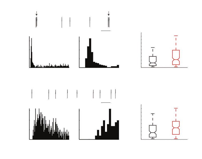
Lei et al
Detailed explanations of these cell-type classification criteria Results
are provided in Appendix 1, available at jpn.ca/180032-a1.
Recording neuronal activity from the lOFC of awake mice
Statistical analysis
To identify changes in individual neuronal activity, we
All analyses used custom Matlab software (H.L.). For LFP recorded extracellular single units and LFP from the lOFC of
oscillation power, the mean baseline firing rate of putative head-fixed, awake adult Sapap3 KO mice (n = 24, 20 males
pyramidal neurons and interneurons, the percentage of and 4 females) and their age-matched, WT littermates (n = 21,
spikes in the bursting mode per neuron and the number of 19 males and 2 females; Fig. 1A and B). Two-way analysis of
bursts per minute per neuron, we determined statistical sig- variance (ANOVA) analysis showed a significant effect of
nificance between WT and Sapap3 KO mice using the genotype but no effect of sex (Appendix 1), so we pooled the
Wilcoxon rank sum test. For LFP oscillation power, n was the data for male and female mice. To minimize stress, the mice
number of animals. For single-unit activity, n was the num- were allowed to behave on an air-supported, frictionless
ber of neurons. We measured the correlation between firing spherical treadmill.38 Because the activity of the lOFC is
rate and the depth of the neurons using the Spearman rank modulated by movement (Fig. 1C and D), we analyzed only
correlation coefficient. the stationary epochs of the recordings to exclude movement
We measured firing variability using CV2.41 We defined CV2 or motor-directed activity confounders.
for spike i as the standard deviation of 2 adjacent interspike
intervals (ISIs) divided by their mean and multiplied by √2. Reduced LFP oscillation power in the lOFC of Sapap3 KO
mice
Brain rhythms are critical to coordinating the activity of
neuronal populations across multiple spatial and temporal
scales, and are involved in a wide range of cognitive and
Each CV2 corresponds to an ISI value that is the mean of the perceptual processes. To assess the rhythmic alterations of
2 adjacent ISIs used to compute CV2. We computed mean CV2 Sapap3 KO mice, we recorded LFP at 64 sites, evenly distrib-
by averaging all CV2 values corresponding to ISIs between a uted in a rectangular plane in the lOFC that was 600 μm in
certain range. The ISI boundaries were logarithmically the medial–lateral dimension and 700 μm in the dorsal–
spaced with a ratio of 1.3. Because of the refractory period, ventral dimension, from a depth of 1300 μm to 2000 μm. We
we set the minimum ISI boundary at 1.69 ms. To access the calculated the LFP power for each mouse by averaging the
significant difference of CV2 for different ISI ranges between recordings across the 64 sites. The lOFC LFP oscillations in
WT and KO mice, we applied a Wilcoxon rank sum test to Sapap3 KO mice exhibited reduced power at all frequency
compare the mean CV2 values that corresponded to each ISI bands compared with their WT littermates (Fig. 2A and B).
range. We then calculated the family-wise error rate to cor- Specifically, the δ (1.5–4 Hz), θ (4–11 Hz), β (11–30 Hz) and γ
rect for multiple comparisons. (30–100 Hz) bands all had reduced power in Sapap3 KO
We calculated the spike-triggered average (STA) of the LFP mice compared with WT mice (δ power normalized to WT
at an interval of −5 s to 5 s, with LFP resampled at 200 Hz, so mean: KO mean ± standard error of the mean [SEM] =
the bin size of the STA was 5 ms. We deemed STA fluctua- 0.57 ± 0.05, WT = 1 ± 0.09, p < 0.001; θ power normalized to
tion to be statistically significant when more than 10 consecu- WT mean: KO = 0.59 ± 0.07, WT = 1 ± 0.09, p < 0.001; β
tive bins (equal to a 50 ms time window) within an interval of power normalized to WT mean: KO = 0.54 ± 0.06, WT = 1 ±
−1 s to 1 s lay outside the minimum/maximum bound of its 0.11, p < 0.001; γ power normalized to WT mean: KO =
values at intervals of −5 s to −1 s and 1 s to 5 s. To access the 0.56 ± 0.06, WT = 1 ± 0.20, p = 0.03; Wilcoxon rank sum test;
significant difference for STA between WT and Sapap3 KO Fig. 2C, D, E and F). This is, to our knowledge, the first re-
mice, we applied the Wilcoxon rank sum test to the data port of LFP alterations in the OFC in an OCD animal model.
points within the same corresponding bins of STA. If 20 or Because brain-rhythm alterations have been associated with
more consecutive bins had p < 0.05, we considered the STA several neuropsychiatric disorders including OCD,42 altera-
during that time window to be significantly different. tions in OFC LFP oscillations may serve as a candidate bio-
marker to be further examined in people with OCD.
Histology
Increased activity of lOFC putative interneurons in Sapap3
To confirm recording location, mice were deeply anesthe- KO mice
tized at the end of each recording (Nembutal, 50–100 mg/kg)
and intracardially perfused with 50 mL 1 × PBS, followed Brain rhythms are generated by the coordinated activity of
by 50 mL 4% paraformaldehyde in PBS. Mouse brains were multiple neuronal populations. The disrupted LFP oscilla-
then postfixed in 4% paraformaldehyde/PBS overnight at tions found in the lOFC of Sapap3 KO mice may indicate
4°C and cryoprotected with 30% sucrose. Coronal sections altered activity of multiple neuronal types. To dissect the role
were cut at 50 mm using a freezing microtome and reacted of individual neurons, we recorded a total of 362 single units
with Hoechst. that were isolated unambiguously using high spike-sort
122 J Psychiatry Neurosci 2019;44(2)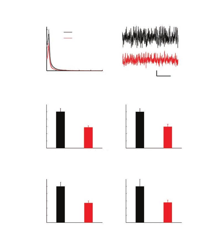
Orbitofrontal dysfunction in an OCD mouse model
quality. Among them, we recorded 215 single units from and depth in either WT or Sapap3 KO mice (Spearman rank
Sapap3 KO mice: 182 were classified as putative pyramidal correlation coefficient: WT putative interneurons r = 0.02, p =
neurons and 30 were classified as putative interneurons, based 0.94; KO putative interneurons r = 0.29, p = 0.12; WT puta-
on action potential waveforms. We recorded 147 single units tive pyramidal neurons r = −0.15, p = 0.11; KO putative
from WT littermates: 112 were classified as putative pyrami- pyramidal neurons r = −0.04, p = 0.57; Fig. 3E and F).
dal neurons and 21 were classified as putative interneurons
(Fig. 3A and B). Increased bursting activity of lOFC putative pyramidal
The firing rate of putative interneurons while the mouse neurons in Sapap3 KO mice
was at rest increased in Sapap3 KO mice (WT mean ± SEM =
18.07 ± 1.50 Hz, median = 17.62 Hz; KO mean ± SEM = 22.56 The overall firing pattern (not merely the firing rate) deter-
± 1.31 Hz, median = 23.30 Hz; p = 0.02 Wilcoxon rank sum mines neuronal function. Although we did not see any
test; Fig. 3C). Interestingly, however, the firing rate of puta- changes in the firing rate of pyramidal cells, we sought to
tive pyramidal neurons at rest was unchanged between WT compare their spike patterns between WT and Sapap3 KO
and Sapap3 KO mice (WT mean ± SEM = 2.75 ± 0.30 Hz, me- mice. In Sapap3 KO mice, lOFC putative pyramidal neurons
dian = 1.62 Hz; KO mean ± SEM = 2.78 ± 0.23 Hz, median = showed a notable enhancement of bursting activity com-
1.50 Hz; p = 0.63 Wilcoxon rank sum test; Fig. 3D), suggesting pared with WT littermates (Fig. 4). Putative pyramidal neur
intricate network imbalances in this mouse model with OCD- ons in Sapap3 KO mice fired more bursts of doublet or triplet
like behaviours. For both putative pyramidal neurons and spikes with very short intra-burst ISI (< 10 ms). The number
interneurons, there was no correlation between firing rate of bursts per minute per neuron was significantly increased
A B
FrA
PrL
mOFC
vOFC
lOFC 350
dlOFC μm
AOD
AOL
AOM
50 μm
AOV
200 μm
C D
Movement
Stationary
frequency (Hz) speed
15
Instant
10 Locomotion
5
0 500 μV
0 50 100 150 200 250 ms
Time (s)
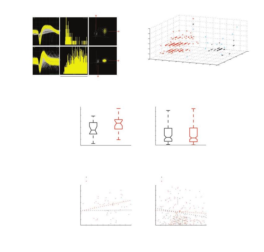
Fig. 1: Recording position and movement modulation of neuronal activity in lOFC. (A) Left: the red shadow summa-
rizes the recording region, which was largely in the lOFC and sometimes also included the very medial portion of the
dlOFC. Right: a histology example showing the tracks of the 4 electrode shanks reviewed by DiI (a fluorescent lipo-
philic cationic indocarbocyanine dye; red). (B) Electrode map. The recording electrodes had 4 shanks spaced by
200 μm. Each shank had 8 recording sites spaced by 50 μm. (C) An example of movement modulation of the spike
activity of a putative pyramidal neuron in the lOFC of a WT mouse. This neuron decreased firing rate during locomo-
tion. Horizontal bars indicate movement bouts. (D) An example of movement modulation of the LFP in the lOFC of a
WT mouse. Black trace in the upper panel shows LFP when stationary. Grey trace in the lower panel shows LFP dur-
ing locomotion. AOD = anterior olfactory area, dorsal part; AOL = anterior olfactory area, lateral part; AOM = anterior
olfactory area, medial part; AOV = anterior olfactory area, ventral part; dlOFC = dorsolateral orbitofrontal cortex; FrA =
frontal association cortex; LFP = local field potential; lOFC = lateral orbitofrontal cortex; mOFC = medial orbitofrontal
cortex; PrL = prelimbic cortex; vOFC = ventral orbitofrontal cortex; WT = wild type.
J Psychiatry Neurosci 2019;44(2) 123Lei et al
in Sapap3 KO mice (WT mean ± SEM = 12.4 ± 1.9 times/min, bursting activity seen in Sapap3 KO mice may enable lOFC
median = 5.8 times/min; KO mean ± SEM = 18.2 ± 2.0 times/ pyramidal neurons to provide a stronger output and drive
min, median = 10.7 times/min; p = 0.001, Wilcoxon rank sum increased activity in the downstream structures of the orbito–
test; Fig. 4C). The percentage of spikes per neuron in the fronto–striatal circuit in this OCD mouse model, reflecting
bursting mode was also significantly increased in Sapap3 KO specific pathologic neural processes in lOFC that underlie
mice (WT mean ± SEM = 22.9 ± 2.1%, median = 16.1%; KO phenotypes relevant to OCD.
mean ± SEM = 32.3 ± 1.8%, median = 29.0%; p < 0.001,
Wilcoxon rank sum test; Fig. 4D). The intra-burst ISI was sig- Increased firing variability for both neuronal types in Sapap3
nificantly shorter in Sapap3 KO mice compared with WT mice KO mice
(WT mean ± SEM = 6.60 ± 0.13 ms; KO mean ± SEM = 6.23 ±
0.09 ms; p = 0.016, Wilcoxon rank sum test). Bursts with short Both lOFC putative pyramidal neurons and interneurons in
intra-burst ISI are more reliable and efficient for eliciting syn- Sapap3 KO mice exhibited enhanced discharge variability
aptic transmission than tonic firing.43 Therefore, the increased compared to WT littermates. To measure firing variability,
A × 108
B
15
WT (n = 21)
KO (n = 24)
10
Power
5
0
10 20 30 40 50 500 μV
5s
Frequency (Hz)
C δ (1.5–4 Hz) D Θ (4–11 Hz)
Normalized power
Normalized power
1 1
0.8 0.8
*** ***
0.6 0.6
0.4 0.4
0.2 0.2
0 0
WT (n = 21) KO (n = 24) WT KO
E β (11–30 Hz) F γ (30–100 Hz)
Normalized power
Normalized power
1 1
0.8 0.8
*** *
0.6 0.6
0.4 0.4
0.2 0.2
0 0
WT KO WT KO
Fig. 2: Reduced lOFC LFP oscillation power in Sapap3 KO mice. (A) Averaged LFP power spectrogram
of WT and Sapap3 KO mice. Shading represents standard error of the mean. (B) Representative LFP
raw recording traces from WT mice (black) and Sapap3 KO mice (red). Traces were from mice with an
LFP power spectrogram closest to the mean values of the corresponding groups. (C, D, E, F) Compari-
son of lOFC LFP oscillation power in δ, θ, β and γ bands between WT and Sapap3 KO mice, respec-
tively. *p < 0.05, ***p < 0.001, Wilcoxon rank sum test. Error bars represent standard error of the mean.
KO = knockout; LFP = local field potential; lOFC = lateral orbitofrontal cortex; WT = wild type.
124 J Psychiatry Neurosci 2019;44(2)Orbitofrontal dysfunction in an OCD mouse model
A Noise
B
Putative pyramidal neurons
Putative interneurons
Single unit
Trough half width (μs)
Unclassified
200
Single unit
100
1
tio
2 k ra
600 400 3 h/pea
Peak half widt
200 ug
h (μs) Tro
0 20 ms Noise
C Interneurons D Pyramidal neurons
* 8
Firing rate (Hz)
Firing rate (Hz)
30
6
20 4
2
10
0
WT (n = 21) KO (n = 30) WT (n = 112) KO (n = 182)
E Interneurons F Pyramidal neurons
WT (n = 21) WT (n = 112)
KO (n = 30) KO (n = 182)
40 8
Firing rate (Hz)
Firing rate (Hz)
30 6
4
20
2
10
0
1.5 1.6 1.7 1.8 1.9 1.5 1.6 1.7 1.8 1.9 2.0
Depth (mm) Depth (mm)
Fig. 3: The mean firing rate of lOFC putative interneurons increased in Sapap3 KO mice, but the mean firing rate of putative pyramidal
neurons did not change. (A, B) Isolation and classification of the recorded single units in lOFC. (A) Top panel: an example of an iso-
lated putative pyramidal single unit in lOFC. Bottom panel: an example of an isolated putative interneuron single unit in lOFC. Left to
right: overlay of the waveforms of the isolated single unit (yellow) and the noise waveforms (grey). Interspike interval histogram. Projec-
tion of the clusters correspondent to the unit and the noise (x axis: PC1, y axis: PC2, z axis: nonlinear energy). (B) Three-dimensional
scatter plot illustrating spike characteristics of all 362 single units recorded in the lOFC of WT and Sapap3 KO mice. Each unit is repre-
sented as a dot for peak width at half-peak amplitude (x axis), trough width at half trough amplitude (y axis) and ratio of trough to peak
amplitude (z axis). We identified 2 major clusters. Putative pyramidal neurons are shown in red. Putative interneurons are shown in
black. Units that did not meet the criteria for these classifications are shown in blue. (C) The mean firing rate of putative interneurons in-
creased in Sapap3 KO mice (red) compared to WT mice (black); *p = 0.02, Wilcoxon rank sum test. (D) The mean firing rate of putative
pyramidal neurons was similar between WT (black) and Sapap3 KO (red) mice; p = 0.63, Wilcoxon rank sum test. The whiskers in the
box plots cover 95% of the data. (E) We found no correlation between interneuron firing rate and depth. Spearman rank correlation
coefficient: WT, r = 0.02, p = 0.94; KO, r = 0.29, p = 0.12. (F) We found no correlation between pyramidal neuron firing rate and depth.
Spearman rank correlation coefficient: WT, r = −0.15, p = 0.11; KO, r = −0.04, p = 0.57. KO = knockout; lOFC = lateral orbitofrontal
cortex; PC = principal component; WT = wild type.
J Psychiatry Neurosci 2019;44(2) 125Lei et al
we adopted a method that was less sensitive to firing rate correct for multiple comparisons; Fig. 5). Cortical neurons
fluctuation over time than the coefficient of variation of ISIs.41 typically fire action potentials with high temporal precision.44
This method compared only adjacent ISIs by calculating CV2 Change of the spike timing influences information coding in
for adjacent ISIs (see Methods). several sensory modalities, such as olfaction,45 gustation,46
Neurons cannot fire as variably at a high rate as at a low audition47 and vision.48 The increased discharge variability of
rate because of the refractory period. To avoid comparing the pyramidal neurons and interneurons interferes with normal
periods when the neuron fires quickly with periods when the information coding and processing in OFC and may reflect
neuron fires slowly, we did not compute the mean CV2 over circuitry abnormalities for OCD-like behaviours.
the entire recording period. Instead, we computed the mean
CV2 for different ISI values. Both lOFC putative pyramidal Altered synchronization of lOFC putative pyramidal neurons
neurons and interneurons in Sapap3 KO mice exhibited en- and interneurons in Sapap3 KO mice
hanced discharge variability for ISIs from 10–190 ms com-
pared to WT mice (pFWE < 0.001 for both putative pyramidal Temporal precision of firing and a tightly maintained balance
neurons and putative interneurons, Wilcoxon rank sum test between excitation and inhibition is critical to normal neural
for each ISI range; family-wise error rate was calculated to synchronization. Because we observed altered firing variability
A
a1
C
a2 a3 0.1 s 50 ***
Number of short ISI
40
60 60
events/minute
30
Counts
40 40
20
20 20 10
0 0 0
0 20 40 60 80 100 0 5 10 15 20 WT (n = 112) KO (n = 182)
Time (ms)
B
b1
D
b2 b3 0.1 s ***
% spikes with short ISI
0.8
15 15
0.6
10 10
Counts
0.4
5 5
0.2
0 0 0
0 20 40 60 80 100 0 5 10 15 20 WT (n = 112) KO (n = 182)
Time (ms)
Fig. 4: The lOFC putative pyramidal neurons showed increased bursting activity in Sapap3 KO mice. (A) A representative example of bursty
pyramidal neurons in Sapap3 KO mice: a1 shows the raw recording trace showing 2 bursts with short intra-burst ISI (intra-burst ISI < 10 ms,
shown by arrows); a2 shows ISI distribution (bin size 1 ms); a3 shows an enlarged view of the ISI distribution from 0–20 ms to better demon-
strate the short intra-burst ISI. (B) A representative example of non-bursty pyramidal neurons in Sapap3 KO mice. (C) The number of bursts
per minute per neuron increased in Sapap3 KO mice; ***p = 0.001, Wilcoxon rank sum test. (D) The percentage of spikes per neuron in the
bursting mode increased in Sapap3 KO mice; ***p < 0.001; Wilcoxon rank sum test. The whiskers in the box plots cover 95% of the data.
KO = knockout; ISI = interspike interval; lOFC = lateral orbitofrontal cortex; WT = wild type.
126 J Psychiatry Neurosci 2019;44(2)Orbitofrontal dysfunction in an OCD mouse model
and distinct perturbations of excitatory and inhibitory neur utes to decreased LFP oscillations in multiple frequency
ons, we then sought to investigate whether the levels of syn- bands is challenging. Nevertheless, changes in neuronal
chronous activity in lOFC would change in Sapap3 KO mice activity synchronization do contribute to LFP oscillation
by calculating the STA of LFP. Because the LFP averages over alterations. We found that putative interneurons experienced
many neurons, STA is more sensitive than cross-correlation in less synchronized inhibition after firing an action potential
detecting local neuronal synchronization.49 Troughs in STA of and putative pyramidal neurons experienced less synchro-
LFP correspond to depolarization of intracellularly measured nized inhibition before firing an action potential. This re-
membrane potential, reflecting summed excitatory events in a duced synchronous activity in the lOFC may contribute to
pool of neurons. Upward deflections in STA of LFP cor decreased LFP oscillation power in multiple frequency
respond to a drop in membrane potential.50 In both WT and bands. Consistent with our results, animal studies have
Sapap3 KO mice, lOFC putative pyramidal neurons and inter- shown that deep-brain stimulation of the nucleus accumbens,
neurons all fired preferentially at the lowest point of the which can effectively alleviate OCD symptoms, elevated
trough. The percentage of cells entrained to the LFP oscilla- spontaneous LFP oscillation power in the δ, β and γ fre-
tions as measured by significant fluctuations in the STA quency bands in the OFC in rats. 51,52 In contrast, low-
around the time of spike was similar between WT and Sapap3 frequency deep-brain stimulation of the nucleus accumbens,
KO mice (WT putative pyramidal neurons 70.6 ± 9.5% mean ± which is ineffective in OCD, exerted no effect on LFP in the
SEM, KO putative pyramidal neurons 72.0 ± 7.0%, p = 0.9, OFC.51,52 Given that LFP oscillation power in the OFC was
Wilcoxon rank sum test; WT putative interneurons 96.4 ± decreased in an OCD mouse model and increased with deep-
3.9%, KO putative interneurons 100%, p = 0.9, Wilcoxon rank brain stimulation of the nucleus accumbens in rats, altera-
sum test). However, the shape of averaged STA of both puta- tions in LFP may serve as a potential neurophysiological bio-
tive pyramidal neurons and interneurons differed in Sapap3 marker to be further examined in people with OCD.
KO mice. The central trough of the STA of both putative pyra- Inhibitory interneurons form reciprocal connections
midal neurons and interneurons was reduced in Sapap3 KO broadly with pyramidal neurons, and so are well positioned
mice (Fig. 6). This reduction may have been due to the re- to coordinate the timing of pyramidal cell activity, regulate
duced power of LFP oscillation in Sapap3 KO mice. In WT information processing and gate information flow. Compro-
mice, the averaged STA of putative interneurons exhibited a mised cortical inhibitory interneurons have been implicated
broad second peak after the central trough (Fig. 6A). This in multiple psychiatric and neurologic disorders, including
peak was significantly reduced in Sapap3 KO mice, indicating schizophrenia,53 autism54 and epilepsy.55 However, little re-
that interneurons experienced less synchronized inhibition search on interneurons has been done in people with OCD or
after firing a spike. The peak ahead of the central trough of the in animal models. It has been reported that Sapap3 KO mice
STA of putative pyramidal neurons was eliminated in Sapap3 have a decreased number of PV-expressing interneurons in
KO mice (Fig. 6B), indicating that synchronized inhibition on the centromedial striatum.37 For the first time, our work
membrane potential, which sculpts the time window when an found elevated spontaneous activity and enhanced discharge
action potential can occur, was reduced in Sapap3 KO mice. variability of putative inhibitory interneurons in the lOFC of
As a result, in Sapap3 KO mice, the spike timing of individual Sapap3 KO mice. Because the lOFC directly projects to the
lOFC pyramidal neurons may become less accurate. This was striatum, inhibition is disrupted in both parts of the cortical–
consistent with our finding that the firing variability of lOFC striatal circuit in this OCD mouse model. Inhibitory inter
putative pyramidal neurons increased in Sapap3 KO mice. neurons are critical for the normal function of the OFC. The
Taken together, the changes in synchrony, along with the activity of inhibitory interneurons in the OFC showed strong
spike activity pattern and LFP changes reported in previous behaviour correlates.56 Compromised inhibitory interneurons
sections, point to the lOFC as a malfunctioning neural sub- in the OFC altered pyramidal neuron activity correlations
strate for behavioural phenotypes relevant to OCD. with decision and reward, and impaired reversal learning.40
The activity alterations of interneurons we found may dis-
Discussion rupt the normal function of the lOFC and help identify one
aspect of malfunction in this region for behaviours relevant
Cognitive and executive function requires the coordinated to OCD. Interneurons also play a fundamental role in rhyth-
activity of large-scale networks. Deficits in temporal coordin mogenesis. The elevated spontaneous activity and discharge
ation in the OFC can lead to disruption of its normal function variability of interneurons may be causally involved in the
and be involved in the pathophysiology of OCD. In the pres- LFP alterations in lOFC, as we found in Sapap3 KO mice.
ent study, we have reported alterations in LFP oscillations in Alternatively, the increased spontaneous activity of inter
the lOFC of an OCD mouse model, to our knowledge, for the neurons could represent adaptive, homeostatic or unrelated
first time. Specifically, we found that Sapap3 KO mice exhib- processes to compensate for other primary abnormalities of
ited reduced power in δ, θ, β and γ oscillations at rest. The the lOFC in Sapap3 KO mice.
neural substrates contributing to the different frequency Although the OFC is thought to play a critical role in OCD,
bands of LFP oscillations and the mechanisms by which these there are discrepancies in the directionality of findings about
oscillations are generated are not well understood. Therefore, how the baseline activity of the OFC is altered in people with
a mechanistic interpretation of how the altered activity pat- OCD or animal models. These discrepancies may be due to
tern of lOFC pyramidal neurons and interneurons contrib- several factors, including the heterogeneity of the disorder,
J Psychiatry Neurosci 2019;44(2) 127Lei et al
c omorbidities, medication history and the different subdivi- dal neurons measured by electrophysiological recording was
sions of OFC analyzed. Our study excluded these confounders similar between WT and Sapap3 KO mice,37 consistent with our
by focusing on the lOFC in an OCD mutant mouse model, and results. The other reported upregulated baseline activity in the
with clear classification of different cell types. Two studies OFC in Slitrk5 KO mice measured by FosB expression.57
have assessed OFC activity change in OCD mouse models. Because this study relied on molecular markers of cell activity,
One found that the baseline activity of lOFC putative pyrami- we do not know the details of activity pattern change for
A Putative pyramidal neurons B Putative interneurons
1.4 1.4
1.2 1.2
1 1
0.8 0.8
CV2
CV2
0.6 0.6
KO (n = 182) KO (n = 30)
0.4 0.4
WT (n = 112) WT (n = 21)
0.2 0.2
0 0
0 50 100 150 200 250 0 50 100 150 200
Mean of ISI pair (ms) Mean of ISI pair (ms)
Fig. 5: The lOFC putative pyramidal neurons and interneurons exhibited enhanced discharge variability in Sapap3 KO mice (red) compared
with WT littermates (black). (A) Mean CV2 of lOFC putative pyramidal neurons plotted against the mean of the 2 adjacent ISIs used to compute
CV2. (B) Mean CV2 of lOFC putative interneurons plotted against the mean of the 2 adjacent ISIs used to compute CV2. The x axis is the mean of
the 2 adjacent ISIs used to compute CV2. The lines are the mean CV2 values in logarithmically spaced bins. The ratio between bin boundaries
was 1.3. We chose logarithmic binning because the upper limit of CV2 at shorter ISIs changes much more rapidly than at longer ISIs. Shading
represents standard error of the mean. KO = knockout; ISI = interspike interval; lOFC = lateral orbitofrontal cortex; WT = wild type.
A Putative interneurons B Putative pyramidal neurons
0 0
STA (μV)
STA (μV)
–20
WT (n = 21) –20 WT (n = 112)
KO (n = 30) KO (n = 182)
–40
–40
–60
–3 –2 –1 0 1 2 3 –3 –2 –1 0 1 2 3
Time (s) Time (s)
Fig. 6: Comparisons of lOFC ensemble synchronization in WT (black) and Sapap3 KO (red) mice. (A) Averaged STA of LFP of lOFC putative
interneurons. (B) Average STA of LFP of lOFC putative pyramidal neurons. Black horizontal bars indicate ranges with significant difference
between WT and Sapap3 KO mice. Shading represents standard error of the mean. KO = knockout; LFP = local field potential; lOFC = lateral
orbitofrontal cortex; STA = spike-triggered average; WT = wild type.
128 J Psychiatry Neurosci 2019;44(2)Orbitofrontal dysfunction in an OCD mouse model
s pecific neuronal types. The increased bursting activity of pu- executive brain functions. Its neuronal activity is modulated
tative pyramidal neurons we found could cause this increase by many behaviours, including grooming-associated move-
in FosB expression. The discrepancy may also result from the ments themselves. To exclude such confounders, we com-
different OCD animal models used (Sapap3 KO v. Slitrk5 KO), pared neuronal activity between WT and Sapap3 KO mice,
different cell types (putative pyramidal neurons v. all cells) recorded only when the mice were stationary. To collect
and different subregions of the OFC examined (lOFC v. the enough data during stationary periods, we applied a head-
entire OFC). The medial and lateral OFC perform different fixed configuration instead of a free-behaviour configuration,
functions, such as processing positive versus negative va- because mice stayed stationary for most of the time during
lence.35,36 The activity of these 2 subregions may be differen- head-fixed recording (percent of time spent stationary, mean
tially affected in OCD mouse models, giving rise to inconsis- ± SEM: WT 90.1 ± 2.3%, KO 90.8 ± 3.5%). In contrast, mice
tent results when examining the lOFC versus the entire OFC. usually moved most of the time during free-movement
Although the mean firing rate of lOFC pyramidal neurons recording (including both locomotion and fine movement),
was similar between WT and Sapap3 KO mice, their activity leaving few stationary periods for effective data analysis.
pattern changed dramatically in Sapap3 KO mice. Specifically, Comparing resting neuronal activity between WT and Sapap3
lOFC pyramidal neurons exhibited significantly increased KO mice required very high recording and spike-sorting
bursting activity with short intra-burst ISI (< 10 ms). Overall quality. We applied very strict criteria for spike sorting and
activity pattern determines neuronal function, not merely included only very well isolated single units to ensure the
firing rate. Bursts with short intra-burst ISI have special im- accuracy of our results. Specifically, the cluster of the isolated
portance in brain function. Compared with tonic firing, burst single unit had to be well separated from other clusters with-
firing is more reliable for eliciting synaptic transmission, pro- out any overlapping of the edge, because edge overlapping
vides stronger output, enhances signal-to-noise ratio and in spike sorting results in contamination by other units or
facilitates synaptic plasticity.43 The increased bursting activity loss of spikes of the isolated unit. Either situation can signifi-
of lOFC pyramidal neurons may provide pathologic stronger cantly affect the measurement of firing rate and firing vari-
output through the OFC–striatal circuit and drive increased ability. Another limitation was that we investigated func-
activity in the striatum of Sapap3 KO mice, as reported in pre- tional abnormalities without establishing causality for these
vious studies.37,58 Another study suggested that sustained in- functional changes and the OCD-like behaviours. Our rea-
crease in synaptic strength from the OFC pyramidal neurons sons were as follows. First, this was a pioneering in vivo elec-
to ventral striatum synapses led to increased repetitive be- trophysiological study of OFC dysfunction in an OCD animal
haviour in mice.59 Many clinical and animal studies have sug- model; characterizing the functional abnormalities was the
gested that hyperactivity in the cortico–striato–thalamo– first step and can serve an important foundation for future
cortical circuit is associated with OCD pathology.2,3,10,11 The work. Second, artificially generating bursts with very short
enhanced bursting activity of lOFC pyramidal neurons may intra-burst ISI without changing the mean firing rate is very
drive hyperactivity in the cortico–striato–thalamo–cortical challenging; we have not found an appropriate way to
circuit and contribute to OCD-like behaviours in Sapap3 KO manipulate the oscillation power of broad frequency bands
mice. A recent study reported that a depression-like state in the LFP without changing the mean firing rate of excit-
depended critically on a bursting mode of firing in the lateral atory neurons. Nevertheless, causal manipulation is defi-
habenula in rats and mice.60 The bursting activity of neurons nitely a key future experiment that may require us to develop
in the lateral habenula was greatly enhanced in rat and new manipulation techniques.
mouse models of depression, and reducing their bursting
activity elicited antidepressant effects. Increasing bursting Conclusion
activity by optogenetics was sufficient to induce depression-
like behaviours. This study suggested that abnormal bursting Here, we have provided the first direct in vivo electrophysio-
activity in a single nucleus could lead to symptoms relevant logical evidence of detailed functional alterations in different
to a psychiatric disorder. Abnormal bursting activity has not neuronal types and local network dysfunction in the lOFC in
been studied in people with OCD or in animal models. In a phenotypes relevant to OCD. These findings advance our
future study, we plan to investigate whether fluoxetine can understanding of the neuropathophysiology and circuitry
suppress the abnormal bursting activity of lOFC pyramidal mechanisms that underlie OCD-like behaviours, and may
neurons, accompanied by alleviation of OCD-like behaviours help generate and refine hypotheses for further investigation.
in Sapap3 KO mice. We also plan to investigate whether artifi- For example, the LFP alterations and increased bursting ac-
cially increasing the bursting activity of lOFC pyramidal tivity may be useful biomarker candidates for further exami-
neurons can induce symptoms relevant to OCD. nation in people with OCD.
Limitations Acknowledgements: We thank Dr. Kathleen B. Quast for her com-
ments on the manuscript. This work was supported by grants from
the National Natural Science Foundation of China (grant no.
One limitation of this study was that we examined activity
31371108, 31171051), the Natural Science Foundation of Beijing
pattern alterations of lOFC neurons only when the mice were (grant no. 5132007, 5112008), the General Program of Science and
resting; we did not examine grooming-related activity. As an Technology Development Project of Beijing Municipal Education
association cortex, the OFC performs complex cognitive and Commission of China (grant no. KM201110025001), the Beijing
J Psychiatry Neurosci 2019;44(2) 129Lei et al
Municipal Technology Foundation for Selected Overseas Chinese 15. Schoenbaum G, Roesch MR, Stalnaker TA, et al. A new perspec-
Scholars, the Simons Initiative on Autism and the Brain Infrastruc- tive on the role of the orbitofrontal cortex in adaptive behaviour.
ture Grant Program. Nat Rev Neurosci 2009;10:885-92.
Affiliations: From the Department of Neurobiology, Beijing Institute 16. Gillan CM, Papmeyer M, Morein-Zamir S, et al. Disruption in the bal-
for Brain Disorders, Beijing Centre of Neural Regeneration and Re- ance between goal-directed behavior and habit learning in obsessive–
compulsive disorder. Am J Psychiatry 2011;168:718-26.
pair, Key Laboratory for Neurodegenerative Diseases of the Ministry
of Education, Capital Medical University, Beijing, China (Lei, Lai, 17. Chamberlain SR, Menzies L, Hampshire A, et al. Orbitofrontal
Sun, Xu); the McGovern Institute for Brain Research, Department of dysfunction in patients with obsessive–compulsive disorder and
Brain and Cognitive Sciences, Massachusetts Institute of Technology, their unaffected relatives. Science 2008;321:421-2.
Cambridge, Massachusetts (Feng); and the Stanley Center for Psychi-
18. Alptekin K, Degirmenci B, Kivircik B, et al. Tc-99m HMPAO brain
atric Research, Broad Institute of MIT and Harvard, Cambridge, perfusion SPECT in drug-free obsessive–compulsive patients with-
Massachusetts (Feng). out depression. Psychiatry Res 2001;107:51-6.
Competing interests: None declared. 19. Baxter LR Jr, Schwartz JM, Mazziotta JC, et al. Cerebral glucose met-
abolic rates in nondepressed patients with obsessive–compulsive
Contributors: H. Lei, Q. Xu and G. Feng designed the study. H. Lei, disorder. Am J Psychiatry 1988;145:1560-3.
J. Lai and X. Sun acquired the data, which H. Lei and J. Lai analyzed.
H. Lei and G. Feng wrote the article, which all authors reviewed. All 20. Rubin RT, Villanueva-Meyer J, Ananth J, et al. Regional xenon 133
authors approved the final version to be published and can certify cerebral blood flow and cerebral technetium 99m HMPAO uptake
that no other individuals not listed as authors have made substantial in unmedicated patients with obsessive–compulsive disorder and
contributions to the paper. matched normal control subjects. Determination by high-resolution
single-photon emission computed tomography. Arch Gen Psychiatry
1992;49:695-702.
References 21. Sawle GV, Hymas NF, Lees AJ, et al. Obsessional slowness. Func-
tional studies with positron emission tomography. Brain 1991;
1. Ruscio AM, Stein DJ, Chiu WT, et al. The epidemiology of obsessive– 114:2191-202.
compulsive disorder in the National Comorbidity Survey Replica- 22. Swedo SE, Schapiro MB, Grady CL, et al. Cerebral glucose metabo-
tion. Mol Psychiatry 2010;15:53-63. lism in childhood-onset obsessive–compulsive disorder. Arch Gen
2. Monteiro P, Feng G. Learning from animal models of obsessive– Psychiatry 1989;46:518-23.
compulsive disorder. Biol Psychiatry 2016;79:7-16. 23. Cottraux J, Gerard D, Cinotti L, et al. A controlled positron emission
tomography study of obsessive and neutral auditory stimulation in
3. Burguiere E, Monteiro P, Mallet L, et al. Striatal circuits, habits, and
obsessive–compulsive disorder with checking rituals. Psychiatry Res
implications for obsessive–compulsive disorder. Curr Opin Neurobiol
1996;60:101-12.
2015;30:59-65.
24. Rauch SL, Jenike MA, Alpert NM, et al. Regional cerebral blood flow
4. Ahmari SE. Using mice to model obsessive compulsive disorder:
measured during symptom provocation in obsessive–compulsive
from genes to circuits. Neuroscience 2016;321:121-37.
disorder using oxygen 15-labeled carbon dioxide and positron emis-
5. Pittenger C, Bloch MH, Williams K. Glutamate abnormalities in sion tomography. Arch Gen Psychiatry 1994;51:62-70.
obsessive compulsive disorder: neurobiology, pathophysiology,
25. Andreou C, Leicht G, Popescu V, et al. P300 in obsessive–compulsive
and treatment. Pharmacol Ther 2011;132:314-32.
disorder: source localization and the effects of treatment. J Psychiatr
6. Boedhoe PS, Schmaal L, Abe Y, et al. Distinct subcortical volume Res 2013;47:1975-83.
alterations in pediatric and adult OCD: a worldwide meta- and 26. Benkelfat C, Nordahl TE, Semple WE, et al. Local cerebral glucose
mega-analysis. Am J Psychiatry 2016;174:60-9. metabolic rates in obsessive–compulsive disorder. Patients treated
7. Anticevic A, Hu S, Zhang S, et al. Global resting-state functional with clomipramine. Arch Gen Psychiatry 1990;47:840-8.
magnetic resonance imaging analysis identifies frontal cortex, stria- 27. Le Jeune F, Verin M, N’Diaye K, et al. Decrease of prefrontal me-
tal, and cerebellar dysconnectivity in obsessive–compulsive disorder. tabolism after subthalamic stimulation in obsessive–compulsive
Biol Psychiatry 2014;75:595-605. disorder: a positron emission tomography study. Biol Psychiatry
8. Breiter HC, Rauch SL. Functional MRI and the study of OCD: from 2010;68:1016-22.
symptom provocation to cognitive-behavioral probes of cortico- 28. Nakao T, Nakagawa A, Yoshiura T, et al. Brain activation of pa-
striatal systems and the amygdala. Neuroimage 1996;4:S127-38. tients with obsessive–compulsive disorder during neuropsycho-
9. Milad MR, Rauch SL. Obsessive–compulsive disorder: beyond logical and symptom provocation tasks before and after symptom
segregated cortico-striatal pathways. Trends Cogn Sci 2012;16:43-51. improvement: a functional magnetic resonance imaging study. Biol
Psychiatry 2005;57:901-10.
10. Del Casale A, Kotzalidis GD, Rapinesi C, et al. Functional neuroimag-
ing in obsessive–compulsive disorder. Neuropsychobiology 2011;64: 29. Saxena S, Brody AL, Maidment KM, et al. Localized orbitofrontal
61-85. and subcortical metabolic changes and predictors of response to
paroxetine treatment in obsessive–compulsive disorder. Neuro-
11. Menzies L, Chamberlain SR, Laird AR, et al. Integrating evidence psychopharmacology 1999;21:683-93.
from neuroimaging and neuropsychological studies of obsessive–
compulsive disorder: the orbitofronto-striatal model revisited. 30. Swedo SE, Pietrini P, Leonard HL, et al. Cerebral glucose metab-
Neurosci Biobehav Rev 2008;32:525-49. olism in childhood-onset obsessive–compulsive disorder. Revisu-
alization during pharmacotherapy. Arch Gen Psychiatry 1992;
12. Menzies L, Achard S, Chamberlain SR, et al. Neurocognitive endophe- 49:690-4.
notypes of obsessive–compulsive disorder. Brain 2007;130:3223-36.
31. Welch JM, Lu J, Rodriguiz RM, et al. Cortico-striatal synaptic
13. Baxter MG, Parker A, Lindner CC, et al. Control of response selec- defects and OCD-like behaviours in Sapap3-mutant mice. Nature
tion by reinforcer value requires interaction of amygdala and orbital 2007;448:894-900.
prefrontal cortex. J Neurosci 2000;20:4311-9.
32. Boardman L, van der Merwe L, Lochner C, et al. Investigating
14. Schoenbaum G, Nugent SL, Saddoris MP, et al. Orbitofrontal SAPAP3 variants in the etiology of obsessive–compulsive disorder
lesions in rats impair reversal but not acquisition of go, no-go odor and trichotillomania in the South African white population. Compr
discriminations. Neuroreport 2002;13:885-90. Psychiatry 2011;52:181-7.
130 J Psychiatry Neurosci 2019;44(2)Orbitofrontal dysfunction in an OCD mouse model
33. Zuchner S, Wendland JR, Ashley-Koch AE, et al. Multiple rare 48. Rathbun DL, Alitto HJ, Weyand TG, et al. Interspike interval
SAPAP3 missense variants in trichotillomania and OCD. Mol analysis of retinal ganglion cell receptive fields. J Neurophysiol 2007;
Psychiatry 2009;14:6-9. 98:911-9.
34. Stewart SE, Yu D, Scharf JM, et al. Genome-wide association study 49. Fries P, Reynolds JH, Rorie AE, et al. Modulation of oscillatory
of obsessive–compulsive disorder. Mol Psychiatry 2013;18:788-98. neuronal synchronization by selective visual attention. Science
2001;291:1560-3.
35. Milad MR, Rauch SL. The role of the orbitofrontal cortex in anxiety
disorders. Ann N Y Acad Sci 2007;1121:546-61. 50. Steriade M. Grouping of brain rhythms in corticothalamic systems.
36. Kringelbach ML, Rolls ET. The functional neuroanatomy of the Neuroscience 2006;137:1087-106.
human orbitofrontal cortex: evidence from neuroimaging and 51. McCracken CB, Grace AA. High-frequency deep brain stimulation
neuropsychology. Prog Neurobiol 2004;72:341-72. of the nucleus accumbens region suppresses neuronal activity and
37. Burguiere E, Monteiro P, Feng G, et al. Optogenetic stimulation of selectively modulates afferent drive in rat orbitofrontal cortex in
lateral orbitofronto-striatal pathway suppresses compulsive be- vivo. J Neurosci 2007;27:12601-10.
haviors. Science 2013;340:1243-6. 52. McCracken CB, Grace AA. Nucleus accumbens deep brain stimulation
38. Dombeck DA, Khabbaz AN, Collman F, et al. Imaging large-scale produces region-specific alterations in local field potential oscillations
neural activity with cellular resolution in awake, mobile mice. and evoked responses in vivo. J Neurosci 2009;29:5354-63.
Neuron 2007;56:43-57.
53. Lewis DA, Hashimoto T, Volk DW. Cortical inhibitory neurons
39. Zhan C, Luo M. Diverse patterns of odor representation by neurons and schizophrenia. Nat Rev Neurosci 2005;6:312-24.
in the anterior piriform cortex of awake mice. J Neurosci 2010;30:
16662-72. 54. Lawrence YA, Kemper TL, Bauman ML, et al. Parvalbumin-,
calbindin-, and calretinin-immunoreactive hippocampal inter-
40. Bissonette GB, Schoenbaum G, Roesch MR, et al. Interneurons are neuron density in autism. Acta Neurol Scand 2010;121:99-108.
necessary for coordinated activity during reversal learning in orbi-
tofrontal cortex. Biol Psychiatry 2015;77:454-64. 55. Aronica E, Redeker S, Boer K, et al. Inhibitory networks in epilepsy-
associated gangliogliomas and in the perilesional epileptic cortex.
41. Holt GR, Softky WR, Koch C, et al. Comparison of discharge vari- Epilepsy Res 2007;74:33-44.
ability in vitro and in vivo in cat visual cortex neurons. J Neurophysiol
1996;75:1806-14. 56. Quirk MC, Sosulski DL, Feierstein CE, et al. A defined network of fast-
spiking interneurons in orbitofrontal cortex: responses to behavioral
42. Pogarell O, Juckel G, Mavrogiorgou P et al. Symptom-specific contingencies and ketamine administration. Front Syst Neurosci
EEG power correlations in patients with obsessive-compulsive 2009;3:13.
disorder. Int J Psychophysiol 2006;62:87-92.
57. Shmelkov SV, Hormigo A, Jing D, et al. Slitrk5 deficiency impairs
43. Lisman JE. Bursts as a unit of neural information: making unreli- corticostriatal circuitry and leads to obsessive–compulsive-like
able synapses reliable. Trends Neurosci 1997;20:38-43. behaviors in mice. Nat Med 2010:16:598-602.
44. Mainen ZF, Sejnowski TJ. Reliability of spike timing in neocortical 58. Mintzopoulos D, Gillis TE, Robertson HR, et al. Striatal magnetic
neurons. Science 1995;268:1503-6. resonance spectroscopy abnormalities in young adult SAPAP3
45. Wehr M, Laurent G. Odour encoding by temporal sequences of knockout mice. Biol Psychiatry Cogn Neurosci Neuroimaging 2016;1:
firing in oscillating neural assemblies. Nature 1996;384:162-6. 39-48.
46. Roussin AT, D’Agostino AE, Fooden AM, et al. Taste coding in the 59. Ahmari SE, Spellman T, Douglass NL, et al. Repeated cortico-striatal
nucleus of the solitary tract of the awake, freely licking rat. J Neurosci stimulation generates persistent OCD-like behavior. Science
2012;32:10494-506. 2013;340:1234-9.
47. Lu T, Liang L, Wang X. Temporal and rate representations of time- 60. Yang Y, Cui YH, Sang KN, et al. Ketamine blocks bursting in the
varying signals in the auditory cortex of awake primates. Nat Neurosci lateral habenula to rapidly relieve depression. Nature 2018;
2001;4:1131-8. 554:317.
J Psychiatry Neurosci 2019;44(2) 131You can also read