Abortive intussusceptive angiogenesis causes multi-cavernous vascular malformations - eLife
←
→
Page content transcription
If your browser does not render page correctly, please read the page content below
RESEARCH ARTICLE

                                       Abortive intussusceptive angiogenesis
                                       causes multi-cavernous vascular
                                       malformations
                                       Wenqing Li1, Virginia Tran1, Iftach Shaked2, Belinda Xue1, Thomas Moore3,
                                       Rhonda Lightle3, David Kleinfeld2,4, Issam A Awad3, Mark H Ginsberg1*
                                       1
                                        Department of Medicine, University of California, San Diego, La Jolla, United
                                       States; 2Department of Physics, University of California, San Diego, La Jolla, United
                                       States; 3Neurovascular Surgery Program, Section of Neurosurgery, Department of
                                       Surgery, University of Chicago School of Medicine and Biological Sciences, Chicago,
                                       United States; 4Section of Neurobiology, University of California San Diego, La
                                       Jolla, United States

                                       Abstract Mosaic inactivation of CCM2 in humans causes cerebral cavernous malformations
                                       (CCMs) containing adjacent dilated blood-filled multi-cavernous lesions. We used CRISPR-Cas9
                                       mutagenesis to induce mosaic inactivation of zebrafish ccm2 resulting in a novel lethal multi-
                                       cavernous lesion in the embryonic caudal venous plexus (CVP) caused by obstruction of blood flow
                                       by intraluminal pillars. These pillars mimic those that mediate intussusceptive angiogenesis;
                                       however, in contrast to the normal process, the pillars failed to fuse to split the pre-existing vessel
                                       in two. Abortive intussusceptive angiogenesis stemmed from mosaic inactivation of ccm2 leading
                                       to patchy klf2a overexpression and resultant aberrant flow signaling. Surviving adult fish
                                       manifested histologically typical hemorrhagic CCM. Formation of mammalian CCM requires the
                                       flow-regulated transcription factor KLF2; fish CCM and the embryonic CVP lesion failed to form in
                                       klf2a null fish indicating a common pathogenesis with the mammalian lesion. These studies describe
*For correspondence:                   a zebrafish CCM model and establish a mechanism that can explain the formation of characteristic
mhginsberg@ucsd.edu                    multi-cavernous lesions.

Competing interests: The
authors declare that no
competing interests exist.
                                       Introduction
Funding: See page 18                   Cerebral cavernous malformations (CCMs) are central nervous system (CNS) vascular anomalies that
Received: 15 August 2020               lead to significant morbidity and mortality (Leblanc et al., 2009). CCMs affect ~1/200 humans and
Accepted: 19 May 2021                  cause a lifelong risk of stroke and other neurological sequelae for which there is no pharmacological
Published: 20 May 2021                 therapy. Heterozygous loss of function mutations of three CCM genes (KRIT1(CCM1), CCM2, and
                                       PDCD10(CCM3)) are associated with development of venous capillary dysplasias with hemorrhage
Reviewing editor: Elisabetta
                                       and increased vascular permeability (Mikati et al., 2015) characteristic of CCM (Leblanc et al.,
Dejana, FIRC Institute of
Molecular Oncology
                                       2009). Heterozygous patients often exhibit a ‘second hit’ on the normal CCM allele in CCM endo-
Foundationtion (IFOM), Italy           thelial cells (Akers et al., 2009; McDonald et al., 2011) and loss of function of P53 or Msh2, genes
                                       that maintain genome stability, ‘sensitize’ Krit1+/- or Pdcd10+/- mice for development of CCM. Thus,
    Copyright Li et al. This article
                                       CCMs are likely to arise following mosaic inactivation of both alleles of a given CCM gene. Neonatal
is distributed under the terms of
                                       endothelial-specific inactivation of murine Krit1, Pdcd10 (Ccm3), or Ccm2 results in cerebellar and
the Creative Commons
Attribution License, which             retinal vascular lesions that resemble CCM (Boulday et al., 2011; Chan et al., 2011; Jenny Zhou
permits unrestricted use and           et al., 2016). Thus, human CCMs arise from venous capillaries as a consequence of mosaic inactiva-
redistribution provided that the       tion of these genes in endothelial cells.
original author and source are            The most striking form of CCM are large complexes containing adjacent dilated blood-filled thin-
credited.                              walled vessels with surrounding hemosiderin deposition indicative of chronic bleeding. These are the

Li et al. eLife 2021;10:e62155. DOI: https://doi.org/10.7554/eLife.62155                                                               1 of 22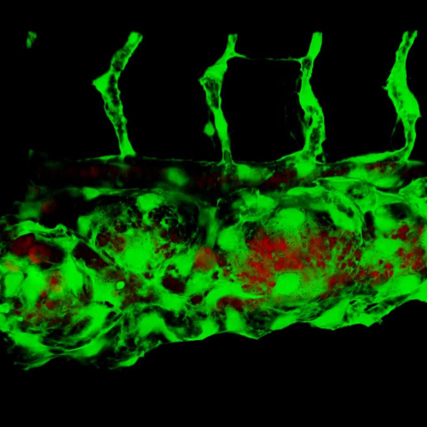
Research Article                                                                                           Developmental Biology

                                    lesions that are surgically resected either to relieve either mass effects or recurrent bleeding and are
                                    therefore clinically relevant. The cellular mechanism whereby these hallmark multi-cavernous lesions
                                    form is unknown and we reasoned that the genetic and experimental accessibility of the zebrafish
                                    and the optical transparency of its embryos and larvae (Gore et al., 2018) could enable in vivo analy-
                                    sis of CCM development. In particular, the fish is amenable to examination of genetic perturbations
                                    by study of mutant fish, CRISPR/Cas9 mutagenesis, or morpholino silencing (Stainier et al., 2017).
                                    The zebrafish ‘heart of glass’ defect in cardiac development led to the discovery of HEG1 (a binding
                                    partner for KRIT1; Gingras et al., 2012) and to the demonstration that loss of KRIT1 (santa) or its
                                    binding partner, CCM2 (valentine), produced an identical cardiac phenotype (Mably et al., 2006).
                                    Importantly, neither mutation nor silencing of any of these genes have been reported to produce
                                    zebrafish CCM. Second, silencing of pdcd10 does not produce either cardiac dilation or CCM in the
                                    fish (Yoruk et al., 2012). Furthermore, there is compelling evidence that HEG1 mutations do not
                                    produce CCM in mice or humans (Zheng et al., 2014). These data indicate important differences
                                    between the consequences of deletion of these genes in the endocardium and in brain endothelial
                                    cells and underscore the need for a zebrafish model of authentic CCM.
                                        Here, we used CRISPR-Cas9 mutagenesis of ccm2 to recapitulate the mosaic inactivation of a
                                    CCM gene. We observed a highly penetrant novel phenotype in embryos. By 2 days post fertilization
                                    (dpf), ~30% of embryos developed striking segmental dilatation of the caudal vein associated with
                                    slowed blood flow and formation of multiple dilated contiguous blood-filled chambers. We show
                                    that these lesions are caused by the intraluminal extension of pillars that lead to physical obstruction
                                    of blood flow. These pillars resemble the first steps of intussusceptive angiogenesis; however, in con-
                                    trast to the physiological process, these pillars fail to organize and fuse together to split the pre-
                                    existing vessel in two. We show that this process of abortive intussusceptive angiogenesis is due to
                                    mosaic inactivation of ccm2 leading to aberrant flow signaling and that, as in CCMs, KLF2 transcrip-
                                    tion factors play an important role in the formation of these lesions. In addition, typical CCM formed
                                    in the brains of all surviving adult and juvenile fish. As in murine CCM models, KLF2 transcription fac-
                                    tors played a key role in the pathogenesis of zebrafish CCMs. Thus, abortive intussusceptive angio-
                                    genesis, as a consequence of aberrant flow signaling, leads to formation of multi-cavernous venous
                                    lesions in zebrafish embryos that resemble human multi-cavernous CCM.

                                    Results
                                    ccm2 CRISPR zebrafish embryos display segmental dilation of the
                                    caudal venous plexus
                                    As noted above, although null mutations of krit1 and ccm2 result in cardiac dilation and some vascu-
                                    lar abnormalities, CCMs have not been observed in zebrafish (Mably et al., 2006; Renz et al.,
                                    2015). We reasoned because humans with CCM are mosaic for homozygous inactivation of CCM1
                                    or CCM2, that induction of such mosaicism using a CRISPR-Cas9 system (Ablain et al., 2015) could
                                    result in lesion formation. To create a mosaic animal, we co-injected Cas9 mRNA and gRNAs target-
                                    ing the ccm2 gene in zebrafish embryos (Figure 1—figure supplement 1A, Supplementary file 2).
                                    As expected, both genomic DNA sequencing and whole mount in situ hybridization showed that
                                    ccm2 was targeted in a variable mosaic pattern affecting all tissues (Figure 1—figure supplement
                                    1B and C). About half of these mosaic embryos displayed lethal cardiovascular defects.
                                       The most prevalent lethal phenotype, observed in ~30% of 2 dpf ccm2 CRISPR embryos, was
                                    localized dilatation of the caudal venous plexus (CVP) associated with erythrocyte accumulation and
                                    sluggish blood flow (Figure 1A and B, Video 1). This phenotype was clearly demonstrated in Tg(fli1:
                                    EGFP)y1 and Tg(gata1:DsRed)sd2 embryos in which erythrocytes are labeled with DsRed and endo-
                                    thelial cells with EGFP (Figure 1C and D). Examination of the dilated CVP revealed multiple large
                                    blood-filled chambers separated by thin-walled partitions that bore a resemblance to Stage 2 human
                                    multi-cavernous CCM (Figure 1C) in contrast to the normal architecture of control embryos
                                    (Figure 1D). In addition, ~5% of ccm2 CRISPR embryos also displayed dilated cranial vessels (CV)
                                    (Figure 1E). We also noted expected phenotypes previously reported in ccm2 morphants and
                                    mutants (Mably et al., 2006; Renz et al., 2015), a small proportion (~10%) of ccm2 CRISPR embryos
                                    exhibited both heart dilation at 2 dpf and increased branching of the subintestinal vein (SIV) at 3 dpf
                                    (Figure 1—figure supplement 2). Co-administration of ccm2 mRNA prevented both CVP dilation

Li et al. eLife 2021;10:e62155. DOI: https://doi.org/10.7554/eLife.62155                                                             2 of 22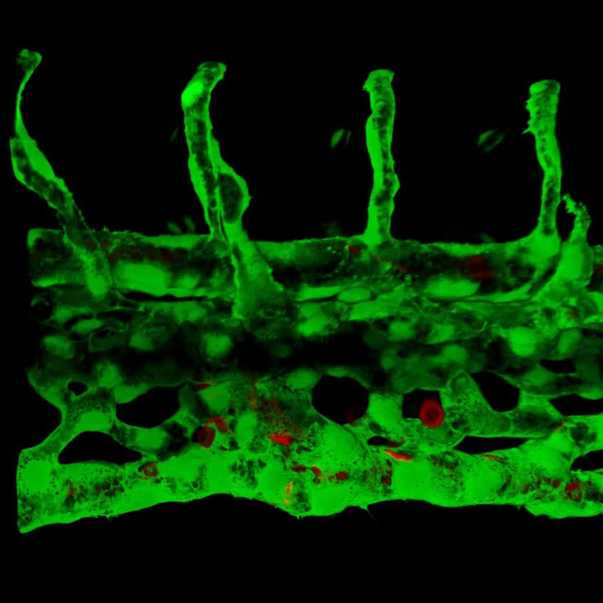
Research Article                                                                                Developmental Biology

                                            ccm2 CRISPR                                    control
                           A                                             B

                           C                                             D
   fli1:EGFP;gata1:DsRed

                           E                                             F
                                              MCeV   PHBC
   fli1:EGFP;gata1:DsRed

                                                                                  MCeV

                                        PMBC
                                                                                                   PHBC
                                                                                 PMBC

                       G                                             H

                                       Dilated CVP   Dilated Heart       Dilated CVP    Dilated Heart       Dilated CV

Figure 1. ccm2 CRISPR zebrafish embryo display novel vascular phenotypes. Endothelial cells and red blood cells were labeled by EGFP and DsRed
respectively in double transgenic Tg(fli1:EGFP)y1;Tg(gata1:DsRed)sd2 embryos. (A) Red blood cells accumulate in dilated segments of the caudal vein of
ccm2 CRISPR fish at 2 days post fertilization (dpf). (B) cas9 mRNA-injected control embryo. (C) ccm2 CRISPR embryos showed accumulation of red
blood cells and intraluminal endothelial cells in a dilated segment of caudal vein in contrast to a control embryo. Note: In this and all succeeding
Figure 1 continued on next page

Li et al. eLife 2021;10:e62155. DOI: https://doi.org/10.7554/eLife.62155                                                                       3 of 22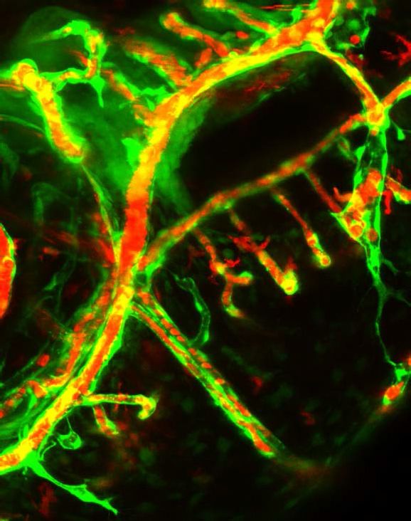
Research Article                                                                                                       Developmental Biology

Figure 1 continued
sagittal views, anterior is to the left (D). (E) ccm2 CRISPR embryos occasionally showed dilations of cerebral veins, whereas control embryos (F) showed
normal development of cerebral veins (F). MCeV: mid-cerebral vein, PMBC: primordial midbrain channel, PHBC: primordial hindbrain channel. (G) The
dilated caudal venous plexus (CVP) and heart of ccm2 CRISPR embryos were rescued by ccm2 mRNA injection. p=0.0336 (dilated CVP), 0.0037 (dilated
heart). p-Values were calculated using an unpaired two-tailed Student’s t-test. (H) Phenotypic distribution of dilated heart, CVP, and cerebral veins (CV)
in ccm2 CRISPR embryos at 2 dpf. p=0.0078 (dilated CVP), 0.0268 (dilated heart), 0.0041 (dilated CV). p-Values were calculated using a paired two-tailed
Student’s t-test. Error bars indicate SD. Scale bar: 1 mm in A and B, and 100 mm in C through F.
The online version of this article includes the following figure supplement(s) for figure 1:
Figure supplement 1. ccm2 CRISPR zebrafish exhibit mosaic expression of CCM2.
Figure supplement 2. Approximately 10% of ccm2 CRISPR zebrafish embryos exhibited dilation of heart and increased branch points of subintestinal
vein.

                                     and heart dilation in ccm2 CRISPR embryos (Figure 1G) confirming that both phenotypes are due to
                                     ccm2 loss. Notably, the dilated heart and CVP dilation appeared to be mutually exclusive, that is, in
                                     over 200 embryos analyzed, we never observed both phenotypes in a single ccm2 CRISPR embryo.
                                     Thus, the localized CVP dilation was the most prevalent phenotype observed in 2 dpf ccm2 CRISPR
                                     embryos in comparison to the cardiac and SIV phenotypes that characterize ccm2 null and ccm2
                                     morphant embryos (Figure 1H; Mably et al., 2006; Renz et al., 2015).

                                     Abortive intussusceptive angiogenesis in the dilated CVP of ccm2
                                     CRISPR embryos
                                     We used confocal microscopy and three-dimensional (3D) reconstruction of the dilated area of the
                                     CVP in Tg(fli1:EGFP;gata1:DsRed) zebrafish to explore their underlying structural defect. We noted
                                     intraluminal endothelial pillars that partitioned the lumen (Figure 2A through C) of the dilated CVP.
                                     In contrast, as expected, a completely patent caudal and ventral vein lumen formed in control
                                     embryos (Figure 2D through F). Furthermore, the ventral vein, which normally forms by a combina-
                                     tion of sprouting and intussusceptive angiogenesis (Karthik et al., 2018), was lost in the dilated
                                     area of the CVP (Figure 2A through F). Importantly, examination of 3D reconstructions of the vessel
                                     revealed that these pillars were associated with pits on the external surface of the dilated CVP
                                     (Figure 2G arrows), a hallmark of the initial phase of intussusceptive angiogenesis (Djonov et al.,
                                     2003). In contrast to normal intussusceptive angiogenesis, wherein transluminal pillars ultimately
                                     fuse to divide vessels longitudinally into new daughter vessels (Djonov et al., 2003), the intussuscep-
                                     tions observed in ccm2 CRISPR embryos were not coordinately formed and failed to fuse resulting in
                                     a honeycombed lumen. This honeycombing created a lumen with multiple chambers filled with red
                                     blood cells (RBCs) associated with sluggish blood flow (Figure 2I through K, Videos 2 and
                                     3), whereas patent lumens and normal blood
                                     flow were observed in control embryos (Video 4).
                                     Thus, in these mosaic ccm2 null zebrafish, an
                                     expanded region of the CVP is formed by multi-
                                     ple dilated erythrocyte-filled chambers and is
                                     associated with evidence of incomplete intus-
                                     susceptive angiogenesis.

                                     Ablation of intravascular pillars
                                     reverts the dilated CVP phenotype
                                     The multiple dilated compartments and sluggish
                                     blood flow suggested that this phenotype could
                                     be due to obstruction of free flow of erythro-
                                     cytes by the meshwork of intravascular pillars. In
                                     support of this idea, we observed that spontane-
                                     ous regression of an existing pillar was accompa-           Video 1. On 2 days post fertilization (dpf), ccm2
                                     nied by reduced dilation of the CVP (Figure 3A              CRISPR embryo displayed cavernoma-like lesion in the
                                     through D). In addition, the trapped erythrocytes           tail. Blood flow was slowed down in the lesion area that
                                     began to circulate freely. This rapid relief of both        contained retained blood cells.
                                     vessel dilation and blood stagnation suggested              https://elifesciences.org/articles/62155#video1

Li et al. eLife 2021;10:e62155. DOI: https://doi.org/10.7554/eLife.62155                                                                           4 of 22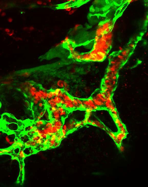
Research Article Developmental Biology
ccm2 CRISPR control
A B C D E F
G H
fli1:EGFP fli1:EGFP
I fli1:EGFP;gata1:DsRed J fli1:EGFP K
X
Y
Z
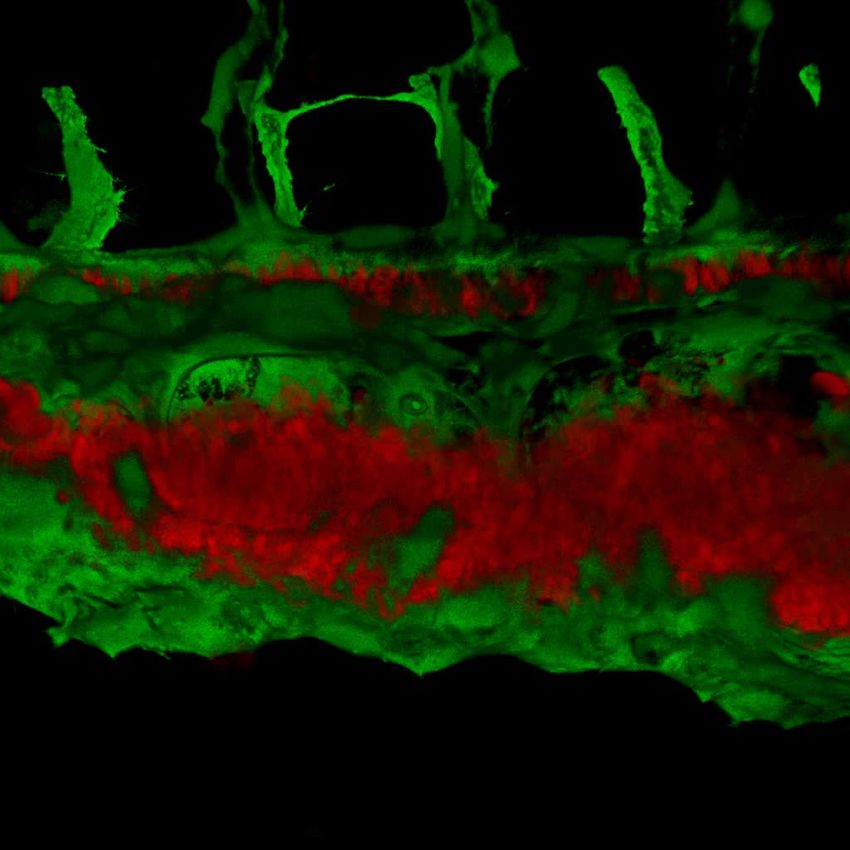
Figure 2. Intravascular pillars honeycomb the lumen of the caudal vein in ccm2 CRISPR embryos. (A–F) XZ planes and three-dimensional (3D) projection
along Y axis of Airyscan images revealed intraluminal endothelial pillars at 2 days post fertilization (dpf) (A–C), whereas Cas9-injected control embryos
displayed a normal patent lumen in both a dorsal and ventral caudal vein (D–F). Endothelial cells were labeled by EGFP in Tg(fli1:EGFP) embryos.
Arrow, arrowhead, and asterisk indicated the dorsal aorta, dorsal vein, and ventral vein, respectively. (G and H) Ventral view of 3D reconstruction show
the irregular surface of the dramatically dilated caudal vein segment in ccm2 CRISPR embryo (G) and normal ventral vein (H). Arrows in G indicate small
pits where the endothelial pillars originate. (I–K) Intraluminal view of 3D reconstruction of ccm2 CRISPR embryo reveals the intraluminal pillars
honeycombing the lumen and the accumulated red blood cells (I). Erythrocytes were not imaged in J to reveal pillars and the area within the box in (J)
was magnified in (K), and arrowhead indicates the intravascular pillar. Endothelial cells and red blood cells were labeled by EGFP or DsRed respectively
in Tg(fli1:EGFP)y1;Tg(gata1:DsRed)sd2 embryos. Scale bar: 20 mm.
that the aberrant pillars may form a physical barrier thus resulting in accumulation of erythrocytes in
dilated cavernous structures. To directly test the role of obstruction by intravascular pillars in dila-
tion, we used targeted short pulses of near-infrared laser light to sever the pillars, a technique that
generates negligible heat transfer and collateral damage to neighboring tissues (Nishimura et al.,
2006). There was near instantaneous reduction of the dilated vessel diameter (93.4 mm) to near-nor-
mal dimensions (69.4 mm) in the example shown (Figure 3E and F, Video 5). In three such indepen-
dent experiments, severing these pillars resulted in a 29 ± 4% reduction in vessel diameter
(p=0.0004, two-tailed t-test). Thus, the pillars are an underlying cause of the CVP dilation observed
in ccm2 CRISPR embryos.
Li et al. eLife 2021;10:e62155. DOI: https://doi.org/10.7554/eLife.62155 5 of 22
Research Article                                                                                                    Developmental Biology

                                    Video 2. Three-dimensional exterior view of caudal        Video 3. Three-dimensional interior view of caudal
                                    venous plexus (CVP) of 2 days post fertilization (dpf)    venous plexus (CVP) of 2 days post fertilization (dpf)
                                    ccm2 CRISPR embryo. Note pits on the surface and          ccm2 CRISPR embryo. Note the endothelial pillars
                                    that the CVP is partitioned into several dilated areas.   within the lumen and accumulated red blood cells.
                                    https://elifesciences.org/articles/62155#video2           https://elifesciences.org/articles/62155#video3

                                    Blood flow and red blood cells are required for CVP dilation
                                    The importance of the pillars in CVP dilation suggested that obstruction of blood flow was responsi-
                                    ble for the phenotype. Consistently, as noted above, ccm2 CRISPR embryos displaying CVP dilation
                                    did not show heart dilation. Conversely, segmental CVP dilation was absent in ccm2 null mutants or
                                    ccm2 morphants that exhibit characteristic heart dilation (Figure 3—figure supplement 1). These
                                    observations suggest that a normally pumping heart and thus normal blood flow is required for CVP
                                    dilation. To investigate the role of blood flow, we took advantage of the capacity of zebrafish
                                    embryos to obtain sufficient oxygen by diffusion to survive temporarily in the absence of circulating
                                                                                       blood. We induced a silent heart phenotype by

                                    Video 4. Three-dimensional view of caudal venous
                                    plexus (CVP) of 2 days post fertilization (dpf) control   Video 5. Laser ablation of intussusception reduced the
                                    embryo.                                                   vessel diameter.
                                    https://elifesciences.org/articles/62155#video4           https://elifesciences.org/articles/62155#video5

Li et al. eLife 2021;10:e62155. DOI: https://doi.org/10.7554/eLife.62155                                                                        6 of 22
Research Article                                                                                          Developmental Biology

                                    using a troponin T (tnnt) morpholino, resulting in ~65% reduction in the frequency of CVP dilation
                                    (Figure 4A). We also reasoned that the meshwork of pillars would not obstruct fluid flow but would
                                    present a barrier to free passage of erythrocytes. Reduction of erythrocytes using morpholinos
                                    directed against gata1(Galloway et al., 2005) or tif1-g (Monteiro et al., 2011) transcription factors
                                    produced a similar dramatic reduction in the CVP dilation (Figure 4A). These data indicate that the
                                    meshwork of pillars obstructs the passage of erythrocytes in flowing blood resulting in multiple
                                    erythrocyte-filled cavernous chambers that dilate the CVP.
                                        As shown in Figure 2, the sprouts that form the ventral vein are lost in the dilated region of the
                                    CVP. Because CVP development in tnnt morphants is nearly normal (Choi et al., 2011), we inspected
                                    the regions of the CVP displaying loss of ventral sprouting in tnnt morphant ccm2 CRISPR embryos.
                                    In 11 such embryos, in spite of the defective ventral sprouting and ventral vein formation, we
                                    observed no intravascular pillars. This result suggests that blood flow, in addition to causing the CVP
                                    dilation, is required for intussusceptive pillar formation (Figure 4B and C) as it is for normal CVP
                                    arborization (Karthik et al., 2018).
                                        Inactivation of either ccm1 or ccm2 markedly upregulates expression of KLF2, a flow-regulated
                                    transcription factor required for normal cardiovascular development and for CCM formation
                                    (Renz et al., 2015; Zhou et al., 2015; Zhou et al., 2016). In situ hybridization revealed that klf2a
                                    was also upregulated in the CVP of ccm2 CRISPR embryos (Figure 4—figure supplement 1). We
                                    used a klf2a reporter line, Tg(klf2a:H2AEGFP), together with an endothelial cell-specific marker line
                                    (Tg(kdrl:mcherry)is5) to observe the activity of the klf2a promoter. Ccm2 morphants displayed a gen-
                                    eralized increase in klf2a reporter expression (Figure 4—figure supplement 2A and A’), whereas
                                    the absence of blood flow in the tnnt morphant caused much reduced reporter expression in endo-
                                    thelial cells (Figure 4—figure supplement 2B and B’; Figure 4—figure supplement 2C and C’).
                                    Consistent with previous reports (Parmar, 2006; Renz et al., 2015), these opposing changes con-
                                    firm that ccm2 and flow can regulate expression of KLF2a. In 23 hpf ccm2 CRISPR embryos, exam-
                                    ined prior to onset of blood flow, a patchy increase in klf2a reporter expression was observed in
                                    ccm2 CRISPR endothelial cells (Figure 4D), whereas reporter expression was uniformly low in control
                                    embryos at the same stage (Figure 4E). A quantitative analysis revealed a subpopulation of high
                                    KLF2a-expressing endothelial cells in ccm2 CRISPR embryos that was absent in control embryos
                                    (Figure 4F). Furthermore, in tnnt morphant 2 dpf ccm2 CRISPR embryos, there was also a striking
                                    mosaic increase in endothelial klf2a reporter expression (Figure 4G). Thus, dilation was associated
                                    with the patchy upregulation of a flow-sensitive transcription factor, KLF2, in the ccm2 CRISPR CVP.
                                    Taken together, these results suggest that patchy KLF2 expression in combination with blood flow
                                    leads to formation of these dilated RBC-filled multi-cavernous lesions in the CVP.

                                    Mosaic upregulation of KLF2a is sufficient for cavernoma formation in
                                    CVP
                                    The patchy increase in KLF2a expression in the CVP endothelial cells of ccm2 CRISPR embryos and
                                    requirement for blood flow suggested the possibility that these two factors led to the formation of
                                    cavernomas in the CVP. To address the role of KLF2, we injected klf2a and klf2b morpholinos and
                                    observed reversal of both CVP dilation and heart dilation in the ccm2 CRISPR embryos (Figure 5A).
                                    Furthermore, ccm2 CRISPR treatment of klf2a-/- embryos caused no CVP dilation (Figure 5—figure
                                    supplement 1). Thus, klf2a is required for the CVP dilation phenotype.
                                       In ccm2 CRISPR embryos, KLF2a was both upregulated in a mosaic fashion and required for CVP
                                    dilation; we therefore asked whether mosaic upregulation of KLF2a expression per se causes caver-
                                    noma formation. Mosaic overexpression was accomplished by injecting a plasmid encoding KLF2a
                                    into Tg(fli1:EGFP)y1 embryos; ~6% of such embryos displayed CVP dilation compared to control
                                    embryos injected with DKLF2a plasmid expressing KLF2a with a deleted DNA binding domain
                                    (Oates et al., 2001; Figure 5B and C). Affected embryos exhibited intussusceptions within the CVP
                                    lumen accompanied by dilation (Figure 5D). These observations show that mosaic upregulation of
                                    KLF2a expression is sufficient for cavernoma formation when blood is flowing.

Li et al. eLife 2021;10:e62155. DOI: https://doi.org/10.7554/eLife.62155                                                            7 of 22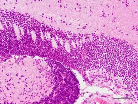
Research Article                                                                                   Developmental Biology

                              A                          B                             C                              D
   kdrl:mcherry; fli1:nEGFP

                              0 min                      40min                          50min                        60min
                              E                              25s before ablation       F                                   25s after ablation
    fli1:EGFP

Figure 3. Intravascular pillars obstruct blood flow leading to vessel dilation in ccm2 CRISPR embryos. (A through D) Time lapse images reveal
spontaneous retraction of an intravascular pillar leading to re-entry of blood cells into circulation and reduced dilation of the caudal vein. Endothelial
cells were labeled by mCherry, and their nucleus and some red blood cells were labeled by EGFP in the Tg(fli1:nEGFP)y7;Tg(kdrl:mcherryras)s896
embryos. The retracted pillar is outlined by dotted lines for emphasis. Note that pillar retraction and vessel dilation were temporally correlated. (E and
F) Laser ablation of pillar reduced caudal venous plexus (CVP) diameter. The diameter of the dilated vein (E) was reduced after ablation (F). Note the
pillars indicated by arrows in (E) are gone after ablation in (F). Dashed line indicates the diameter of the vein before and after ablation. Scale bar: 50
mm.
The online version of this article includes the following figure supplement(s) for figure 3:
Figure supplement 1. Both ccm2 null mutants and morphants displayed heart dilation but no caudal venous plexus (CVP) dilation on 2 days post
fertilization (dpf).

                                                     Mosaic expression of ccm2 causes KLF2a-dependent cavernoma
                                                     formation
                                                     ccm2 CRISPR caused mosaic inactivation of ccm2 and the dilated CVP phenotype, whereas global
                                                     inactivation of ccm2 in ccm2 null mutants or ccm2 morphants does not. We therefore questioned
                                                     whether mosaicism, per se, played a role in the CVP dilation. To test this idea, we globally reduced
                                                     ccm2 expression by co-injecting a sublethal dose of ccm2 morpholino with the ccm2 gRNA CRISPR
                                                     mixture. The chosen morpholino dose did not increase the frequency of observable heart defects;
                                                     however, the percentage of embryos displaying CVP dilation decreased dramatically (Figure 6A).
                                                     We then reasoned that because ccm2 acts as a scaffold connecting krit1 to ccm3 (Stahl et al.,
                                                     2008), the overexpression of ccm2 might have a dominant negative effect. Indeed, when we injected
                                                     linearized DNA containing ccm2 fused to m-Orange, ccm2 mosaic overexpression led to CVP dila-
                                                     tion and aberrant intussusceptions similar to those observed in ccm2 CRISPR embryos in ~8% of
                                                     embryos (Figure 6B and B’). In sharp contrast, injection of a plasmid encoding a loss of krit1 binding
                                                     function ccm2(L197R) mutant (Kleaveland et al., 2009) resulted in of embryos displaying a normal
                                                     vascular development (Figure 6C and C’). Importantly, mosaic overexpression of ccm2 caused

Li et al. eLife 2021;10:e62155. DOI: https://doi.org/10.7554/eLife.62155                                                                            8 of 22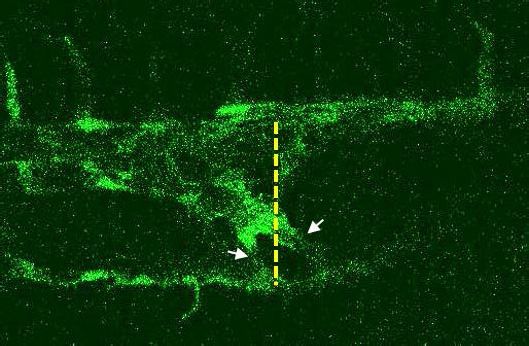
Research Article                                                                                             Developmental Biology

                                    significantly less CVP dilation in klf2a-/- embryos (Figure 6—figure supplement 1). Thus, mosaicism
                                    for ccm2 expression causes klf2a-dependent formation of multi-cavernous erythrocyte-filled struc-
                                    tures in the CVP. Combined with the capacity of mosaic expression of klf2a to cause CVP dilation,
                                    these results show that mosaic expression of CCM2 leads to mosaic KLF2a expression and abortive
                                    intussusceptive angiogenesis that obstructs the lumen to form these cavernoma-like lesions.

                                    CCMs in adult zebrafish
                                    The foregoing data indicated that mosaic inactivation of ccm2 results in a multi-cavernous lesion in
                                    the embryonic CVP that resembles mammalian CCM in gross architecture and dependence on KLF2.
                                    We then asked if authentic CCM would develop in the ~50% of ccm2 CRISPR embryos that devel-
                                    oped with a normal gross morphology and survived to adulthood. Brain vascular lesions were
                                    observed in virtually all of these adult ccm2 CRISPR zebrafish (Figure 7A and C) and not in control
                                    fish (Figure 7E and G). In order to image the lesions at the whole brain level, clear, unobstructed
                                    brain imaging cocktails and computational analysis (CUBIC) was applied to these brains, and the
                                    transparent brains were scanned by light sheet microscopy (Figure 7B,D,F and H). The distribution
                                    of lesions included cerebrum, cerebellum, brain stem, and, in some fish, the spinal cord (Figure 7I).
                                    This distribution pattern is similar to that found in patients (Goldstein and Solomon, 2017). Hema-
                                    toxylin and eosin stained sections showed dilated multi-cavernous vascular channels filled with nucle-
                                    ated erythrocytes and lacking mature vessel wall angioarchitecture (Figure 7J). Perl’s Prussian blue
                                    staining indicated prior hemorrhage adjacent to the lesions (Figure 7K). These histological findings
                                    were absent in control fish (Figure 7L and M) and resemble those in CCM patients (Figure 7N and
                                    O; Cox et al., 2017). A dramatic reduction in CCM was seen in ccm2 CRISPR in klf2a-/- zebrafish
                                    (Steed et al., 2016; Figure 7P) consistent with previous murine studies in which inactivation of Klf2
                                    prevented CCM formation (Zhou et al., 2016). In addition, similar to humans, these adult zebrafish
                                    also developed extracranial lesions (Figure 7—figure supplement 1).

                                    Discussion
                                    Familial CCM lesions form as a consequence of mosaic complete inactivation of CCM1, -2, or -3.
                                    Here, we have used Cas9-CRISPR mutagenesis to create such a mosaicism for ccm2 in zebrafish and
                                    show that surviving adult ccm2 CRISPR animals develop brain and extracranial lesions that closely
                                    resemble those observed in humans with CCM. In ~30% of embryos, we observed a novel pheno-
                                    type, the formation of segmental dilatation of the caudal vein associated with slowed blood flow
                                    and formation of multiple markedly dilated blood-filled chambers, resembling a multi-cavernous
                                    CCM. These lesions are caused by intussusceptive intraluminal pillars that obstruct the passage or
                                    erythrocytes resulting in the development of multiple dilated blood-filled chambers. These pillars
                                    form as a consequence of a combination of blood flow and mosaic overexpression of a flow-depen-
                                    dent transcription factor, KLF2a, leading to aberrant flow sensing in the developing CVP. In sum, our
                                    studies describe a zebrafish model for CCM and provide a new mechanism that can explain the for-
                                    mation of the characteristic multi-cavernous lesions seen in humans.

                                    The role of blood flow in CVP dilation
                                    The segmental dilation of the CVP is due to intussusceptive pillars that fail to fuse normally, thus
                                    honeycombing the vein lumen and obstructing the free flow of erythrocytes. Evidence for the role of
                                    obstruction includes the marked slowing of blood flow within the lesions, dependence of dilation on
                                    blood flow and erythrocytes, and the relief of dilation by spontaneous or induced regression of the
                                    pillars. Intussusceptive angiogenesis differs from sprouting angiogenesis by splitting the existing ves-
                                    sel intraluminally as a response to increased blood flow (Djonov et al., 2003; Egginton et al.,
                                    2001). Recent elegant studies have shown that localized reduction in fluid shear stress occurs adja-
                                    cent to intussusceptive pillars and is associated with the formation of new pillars that align with exist-
                                    ing pillars (Karthik et al., 2018). These observations suggested that these blood flow patterns are
                                    responsible for the formation of the aligned pillars required for orderly fusion to split the vessel in
                                    two (Karthik et al., 2018). Similar to physiological intussusceptive angiogenesis, the hallmark pits
                                    and intraluminal pillars were also observed in the CVP of ccm2 CRISPR embryos; however, these pil-
                                    lars failed to undergo orderly fusion to split the vessel. We propose that this failure to undergo
                                    orderly fusion and CVP arborization is due to mosaic overexpression of klf2a, a flow-sensitive

Li et al. eLife 2021;10:e62155. DOI: https://doi.org/10.7554/eLife.62155                                                               9 of 22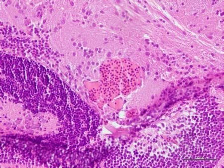
Research Article                                                                                         Developmental Biology

        A                                                                                                        B     ccm2 CRISPR+tnnt MO

                                                                                                                 C   ccm2 CRISPR+control MO

                                               tnnt     tif1γ       gata1   control
       klf2a:H2b-EGFP;kdrl:mcherry

                                     D                          ccm2 CRISPR                                  E                  cas9 mRNA

                                      A

                 F                                                                                           G        ccm2 CRISPR+ tnnt MO
                                                                               klf2a:H2b-EGFP;kdrl:mcherry

                                               CRISPR     control

Figure 4. Blood flow is required for pillar formation and vessel dilation. Morpholinos targeting tnnt, gata1, tif1gamma, or a control morpholino were
co-injected with ccm2 guide and Cas9 RNA. (A) Reduction of blood flow in tnnt morphants (A, B, C) resulted in reduced caudal venous plexus (CVP)
dilation (A) and intravascular honeycombing (B, C) in 2 days post fertilization (dpf) ccm2 CRISPR Tg(fli1:EGFP) embryos. Arrows indicate
intussusceptions. Scale bar: 100 mm. (A) Loss of erythrocytes in gata1 or tif1gamma morphant ccm2 CRISPR embryos also reduced the incidence of CVP
dilation. p-Values were calculated using one-way ANOVA. **p
Research Article Developmental Biology
A B klf2a
Δklf2a
D klf2a
C
Δklf2a
klf2a Δklf2a
Figure 5. Mosaic KLF2a expression caused caudal venous plexus (CVP) dilation. (A) Both the CVP dilation and heart dilation were rescued by injection
of klf2 morpholinos in 2 days post fertilization (dpf) ccm2 CRISPR embryos. **pResearch Article Developmental Biology
A
Dilated CVP Dilated Heart
B ccm2 C ccm2(L197R)
fli1:EGFP
B’ C’
cmv:morange
Figure 6. Mosaic ccm2 expression caused caudal venous plexus (CVP) dilation. (A) Low-dose ccm2 morpholino
reduced the incidence of CVP dilation but did not significantly increase heart dilation in ccm2 CRISPR embryos. (B
and C) Mosaic ccm2 but not inactive ccm2(L197E) overexpression caused CVP dilation. Arrows indicate pillars in
the CVP. (B’ and C’) Mosaic expression of mOrange-tagged ccm2 or ccm2(L197E). Scale bar: 100 mm. Error bars
are ± SD.
The online version of this article includes the following figure supplement(s) for figure 6:
Figure supplement 1. Reduced caudal venous plexus (CVP) dilation in CCM2 over expressing klf2a-/- embryos.
Li et al. eLife 2021;10:e62155. DOI: https://doi.org/10.7554/eLife.62155 12 of 22Research Article Developmental Biology
Dorsal Ventral
A B C D
ccm2 CRISPR
L R L R R L R L
E F G H
control
L R L R R L R L
I
J H&E K prussian blue
Distribution of CCM (%)
L H&E M prussian blue
P
6
Fish displaying CCM(%)
N H&E O prussian blue
10
**
klf2a-/- klf2a+/+
Figure 7. Adult ccm2 CRISPR zebrafish develop typical cerebral cavernous malformation (CCM) lesions. The ~50% of ccm2 CRISPR fish that survived
developed highly penetrant CCMs (A and C). Arrows indicate superficial lesions on dorsal (A) and ventral (C) surface of the brain. Note hemorrhage into
the ventricles. Lesions are absent in control embryos (E and G). Clear, unobstructed brain imaging cocktails and computational analysis (CUBIC)
clearing (B, D, F, H) enables visualization of CCM burden by light sheet microscopy. Arrows indicate the lesions that corresponded to those seen in
Figure 7 continued on next page
Li et al. eLife 2021;10:e62155. DOI: https://doi.org/10.7554/eLife.62155 13 of 22Research Article Developmental Biology
Figure 7 continued
bright field, and arrowhead indicates a deeper lesion. L: left, R: right. Scale bar: 1 mm. (I) Cavernomas were dispersed throughout the central nervous
system including cerebrum, cerebellum, brain stem, and spinal cord. (J) Hematoxylin and eosin (H&E) stained brain section reveals nucleated
erythrocytes filling a dilated vessel with adjacent Prussian blue stained iron deposition (K) in ccm2 CRISPR fish and the absence of lesions or iron
deposition in control fish (L, M). (N, O) A CCM from a patient stained with H&E (N) or Prussian blue (O). Note similar appearance to the zebrafish lesion
shown in (J, K). Arrow indicates dilated vessel. Scale bar: 50 mm. (P) CCMs were significantly reduced in ccm2 CRISPR adult fish on klf2a-/- background
compared to that on klf2a+/+ background. Total number of embryos in each group is indicated. p=0.0076. Two-tailed Fisher’s exact test was used for
comparison.
The online version of this article includes the following figure supplement(s) for figure 7:
Figure supplement 1. Ccm2 CRISPR adult fish displayed body wall lesions.
Figure supplement 2. Inhibiting Rho kinase blocks caudal venous plexus (CVP) cavernoma formation in ccm2 CRISPR zebrafish.
Ccm2 mosaicism causes multi-cavernous malformations
Initially we ascribed the absence of segmental CVP dilation in ccm2 null fish (Mably et al., 2006;
Renz et al., 2015) solely to the reduced blood flow caused by the dilated heart. This explanation is
insufficient because rescue of the heart phenotype in global krit1 (ccm1) null fish was not reported
to cause segmental CVP dilation or CCMs (Rödel et al., 2019). Normal intussusceptive angiogenesis
requires an orderly patterning of high and low flow signaling (Karthik et al., 2018). Ccm2 mosaicism
causes a random upregulation of klf2a, a key effector of flow signaling, thereby disrupting this
orderly patterning of flow signaling. In contrast, the global knockout uniformly upregulates klf2a so
that the patterning of other flow-sensitive signals can guide the completion of the intussusceptive
arborization.
The dilated multi-cavernous CVP lesions described here resemble multi-cavernous CCM
(McDonald et al., 2011) and their formation required mosaicism. Recent studies found that multi-
cavernous murine CCMs are mosaic for inactivation of Ccm3 (Detter et al., 2018; Malinverno et al.,
2019). The mouse studies emphasized that simply dilated vessels are not mosaic and contained only
Ccm3 null endothelial cells (Detter et al., 2018; Malinverno et al., 2019). Mosaicism in multi-cav-
ernous murine CCM was ascribed to recruitment of wild-type cells to the clonal CCM (Detter et al.,
2018; Malinverno et al., 2019). Importantly, the mouse studies did not address the mechanism by
which multi-cavernous lesions form. Here, we have shown that mosaicism is a prerequisite for forma-
tion of multi-cavernous CVP lesions because it disorganizes the flow signaling required for orderly
sprouting and intussusceptive angiogenesis that remodel the CVP.
Ccm2 CRISPR zebrafish are an authentic CCM model
As in humans (Akers et al., 2009; McDonald et al., 2011), the fish CCM lesions arise as a conse-
quence of mosaic inactivation of CCM genes. Second, as in humans, chronic bleeding leads to iron
deposition; this finding contrasts with the lack of iron deposition seen in acute mouse CCM models
(Zeineddine et al., 2019). Third, similar to humans, histologically typical lesions are distributed
throughout the CNS in contrast to the hindbrain-restricted lesions in acute mouse
models (Zeineddine et al., 2019). Fourth, as is true in mouse models (Cuttano et al., 2016;
Zheng et al., 2014), the development of CCM depends on klf2a, the orthologue of murine Klf2 and
paralogue of Klf4, indicating that they form by the same pathogenetic mechanism. That said, in con-
trast to the KLF2 dependence of CVP dilation, injection of a KLF4 morpholino (Li et al., 2011) did
not rescue this lesion (our unpublished data). There are chronic sensitized mouse models which do
exhibit hemosiderin deposits and lesions throughout the CNS (McDonald et al., 2011); however,
these models require cumbersome breeding schemes and mice of more than 3 months of age. That
said, a recent report that postnatal induction of brain endothelial cell-specific ablation of the Ccm2
gene using the inducible Slco1c1-CreERT2 mouse results in iron deposits around CCM throughout
the murine brain at 3 months of age has great promise (Cardoso et al., 2020). In contrast to existing
mouse models, the present model uses CRISPR-Cas9 to generate highly penetrant typical lesions
throughout the CNS, requires about 2 months, and can be induced in mutant strains without addi-
tional breeding. Thus, this model should be a useful tool in future studies to assess the effect of the
many genetic manipulations possible in zebrafish (Gore et al., 2018) on the pathogenesis of CCM
Li et al. eLife 2021;10:e62155. DOI: https://doi.org/10.7554/eLife.62155 14 of 22Research Article Developmental Biology
and to provide a complement to pharmacological screens directed at the dilated heart phenotype
of ccm1 or ccm2 mutant fish (Otten et al., 2018).
In sum, the present work reveals a new embryonic vascular malformation, a multi-cavernous dila-
tion of the CVP that resembles multi-cavernous Stage 2 CCM. The CVP malformation requires blood
flow and mosaic inactivation of ccm2 and is caused by abortive intussusceptive angiogenesis as a
consequence of imbalanced flow signaling. The high penetrance and resemblance of the embryonic
CVP malformation to multi-cavernous CCM suggest that it will be a useful phenotype for pharmaco-
logical or morpholino-based analyses. That said, the CVP does lack CNS accessory cells, such as
astrocytes (Lopez-Ramirez et al., 2021), that promote CCM development. Indeed, we recently
reported that propranolol blocks the embryonic CVP malformation by b1 adrenergic receptor
antagonism (Li et al., 2021), a result that comports with the beneficial effects of propranolol in
murine CCM models (Li et al., 2021; Oldenburg et al., 2021) and in anecdotal reports in
humans (Lanfranconi et al., 2020; Reinhard et al., 2016). We have found that blockade or Rho
kinase also ameliorates the CVP lesion (Figure 7—figure supplement 2) as it does murine
CCM (McDonald et al., 2012). In addition, we report a tractable zebrafish model of CNS CCM that
mimics the mammalian disease in mosaicism, lesion histology and distribution, and dependence on
KLF2 transcription factors. A particularly appealing feature of these two new zebrafish models is that
disease pathogenesis can be studied on mutant backgrounds without the need for additional breed-
ing. Manipulations that ameliorate the embryonic lesion can then readily be tested for effects on the
formation of brain CCMs that occur in the 50% of ccm2 CRISPR embryos that survive to adulthood.
Materials and methods
Key resources table
Reagent type
(species) or
resource Designation Source or reference Identifiers Additional information
Gene (Danio ccm2 http://www.ensembl.org/ ENSDARG00000013705
rerio)
Gene (Danio klf2a http://www.ensembl.org/ ENSDARG00000042667
rerio)
Strain, strain ccm2m201 zfin.org ZDB-ALT-980203–523
background
(Danio rerio)
Strain, strain klf2aig4 zfin.org ZDB-ALT-161103–5
background
(Danio rerio)
Strain, strain Tg(fli1:EGFP)y1 zfin.org ZDB-ALT-011017–8
background
(Danio rerio)
Strain, strain Tg(gata1:dsred)sd2 zfin.org ZDB-ALT-051223–6
background
(Danio rerio)
Strain, strain Tg(klf2a:H2b-EGFP) zfin.org ZDB-ALT-161017–10
background
(Danio rerio)
Strain, strain Tg(fli1:negfp)y7 zfin.org ZDB-ALT-060821–4
background
(Danio rerio)
Strain, strain Tg(kdrl:mcherry)is5 zfin.org ZDB-ALT-110127–25
background
(Danio rerio)
Recombinant pCS2-nls-zCas9-nls addgene.org 47929
DNA reagent
Recombinant pT7-gRNA addgene.org 46759
DNA reagent
Continued on next page
Li et al. eLife 2021;10:e62155. DOI: https://doi.org/10.7554/eLife.62155 15 of 22Research Article Developmental Biology
Continued
Reagent type
(species) or
resource Designation Source or reference Identifiers Additional information
Commercial mMESSAGE Thermo Fisher AM1340
assay or kit mMACHINE Scientific
SP6 Transcription Kit Wlatham, MA
Commercial MEGAshortscript T7 Thermo Fisher AM1333
assay or kit Transcription kit Scientific, Waltham, MA
Sequence-based crRNA-1 This paper ccm2 gRNA GGTGTTTCTGAAAGGGGAGA
reagent
Sequence-based crRNA-2 This paper ccm2 gRNA GGAGAAGGGTAGGGATAAGA
reagent
Sequence-based crRNA-3 This paper ccm2 gRNA GGGTAGGGATAAGAAGGCTC
reagent
Sequence-based crRNA-4 This paper ccm2 gRNA GGACAGCTGACCTCAGTTCC
reagent
Chemical ccm2-MO zfin.org ZDB-MRPHLNO-060821–3 GAAGCTGAGTAATACCTTAACTTCC
compound, drug
Chemical tnnt-MO zfin.org ZDB-MRPHLNO-060317–4 CATGTTTGCTCTGATCTGACACGCA
compound, drug
Chemical gata1-MO zfin.org ZDB-MRPHLNO-050208–10 CTGCAAGTGTAGTATTGAAGATGTC
compound, drug
Chemical tif1g -MO afin.org ZDB-MRPHLNO-110321–1 GCTCTCCGTACAATCTTGGCCTTTG
compound, drug
Chemical klf2a-MO afin.org ZDB-MRPHLNO-100610–8 GGACCTGTCCAGTTCATCCTTCCAC
compound, drug
Chemical klf2b-MO zfin.org ZDB-MRPHLNO-150427–1 AAAGGCAAGGTAAAGCCATGTCCAC
compound, drug
Software Volocity PerkinElmer Volocity
Waltham, MA
Software ZEN Zeiss, Oberkochen, German ZEN 2.3 SP1
Software ImageJ ImageJ RRID:SCR_003070
software (http://imagej.nih.gov/ij/)
Software GraphPad GraphPad Prism Prism five for Windows Version 5.01
Prism software (https://graphpad.com)
Zebrafish lines and husbandry
Zebrafish were maintained and with approval of Institutional Animal Care and Use Committee of the
University of California, San Diego. The following mutant and transgenic lines were maintained under
standard conditions: ccm2m201 (Mably et al., 2006), klf2aig4 (Steed et al., 2016), Tg(fli1:EGFP)y1
(Lawson and Weinstein, 2002), Tg(gata1:dsred)sd2 (Traver et al., 2003), Tg(fli1:negfp)y7
(Roman et al., 2002), Tg(klf2a:H2b-EGFP) (Heckel et al., 2015), Tg(kdrl:mcherry)is5 (Jin et al.,
2005), and casper (White et al., 2008). See Expanded Materials and Methods for morpholino injec-
tions. Morpholinos sequences are shown in Supplementary file 1 (Morpholino sequences).
Plasmids pCS2-nls-zCas9-nls (47929) and pT7-gRNA (46759) were bought from Addgene. Crispr
RNA (crRNA) sequences were listed in Supplementary file 2 (crRNA sequences for zebrafish ccm2).
Target gRNA constructs were generated as described before (Jao et al., 2013). PCS2-moran-
geccm2, pCS2-morangeccm2 mutant(L197R), pCS2-morangeklf2a, pCS2-morangeDklf2a were
cloned by infusion (Clontech) as follows: mOrange was cloned into ClaI, and linker sequence (5’-
ggcagcgcgggcagcgcggcgggcagcggcgaattt-3’) between ClaI and EcoRI. Then ccm2, L197R mutant,
klf2a or Dklf2a sequence were cloned into EcoRI, respectively. These plasmids were then double-
digested by SalI and NotI (NEB), and the fragment containing CMV promoter and coding sequence
were purified and 0.5 nl of a 200 ng/ml solution was injected into single cell embryos. Primer sequen-
ces are listed in Supplementary file 3 (Primers for template DNA synthesis).
Li et al. eLife 2021;10:e62155. DOI: https://doi.org/10.7554/eLife.62155 16 of 22Research Article Developmental Biology
RNA synthesis
For cas9 mRNA, pCS2-nls-zCas9-nls was digested by NotI and then purified by column (Macherey-
Nagel) as template. Capped nls-zCas9-nls RNA was synthesized using mMESSAGE mMACHINE SP6
Transcription Kit (Thermo Fisher Scientific) and purified through lithium chloride precipitation
described in the same kit. For gRNA synthesis, gRNA constructs were linearized by BamHI digestion
and purified by column (Macherey-Nagel). gRNA was synthesized by in vitro transcription using
MEGAshortscript T7 Transcription kit (Thermo Fisher Scientific) and purified by alcohol precipitation
described in the same kit. The concentration of nls-zCas9-nls RNA and gRNA were measured by
NanoDrop 1000 Spectrophotometer (Thermo Fisher Scientific), and their quality was confirmed by
electrophoresis through a 1% (wt/vol) agarose gel. The final concentrations for RNA injection are as
follows: cas9 750 ng/ml, gRNA 120 ng/ml, and injection volume is 0.5 nl.
Whole mount in situ hybridization
Zebrafish embryos were collected at 48 hpf and fixed with 4% paraformaldehyde overnight. In situ
hybridization was performed as described before (Thisse and Thisse, 2008). The hybridization tem-
perature is 68˚C, and the probe concentration is 1 ng/ml. For primers used to amplify the template
DNA for probe synthesis, see Expanded Materials and Methods. The images for in situ hybridization
were captured by Olympus MVX10, Macro-view.
Airyscan imaging and 3D reconstruction
Embryos for imaging were anesthetized with egg water containing 0.016% tricaine (3-amino benzoic
acid ethyl ester, Sigma-Aldrich) and then embedded in 1% low melting point agarose (Invitrogen
16520050). Imaging was performed with Zeiss 880 Airyscan confocal under the standard Airyscan
mode, and a 20/NA 0.8 objective was used. Maximum projection was performed with ZEN (Zeiss).
3D reconstruction was performed with Volocity (PerkinElmer).
Laser ablation of intravascular pillars
Laser ablation of intravascular pillars was performed using targeted ultrafast laser pulses that were
generated with a multi-pass Ti:Al2O3 amplifier of local construction that followed a previously pub-
lished design (Nishimura et al., 2006) and operated at a 5 kHz pulse rate. The ablation beam and
the imaging beam were combined with a polarizing beamsplitter (Nishimura et al., 2006) prior to
the microscope objective. The two beams were focused in the same focal plane and the ablation
beam was centered in the area that is raster-scanned by the imaging beam so that ablation occurred
at the center of the TPLSM imaging field. The energy per pulse of the ablation beam was tuned with
neutral density filters and the quantity of pulses was controlled by a mechanical shutter (Uniblitz
LS3Z2 shutter and VMM-D1 driver; Vincent). The energy and number of pulses was adjusted based
on damage evaluated from the real-time TPLSM images and ranged between 0.2 and 0.4 mJ.
Live imaging of endothelial pillar ablation
Live images of the fish vessels were obtained with a two-photon laser scanning microscope of local
design (Nishimura et al., 2006), which was adapted to include an ablation beam. Low-energy,
100 fs, 76 MHz pulses for TPLSM were generated by a Titanium:Sapphire laser oscillator (Mira F-900;
Coherent Inc) that was pumped by a continuous wave laser (Verdi V-10 Nd:YVO4 laser; Coherent
Inc). The imaging laser pulses were scanned in a raster pattern by galvanometric mirrors that are
relay-imaged to the rear aperture of the objective. The two-photon excited fluorescence is reflected
by a dichroic mirror and transmitted to a photomultiplier tube. To produce laser pulses for ablation
while imaging, we employed a Pockels cell (QS-3 with NVP-525D driver and DD1 timing circuit;
Quantum Technologies) to reroute 1 in 76,000 pulses from the oscillator pulse train to seed a multi-
pass Titanium:Sapphire amplifier that is pumped by a Q-switched laser (Corona; Coherent). A half-
wave plate (l/2) rotates the polarization of the amplified pulses to lie perpendicular to that of the
laser oscillator and thus permits both the ablation beam and the imaging beam to be routed to the
microscope objective with a polarizing beamsplitter. We used a 25/NA 0.95, water immersion
objective (Olympus) for imaging and ablation.
Li et al. eLife 2021;10:e62155. DOI: https://doi.org/10.7554/eLife.62155 17 of 22Research Article Developmental Biology
Histology
Hematoxylin and eosin stain and Perl’s Prussian blue stain were performed as
described (Zeineddine et al., 2019).
Zebrafish brain dissection, CUBIC treatment, and light sheet imaging
Zebrafish brain dissection was performed as previously described (Gupta and Mullins, 2010). CUBIC
was optimized on the basis of previous report (Susaki et al., 2015). The brains were fixed with
pH 7.5 4% PFA for 24 hr and then washed with PBS for 24 hr. After PBS wash, CUBICR1 treatment
was then performed at 37˚C in water bath for 42 hr. Samples were imaged in CUBICR2 as medium
with ZEISS Lightsheet Z.1. Scanning was performed with 5 dual illumination optics and 5
objective.
Statistical analysis
Statistical analysis was performed with GraphPad Prism. p-Values were calculated by paired two-
tailed Student’s t-test unless otherwise specifically indicated. The mean and SD were shown in the
bar graphs.
Acknowledgements
We gratefully acknowledge Brant Weinstein for sharing a CUBIC protocol, David Traver, Miguel
Lopez-Ramirez, Alexandre Gingras, and Sara McCurdy for valuable discussion and criticism,
and Jennifer Santini and Marcy Erb for microscopy technical assistance. We also acknowledge
resources provided by the UCSD School of Medicine Microscopy Core (NINDS P30 NS047101).
Additional information
Funding
Funder Grant reference number Author
National Heart, Lung, and HL 139947 Mark H Ginsberg
Blood Institute
National Institutes of Health NS 92521 Thomas Moore
Rhonda Lightle
Issam A Awad
Mark H Ginsberg
National Institute of Mental R35 NS097265 David Kleinfeld
Health
National Institutes of Health R01 NS108472 Iftach Shaked
Be Brave for Life Wenqing Li
The funders had no role in study design, data collection and interpretation, or the
decision to submit the work for publication.
Author contributions
Wenqing Li, Conceptualization, Data curation, Formal analysis, Investigation, Methodology; Virginia
Tran, Belinda Xue, Thomas Moore, Rhonda Lightle, Investigation; Iftach Shaked, David Kleinfeld,
Investigation, Methodology; Issam A Awad, Formal analysis; Mark H Ginsberg, Conceptualization,
Data curation, Formal analysis, Funding acquisition, Project administration
Author ORCIDs
David Kleinfeld https://orcid.org/0000-0001-9797-4722
Mark H Ginsberg https://orcid.org/0000-0002-5685-5417
Li et al. eLife 2021;10:e62155. DOI: https://doi.org/10.7554/eLife.62155 18 of 22Research Article Developmental Biology
Ethics
Animal experimentation: This study was performed in strict accordance with the recommendations
in the Guide for the Care and Use of Laboratory Animals of the National Institutes of Health. All of
the animals were handled according to approved institutional animal care and use committee
(IACUC) protocols (#S14135 ) of the University of California San Diego.
Decision letter and Author response
Decision letter https://doi.org/10.7554/eLife.62155.sa1
Author response https://doi.org/10.7554/eLife.62155.sa2
Additional files
Supplementary files
. Supplementary file 1. Morpholino sequences.
. Supplementary file 2. crRNA sequence for zebrafish ccm2.
. Supplementary file 3. Primers for template DNA synthesis.
. Transparent reporting form
Data availability
Raw phenotype counts have been provided in figures and figure legends.
References
Ablain J, Durand EM, Yang S, Zhou Y, Zon LI. 2015. A CRISPR/Cas9 vector system for tissue-specific gene
disruption in zebrafish. Developmental Cell 32:756–764. DOI: https://doi.org/10.1016/j.devcel.2015.01.032,
PMID: 25752963
Akers AL, Johnson E, Steinberg GK, Zabramski JM, Marchuk DA. 2009. Biallelic somatic and germline mutations
in cerebral cavernous malformations (CCMs): evidence for a two-hit mechanism of CCM pathogenesis. Human
Molecular Genetics 18:919–930. DOI: https://doi.org/10.1093/hmg/ddn430, PMID: 19088123
Boulday G, Rudini N, Maddaluno L, Blécon A, Arnould M, Gaudric A, Chapon F, Adams RH, Dejana E, Tournier-
Lasserve E. 2011. Developmental timing of CCM2 loss influences cerebral cavernous malformations in mice.
Journal of Experimental Medicine 208:1835–1847. DOI: https://doi.org/10.1084/jem.20110571
Cardoso C, Arnould M, De Luca C, Otten C, Abdelilah-Seyfried S, Heredia A, Leutenegger AL, Schwaninger M,
Tournier-Lasserve E, Boulday G. 2020. Novel chronic mouse model of cerebral cavernous malformations. Stroke
51:1272–1278. DOI: https://doi.org/10.1161/STROKEAHA.119.027207, PMID: 31992178
Chan AC, Drakos SG, Ruiz OE, Smith ACH, Gibson CC, Ling J, Passi SF, Stratman AN, Sacharidou A, Revelo MP,
Grossmann AH, Diakos NA, Davis GE, Metzstein MM, Whitehead KJ, Li DY. 2011. Mutations in 2 distinct
genetic pathways result in cerebral cavernous malformations in mice. Journal of Clinical Investigation 121:
1871–1881. DOI: https://doi.org/10.1172/JCI44393
Choi J, Mouillesseaux K, Wang Z, Fiji HD, Kinderman SS, Otto GW, Geisler R, Kwon O, Chen JN. 2011. Aplexone
targets the HMG-CoA reductase pathway and differentially regulates arteriovenous angiogenesis.
Development 138:1173–1181. DOI: https://doi.org/10.1242/dev.054049, PMID: 21307094
Cox EM, Bambakidis NC, Cohen ML. 2017. Pathology of cavernous malformations. In: Cox E. M (Ed). Handbook
of Clinical Neurology. Elsevier. p. 267–277. DOI: https://doi.org/10.1016/B978-0-444-63640-9.00025-4
Cuttano R, Rudini N, Bravi L, Corada M, Giampietro C, Papa E, Morini MF, Maddaluno L, Baeyens N, Adams RH,
Jain MK, Owens GK, Schwartz M, Lampugnani MG, Dejana E. 2016. KLF4 is a key determinant in the
development and progression of cerebral cavernous malformations. EMBO Molecular Medicine 8:6–24.
DOI: https://doi.org/10.15252/emmm.201505433, PMID: 26612856
Detter MR, Snellings DA, Marchuk DA. 2018. Cerebral cavernous malformations develop through clonal
expansion of mutant endothelial cells. Circulation Research 123:1143–1151. DOI: https://doi.org/10.1161/
CIRCRESAHA.118.313970, PMID: 30359189
Djonov V, Baum O, Burri PH. 2003. Vascular remodeling by intussusceptive angiogenesis. Cell and Tissue
Research 314:107–117. DOI: https://doi.org/10.1007/s00441-003-0784-3, PMID: 14574551
Egginton S, Zhou AL, Brown MD, Hudlická O. 2001. Unorthodox angiogenesis in skeletal muscle. Cardiovascular
Research 49:634–646. DOI: https://doi.org/10.1016/S0008-6363(00)00282-0, PMID: 11166277
Galloway JL, Wingert RA, Thisse C, Thisse B, Zon LI. 2005. Loss of gata1 but not gata2 converts erythropoiesis
to myelopoiesis in zebrafish embryos. Developmental Cell 8:109–116. DOI: https://doi.org/10.1016/j.devcel.
2004.12.001, PMID: 15621534
Li et al. eLife 2021;10:e62155. DOI: https://doi.org/10.7554/eLife.62155 19 of 22Research Article Developmental Biology
Gingras AR, Liu JJ, Ginsberg MH. 2012. Structural basis of the junctional anchorage of the cerebral cavernous
malformations complex. Journal of Cell Biology 199:39–48. DOI: https://doi.org/10.1083/jcb.201205109
Goldstein HE, Solomon RA. 2017. Epidemiology of cavernous malformations. In: Goldstein H. E (Ed). Handbook
of Clinical Neurology. Elsevier. p. 241–247. DOI: https://doi.org/10.1016/B978-0-444-63640-9.00023-0
Gore AV, Pillay LM, Venero Galanternik M, Weinstein BM. 2018. The zebrafish: a fintastic model for
hematopoietic development and disease. WIREs Developmental Biology 7:e312. DOI: https://doi.org/10.1002/
wdev.312, PMID: 29436122
Gupta T, Mullins MC. 2010. Dissection of organs from the adult zebrafish. Journal of Visualized Experiments 1:
1717. DOI: https://doi.org/10.3791/1717
Heckel E, Boselli F, Roth S, Krudewig A, Belting HG, Charvin G, Vermot J. 2015. Oscillatory flow modulates
mechanosensitive klf2a expression through trpv4 and trpp2 during heart valve development. Current Biology
25:1354–1361. DOI: https://doi.org/10.1016/j.cub.2015.03.038, PMID: 25959969
Jao LE, Wente SR, Chen W. 2013. Efficient multiplex biallelic zebrafish genome editing using a CRISPR nuclease
system. PNAS 110:13904–13909. DOI: https://doi.org/10.1073/pnas.1308335110, PMID: 23918387
Jenny Zhou H, Qin L, Zhang H, Tang W, Ji W, He Y, Liang X, Wang Z, Yuan Q, Vortmeyer A, Toomre D, Fuh G,
Yan M, Kluger MS, Wu D, Min W. 2016. Endothelial exocytosis of angiopoietin-2 resulting from CCM3
deficiency contributes to cerebral cavernous malformation. Nature Medicine 22:1033–1042. DOI: https://doi.
org/10.1038/nm.4169, PMID: 27548575
Jin SW, Beis D, Mitchell T, Chen JN, Stainier DY. 2005. Cellular and molecular analyses of vascular tube and
lumen formation in zebrafish. Development 132:5199–5209. DOI: https://doi.org/10.1242/dev.02087,
PMID: 16251212
Karthik S, Djukic T, Kim JD, Zuber B, Makanya A, Odriozola A, Hlushchuk R, Filipovic N, Jin SW, Djonov V. 2018.
Synergistic interaction of sprouting and intussusceptive angiogenesis during zebrafish caudal vein plexus
development. Scientific Reports 8:9840. DOI: https://doi.org/10.1038/s41598-018-27791-6, PMID: 29959335
Kleaveland B, Zheng X, Liu JJ, Blum Y, Tung JJ, Zou Z, Sweeney SM, Chen M, Guo L, Lu MM, Zhou D, Kitajewski
J, Affolter M, Ginsberg MH, Kahn ML. 2009. Regulation of cardiovascular development and integrity by the
heart of glass-cerebral cavernous malformation protein pathway. Nature Medicine 15:169–176. DOI: https://
doi.org/10.1038/nm.1918, PMID: 19151727
Lanfranconi S, Scola E, Bertani GA, Zarino B, Pallini R, d’Alessandris G, Mazzon E, Marino S, Carriero MR, Scelzo
E, Faragò G, Castori M, Fusco C, Petracca A, d’Agruma L, Tassi L, d’Orio P, Lampugnani MG, Nicolis EB,
Vasamı̀ A, et al. 2020. Propranolol for familial cerebral cavernous malformation (Treat_CCM): study protocol for
a randomized controlled pilot trial. Trials 21:401. DOI: https://doi.org/10.1186/s13063-020-4202-x, PMID: 323
98113
Lawson ND, Weinstein BM. 2002. In vivo imaging of embryonic vascular development using transgenic zebrafish.
Developmental Biology 248:307–318. DOI: https://doi.org/10.1006/dbio.2002.0711, PMID: 12167406
Leblanc GG, Golanov E, Awad IA, Young WL, Biology of Vascular Malformations of the Brain NINDS Workshop
Collaborators. 2009. Biology of vascular malformations of the brain. Stroke 40:694–702. DOI: https://doi.org/
10.1161/STROKEAHA.109.563692, PMID: 19834013
Li IC, Chan CT, Lu YF, Wu YT, Chen YC, Li GB, Lin CY, Hwang SP. 2011. Zebrafish Krüppel-Like factor 4a
represses intestinal cell proliferation and promotes differentiation of intestinal cell lineages. PLOS ONE 6:
e20974. DOI: https://doi.org/10.1371/journal.pone.0020974, PMID: 21687630
Li W, Shenkar R, Detter MR, Moore T, Benavides C, Lightle R, Girard R, Hobson N, Cao Y, Li Y, Griffin E, Gallione
C, Zabramski JM, Ginsberg MH, Marchuk DA, Awad IA. 2021. Propranolol inhibits cavernous vascular
malformations by b1 adrenergic receptor antagonism in animal models. Journal of Clinical Investigation 131:
JCI144893. DOI: https://doi.org/10.1172/JCI144893
Lopez-Ramirez MA, Soliman SI, Hale P, Lai CC, Pham A, Estrada E, McCurdy S, Girard R, Verma R, Moore T,
Lightle R, Hobson N, Shenkar R, Poulsen O, Haddad GG, Daneman R, Gongol, H B. 2021. Non cell-
autonomous effect of astrocytes on cerebral cavernous malformations. bioRxiv. DOI: https://doi.org/10.1101/
2021.01.29.428891
Mably JD, Chuang LP, Serluca FC, Mohideen MA, Chen JN, Fishman MC. 2006. Santa and valentine pattern
concentric growth of cardiac myocardium in the zebrafish. Development 133:3139–3146. DOI: https://doi.org/
10.1242/dev.02469, PMID: 16873582
Malinverno M, Maderna C, Abu Taha A, Corada M, Orsenigo F, Valentino M, Pisati F, Fusco C, Graziano P,
Giannotta M, Yu QC, Zeng YA, Lampugnani MG, Magnusson PU, Dejana E. 2019. Endothelial cell clonal
expansion in the development of cerebral cavernous malformations. Nature Communications 10:2761.
DOI: https://doi.org/10.1038/s41467-019-10707-x, PMID: 31235698
McDonald DA, Shenkar R, Shi C, Stockton RA, Akers AL, Kucherlapati MH, Kucherlapati R, Brainer J, Ginsberg
MH, Awad IA, Marchuk DA. 2011. A novel mouse model of cerebral cavernous malformations based on the
two-hit mutation hypothesis recapitulates the human disease. Human Molecular Genetics 20:211–222.
DOI: https://doi.org/10.1093/hmg/ddq433, PMID: 20940147
McDonald DA, Shi C, Shenkar R, Stockton RA, Liu F, Ginsberg MH, Marchuk DA, Awad IA. 2012. Fasudil
decreases lesion burden in a murine model of cerebral cavernous malformation disease. Stroke 43:571–574.
DOI: https://doi.org/10.1161/STROKEAHA.111.625467, PMID: 22034008
Mikati AG, Khanna O, Zhang L, Girard R, Shenkar R, Guo X, Shah A, Larsson HB, Tan H, Li L, Wishnoff MS, Shi C,
Christoforidis GA, Awad IA. 2015. Vascular permeability in cerebral cavernous malformations. Journal of
Cerebral Blood Flow & Metabolism 35:1632–1639. DOI: https://doi.org/10.1038/jcbfm.2015.98, PMID: 25966
944
Li et al. eLife 2021;10:e62155. DOI: https://doi.org/10.7554/eLife.62155 20 of 22You can also read