Melanin Pathway Genes Regulate Color and Morphology of Butterfly Wing Scales - It works!
←
→
Page content transcription
If your browser does not render page correctly, please read the page content below
Article

Melanin Pathway Genes Regulate Color and
Morphology of Butterfly Wing Scales
Graphical Abstract                                               Authors
                                                                 Yuji Matsuoka, Antónia Monteiro

                                                                 Correspondence
                                                                 dbsyuji@nus.edu.sg (Y.M.),
                                                                 antonia.monteiro@nus.edu.sg (A.M.)

                                                                 In Brief
                                                                 Matsuoka and Monteiro discover that
                                                                 deletions of the yellow and DDC melanin
                                                                 genes alter both the color and the
                                                                 morphology of Bicyclus anynana wing
                                                                 scales. This study identifies genes that
                                                                 regulate the intricate morphology of wing
                                                                 scales.

Highlights
d   Melanin pathway products regulate both the color and
    morphology of butterfly wing scales

d   The link between color and morphology underscores a
    potential bias and/or constraint

d   Different melanin genes and/or products produce each color
    on the wings of Bicyclus anynana

d   Female wing scales are larger than male wing scales

           Matsuoka & Monteiro, 2018, Cell Reports 24, 56–65
           July 3, 2018 ª 2018 The Authors.
           https://doi.org/10.1016/j.celrep.2018.05.092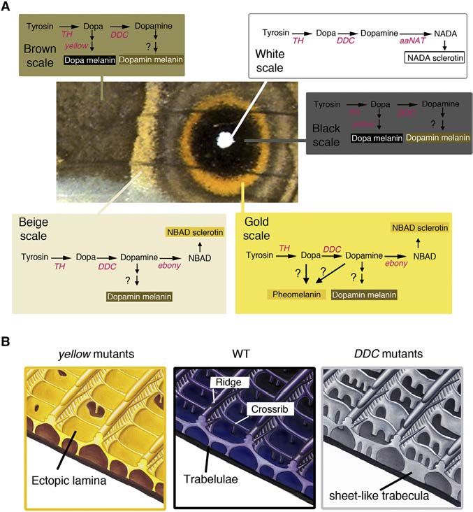
Cell Reports

                                                                                                                   Article

Melanin Pathway Genes Regulate Color
and Morphology of Butterfly Wing Scales
Yuji Matsuoka1,* and Antónia Monteiro1,2,3,*
1Department    of Biological Sciences, National University of Singapore, 14 Sciences Drive 4, Singapore 117543, Singapore
2Science  Division, Yale-NUS College, 10 College Avenue West, Singapore 138609, Singapore
3Lead Contact

*Correspondence: dbsyuji@nus.edu.sg (Y.M.), antonia.monteiro@nus.edu.sg (A.M.)
 https://doi.org/10.1016/j.celrep.2018.05.092

SUMMARY                                                               thetic pathways affect both the spatial patterns of cuticle depo-
                                                                      sition and its coloration.
The cuticular skeleton of a butterfly wing scale cell is                 We chose to examine the effects of pigment enzyme muta-
an exquisitely finely sculpted material that can                      tions on both the color and intricate morphology of butterfly
contain pigments, produce structural colors, or                       wing scales because scales of different color within a wing often
both. While cuticle rigidity and pigmentation depend                  display different cuticular micromorphologies (Ghiradella et al.,
on the products of the melanin pathway, little is                     1972; Vukusic et al., 2000; Janssen et al., 2001; Stavenga
                                                                      et al., 2004; Siddique et al., 2015). This suggests that the two
known about whether genes in this pathway also
                                                                      processes may be linked genetically. Furthermore, scale
play a role in the development of specific scale mor-                 pigmentation and morphological patterning coincide during
phologies. Here, we use CRISPR/Cas9 to show that                      pupal wing development. For instance, melanin pigments are
knockout mutations in five genes that function in                     deposited at late pupal development stages, with a subtle time
the melanin pathway affect both the fine structure                    lag between the deposition of yellow, brown, and black melanin
and the coloration of the wing scales. Most dramati-                  pigments (Koch et al., 1998; Wittkopp and Beldade, 2009), and
cally, mutations in the yellow gene lead to extra                     this process correlates with the development of the fine morpho-
horizontal laminae on the surface of scales, whereas                  logical features of scales such as the longitudinal ridges and
mutations in DDC gene lead to taller and sheet-like                   crossribs (Waku and Kitagawa, 1986; Ghiradella, 1989) (Figure 1).
vertical laminae throughout each scale. We identify                   In addition, most melanin pigmentation enzymes are expressed
genes affecting the development of color and scale                    at these same late stages (Nishikawa et al., 2013; Connahs et al.,
                                                                      2016; Zhang et al., 2017a), indicating that they may play a dual
morphology, the regulation and pleiotropic effects
                                                                      role in scale construction and pigmentation. We hypothesized,
of which may be important in creating and limiting                    therefore, that molecules that determine color in butterfly wings
the diversity of the structural and pigmentary colors                 also may regulate scale morphology.
observed in butterflies.                                                 We focused on enzymes from two main pigmentation path-
                                                                      ways, the melanin pathway and the ommochrome synthesis
INTRODUCTION                                                          pathway. The melanin biosynthetic pathway comprises a well-
                                                                      studied branched series of chemical reactions that can produce
The exoskeleton of insects provides important structural support      five different final molecules: two types of eumelanin (dopa-
and is used as a canvas for the deposition of pigments or for the     melanin and dopamine-melanin), pheomelanine, N-b-alanyldop-
sculpting of structural colors used in camouflage and mate            amine (NBAD), and N-acetyldopamine (NADA) (Galván et al.,
signaling. The skeleton is made up of cuticle, an extracellular ma-   2015; Arakane et al., 2009; Figure 2A). Eumelanins are made
trix comprising chitin fibers, cuticular proteins, lipids, and pig-   from dihydroxyphenylalanine (DOPA) or from dopamine through
ments (Moussian, 2010). One of the most common pigments               additional reactions catalyzed by the laccase2 and yellow family
found in insect cuticle is melanin, and some melanin pathway          gene products to produce black (dopa-melanine) and brown
products also take part in cuticle hardening or sclerotization,       (dopamine-melanin) pigments (Andersen, 2005; Figure 2A).
causing cuticular melanization in insects to be tightly linked        Recently, pheomelanine (a reddish-yellow pigment) was identi-
with cuticular sclerotization. For example, the cross-linking of      fied as an additional final product of the melanin pathway in
melanin pathway molecules with nucleophilic amino acid resi-          insects (Speiser et al., 2014; Galván et al., 2015; Jorge Garcı́a
dues and cuticular proteins confers cuticle stiffness (Xu et al.,     et al., 2016; Polidori et al., 2017; Figure 2A). NBAD and NADA
1997; Kerwin et al., 1999; Andersen, 2005; Suderman et al.,           are sclerotizing precursor molecules that have a yellow color
2006; Noh et al., 2016). The interaction among all of these cuticle   and are colorless, respectively. These molecules are made
components is therefore important in the assembly of the insect       from dopamine through a reaction catalyzed by the ebony
exoskeleton and in its coloration (Xiong et al., 2017).               gene product and the aaNAT gene product, respectively (Fig-
   Given the interdependency of coloring and hardening cuticle        ure 2A). The ommochrome pathway typically produces yellow,
we sought to explore whether mutations in the enzymes that            orange, and red pigments (Reed et al., 2008; Ferguson and Jig-
control the flux of chemical precursors across pigment biosyn-        gins, 2009). The vermilion gene product, tryptophan oxidase, is

56 Cell Reports 24, 56–65, July 3, 2018 ª 2018 The Authors.
This is an open access article under the CC BY license (http://creativecommons.org/licenses/by/4.0/).
Figure 1. Wing Scale Arrangement and
                                                                                             Scale Structure of B. anynana
                                                                                             (A) Arrangement of butterfly wing scales with cover
                                                                                             (tan) and ground (green) scales alternating along
                                                                                             vertical rows.
                                                                                             (B) Individual ground scale, where the white square
                                                                                             indicates the region magnified in (C).
                                                                                             (C) Ridges run along the longitudinal axis of the
                                                                                             scale and crossribs connect neighboring ridges,
                                                                                             resulting in the delineation of open spaces, or
                                                                                             windows (blue).
                                                                                             (D) Cross-sectional view of a similar area as in (C),
                                                                                             showing a smooth lower lamina (blue) and a more
                                                                                             intricately patterned upper lamina (green). The
                                                                                             upper lamina connects to the lower lamina via the
                                                                                             trabeculae.
                                                                                             Scales and features of scales are artificially
                                                                                             colored.

the most upstream enzyme in the pathway, whereas white and            gan, 1976; Wasik et al., 2014) (Figure 1). Each longitudinal ridge
scarlet code for a transporter protein that allow ommochrome          is connected via thinner crossribs, resulting in the delineation of
pigment precursors to be incorporated into pigment granules           open spaces, or windows (Figure 1). Butterflies also have two
(Mackenzie et al., 2000; Figure 2B).                                  types of scales, cover and ground, that alternate along a row
   We chose to examine the functions of five melanin and three        and can differ in both structure and pigmentation. We found
ommochrome biosynthesis genes in controlling the color and            that brown and beige scales have much larger window sizes
morphology of the wing scales of Bicyclus anynana, an African         than do eyespot-forming scales (white, black, and gold) (Fig-
butterfly (Lepidoptera; Nymphalidae; Satyrinae). The brown,           ure S1F; Table S4). We then examined each of these scale types
beige, black, and gold colors on the wings of B. anynana likely       independently in each sex. We found a variety of differences be-
result from pigments in these pathways (Figure 2). We first exam-     tween the sexes in scale size and morphology but no difference
ined differences in scale color and morphology across the sexes.      in color (Figure S1; Table S4). The trend was similar in ground
Then, we used CRISPR/Cas9 to knock out these eight genes.             scales (Figure S2; Table S5). Based on these results, we decided
Recent CRISPR/Cas9 mutagenesis in B. anynana as well as in            to focus exclusively on males for the subsequent study of the
other butterfly species targeted a few melanin genes and a candi-     effects of mutations of melanin and ommochrome pathway
date ommochrome regulator, and color disruptions were re-             genes on color and scale morphology.
ported in all of the species (Zhang et al., 2017a, 2017b). Here,
in addition to the previously described color disruptions in yellow   Targeted Mutagenesis against Melanin and
and ebony in B. anynana (Zhang et al., 2017a), we targeted the        Ommochrome Biosynthesis Genes
function of three other melanin biosynthesis pathway genes, tyro-     Using CRISPR/Cas9 we disrupted the functions of five melanin
sine hydroxylase (TH), DOPA decarboxylase (DDC), and arylalkyl-       biosynthesis pathway genes—TH, DDC, yellow, ebony, and
amine N-acetyltransferase (aaNAT), and three ommochrome               aaNAT—and three ommochrome pathway genes—vermilion,
biosynthesis pathway genes, vermilion, white, and scarlet, with       white, and scarlet (injection details, numbers, etc., are summa-
the aim of identifying the enzymes and biosynthesis products          rized in Table S1). Single-guide RNA (sgRNA) activity was
needed for generating each of the scale colors in B. anynana.         confirmed as seen in Figure S3. We scored phenotypes in G0
Finally, we examined whether some of these enzymes and mole-          mosaic individuals.
cules contribute to both color and morphology of scales.              TH Mutants
                                                                      TH catalyzes tyrosine into DOPA, which is the initial step of the
RESULTS                                                               melanin pathway (Figure 2A). Removing TH function is known to
                                                                      eliminate all of the melanin pathway products in a variety of
Morphology but Not Color Differs between Male and                     arthropods, including in B. anynana (True et al., 1999; Liu
Female Scales                                                         et al., 2010, 2014; Zhang et al., 2017a; Figure 2A). Of 21 larvae,
Before conducting analyses on pigment biosynthesis gene               3 showed a lack of black pigment on the head capsule, which is
mutants, we analyzed the differences between wild-type male           usually black in the first and second instars (Figure S4A). Three
and female scales. The scales of B. anynana represent typical         out of six butterflies successfully emerged from pupae. Wing
butterfly scales, containing a smooth lower lamina and a more         scales of all colors were missing in some of the TH mutant
intricately patterned upper lamina with longitudinal ridges that      clones (Figure 3A). In other clones (perhaps heterozygous
run along the proximal-distal (P-D) axis and connect to the lower     clones with a single copy of TH mutated), scales were present
lamina via short pillars called trabeculae (Ghiradella and Radi-      but had less pigment and were curled. White scales, even in

                                                                                                Cell Reports 24, 56–65, July 3, 2018 57
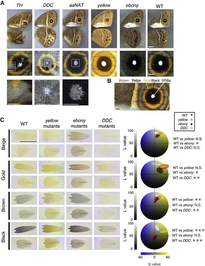
Figure 2. Proposed Melanin and Ommochrome Biosynthesis Pathways in Insects
(A) The melanin biosynthesis pathway produces up to five different molecules: pheomelanin, dopa-melanin, dopamine-melanin, NADA sclerotin, and NBAD
sclerotin. NADA and NBAD sclerotin contribute to the sclerotization of cuticle when cross-linked with cuticular proteins. Dopa-melanin and dopamine-melanin are
thought to be generated by the oxidative polymerization of 1-carboxyl 5,6-dihydroxyindole-2-carboxylic acid (DHI) and 5,6-dihydroxyindole (DHICA), respec-
tively. Pheomelanin is generated by the oxidative polymerization of cysteinyldopa or cysteinyldopamine in the presence of cysteine (Ito and Wakamatsu [2008]).
The scheme is modified from Galván et al. (2015) and Arakane et al. (2009).
(B) The ommochrome pathway produces up to three different pigments: 3-hydroxykynurenine (3-OHK), xanthommatin, and dihydroxanthommatin.
(C) Functional redundancy of ABC transporter proteins. The White transporter protein also can form heterodimers with OK and Brown to incorporate different
precursors into the pigment granule used in the production of other pigments such as urate granules or pteridines. Highlighted in red are the enzymes investigated
in this study.

putatively heterozygous clones that are contiguous with other                     aaNAT Mutants
colored regions, were affected in more extreme ways and                           aaNAT catalyzes the conversion of dopamine to NADA sclerotin
were always absent. Together, these results indicate that TH                      (colorless) (Figure 2A). WT B. anynana naturally change their
is required for larval head pigmentation and for scale overall                    head capsule color from black in the third instar to light brown
development, pigmentation, and structural rigidity. These phe-                    in the fourth instar (Figure S4B). From 20 larvae showing a normal
notypes are similar to those observed in other butterfly species                  black head as third instars, 16 retained their black head capsule
(Zhang et al., 2017a).                                                            color until the last larval instar (Figure S4B). This phenotype is
DDC Mutants                                                                       similar to the phenotype observed in Bombyx aaNAT RNAi larvae
DDC catalyzes the production of dopamine from DOPA (Fig-                          (Long et al., 2015). 4 out of 20 butterflies lacked white scales in
ure 2A). By disrupting the function of DDC, butterflies are                       multiple eyespot centers (Figure 3A), but no other scale abnor-
expected to be able to produce only dopa-melanin (black                           malities were detected. These results indicate that aaNAT is
pigment); however, they may still be able to produce pheomela-                    required for brown head capsule pigmentation in late larvae,
nin if that part of the pathway is active in B. anynana wing scale                the lighter color achieved presumably by shunting DOPA away
cells. 5 out of 42 larvae lacked black pigment on their heads (Fig-               from dopa-melanin production and into NADA sclerotin produc-
ure S4A). From two of the seven butterflies that emerged, the                     tion. aaNAT also is required for the development of the white,
black eyespot scales became gray, the brown and beige scales                      structurally colored scales of the eyespot centers.
became whitish, the gold ring region became paler while still re-                 yellow and ebony Mutants
taining a gold color, and the white eyespot center scales were                    Color changes in both yellow and ebony mutants in B. anynana
absent (Figure 3A). Furthermore, all of the scales were mostly                    were previously broadly described (Zhang et al., 2017a), but
curled. All of these phenotypes were similar to the mild TH                       here we quantified those changes using the L*a*b color space
mutant phenotypes. L*a*b color space quantitative analyses                        analysis. In yellow mutants, the darker-colored scales (black
(Hirschler, 2010) supported the visible color changes described:                  and brown) became significantly lighter relative to WT scales,
DDC mutant scales, other than beige scales, were significantly                    whereas the lighter-colored scales (gold, beige, and white) did
lighter (higher L value) and less yellowish in color (lower b value)              not change in color (Figures 3A and 3C; Table S6). This indi-
compared to wild-type (WT) scales (Figure 3C; Table S6). Our re-                  cates that yellow is active only in darker-colored scales. We
sults indicate that dopamine is an important precursor for the                    infer that in these scales yellow is acting to convert DOPA to
pigments contained in most wing scales and a required molecule                    dopa-melanin.
in the development of white scales. The persistence of black and                     In ebony mutants the result was opposite that of yellow mu-
gold pigments in the black and gold mutated scales, respec-                       tants; in other words, the lighter-colored scales (gold and beige)
tively, indicate that dopa-melanin is likely present in the black                 became significantly darker relative to WT scales, whereas the
scales, and other non-dopamine-derived pigments (possibly                         darker-colored scales (black and brown) did not change in color
pheomelanin or an ommochrome pigment) are present in the                          (Figure 3A). This indicates that ebony is active only in the lighter-
gold scales. These results also indicate that DDC, similar to                     colored scales to convert dopa-melanin to NBAD sclerotin
TH, is required for scale hardening across all of the colored                     (yellow). The removal of ebony makes these scales less yellow
scales.                                                                           (lower b) and darker (lower L) (Figure 3C; Table S6).

58 Cell Reports 24, 56–65, July 3, 2018
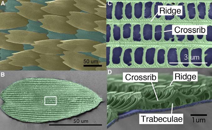
Figure 3. Melanin Biosynthesis Gene Mu-
                                                                                           tants in B. anynana
                                                                                           (A) Representative pictures of each melanin gene
                                                                                           mutant. TH, scales of all colors failed to develop in
                                                                                           mutant TH clonal tissue. DDC, scale development
                                                                                           also was disrupted but not as frequently as in TH
                                                                                           mutants. White scale development was always
                                                                                           disrupted in mutant clones, but gold, brown, and
                                                                                           black scales became paler and curled in pre-
                                                                                           sumed heterozygous mutant clones. In aaNAT
                                                                                           mutants, white scale development alone was dis-
                                                                                           rupted. In yellow mutants, brown and black scales
                                                                                           became lighter, and in ebony mutants, beige and
                                                                                           gold scales became darker. Scale bar indicates
                                                                                           5 mm in low-magnification images (top row) and
                                                                                           1 mm in higher-magnification images (middle and
                                                                                           bottom rows).
                                                                                           (B) Colored areas that were sampled for individual
                                                                                           scale measurements. Scale bar indicates 1 mm.
                                                                                           (C) Transmission photos of individual mutant and
                                                                                           wild-type (WT) cover and ground scales. Scale bar
                                                                                           indicates 100 mm. Diagrams at right show the
                                                                                           color space occupied by cover scales on the L*a*b
                                                                                           color space. Each colored ellipse represents
                                                                                           measurements from five scales from three
                                                                                           different male individuals (N = 3 for statistical an-
                                                                                           alyses). *p % 0.05, **p % 0.01, and ***p % 0.001.
                                                                                           See also Figures S3 and S4 and Tables S1 and S6.

                                                                                           observed in a single adult eye of a scarlet
                                                                                           mosaic mutant (Figure S4C). We did not
                                                                                           detect any phenotypes from vermilion
                                                                                           mutants in larvae or adults, even though
                                                                                           a previous study detected vermilion
                                                                                           expression in developing wing tissue in
                                                                                           this species (Beldade et al., 2005).

                                                                                           Deletion of Melanin Pathway Genes
                                                                                           Affected Scale Morphology
                                                                                           To examine the contribution of melanin
                                                                                           biosynthesis pathway products to the
                                                                                           development of scale size and the cutic-
                                                                                           ular microstructures of wing scales, we
Ommochrome Biosynthesis Pathway May Not                             took scanning electron microscopy (SEM) images of individual
Contribute to Wing Coloration in B. anynana                         scales of different colors from male mutants and compared
To examine whether the ommochrome pathway contributes to            these to WT scales. In yellow mutants, the windows of the black
the production of the gold color in B. anynana, we individually     and brown scales were covered broadly by a supernumerary
disrupted the function of three ommochrome biosynthesis             lamina (Figure 4A), which only rarely occurs in WT scales, with
pathway genes, vermilion, white, and scarlet. The vermilion         the exception of white scales, which naturally display parts of
gene product catalyzes the most upstream reaction of ommo-          such lamina. White, gold, and beige scales showed no marked
chrome pathway while White and Scarlet are ATP-binding              differences in these mutants. The crossribs flanking these closed
cassette (ABC) transporters that form heterodimers to selec-        windows in mutant scales were thinner and the crossrib spacing
tively incorporate the ommochrome precursor into the pigment        was significantly denser in black and brown scales (Figure 4B;
granules inside cells (Mackenzie et al., 1999; Figure 2B). We       Tables S9 and S10). The overall scale size and the distance
confirmed the presence of indel mutations in groups of embryos,     between longitudinal ridges were unchanged (Figure 4B; Tables
after injection of sgRNA targeting each of these genes along with   S7 and S8).
Cas9 (Figure S3), but we did not observe any wing phenotypes in        In DDC mutants, the arrangement of crossribs on all of the
adults. The white mosaic mutants, however, showed disruption        color scales became disordered, and neighboring crossribs
of larval body color and lack of pigmentation in the adult eyes     often were fused, resulting in larger windows (Figure 4A).
(Figures S4B and S4C). Lack of eye pigmentation also was            Sheet-like trabeculae, taller than the feet-like trabeculae of WT

                                                                                              Cell Reports 24, 56–65, July 3, 2018 59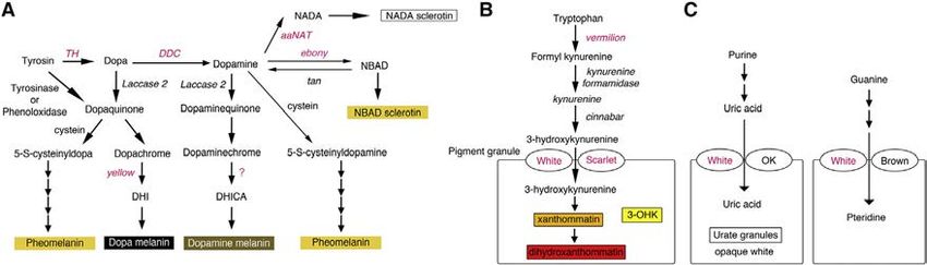
Figure 4. Scanning EM Images of Individual
                                                                                            Scales from WT, yellow, and DDC Mutants
                                                                                            and Detailed Morphological Measurements
                                                                                            from These and ebony Mutants
                                                                                            (A) Representative images of individual scales from
                                                                                            WT, yellow mutants, and DDC mutants. In yellow
                                                                                            mutants, there is a thin lamina that covers the
                                                                                            windows of darker-colored scales (black and
                                                                                            brown) (the ectopic lamina was colored in orange
                                                                                            in the scanning EM images). The morphology of the
                                                                                            lighter-colored scales (gold, beige, and white) is
                                                                                            unchanged. In DDC mutants, crossribs are more
                                                                                            disorganized and sometimes fuse with neigh-
                                                                                            boring crossribs. In addition, trabeculae become
                                                                                            sheet-like underneath the crossribs. Scale bars
                                                                                            indicate 10 mm in low-magnification images of
                                                                                            whole scales and 1 mm in high-magnification
                                                                                            images (top row).
                                                                                            (B) Several measurements of individual scale fea-
                                                                                            tures. We plotted all of the measurements taken
                                                                                            from five scales of the same type from three
                                                                                            different individuals, but statistics used N = 3,
                                                                                            where the five measurements within an individual
                                                                                            were averaged. Error bars represent 95% confi-
                                                                                            dence intervals of means. *p % 0.05, **p % 0.01,
                                                                                            and ***p % 0.001.
                                                                                            See also Figure S5 and Tables S7, S8, S9, and S10.

                                                                                            tive to WT scales. We also detected
                                                                                            smaller changes in scale morphology in
                                                                                            the ebony mutants, but these were small
                                                                                            compared to the observed changes in
                                                                                            color in the same mutants.

                                                                                            DISCUSSION

                                                                                          In this study, we generated mosaic mu-
                                                                                          tants for five of the known melanin biosyn-
                                                                                          thesis pathway genes and three of the
scales, appeared beneath the crossribs (Figure 4A). The width of     ommochrome pathway genes using CRISPR/Cas9. This allowed
each crossrib also became thinner (Figure 4B; Table S9); how-        us to identify which pigment pathway and which genes are
ever, the scale size and the distance between longitudinal ridges    needed for producing each of the colored scales on the wings
were unchanged (Tables S7 and S8). These phenotypes were             of B. anynana and how these genes affect scale color and
stronger in darker-colored scales (black) and weaker in lighter-     morphology.
colored scales (gold and beige).
   In ebony mutants, we did not detect dramatic changes in           The Role of Melanin Biosynthesis Pathway Genes in the
morphology, but the scale size was slightly larger in black and      Development of Pigmentary Colors
beige mutant scales, the thickness of the crossribs was slightly     The melanin pathway mutants displayed both expected color
higher in brown mutant scales, and the distance between longi-       alterations (i.e., those observed in similar experiments in other
tudinal ridges was slightly larger in white and gold mutant scales   insects and butterflies) (True et al., 1999; Wittkopp et al., 2002;
relative to WT scales (Figure 4B; Tables S7, S8, and S9). Most of    Arakane et al., 2009; Liu et al., 2010, 2014, 2016; Li et al.,
these morphological changes had a small effect size, took place      2015; Perry et al., 2016; Zhang and Reed, 2016; Beldade and
in scales where there was no significant effect on color, did not    Peralta, 2017; Zhang et al., 2017a) and previously undescribed
transform the morphology of one colored scale into that of           color changes. A surprising discovery was that aaNAT is neces-
another colored scale, and may be simply due to slight differ-       sary for generating the complete skeleton of white eyespot cen-
ences in genetic background between the six animals measured.        ter scales in B. anynana, which produce a white structural color
   In summary, the color and the morphology of the darker scales     (Monteiro et al., 2015; Figure 3A). In Bombyx, Oncopeltus, and
in yellow mutants and the color and morphology of all of the         Tribolium, loss or downregulation of aaNAT caused increased
colored scales in DDC mutants were changed dramatically rela-        cuticle melanization (Dai et al., 2010; Zhan et al., 2010; Liu

60 Cell Reports 24, 56–65, July 3, 2018
Figure 5. Cell-Specific Expression and
Function of Melanin Biosynthesis Pathway
Genes in Wing Scales of B. anynana Control
Scale Color and Morphology
(A) Summary of the scale cell-specific expression
of each of the examined melanin pathway genes,
inferred from the loss of function mutants, on the
wings of B. anynana, and how their combinatorial
expression contributes to the production of
different pigments in each scale cell.
(B) Schematic diagram of the morphology of a
black scale from WT, yellow, and DDC mutants. In
yellow mutants, black scales become yellow-
brown, the normally open windows become
covered by an ectopic cuticular lamina, and the
adjacent crossribs become thinner, implicating the
gene yellow in the creation of both dark pigmen-
tation and in opening windows. In DDC mutants,
the black scales become grayish, the trabeculae
became sheet-like and taller, and the adjacent
crossribs become thinner, implicating the gene
DDC in the creation of dark pigmentation and short
and pillar-like trabeculae. (Hand illustrations by
Katerina Evangelou.)
different heterodimers selectively incor-
porate different pigment precursors into
the pigment granules (Figures 2B and
2C). For instance, in Drosophila, the White
protein forms a heterodimer with the
Brown protein and incorporates a pteri-
dine precursor into the pigment granule
(Mackenzie et al., 1999; Figure 2C). The
product of a brown gene paralog, the
OK protein in Bombyx and Helicoverpa
et al., 2016; Noh et al., 2016). We speculate that in those sys- moths, forms a heterodimer with White to transport uric acid
tems, aaNAT is co-expressed with yellow in the same cells, lead- into the pigment granules of the larval epidermis to make whitish
ing to the redirection of the common dopamine precursor used urate granules (Wang et al., 2013; Khan et al., 2017; Figure 2C).
by both enzymes to the exclusive use by yellow, leading to the The loss of epidermal color in white mutants in B. anynana is
production of more dopamine-melanin and a darker color. likely due to the failure of uric acid uptake into epidermal cells
The color of the gold scales in B. anynana was affected by as observed in Bombyx ok mutants (Wang et al., 2013). The
mutations in ebony (Figure 3A), suggesting that NBAD sclerotin eye phenotype in adult white B. anynana mutants can be due
contributes to this color. We observed that in DDC mutants, to a disruption in either an ommochrome precursor uptake
gold color persisted in the affected regions. These results sug- (Mackenzie et al., 1999; Quan et al., 2002; Tatematsu et al.,
gest that additional pigments also may be present in this area. 2011), a pteridine precursor uptake (Dreesen et al., 1988; Grubbs
We speculate that pheomelanin molecules may be contributing et al., 2015), or a uric acid uptake (Khan et al., 2017). These
to this color in B. anynana. Despite the belief that invertebrates results suggest that either the ommochrome pathway may not
do not produce pheomelanin, this product was detected in be playing a role in the development of wing pigmentation in
mollusks, grasshoppers, butterflies, bumblebees, and wasps B. anynana or that cells expressing these genes, especially cells
(Speiser et al., 2014; Galván et al., 2015; Jorge Garcı́a et al., of the gold ring in the eyespots, were not hit with mutations in
2016; Polidori et al., 2017) and may be present in the gold wing these genes.
scales of B. anynana. Color on the wings of B. anynana appears to derive primarily
We targeted two transporter protein genes, white and scarlet, from melanin pathway products, whereas the different color pat-
as well as the enzyme vermilion. None of those ommochrome terns likely result from the different spatial expressions and
pathway gene mutations affected coloration on the wings of perhaps also the levels of expression of the different melanin
B. anynana, whereas mutations in white led to changes in eye pathway genes in each scale. By examining the affected colored
and larval coloration, probably via disruptions to two other regions in each mutant, we inferred the spatial expression for
pigment pathways (Figures 2C, S4B, and S4C). ABC trans- each of these enzymes (Figure 5A) and proposed how these en-
porters, including White, are diverse (Merzendorfer, 2014), and zymes may be regulated by previously mapped transcription
Cell Reports 24, 56–65, July 3, 2018 61factors. In particular, DDC being expressed overall on the wing these areas would help polymerize cuticle more tightly under
and yellow being expressed in darker-colored scales correlates and around each crossrib to create short and thick pillars under
nicely with functional and quantitative Distal-less gene expres- the ridges, the trabeculae, and thicker crossribs. At later stages,
sion (Brakefield et al., 1996; Monteiro et al., 2013; Connahs the cytoplasm of the scale cells disappears, leaving behind the
et al., 2017), suggesting that DDC and yellow expression may cuticular skeleton (Ghiradella, 1989). Cells that do not express
be regulated by Distal-less. Any one of the 186 genes recently DDC leave behind tall, sheet-like trabeculae, and cells that do
found to be differentially expressed in the central cells of an not express yellow leave behind unaggregated cuticle as a thin
eyespot during the early pupal stage (Özsu and Monteiro, lamina covering the windows (Figure 5B). We propose that the
2017) or at other time points (Monteiro et al., 2006; Özsu et al., polymerization and interaction of subcellular compartmentalized
2017) could be upregulating aaNAT expression specifically in dopa-melanin and dopamine-melanin molecules with chitin or
these cells. Expression of ebony in the gold ring could be upre- other cuticular proteins are important for constructing specific
gulated by engrailed (Brunetti et al., 2001), but it is still unclear cuticular morphologies in the scale cell, especially involving
what could be regulating this gene in the beige transversal cuticle aggregation around the trabeculae and crossribs.
band. Finally, TH (and DDC) may not be spatially regulated by Mutations in melanin pathway genes also affected scale
any of the above pattern-associated transcription factors. morphology in subtler ways, such as in altering the spacing
between ridges and crossribs, but these changes were not
Subcellular Localization and Functions of Melanin sufficient to explain why scales of different colors exhibit these
Pathway Products during Wing Scale Development different morphologies (Janssen et al., 2001; Figures S1 and
Butterfly wing scale development has been visualized largely via S2). These results suggest that other factors besides melanin
transmission EM (TEM) images of fixed developing scale cells pathway products regulate these subtler quantitative differences
(Ghiradella et al., 1972; Greenstein 1972a, 1972b; Ghiradella, in scale morphology between scales of different colors. Some of
1989), but more recently fluorescent microscopy also was these factors may involve overall concentration of the cuticle in-
used (Dinwiddie et al., 2014). In TEM images of early scale devel- side the cell and type of cuticular proteins expressed in each
opment of Ephestia kuehniella moths, microfibril bundles, scale. Ohkawa et al. (2004) demonstrated that electrospun fibers
aligned with regular spacing, were observed on the surface of formed by lower concentrations of chitosan, a derivate of chitin,
scale cells as the scale started to flatten (Overton, 1966; Ghira- were thinner than those formed at higher concentrations. It is
della, 1974). Greenstein (1972b) proposed that the scale ridges possible, then, that eyespot-forming scales showing a higher
of the giant silkmoth, Hyalophora cecropia, were likely to develop density of crossribs may have higher concentrations of cuticle-
in between these microfibril bundles by buckling of the expand- building materials than beige or brown scales. Finally, the
ing extracellular cuticle. Dinwiddie et al. (2014) showed that amount and/or the spatial distribution of the large number of
cuticle deposition along the future longitudinal ridges happened cuticle proteins found in lepidopterans (Liang et al., 2010), which
in between flanking longitudinal actin bundles inside the devel- have been implicated in the joint regulation of larval body cuticle
oping scale, and disruption of actin filament formation led to thickness and its coloration (Xiong et al., 2017), also may
disorganized scales. Together, these studies indicate that scale contribute to particular scale morphologies.
ridge development likely depends on the interaction of actin fila- By disrupting melanin and ommochrome pathway genes, we
ments and cuticle. demonstrated in this study that most of the color observed on
Our study shows that melanin pathway products also play a the wings of B. anynana comprises melanin biosynthesis
role during butterfly wing scale development. The development pathway products. We also showed how each of the colors is
of B. anynana wing scales appears to require both TH and the result of the expression of a specific subset of melanin
DDC, two early components of the melanin pathway that also pathway enzymes in each scale cell that channel the flux of pre-
severely affected scale sclerotization. Without TH, scale cells cursors in the pathway into specific end pigments (Figure 5A).
were unable to produce a cuticular skeleton (Figure 3A). Without This work contributes to our understanding of the genetic mech-
adequate levels of either TH or DDC, scales developed a curly anisms that establish complex color patterns in butterfly wings.
appearance and had overall lower levels of pigmentation. These We also showed that melanin biosynthesis pathway products
results indicate that the expression of both genes is important in are acting both as pigments and as scaffolds to build the fine
the development of the cuticular skeleton as well as in the devel- cuticular structures of a wing scale (Figure 5B) and may therefore
opment of melanin pigments. be important in creating as well as limiting the diversity of the
The yellow and DDC mutants described here show how structural and pigmentary colors observed in butterflies. These
disruptions to the melanin synthesis pathway affect both scale molecules, together with actin (Dinwiddie et al., 2014), affect
color and specific details of the scale morphology (Figures 3 the microstructures on a wing scale. The genes examined here
and 4). We infer that loss of dopa-melanin and dopamine- should perhaps also be investigated in other butterflies, in partic-
melanin cause the yellow and DDC color and scale structural ular those that show complex morphologies (Saranathan et al.,
changes, respectively. We speculate that cuticle is first distrib- 2010) and iridescent structural colors (Ghiradella et al., 1972;
uted at the periphery of the developing scale, including in the Vukusic et al., 2000) because they may contribute to the
window regions, and the presence of dopa-melanin helps cuticle construction of complex photonic materials. Uncovering the
polymerize around the crossribs to create windows devoid of genetic basis of such finely sculpted biophotonic materials
cuticle. Similarly, cuticle would be present initially in vertical walls may eventually pave the way for a live-cell bioengineering pro-
below the crossribs, and the presence of dopamine-melanin in cess of fabricating these materials.
62 Cell Reports 24, 56–65, July 3, 2018EXPERIMENTAL PROCEDURES same individuals, and three different male and female individuals were
measured for statistical purposes. ANOVAs were used to test for significant
Butterfly Husbandry differences in mean color values between the sexes, and multivariate analyses
B. anynana, originally collected in Malawi, have been reared in the laboratory of variance (MANOVAs) were used to test for significant differences between
since 1988. Larvae were fed young corn plants and adults were fed mashed color of WT males and each of the male melanin mutants using IBM-SPSS
bananas. B. anynana were reared at 27! C and 60% humidity in a 12:12-hr Statistics version 21 software.
light:dark cycle.
Scanning EM and Sample Preparation
sgRNA Design and Production Small pieces of wing (5 mm 3 5 mm) were cut under a dissecting microscope.
Sequence choice for sgRNAs and sgRNA and Cas9 mRNA production was Fifty percent ethanol/MQ water was dropped onto the wing pieces, and the
performed as previously described by Zhang et al. (2017a) and in the Supple- pieces were dipped into liquid nitrogen for 5 min. The wing pieces were
mental Information. The sequences of sgRNAs used in this study are listed in removed from liquid nitrogen and evaporated at room temperature. The pieces
Table S2. of wing or individual scales were moved using a sharpened tungsten needle
onto double-stick carbon tape and pressed gently onto the metal scanning
Microinjection EM stub. After enough desiccation (overnight), platinum conductive coating
Eggs were laid on corn leaves for 30 min. We co-injected 0.5 mg/mL final was performed for 60 s at 20 mA using an Auto Fine Coaters JFC-1600
concentration of sgRNA and 0.5 mg/mL final concentration of Cas9 mRNA (JEOL). Images were taken using a JSM-6701F scanning electron microscope.
into embryos within 1 to 3 hr after egg laying. The eggs were sunk into PBS, The method for imaging cross-sections of single scales (shown in Figure 1D)
and injection was performed inside the PBS. Food dye was added to the injec- was previously described by Wasik et al. (2014).
tion solution for visualization. Injected embryos were incubated at 27! C in
PBS, transferred to the corn silk the next day, and further incubated at 27! C. Scale Measurements
After hatching, larvae were moved to corn leaves and reared at 27! C with a We compared several traits between male and female scales such as area,
12:12-hr light:dark cycle and 60% relative humidity. length, width, average distance between longitudinal ribs, density of cross-
ribs along a standard length, and width of each crossrib in both cover and
Detection of Indel Mutations ground scales of different colors. For crossrib thickness and distance be-
We assessed successful disruptions of the target genes using a T7 endonu- tween each crossrib, we took the average of 10 measurements in a different
clease I assay (NEB) (in injected embryos), followed by Sanger sequencing area of the scale. For distance between ridges, we took the average of three
of target regions of the DNA in adults, as previously described (Zhang et al., measurements in a different area of the scale. For scale area size, scale
2017a). Detailed procedures are described in the Supplemental Experimental width, scale length, and scale density, we took the average of one measure-
Procedures. The primer sequences used in this study are listed in Table S2. ment in a different area of the scale, and then the average of five individual
scales of the same color from the same individual. Our sample size, for sta-
Acquisition of Colored Images tistical purposes, was equal to 3—in other words, the measurements from
The images of butterfly wings and larvae were taken with a Leica DMS1000 three distinct individual animals of each sex. We measured each trait using
microscope camera. Color images of individual scales were taken after ImageJ software. ANOVAs were used to test for significant differences in
embedding scales in clove oil. This oil matches the refractive index of the scale measurements between the sexes and between WT males and
insect cuticle and allows pigmentary color to be measured precisely (Wasik each of the male melanin mutants using IBM-SPSS Statistics version 21
et al., 2014). In particular, this oil treatment removes any contribution of struc- software.
tural colors that may result from light constructively interfering when crossing
finely patterned materials with different refractive indexes, such as cuticle and
SUPPLEMENTAL INFORMATION
air. We put one drop of clove oil on a glass slide. Wing scales were taken from a
mutated site (from the area depicted in Figure 3B) by using a fine tungsten nee-
Supplemental Information includes Supplemental Experimental Procedures,
dle and dipped into the clove oil. Once enough scales were transferred, a cover
five figures, and ten tables and can be found with this article online at
glass was placed on top of the oil and the scales. Additional clove oil was sup-
https://doi.org/10.1016/j.celrep.2018.05.092.
plied from the edge of the cover glass to fill the space, and this preparation was
sealed with nail polish. Images of wing scales were taken via light transmission
using an Axioskop2 mot plus (Carl Zeiss) microscope and an Axiocam ERc 5s ACKNOWLEDGMENTS
(Carl Zeiss).
We thank Katerina Evangelou (PhD student, University of the Arts London,
Color Measurements Central Saint Martins College, katerinaev@hotmail.com) for producing 3D
Light transmission images taken using the above method were not entirely hand-drawn illustrations of wing scales and also thank Monteiro Lab members
consistent for brightness, so images were edited using the Digital Color Meter for their help and discussion. The work was partially funded by the Ministry of
(a pre-installed Macintosh application [Apple]) to make the background bright- Education, Singapore grant MOE2015-T2-2-159 and the LHK Fund from the
ness similar across all of the images. The same relative region in a scale (in the Department of Biological Sciences, NUS (R-154-000-608-651).
middle) was selected with a rectangular marquee of a constant size, and the
color was averaged inside the selected area using the ‘‘average’’ tool in Adobe AUTHOR CONTRIBUTIONS
Photoshop (CS3). For the quantitative assessment of color, we conducted
L*a*b color space analysis, which is a way of mathematically describing color Y.M. and A.M. designed the study. Y.M. conducted the study and analyzed the
in three dimensions with three parameters. The L value (0–100) indicates how data. Y.M. and A.M. wrote the manuscript.
bright the color is, with higher values indicating a brighter/lighter color; the a
value ("60 to 60) indicates where the color is located between red (negative DECLARATION OF INTERESTS
values) and green (positive values) opponent colors; and the b value ("60 to
60) indicates where the color is located between blue (negative) and yellow The authors declare no competing interests.
(positive) opponent colors. The L*a*b color space is used in industry and in bio-
logical color assessment. Measurements were conducted using the Digital Received: January 4, 2018
Color Meter. In this study, we plotted only the L and b values because the a Revised: March 26, 2018
value (red–green) was similar among all of the measured scales. Color mea- Accepted: May 29, 2018
surements were averaged across five scales of the same color from the Published: July 3, 2018
Cell Reports 24, 56–65, July 3, 2018 63REFERENCES Ito, S., and Wakamatsu, K. (2008). Chemistry of mixed melanogenesis—
pivotal roles of dopaquinone. Photochem. Photobiol. 84, 582–592.
Andersen, S.O. (2005). Cuticular sclerotization and tanning. In Comprehensive Janssen, J.M., Monteiro, A., and Brakefield, P.M. (2001). Correlations between
Molecular Insect Science, L.I. Gilbert, K. Iatrou, and S.S. Gill, eds. (Elsevier scale structure and pigmentation in butterfly wings. Evol. Dev. 3, 415–423.
Pergamon Press), pp. 145–170.
Jorge Garcı́a, A., Polidori, C., and Nieves-Aldrey, J.L. (2016). Pheomelanin in
Arakane, Y., Lomakin, J., Beeman, R.W., Muthukrishnan, S., Gehrke, S.H., Ka- the secondary sexual characters of male parasitoid wasps (Hymenoptera:
nost, M.R., and Kramer, K.J. (2009). Molecular and functional analyses of Pteromalidae). Arthropod Struct. Dev. 45, 311–319.
amino acid decarboxylases involved in cuticle tanning in Tribolium castaneum.
Kerwin, J.L., Turecek, F., Xu, R., Kramer, K.J., Hopkins, T.L., Gatlin, C.L., and
J. Biol. Chem. 284, 16584–16594.
Yates, J.R., 3rd. (1999). Mass spectrometric analysis of catechol-histidine
Beldade, P., and Peralta, C.M. (2017). Developmental and evolutionary mech- adducts from insect cuticle. Anal. Biochem. 268, 229–237.
anisms shaping butterfly eyespots. Curr. Opin. Insect Sci. 19, 22–29.
Khan, S.A., Reichelt, M., and Heckel, D.G. (2017). Functional analysis of the
Beldade, P., Brakefield, P.M., and Long, A.D. (2005). Generating phenotypic ABCs of eye color in Helicoverpa armigera with CRISPR/Cas9-induced muta-
variation: prospects from ‘‘evo-devo’’ research on Bicyclus anynana wing tions. Sci. Rep. 7, 40025.
patterns. Evol. Dev. 7, 101–107.
Koch, P.B., Keys, D.N., Rocheleau, T., Aronstein, K., Blackburn, M., Carroll,
Brakefield, P.M., Gates, J., Keys, D., Kesbeke, F., Wijngaarden, P.J., Monteiro, S.B., and ffrench-Constant, R.H. (1998). Regulation of dopa decarboxylase
A., French, V., and Carroll, S.B. (1996). Development, plasticity and evolution expression during colour pattern formation in wild-type and melanic tiger swal-
of butterfly eyespot patterns. Nature 384, 236–242. lowtail butterflies. Development 125, 2303–2313.
Brunetti, C.R., Selegue, J.E., Monteiro, A., French, V., Brakefield, P.M., and Li, X., Fan, D., Zhang, W., Liu, G., Zhang, L., Zhao, L., Fang, X., Chen, L., Dong,
Carroll, S.B. (2001). The generation and diversification of butterfly eyespot Y., Chen, Y., et al. (2015). Outbred genome sequencing and CRISPR/Cas9
color patterns. Curr. Biol. 11, 1578–1585. gene editing in butterflies. Nat. Commun. 6, 8212.
Connahs, H., Rhen, T., and Simmons, R.B. (2016). Transcriptome analysis of Liang, J., Zhang, L., Xiang, Z., and He, N. (2010). Expression profile of cuticular
the painted lady butterfly, Vanessa cardui during wing color pattern develop- genes of silkworm, Bombyx mori. BMC Genomics 11, 173.
ment. BMC Genomics 17, 270.
Liu, C., Yamamoto, K., Cheng, T.C., Kadono-Okuda, K., Narukawa, J., Liu,
Connahs, H., Tlili, S., van Creij, J., Loo, T.Y.J., Banerjee, T., Saunders, T.E., S.P., Han, Y., Futahashi, R., Kidokoro, K., Noda, H., et al. (2010). Repression
and Monteiro, A. (2017). Disrupting different Distal-less exons leads to ectopic of tyrosine hydroxylase is responsible for the sex-linked chocolate mutation
and missing eyespots accurately modeled by reaction-diffusion mechanisms. of the silkworm, Bombyx mori. Proc. Natl. Acad. Sci. USA 107, 12980–12985.
bioRxiv. https://doi.org/10.1101/183491.
Liu, J., Lemonds, T.R., and Popadic!, A. (2014). The genetic control of apose-
Dai, F.Y., Qiao, L., Tong, X.L., Cao, C., Chen, P., Chen, J., Lu, C., and Xiang, matic black pigmentation in hemimetabolous insects: insights from Oncopel-
Z.H. (2010). Mutations of an arylalkylamine-N-acetyltransferase, Bm-iAANAT, tus fasciatus. Evol. Dev. 16, 270–277.
are responsible for silkworm melanism mutant. J. Biol. Chem. 285, 19553–
Liu, J., Lemonds, T.R., Marden, J.H., and Popadic!, A. (2016). A pathway anal-
19560.
ysis of melanin patterning in a hemimetabolous insect. Genetics 203, 403–413.
Dinwiddie, A., Null, R., Pizzano, M., Chuong, L., Leigh Krup, A., Ee Tan, H., and
Long, Y., Li, J., Zhao, T., Li, G., and Zhu, Y. (2015). A new arylalkylamine N-ace-
Patel, N.H. (2014). Dynamics of F-actin prefigure the structure of butterfly wing
tyltransferase in silkworm (Bombyx mori) affects integument pigmentation.
scales. Dev. Biol. 392, 404–418.
Appl. Biochem. Biotechnol. 175, 3447–3457.
Dreesen, T.D., Johnson, D.H., and Henikoff, S. (1988). The brown protein of
Mackenzie, S.M., Brooker, M.R., Gill, T.R., Cox, G.B., Howells, A.J., and Ewart,
Drosophila melanogaster is similar to the white protein and to components
G.D. (1999). Mutations in the white gene of Drosophila melanogaster affecting
of active transport complexes. Mol. Cell. Biol. 8, 5206–5215.
ABC transporters that determine eye colouration. Biochim. Biophys. Acta
Ferguson, L.C., and Jiggins, C.D. (2009). Shared and divergent expression 1419, 173–185.
domains on mimetic Heliconius wings. Evol. Dev. 11, 498–512.
Mackenzie, S.M., Howells, A.J., Cox, G.B., and Ewart, G.D. (2000). Sub-
Galván, I., Jorge, A., Edelaar, P., and Wakamatsu, K. (2015). Insects synthesize cellular localisation of the white/scarlet ABC transporter to pigment granule
pheomelanin. Pigment Cell Melanoma Res. 28, 599–602. membranes within the compound eye of Drosophila melanogaster. Genetica
Ghiradella, H. (1974). Development of ultraviolet-reflecting butterfly scales: 108, 239–252.
how to make an interference filter. J. Morphol. 142, 395–409. Merzendorfer, H. (2014). ABC transporters and their role in protecting insects
Ghiradella, H. (1989). Structure and development of iridescent butterfly scales: from pesticides and their metabolites. Adv. Insect Physiol. 46, 1–72.
lattices and laminae. J. Morphol. 202, 69–88. Monteiro, A., Glaser, G., Stockslager, S., Glansdorp, N., and Ramos, D. (2006).
Ghiradella, H., and Radigan, W. (1976). Development of butterfly scales. II. Comparative insights into questions of lepidopteran wing pattern homology.
Struts, lattices and surface tension. J. Morphol. 150, 279–298. BMC Dev. Biol. 6, 52.
Ghiradella, H., Aneshansley, D., Eisner, T., Silberglied, R.E., and Hinton, H.E. Monteiro, A., Chen, B., Ramos, D.M., Oliver, J.C., Tong, X., Guo, M., Wang,
(1972). Ultraviolet reflection of a male butterfly: interference color caused by W.K., Fazzino, L., and Kamal, F. (2013). Distal-less regulates eyespot patterns
thin-layer elaboration of wing scales. Science 178, 1214–1217. and melanization in Bicyclus butterflies. J. Exp. Zool. B Mol. Dev. Evol. 320,
Greenstein, M.E. (1972a). The ultrastructure of developing wings in the giant 321–331.
silkmoth, Hyalophora cecropia. I. Generalized epidermal cells. J. Morphol. Monteiro, A., Tong, X., Bear, A., Liew, S.F., Bhardwaj, S., Wasik, B.R., Dinwid-
136, 1–21. die, A., Bastianelli, C., Cheong, W.F., Wenk, M.R., et al. (2015). Differential
Greenstein, M.E. (1972b). The ultrastructure of developing wings in the giant expression of ecdysone receptor leads to variation in phenotypic plasticity
silkmoth, Hyalophora cecropia. II. Scale-forming and socket-forming cells. across serial homologs. PLoS Genet. 11, e1005529.
J. Morphol. 136, 23–51. Moussian, B. (2010). Recent advances in understanding mechanisms of insect
Grubbs, N., Haas, S., Beeman, R.W., and Lorenzen, M.D. (2015). The ABCs of cuticle differentiation. Insect Biochem. Mol. Biol. 40, 363–375.
eye color in Tribolium castaneum: orthologs of the Drosophila white, scarlet, Nishikawa, H., Iga, M., Yamaguchi, J., Saito, K., Kataoka, H., Suzuki, Y., Su-
and brown genes. Genetics 199, 749–759. gano, S., and Fujiwara, H. (2013). Molecular basis of wing coloration in a
Hirschler, R. (2010). Electronic colour communication in the textile and apparel Batesian mimic butterfly, Papilio polytes. Sci. Rep. 3, 3184.
industry. file:///C:/Users/Owner/Downloads/Electronic_colour_communication_ Noh, M.Y., Koo, B., Kramer, K.J., Muthukrishnan, S., and Arakane, Y. (2016).
in_the_textile_and.pdf. Arylalkylamine N-acetyltransferase 1 gene (TcAANAT1) is required for cuticle
64 Cell Reports 24, 56–65, July 3, 2018morphology and pigmentation of the adult red flour beetle, Tribolium casta- Positional cloning of silkworm white egg 2 (w-2) locus shows functional
neum. Insect Biochem. Mol. Biol. 79, 119–129. conservation and diversification of ABC transporters for pigmentation in
Ohkawa, K., Cha, D., Kim, H., Nishida, A., and Yamamoto, H. (2004). Electro- insects. Genes Cells 16, 331–342.
spinning of chitosan. Macromol. Rapid Commun. 25, 1600–1605. True, J.R., Edwards, K.A., Yamamoto, D., and Carroll, S.B. (1999). Drosophila
Overton, J. (1966). Microtubules and microfibrils in morphogenesis of the scale wing melanin patterns form by vein-dependent elaboration of enzymatic pre-
cells of Ephestia ku€hniella. J. Cell Biol. 29, 293–305. patterns. Curr. Biol. 9, 1382–1391.
Özsu, N., and Monteiro, A. (2017). Wound healing, calcium signaling, and other Vukusic, P., Sambles, J.R., and Lawrence, C.R. (2000). Colour mixing in wing
novel pathways are associated with the formation of butterfly eyespots. BMC scales of a butterfly. Nature 404, 457.
Genomics 18, 788. Waku, Y., and Kitagawa, M. (1986). Developmental process of scale pigment
Özsu, N., Chan, Q.Y., Chen, B., Gupta, M.D., and Monteiro, A. (2017). Wingless granules in the cabbage butterfly, Pieris rapae crucivora. Jpn. J. Appl. Entomol.
is a positive regulator of eyespot color patterns in Bicyclus anynana butterflies. Zool. 30, 35–42.
Dev. Biol. 429, 177–185. Wang, L., Kiuchi, T., Fujii, T., Daimon, T., Li, M., Banno, Y., Kikuta, S., Kika-
Perry, M., Kinoshita, M., Saldi, G., Huo, L., Arikawa, K., and Desplan, C. (2016). wada, T., Katsuma, S., and Shimada, T. (2013). Mutation of a novel ABC trans-
Molecular logic behind the three-way stochastic choices that expand butterfly porter gene is responsible for the failure to incorporate uric acid in the
colour vision. Nature 535, 280–284. epidermis of ok mutants of the silkworm, Bombyx mori. Insect Biochem.
Polidori, C., Jorge, A., and Ornosa, C. (2017). Eumelanin and pheomelanin are Mol. Biol. 43, 562–571.
predominant pigments in bumblebee (Apidae: Bombus) pubescence. PeerJ 5, Wasik, B.R., Liew, S.F., Lilien, D.A., Dinwiddie, A.J., Noh, H., Cao, H., and
e3300. Monteiro, A. (2014). Artificial selection for structural color on butterfly wings
^ moto, N., Sezutsu, H., Ote, M., Shimada, T., Kanda, T.,
Quan, G.X., Kim, I., Ko and comparison with natural evolution. Proc. Natl. Acad. Sci. USA 111,
Mita, K., Kobayashi, M., and Tamura, T. (2002). Characterization of the kynur- 12109–12114.
enine 3-monooxygenase gene corresponding to the white egg 1 mutant in the Wittkopp, P.J., and Beldade, P. (2009). Development and evolution of insect
silkworm Bombyx mori. Mol. Genet. Genomics 267, 1–9. pigmentation: genetic mechanisms and the potential consequences of pleiot-
Reed, R.D., McMillan, W.O., and Nagy, L.M. (2008). Gene expression underly- ropy. Semin. Cell Dev. Biol. 20, 65–71.
ing adaptive variation in Heliconius wing patterns: non-modular regulation of Wittkopp, P.J., True, J.R., and Carroll, S.B. (2002). Reciprocal functions of the
overlapping cinnabar and vermilion prepatterns. Proc. Biol. Sci. 275, 37–45. Drosophila yellow and ebony proteins in the development and evolution of
Saranathan, V., Osuji, C.O., Mochrie, S.G., Noh, H., Narayanan, S., Sandy, A., pigment patterns. Development 129, 1849–1858.
Dufresne, E.R., and Prum, R.O. (2010). Structure, function, and self-assembly Xiong, G., Tong, X., Gai, T., Li, C., Qiao, L., Monteiro, A., Hu, H., Han, M., Ding,
of single network gyroid (I4132) photonic crystals in butterfly wing scales. Proc. X., Wu, S., et al. (2017). Body shape and coloration of silkworm larvae are influ-
Natl. Acad. Sci. USA 107, 11676–11681. enced by a novel cuticular protein. Genetics 207, 1053–1066.
Siddique, R.H., Gomard, G., and Hölscher, H. (2015). The role of random nano- Xu, R., Huang, X., Hopkins, T.L., and Kramer, K.J. (1997). Catecholamine and
structures for the omnidirectional anti-reflection properties of the glasswing histidyl protein cross-linked structures in sclerotized insect cuticle. Insect
butterfly. Nat. Commun. 6, 6909. Biochem. Mol. Biol. 27, 101–108.
Speiser, D.I., DeMartini, D.G., and Oakley, T.H. (2014). The shell-eyes of the Zhan, S., Guo, Q., Li, M., Li, M., Li, J., Miao, X., and Huang, Y. (2010). Disrup-
chiton Acanthopleura granulata (Mollusca, Polyplacophora) use pheomelanin tion of an N-acetyltransferase gene in the silkworm reveals a novel role in
as a screening pigment. J. Nat. Hist. 48, 2899–2911. pigmentation. Development 137, 4083–4090.
Stavenga, D.G., Stowe, S., Siebke, K., Zeil, J., and Arikawa, K. (2004). Butterfly Zhang, L., and Reed, R.D. (2016). Genome editing in butterflies reveals that
wing colours: scale beads make white pierid wings brighter. Proc. Biol. Sci. spalt promotes and Distal-less represses eyespot colour patterns. Nat. Com-
271, 1577–1584. mun. 7, 11769.
Suderman, R.J., Dittmer, N.T., Kanost, M.R., and Kramer, K.J. (2006). Model Zhang, L., Martin, A., Perry, M.W., van der Burg, K.R., Matsuoka, Y., Monteiro,
reactions for insect cuticle sclerotization: cross-linking of recombinant A., and Reed, R.D. (2017a). Genetic basis of melanin pigmentation in butterfly
cuticular proteins upon their laccase-catalyzed oxidative conjugation with wings. Genetics 205, 1537–1550.
catechols. Insect Biochem. Mol. Biol. 36, 353–365. Zhang, L., Mazo-Vargas, A., and Reed, R.D. (2017b). Single master regulatory
Tatematsu, K., Yamamoto, K., Uchino, K., Narukawa, J., Iizuka, T., Banno, Y., gene coordinates the evolution and development of butterfly color and irides-
Katsuma, S., Shimada, T., Tamura, T., Sezutsu, H., and Daimon, T. (2011). cence. Proc. Natl. Acad. Sci. USA 114, 10707–10712.
Cell Reports 24, 56–65, July 3, 2018 65You can also read