First comprehensive TSC1/TSC2 mutational analysis in Mexican patients with tuberous Sclerosis complex reveals numerous novel pathogenic variants ...
←
→
Page content transcription
If your browser does not render page correctly, please read the page content below
www.nature.com/scientificreports

            OPEN           First comprehensive TSC1/TSC2
                           mutational analysis in Mexican
                           patients with Tuberous Sclerosis
                           Complex reveals numerous novel
                           pathogenic variants
                           Miriam E. Reyna-Fabián1, Nancy L. Hernández-Martínez1, Miguel A. Alcántara-Ortigoza1,
                           Jorge T. Ayala-Sumuano2, Sergio Enríquez-Flores3, José A. Velázquez-Aragón1,
                           Alfredo Varela-Echavarría4, Carlos G. Todd-Quiñones5,6 & Ariadna González-del Angel1*
                           The aim of this study was to improve knowledge of the mutational spectrum causing tuberous sclerosis
                           complex (TSC) in a sample of Mexican patients, given the limited information available regarding this
                           disease in Mexico and Latin America. Four different molecular techniques were implemented to identify
                           from single nucleotide variants to large rearrangements in the TSC1 and TSC2 genes of 66 unrelated
                           Mexican-descent patients that clinically fulfilled the criteria for a definitive TSC diagnosis. The mutation
                           detection rate was 94%, TSC2 pathogenic variants (PV) prevailed over TSC1 PV (77% vs. 23%) and a
                           recurrent mutation site (hotspot) was observed in TSC1 exon 15. Interestingly, 40% of the identified
                           mutations had not been previously reported. The wide range of novels PV made it difficult to establish
                           any genotype-phenotype correlation, but most of the PV conditioned neurological involvement
                           (intellectual disability and epilepsy). Our 3D protein modeling of two variants classified as likely
                           pathogenic demonstrated that they could alter the structure and function of the hamartin (TSC1) or
                           tuberin (TSC2) proteins. Molecular analyses of parents and first-degree affected family members of the
                           index cases enabled us to distinguish familial (18%) from sporadic (82%) cases and to identify one case
                           of apparent gonadal mosaicism.

                           Tuberous sclerosis complex (TSC; MIM #191100, MIM #613254) is an autosomal dominant syndrome character-
                           ized by the presence of multiple hamartomas in different organs and systems. The incidence is about 0.1–1/10,000
                           births and the prevalence varies from 1/6,000 to 1/10,000 among different populations1–3. The manifestations
                           of TSC are highly variable among individuals and even within the same family4, but the most common clinical
                           features are localized in skin and central nervous system5–7.
                               TSC is caused by pathogenic variants (PV) in the tumor suppressor-genes, TSC1 (tuberous sclerosis com-
                           plex 1, MIM *605284, 9q34.13) and TSC2 (tuberous sclerosis complex 2, MIM *191092, 16p13.3). These PV
                           can be detected by various molecular techniques, such as single-strand conformational polymorphism (SSCP),
                           direct Sanger sequencing (SS), multiplex ligation-dependent probe amplification (MLPA) and next-generation
                           sequencing (NGS). There are currently more than 2,000 pathogenic TSC1/TSC2 variants described in the Leiden
                           Open Variation Database (www.lovd.nl/TSC1 and www.lovd.nl/TSC2)8; of them, 21–26% are located in TSC1
                           and 69–79% in TSC29,10. In approximately 5–25% of the analyzed TSC cases, a PV could not be identified in either
                           gene9,11,12. The emergence of new techniques, such as NGS, has significantly increased the mutation detection rate

                           1
                            Laboratorio de Biología Molecular, Instituto Nacional de Pediatría, Secretaría de Salud, Ciudad de México, México.
                           2
                            IDIX SA de CV., Querétaro, México. 3Grupo de Investigación en Biomoléculas y Salud Infantil, Laboratorio de Errores
                           Innatos del Metabolismo y Tamiz, Instituto Nacional de Pediatría, Ciudad de México, México. 4Departamento de
                           Neurobiología del Desarrollo y Neurofisiología, Instituto de Neurobiología, Universidad Nacional Autónoma de
                           México, Querétaro, México. 5Posgrado en Biología Experimental, Universidad Autónoma Metropolitana-Iztapalapa,
                           Ciudad de México, México. 6Laboratorio de Biología Molecular, Departamento de Genética Humana, Hospital de Alta
                           Especialidad de Veracruz, Veracruz, México. *email: ariadnagonzalezdelangel@gmail.com

Scientific Reports |   (2020) 10:6589 | https://doi.org/10.1038/s41598-020-62759-5                                                           1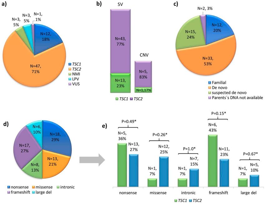
www.nature.com/scientificreports/                                                              www.nature.com/scientificreports

                           in cases where conventional tests were not successful (i.e., by identifying low rate somatic mosaic variants)11–13,
                           but it remains difficult to establish any phenotype-genotype correlation in TSC. It has been proposed that the
                           more severe phenotypes (in terms of the quantity or severity of the clinical features) are mainly TSC2-related.
                           Certain other clinical manifestations, including subependymal giant cell astrocytoma (SEGA), renal angiomyoli-
                           pomas and cardiac rhabdomyomas, are more common in patients with TSC2 variants14–16.
                                Most of the studies on TSC have been implemented in populations of Europe, USA, Canada and Brazil; hence,
                           it is relevant to include more cases from other Latin American countries in order to investigate and expand
                           the responsible TSC genotype, better delineate the clinical features and contribute to possibly identifying
                           genotype-phenotype correlations. To our knowledge, there are only two previous published molecular studies of
                           TSC patients in Mexico: one involving three patients with early-stage polycystic kidney disease that were molecu-
                           larly confirmed to represent TSC2/PKD1 contiguous gene syndrome cases17 and one involving three TSC2-cases
                           with prenatally documented cardiac rhabdomyomas18. Therefore, in order to improve our knowledge of this dis-
                           ease and to spread the use of innovative and highly sensitive molecular techniques such as MLPA and NGS for the
                           diagnosis of TSC in countries where the disease has been under-studied, we used a combined molecular strategy
                           to analyze the mutational spectrum of TSC1 and TSC2 and the principal clinical features of 66 Mexican-descent
                           unrelated cases of TSC.

                           Results
                           Patients. This study included 66 unrelated patients recruited between 2008 and 2017 in the genetics service
                           at the National Institute of Pediatrics in Mexico City, Mexico. All patients were clinically classified as definitive
                           TSC cases according to the most recent diagnostic criteria19. Thirty-six cases were male (55%) and 30 were female
                           (45%), and the mean age at diagnosis was 6 years 6 months (range: 1 month – 24 years of age) with a median age
                           of 6 years. Most patients were diagnosed during childhood (N = 36, 55%); the rest were diagnosed in infancy and
                           adolescence (N = 15, 23%; N = 14, 21% respectively), and only one in adulthood (N = 1, 1.5%).
                               Clinical evaluation by a medical geneticist and imaging studies (cranial computed tomography and renal
                           ultrasonography) on available parents and potentially affected family members from the 66 index cases allowed
                           us to classify 54 cases as sporadic (without any family history of TSC) and 12 cases as familial (with one or more
                           affected members). One of the familial cases was considered to represent possible gonadal mosaicism (two sib-
                           lings were affected while both parents were healthy).

                           Mutational analyses of TSC1 and TSC2. SSCP and SS. Genomic DNA samples derived from all 66
                           cases were initially subjected to mutational analyses of TSC1 and TSC2 by a SSCP assay followed by SS confirma-
                           tion in 61 cases and direct SS in the remaining five cases (Fig. 1). Both assays included all coding and non-coding
                           (20 bp at the exon-intron boundaries) regions of the TSC1 (NM_000368.4) and TSC2 (NM_000548.3) genes.
                           These analyses identified a clear disease-causing PV in 40/61 cases studied by SSCP/SS and in two of five cases
                           studied by direct SS (Table 1). Three other variants were classified as likely pathogenic variants (LPV) according
                           to guidelines of the American College of Medical Genetics and Genomics and the Association for Molecular
                           Pathology (ACMG/AMP)20. The two LPV in TSC1 were c.737+3A>G and p.(Leu112_Leu113delinsLysGluVal)
                           from cases ET75 and ET201, respectively, and the single in-frame LPV in TSC2: p.(His1746_Arg1751dup) from
                           case ET171. From these three cases, solely in case ET75, analysis of the proband’s paternity and maternity (crite-
                           rion PS2)20 using 15 short tandem repeat markers (13 of them belong to the CODIS system) could be performed
                           and confirmed parentage, but this ACMG/AMP criterion was not enough to re-classify the LPV as pathogenic
                           (Table 2). Therefore, our analysis identified a PV or LPV in 42/61 cases studied by SSCP/SS and in three of five
                           cases studied by direct SS (Fig. 1).

                           MLPA to identify deletions or duplications. In order to identify copy number variants (CNV) at both genes in
                           the remaining 21 cases, we performed MLPA (Fig. 1). The utilized TSC2 probemix contained one probe (exon
                           40) for the TSC2-adjacent gene, PKD1 (polycystic kidney disease 1, MIM *601313), whose mutation cause auto-
                           somal dominant polycystic kidney disease (ADPKD, MIM#173900). Our MLPA results identified six heterozy-
                           gous gross deletions: one in TSC1 and five in TSC2 (Table 1, Fig. 2b). Of the five gross deletions in TSC2, three
                           involved at least exons 40–46 of PKD1 (Fig. 3) resulting in the TSC2-PKD1 contiguous gene syndrome (CGS,
                           MIM #600273), which were previously published17. Interestingly, one of those TSC2-PKD1 CGS cases (ET178)
                           showed an atypical mild polycystic kidney phenotype.

                           Next-generation sequencing. Finally, an NGS study examining TSC1 and TSC2 coding exons and intron-exon
                           boundaries (150 bp) was carried out in the remaining 15 cases (Fig. 1). The median depth of coverage was
                           639×(range 86×–1940×) with a 99.9% width of coverage. A customized bioinformatic analysis enabled us to
                           identify a PV in 10 cases; we also found one case (ET243) with a missense variant p.(Trp1060Ser) in TSC2 that
                           was classified as an LPV20 and one case (ET81) with an intronic variant c.3815–21G>A in TSC1 that was classi-
                           fied [PP3, PP4]20 as a variant of uncertain significance or VUS (Tables 1,2). All NGS-identified variants were con-
                           firmed by SS in the index cases and their available parents. As the missense TSC2 p.(Trp1060Ser) LPV from case
                           ET243 was not reported in the main genotype databases and we did not find it in 212 alleles of healthy and ethni-
                           cally matched individuals assessed by a specific-allele PCR assay (data not shown), we were able to re-classify it as
                           a pathogenic variant (IIIa) [PM2, PS2, PS4, PP3, PP4]20. In the remaining three cases (ET44, ET61 and ET223),
                           no mutation was identified (NMI; lacking any LPV, VUS or pathogenic genotype) by the implemented molecular
                           technologies (Fig. 1).
                               To summarize, we were able to identify a PV or LPV in 62 cases and we could not identify a PV or LPV in four
                           cases, although one of them (case ET81) was found to harbor a VUS in TSC2 (c.3815–21G>A) (Figs. 2a and 3).
                           Of the identified changes, 56 (90%) corresponded to small variants (SV) such as point mutations, deletions,

Scientific Reports |   (2020) 10:6589 | https://doi.org/10.1038/s41598-020-62759-5                                                            2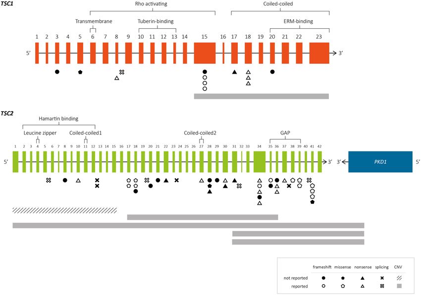
www.nature.com/scientificreports/ www.nature.com/scientificreports
Figure 1. Molecular algorithm used in 66 definitive TSC patients. Abbreviations: CGS: TSC2-PKD1 contiguous
gene syndrome; LPV: likely pathogenic variant; PV: pathogenic variant; SS: Sanger sequencing; SSCP: single-
strand conformation polymorphism; VUS: variant of uncertain significance.
small insertions/deletions (InDels) and duplications, while six were large deletions (10%). Far more of the iden-
tified changes were found in TSC2 (N = 48) than TSC1 (N = 14, Fig. 2b). The mutational proportions for TSC1
and TSC2 are shown in Fig. 2d,e. Eight intronic variants were identified at both genes; five affected canonical
splice sites (in TSC2) and three affected intronic splicing enhancers sequences (TSC1: c.737+3A>G; TSC2:
c.481+5G>T and c.5160+5G>T).
Based on our review of the literature and public databases, including the Leiden Open Variation Database
(LOVD, www.lovd.nl/), dbSNP (https://www.ncbi.nlm.nih.gov/projects/SNP), Exome Aggregation Consortium
(http://exac.broadinstitute.org), Genome Aggregation Database (gnomad.broadinstitute.org/), ClinVar (https://
www.ncbi.nlm.nih.gov/clinvar/), and Human Genome Mutation Database (http://www.hgmd.cf.ac.uk/), we
determined that 25 of the 62 (40%) PV or LPV identified herein (six in TSC1 and 19 in TSC2) had not been
previously reported. All of them have been submitted to LOVD (Table 1). Of these 25 novel variants, 24 were
considered pathogenic and one was an LPV20 in TSC1 [p.(Leu112_Leu113delinsLysGluVal)].
Direct molecular screening in parents (when available) of the 62 cases with a PV/LPV showed that the path-
ogenic allele was absent from both parents for 33 patients (de novo cases). However, in 12 cases with one or more
clinically affected family members, we confirmed the same PV in the available affected cases (familial cases, see
Table 1). We suspect gonadal mosaicism in familial case ET28 as we identified a novel heterozygous PV in TSC2:
c.3624G>A or p.(Trp1208*) in two affected siblings but failed to find this allele in peripheral blood leukocyte
DNA of both clinical healthy parents. This argument was further strengthened when we confirmed the proband’s
paternity and maternity by DNA profiling (data not shown). In 15 cases, we could not analyze the father’s DNA
but there was no reported family history of TSC, so we designated these as suspected de novo cases. In the remain-
ing two cases, the mother’s and father’s DNA samples were not available for testing (Table 1, Fig. 2c).
Protein modeling of two missense LPV. To examine possible functional and structural consequences
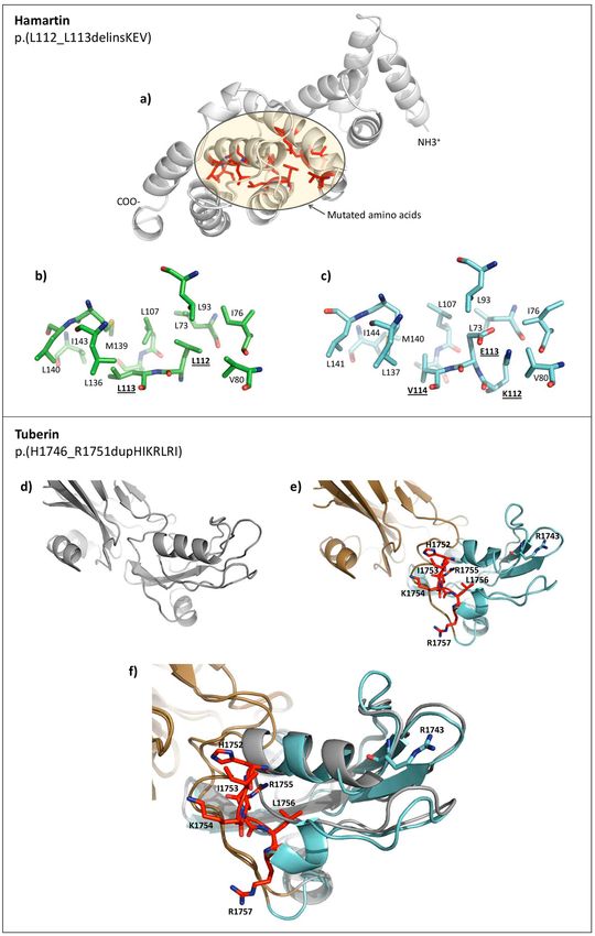
of the two in-frame variants that were classified as LPV [TSC1: c.333_337delinsAAAAGAGG or p.(Leu112_
L113delinsLysGluVal) and TSC2: c.5238_5255dup or p.(His1746_Arg1751dup)], we modeled the protein
structures of the N-terminal region of wild-type (WT) and mutated (MUT) p.(Leu112_L113delinsLysGluVal)
hamartin variant and the C-terminal region of WT and MUT p.(His1746_Arg1751dup) tuberin variant (Fig. 4).
Scientific Reports | (2020) 10:6589 | https://doi.org/10.1038/s41598-020-62759-5 3www.nature.com/scientificreports/ www.nature.com/scientificreports
TSC1 gene
Previous reports LOVD/dbSNP
Nucleotide change Codon change Clinical Molecular /ExAC/gnomAD/ClinVar/
Location (NM_000368.4) (NP_000359.1) significancea technique Inheritance b
HGMD/Literature Case Sex Age♦
Familial (mother
Exon 3 c.89_102del p.(Lys30Ilefs*2) Pathogenic SSCP and brother NPR (LOVD: TSC1_001334) ET173 M 12 y
heterozygous)
p.(Leu112_ Likely
Exon 5 c.333_337delinsAAAAGAGG SSCP suspected de novo NPR (LOVD: TSC1_001335) ET201 M 7y
Leu113delinsLysGluVal) Pathogenic
TSC1_000037 (+/+)/
Exon 8 c.682C>T p.(Arg228*) Pathogenic SSCP de novo rs118203427/NR/NR/49083/ ET25 M 5y
CM981931
Likely TSC1_000041 (-/+?)/
Intron 8 c.737+3A>G — SS de novo ET75 M 10 y
Pathogenic rs118203439/NR/NR/49093/NR
Exon 15 c.1458_1461del p.(Ser487Argfs*44) Pathogenic SS suspected de novo NPR (LOVD: TSC1_001336) ET157 M 6y
Familial (mother TSC1_000116 (+/+)/
Exon 15 c.1888_1891del p.(Lys630Glnfs*22) Pathogenic SSCP and brother rs118203595/NR/NR/5097/ ET93 F 2y
heterozygous) CD972488
TSC1_000116(+/+)/
Exon 15 c.1888_1891del p.(Lys630Glnfs*22) Pathogenic SSCP de novo rs118203595/NR/NR/5097/ ET249 M 10 y
CD972488
Familial TSC1_000121 (+/+)/
Exon 15 c.1959dup p.(Gln654Thrfs*34) Pathogenic NGS (daughter rs118203603/NR/NR/48857/ ET264 M 16 y
heterozygous) CI067260
Exon 17 c.2101C>T p.(Gln701*) Pathogenic NGS de novo NPR (LOVD: TSC1_000876) ET107 M 2y
Familial (father TSC1_000145(+/+)/
Exon 18 c.2227C>T p.(Gln743*) Pathogenic NGS and sister rs118203661/NR/NR/48921/ ET190 M 4y
heterozygous) CM971522
Familial (father TSC1_000155 (+/+)/
Exon 18 c.2341C>T p.(Gln781*) Pathogenic SSCP and sister rs118203680/NR/NR/48941/ ET130 F 4Y
heterozygous) CM052373
TSC1_000156(+/+)/
No parental DNA
Exon 18 c.2356C>T p.(Arg786*) Pathogenic SSCP rs118203682/NR/NR/48943/ ET213 M 24 y
for testing
CM971523
Exon 20 c.2596_2600dup p.(Gln867Hisfs*13) Pathogenic SSCP de novo NPR (LOVD: TSC1_001338) ET117 M 9y
c.(1439+1_1997-1)_
Exon 15–23 — Pathogenic MLPA de novo NPR (LOVD: TSC1_001339) ET254 F 7y
(2976 + 1_*4888)del
TSC2 gene
Previous reports LOVD/
Nucleotide change Codon change Clinical Molecular
Location Inheritanceb dbSNP/ExAC/gnomAD/ Case Sex Age
(NM_000548.4) (NP_000539.2) significancea technique
ClinVar/HGMD/Literature
TSC2_000966 (+/+?)/
Intron 5 c.481+5G>T — Pathogenic SSCP de novo rs137854135/NR/NR/49825/ ET96 F 6y
NR/Tybuczy et al., 201512
Exon 8 c.668dup p.(Asp223Glufs*12) Pathogenic SSCP suspected de novo NPR (LOVD: TSC2_004257) ET166 M 6y
TSC2_001218 (+/+)/
Familial (father
Exon 10 c.912G>A p.(Trp304*) Pathogenic SSCP rs397514884/NR/NR/64852/ ET236 M 8y
heterozygous)
CM010495
Intron 12 c.1258-1G>C — Pathogenic SSCP de novo NPR (LOVD: TSC2_004258) ET41 M 10 y
No parental DNA
Intron 12 c.1258-2A>G — Pathogenic SSCP NPR (LOVD: TSC2_002492) ET200 M 1m
for testing
TSC2_000053(+/+)/
Exon 17 c.1831C>T p.(Arg611Trp) Pathogenic SSCP de novo rs45469298/NR/NR/49643/ ET161 F 3y
CM961387
TSC2_000105 (+/+)/
Exon 17 c.1832G>A p.(Arg611Gln) Pathogenic SSCP de novo rs28934872/NR/NR/12397/ ET72 M 1y
CM981945
TSC2_000188 (+/+?)(+?/+?)/
Exon 18 c.1841C>A p.(Ala614Asp) Pathogenic SSCP suspected de novo rs45454398/NR/NR/49721/ ET120 M 9y
CM991204
TSC2_000188(+/+?)(+?/+?)/
Exon 18 c.1841C>A p.(Ala614Asp) Pathogenic SSCP de novo rs45454398/NR/NR/49721/ ET148 F 8m
CM991204
Exon 18 c.1881_1882dup p.(Arg628Profs*71) Pathogenic NGS de novo NPR (LOVD: TSC2_004259) ET32 F 1y
TSC2_000439(+/+)/
Intron 19 c.2098-1G>A — Pathogenic SSCP suspected de novo rs45517212/NR/NR/49730/ ET232 F 9y
CS010577
Exon 20 c.2172dup p.(Thr725Tyrfs*37) Pathogenic SSCP de novo NPR (LOVD: TSC2_004260) ET238 M 3y
Exon 21 c.2309_2315del p.(Leu770Hisfs*2) Pathogenic SSCP de novo NPR (LOVD: TSC2_004261) ET53 F 8m
Exon 22 c.2448dup p.(Asp817*) Pathogenic NGS suspected de novo NPR (LOVD: TSC2_004262) ET122 F 3y
Intron 23 c.2640-1G>T — Pathogenic SSCP suspected de novo NPR (LOVD: TSC2_004263) ET159 F 2y
Continued
Scientific Reports | (2020) 10:6589 | https://doi.org/10.1038/s41598-020-62759-5 4www.nature.com/scientificreports/ www.nature.com/scientificreports
TSC2 gene
Previous reports LOVD/
Nucleotide change Codon change Clinical Molecular
Location Inheritanceb dbSNP/ExAC/gnomAD/ Case Sex Age
(NM_000548.4) (NP_000539.2) significancea technique
ClinVar/HGMD/Literature
TSC2_000492 (+/+)/
Exon 27 c.3094C>T p.(Arg1032*) Pathogenic SS suspected de novo rs45465195/NR/NR/49240/ ET277 M 9m
CM001801
Exon 28 c.3134_3136delinsTTTT p.(Ser1045Phefs*123) Pathogenic NGS suspected de novo NPR (LOVD: TSC2_004264) ET278 F 7m
Exon 28 c.3179G>C p.(Trp1060Ser) Pathogenic NGS de novo NPR (LOVD: TSC2_004265) ET243 M 5y
Exon 28 c.3277G>T p.(Glu1093*) Pathogenic SSCP de novo NPR (LOVD: TSC2_004266) ET87 M 4y
Familial (father
and cousin from
Exon 29 c.3371_3381del p.(Ala1124Glyfs*40) Pathogenic SSCP NPR (LOVD: TSC2_004267) ET175 M 11 y
the father´s side
heterozygous)
TSC2_000269(+/+)/NR/NR/
Exon 30 c.3532C>T p.(Gln1178*) Pathogenic SSCP de novo ET22 F 12 y
NR/49263/CM992688
Familial (mother
Exon 30 c.3538A>T p.(Lys1180*) Pathogenic SSCP NPR (LOVD: TSC2_004268) ET145 F 2m
heterozygous)
Familial (brother
heterozygous);
Exon 31 c.3624G>A p.(Trp1208*) Pathogenic SSCP NPR (LOVD: TSC2_002982) ET28 F 14 y
gonadal
mosaicism
TSC2_000563 (+/+)/
Exon 34 c.4174C>T p.(Gln1392*) Pathogenic SSCP de novo rs45517330/NR/NR/49806/ ET124 F 17 y
CM091103
TSC2_000565 (+/+)/
Exon 34 c.4180_4181delCT p.(Leu1394Alafs*19) Pathogenic SSCP suspected de novo ET56 M 6m
rs137854363/NR/NR/50061/NR
TSC2_000860 (+/+)/
Exon 34 c.4318C>T p.(Gln1440*) Pathogenic NGS de novo rs45517337/NR/NR/49524/ ET241 M 14 y
CM078630
Familial (father
Exon 34 c.4367_4385del p.(Leu1456Profs*14) Pathogenic SSCP NPR (LOVD: TSC2_004270) ET168 M 6y
heterozygous)
TSC2_000221 (+/+)/
Familial (mother
Exon 34 c.4375C>T p.(Arg1459*) Pathogenic SSCP rs45517340/NR/ ET188 F 17 y
heterozygous)
rs45517340/49986/CM991214
TSC2_002387 (+/+)/
Exon 35 c.4496dup p.(Val1500Argfs*24) Pathogenic SSCP de novo ET35 M 3y
rs397515194/NR/NR/65267/NR
Exon 35 c.4560del p.(Asn1522Metfs*54) Pathogenic SSCP de novo NPR (LOVD: TSC2_004271) ET16 F 9y
Exon 36 c.4581del p.(Phe1527Leufs*49) Pathogenic NGS de novo NPR (LOVD: TSC2_004272) ET146 M 1y
TSC2_000595(+/+)/
Exon 36 c.4620C>A p.(Tyr1540*) Pathogenic SSCP de novo rs45455897/NR/NR/49263/ ET19 F 12 y
CM091132
TSC2_002901 (+/+)/NR/NR/
Exon 36 c.4660C>T p.(Gln1554*) Pathogenic SSCP suspected de novo ET195 F 8y
NR/NR/NR
TSC2_000615 (+/+)/
Exon 37 c.4830G>A p.(Trp1610*) Pathogenic SSCP de novo rs45517372/NR/NR/49841/ ET127 F 9m
CM091137
Intron 37 c.4849+2_4849+11del — Pathogenic SSCP suspected de novo NPR (LOVD: TSC2_004273) ET114 M 3y
TSC2_000598 (+/+?)/
rs45485092/NR/NR/49333/
Exon 38 c.4918C>T p.(His1640Tyr) Pathogenic♣ NGS suspected de novo ET7 F 12 y
CM090851/Coevoets et al.,
200960
TSC2_000033(+/+)/
Exon 39 c.5024C>T p.(Pro1675Leu) Pathogenic SSCP de novo rs45483392/NR/NR/12393/ ET66 F 2y
CM971532
TSC2_000033(+/+)/
Exon 39 c.5024C>T p.(Pro1675Leu) Pathogenic SSCP de novo rs45483392/NR/NR/12393/ ET154 M 8y
CM971532
TSC2_000651 (+/+)(+?/+)/
Intron 40 c.5160+5G>T — Pathogenic♣ NGS de novo rs45515392/NR/NR/49430/ ET4 F 6y
CS091153/Avgeris et al., 201732
TSC2_000149 (+/+)/
Exon 41 c.5238_5255del p.(His1746_Arg1751del) Pathogenic SSCP de novo rs137854218/NR/NR/12402/ ET139 M 20 y
CD982991
TSC2_000149 (+/+)/
Exon 41 c.5238_5255del p.(His1746_Arg1751del) Pathogenic SSCP de novo rs137854218/NR/NR/12402/ ET142 F 1y
CD982991
TSC2_000149 (+/+)/
Exon 41 c.5238_5255del p.(His1746_Arg1751del) Pathogenic SSCP de novo rs137854218/NR/NR/12402/ ET151 F 5y
CD982991
p.(His1746_ Likely TSC2_004274/rs137854218/
Exon 41 c.5238_5255dup SSCP suspected de novo ET171 M 7m
Arg1751dup) Pathogenic NR/rs137854218/NR/NR
Continued
Scientific Reports | (2020) 10:6589 | https://doi.org/10.1038/s41598-020-62759-5 5www.nature.com/scientificreports/ www.nature.com/scientificreports
TSC2 gene
Previous reports LOVD/
Nucleotide change Codon change Clinical Molecular
Location Inheritanceb dbSNP/ExAC/gnomAD/ Case Sex Age
(NM_000548.4) (N P_000539.2) si gnificancea technique
ClinVar/HGMD/Literature
Deletion exons 1-15 NG_005895.1(NM_000548.4):c. TSC2_001076 (+/+)
Exon 1—15 (?_−106)_(1444 + 1_1599-1)del GRCh38 Chr16 Pathogenic MLPA de novo (+?/+)/NR/NR/NR/NR/ ET104 M 1m
NC_000016:g.(?_2047464)_(2064272-2064427) CG015688,CG015689
Deletion exons 17-36 NM_000548.4: c.(1716 + 1_1717- Familial (mother
Exon 17—36 1)_(4662 + 1_4663-1)del NC_000016: Pathogenic MLPA and brother NPR (LOVD: TSC2_004276) ET90 F 13 y
g(2070456_2085322)del (GRCh38) heterozygous)
Complete TSC2 deletion + PKD1 (Exons 20-46)
arr[hg38] 16p13.3(1,875,332-2,106,147)x1/HS3ST6, MLPA NR/NR/NR/NR/NR/NR/Reyna-
Exon 1—42 Pathogenic de novo ET178 M 3.5 y
MSRB1, RPL3L, NDUFB10, RPS2, RNF151, NOXO1, CMA Fabián et al., 201917
GFER, SYNGR3, ZNF598, NPW, NTHL,SLC9A3R2.
DeletionTSC2 (Exons 31-42) + PKD1 (Ex 46-40)
NG_005895.1(NM_000548.4):c.(3610 + 1_3611- NR/NR/NR/NR/NR/NR/Reyna-
Exon 31—42 Pathogenic MLPA suspected de novo ET183 M 7m
1)_(5260_*102)del NG_008617.1(NM_001009944.2):c. Fabián et al., 201917
(?_11411)_(12445_*1017)del
DeletionTSC2 (Exons 31-42) + PKD1 (Ex 46-40)
NG_005895.1(NM_000548.4):c.(3610 + 1_3611- NR/NR/NR/NR/NR/NR/Reyna-
Exon 31—42 Pathogenic MLPA de novo ET1 F 17 y
1)_(5260_*102)del NG_008617.1(NM_001009944.2):c. Fabián et al., 201917
(?_11411)_(12445_*1017)del
TSC2_004269/
Intron 31 c.3815-21G>A — VUS NGS suspected de novo rs778201014/A = 0.0002/19/NR/ ET81 M 8y
rs778201014/NR/NR
Table 1. General information for the 63 TSC patients in whom we identified a pathogenic variant (PV), likely
pathogenic variant (LPV) or variant of unknown significance (VUS). Symbols: aclassified according to ACMG/
AMP criteria20; bassigned by molecular study of parents (if available) ♦ age at diagnosis; ♣ variant effect
assigned by functional studies. Abbreviations: CMA: chromosomal microarray analysis; F: female; m: months;
M: male; MLPA: multiplex ligation-probe amplification; NGS: next-generation sequencing; NPR: not previously
reported in any public Database or literature; NR: not reported; SS: Sanger sequencing; SSCP: single-strand
conformation polymorphism; y: years.
The modeled hamartin WT and MUT 3D structures showed that the amino acid residues surrounding the inser-
tion/deletion region have a hydrophobic character in the WT protein, and the insertion of Lys112Glu113Val114
(two of which are ionizable) could alter the stability of this hydrophobic region. Previous work showed that
the incorporation of negatively charged residues in proteins with hydrophobic clusters can provoke a signifi-
cant structural alteration, and that such residues are therefore usually excluded from hydrophobic pockets21.
Our modeling of tuberin revealed that the six duplicated amino acid residues (HisIleLysArgLeuArg at positions
1752–1757) drastically altered the secondary structure of the C-terminal end region of the MUT protein com-
pared to the WT protein (Fig. 4f). The mutated region was found to lie in close contact with the GAP domain,
suggesting that the inserted amino acids could significantly alter the GAP domain contacts. Notably, the inserted
amino acids are located close to Arg1743 in the primary sequence, and a previous report showed that the Pro1743
mutation can abolish the GAP activity of tuberin22. Hence, this region seems to be critical for the correct function
of tuberin.
Clinical manifestations in patients with novel genetic variants. We identified novel PV in 24 TSC
cases and had detailed TSC clinical information for 22 of them (see Supplementary Table S1). We were not able to
identify a clear phenotype-genotype correlation since each variant was unique. However, if we exclude the single
neonatal patient ET200, most of the cases showed neurological involvement (N = 21/21), including intellectual
disability/developmental delay (N = 20/21), epilepsy (N = 21/21) and/or behavioral abnormalities (N = 8/21);
meanwhile, only one case (familial, ET173; having a PV in TSC1) presented epilepsy without intellectual disabil-
ity (Supplementary Table S1).
The presence of cardiac rhabdomyoma was observed in eight of the 22 above-described patients (36%), one
with a PV in TSC1 and the remaining seven with alterations in TSC2. In four of those cases, the rhabdomyoma
presented complete regression (ET107, ET238, ET159, ET87), while the remaining four cases did not require
medical or surgical management. In a single case (ET200), the rhabdomyoma was detected prenatally. Renal
angiomyolipomas were identified by ultrasound in five cases (5/22; 23%), only one of which harbored a PV in
TSC1. Case ET171 (harboring an LPV) was the only patient in our series that died during the study period; this
occurred due to bronchopneumonia at 1 year 9 months of age. Variable expressivity could be corroborated in
five out of six familial cases that had detailed TSC clinical information available and harbored a previously unre-
ported PV (Supplementary Table S1). In the putative gonadal mosaicism case (ET28), the index case displayed a
mild intellectual disability and epilepsy, while the brother reportedly exhibited psychotic episodes with moderate
intellectual disability. In three cases with a novel PV, the parents showed multiple dental pits (mothers of ET117
and ET122, and father of ET243) or hypopigmented macules and learning disability (mother of ET201, who had
an LPV). However, SS analysis led us to exclude minimal expression of the TSC phenotype.
We were unable to identify a PV, LPV or VUS in patients ET61, ET44 and ET223; interestingly, all three of
them had epilepsy and cardiac rhabdomyomas that persisted at 17, 6 and 10 years of age, respectively and only
one of them showed severe intellectual disability (ET223).
Scientific Reports | (2020) 10:6589 | https://doi.org/10.1038/s41598-020-62759-5 6www.nature.com/scientificreports/ www.nature.com/scientificreports
Overall, of the 66 cases studied herein, six presented with SEGA (ET22, ET41, ET56, ET104, ET277, ET278),
three were TSC2-PKD1 CGS cases (ET1, ET178, ET183) and one case showed an atypical large and bilateral
fibrous cephalic plaque (ET159).
Discussion
The clinical characterization of early-stage TSC has proven challenging due to the variable expressivity of the
disease and the absence of any clear genotype-phenotype correlation. Most of the cases examined herein were
diagnosed before 10 years of age (N = 51/66; 77%); this was similar to a previous study with a larger sample size
(N = 197/243; 81%)23 performed at two different Hospitals in Boston, and there was no statistically significant
difference in the age of diagnosis between the two studies (P = 0.48, Fisher’s exact test, 2-tailed). However, as only
four of our TSC cases were diagnosed in the first 6 months of life, it could be useful for clinicians in Mexico to
monitor specific clinical signs that have recently been reported to be useful for an earlier TSC diagnosis (before
6 months)1,24.
The emergence and routine implementation of new molecular techniques, such as MLPA and NGS, have rev-
olutionized TSC diagnosis and increased the mutation detection rate to ~80–96%12,14,15,25–29. In this study, a PV
or LPV was identified in 62 (94%) of the 66 included cases that fulfilled definitive TSC diagnosis criteria. Most
of the 62 PV/LPV were present in TSC2 (77% compared to 23% in TSC1) and there was a greater proportion of
SV (90%) compared to CNV (10%). These data agree with the findings of multiple previous studies in other pop-
ulations, which showed that the causative mutation rate was 77–85% in TSC2 vs. 15–23% in TSC1, and that the
mutations were 87–94% SV compared to only 6–13% large deletions12,14,25,26.
Regarding the mutational spectrum, frameshift and nonsense mutations were the most common variants in
both genes (see Figure 2), whereas there were few intronic variants in TSC1 or TSC2 (7% vs. 10%, respectively).
For CNV, large deletions were more common in TSC2 than in TSC1 (8%, N = 5/62 vs. 1.6%, N = 1/62), and
showed proportions similar to those in other reports (5.6–7% vs. 0.5%)5,30. We did not find any other significant
difference in the mutational spectrum between TSC1 and TSC2 (P > 0.5, Fisher’s exact test, 2-tailed Fig. 2d).
In terms of the genetic distributions of SV and CNV found in this study, four of the 13 SV found in TSC1
(N = 4/13; 31%) were located in exon 15, which agrees with that reported in LOVD and various other publications
(9.5–34%)5,8,10,14,25. This apparent accumulation of variants could be because exon 15 is the largest coding exon
(559 bp) in TSC1. Four other SV were identified in exons 17 and 18, which form part of the coiled-coiled domain;
together, these three exons (15, 17 and 18) presented the highest mutation frequency in TSC1 (62%). Similarly,
Hung et al.31 found that up to 89% of the identified PV localized to this region in Taiwanese TSC families. In the
tuberin-binding domain (exons 10–13), in contrast, no PV was identified in our patients. There is debate as to
whether this domain is a mutation region: some studies showed it to be a low-frequency mutation site25,31–35,
while others found the opposite5,14,15,36. In TSC2, the GTPase-activating protein (GAP) binding domain (exons
35–39) contained 10 out of the 43 total SV (23%) identified herein, and the remaining were distributed through-
out the gene.
In this study, recurrent PV were observed in both TSC1 and TSC2. In TSC1, the frameshift p.(Lys630Gl-
nfs*22) PV, which was previously reported as one of the most common mutations in that gene10, was seen in two
of the 13 SV (15%) identified at this locus. In TSC2, the missense variants, p.(Ala614Asp) and p.(Pro1675Leu),
were identified in two cases each (N = 2/43; 4.7%), while an in-frame microdeletion p.(His1746_Arg1751del)
was seen in three cases (N = 3/43; 7%). The latter is the most frequently reported TSC2 variant in the literature
(N = 5/182, 2.8%; N = 4/98, 4.1%; N = 9/158, 5.7%)25,36,37 and could therefore be considered a potential hotspot.
In this context, it is notable that we observed a novel microduplication affecting the same nucleotides and amino
acids p.(His1746_Arg1751dup) in patient ET171. Our detailed analysis revealed that the microdeletion involved
the CCG motif located three nucleotides upstream of the 5’ breakpoint and the microduplication involved the
GTA motif located four nucleotides downstream of the 3’ breakpoint. These motifs are thought to favor replica-
tion slippage and are overrepresented in the close vicinity of microdeletions and/or microduplications38. Also,
the ACTTAC motif located downstream of the 3’ breakpoint near the donor splice site, may promote second-
ary structure formation at the DNA level, increasing the potential for microdeletions and microinsertions38.
Therefore, this region is prone to microdeletions (113 reported patients in LOVD: TSC2_00149) and microdupli-
cation (one case reported herein) due to its particular DNA architecture and could be considered a TSC2 hotspot.
Regarding TSC inheritance, it is more often found sporadic cases (~85%) than familial ones25. We describe
a similar proportion herein: 54 cases (82%) lacked any family history of TSC and 12 cases were familial (18%).
When we examine only the de novo cases, which were defined as patients for whom the molecular study discarded
the presence of a PV in either parent (N = 34), there were approximately four times more PV in TSC2 than in
TSC1 (28 vs. 6 cases, respectively). In contrast, the familial cases showed similar proportions of PV in TSC1 ver-
sus TSC2 (5 vs. 7 cases; no significant difference, P = 0.1235 by Fisher’s exact test). These findings are comparable
to previous reports that 67–85% of TSC cases were found to be caused by de novo germline mutations, mostly
located in TSC214,15,25,39,40 (two to ten times more often than in TSC114). The familial cases showed no difference
in the mutation frequency between TSC1 and TSC214,25,36, but Dabora et al.25 pointed out that the reported fre-
quencies of TSC1 and TSC2 mutations in familial cases could be biased by the small number of families studied.
Germline mosaicism was suggested in one of the familial cases (ET28) and even though germline mosaicism is
rarely seen in TSC (6%) and we found a somewhat lower rate (1/66 cases; 1.5%), a conservative 2–3% recurrence
risk should be advised for apparently sporadic TSC families41
Our search of the literature and public databases for previous reports of the 62 mutations found in TSC1/TSC2
allowed us to determine that 25 (40%) of the PV/LPV found in the present work were novel, which was a higher
proportion than those found in previous studies (38%, 29%, 22%) using Greek and Malaysian populations15,26,32.
This is expected since this disease presents high allelic and locus heterogeneity, and emphasizes the importance
Scientific Reports | (2020) 10:6589 | https://doi.org/10.1038/s41598-020-62759-5 7www.nature.com/scientificreports/ www.nature.com/scientificreports
(A) not previously reported variants
Clinical In silico
Case Inheritance Gene Location Identified Variant significance† analyses
PROVEAN Mutation
Score1 Taster2
Likely
suspected de c.333_337delinsAA pathogenic 0.9989
−11.94
201a novo (father TSC1 Exon 5 AAGAGGp.(Leu112_ (V) [PM2, disease
deleterious
not studied) Leu113delinsLysGluVal) PM5, PP3, causing
PP4]
(B) variants previously reported in public Databases
In silico analyses GnomAd database,
Clinical Splice Site PROVEAN Allele frequency
MaxEntScan NNSPLICE GeneSplicer
Case Inheritance Gene Location Identified Variant significance† Finder Score1 Total/Latino
Drastically
Likely diminishing
Natural Natural
pathogenic (−74.7%) Natural donor
donor donor rs118203439, no data
75 de novo TSC1 Intron 8 c.737+3 A>G (II) [PM2, natural splicing site —
splicing site splicing site available
PS2, PP3, donor abolished
abolished abolished
PP4] splicing site
recognition
Likely
suspected de pathogenic
c.5238_5255dupp. −9.222 rs1236719116,
171a novo (father TSC2 Exon 41 (IV) [PM1, — — — —
(His1746_Arg1751dup) deleterious 0.000003998/0.000
not studied) PM2, PM4,
PP3, PP4]
Slight
Strengthened
decrease
Variant of recognition
(−6.9%) in
suspected de uncertain (35%) for rs778201014,
81 TSC2 Intron 32 c.3815-21 G > A No change No change recognition —
novo significance natural 0.0001279/0.0009600
of natural
[PP3, PP4] acceptor
acceptor
splicing site
splicing site
Table 2. Molecular information and in silico evaluation of the three LPV and one VUS. Symbols: 1 value < −2.5
is deleterious; 2 value close to 1 indicates a high ‘security’ of the prediction; aprotein modeling was performed in
these variants; †classified according to ACMG/AMP criteria20.
of implementing multiple and diverse molecular techniques to evaluate coding and non-coding regions in both
genes, and to discriminate SV from CNV. Our results are similar to those of Yu et al.42, who found a high percent-
age (54%) of new TSC variants but included a very limited number of cases (N = 11).
The molecular algorithms for detecting mutations in TSC1 and TSC2 by combining direct SS, NGS and MLPA
techniques have been shown to achieve a very high mutation detection power15,26,32. Here, although we used a
combined molecular methodology, there were three cases (ET44, ET61 and ET223) that fulfilled the criteria for
a definitive TSC diagnosis but in whom no mutation was identified (NMI; 4.5%). Our NMI cases could have
mutations in regions not covered by the SS and NGS techniques (promoters, regulatory regions and deep intronic
mutations affecting splicing and branch point sites), mosaicism at a very low allelic frequency that could not be
detected by the implemented bioinformatic algorithm and/or epigenetic modifications leading to transcriptional
silencing11.
The protein modeling of the two missense LPV (cases ET201 and ET171) showed that these changes could
induce potential structural alterations in important functional regions of the hamartin and tuberin proteins. In
hamartin, the insertion of Lys112Glu113Val114 occurred at a potentially hydrophobic region. The residues were
predicted to be buried in relatively rare hydrophobic cavities and would not be compatible with the hydrophobic
interior of proteins43,44, and consequently would be likely to alter the structure and function of the protein. In
tuberin, the introduction of HisIleLysArgLeuArg at C-terminal positions 1752–1757 appears very likely to alter
the GAP domain. This region is important, since it regulates the GTP-binding domain and hydrolyzes Ras super-
family proteins that contribute to regulating cell growth regulation, proliferation and differentiation5. Moreover, it
has been demonstrated that the C-terminal region of tuberin contains various important zones, including amino
acids that are relevant for calmodulin binding (amino acids 1740–1757), a region that overlaps with estrogen
receptor-α (amino acids 734–1807) and a nuclear localization signal (amino acids 1743–1755). All these regions
are close to the amino acids that are inserted in our case, and their functions could potentially be affected.
Here, the intronic c.3815–21G>A variant was classified as a VUS. It was previously reported in human subject
databases [e.g., dbSNP (rs778201014) and ExAC] at very low allelic frequencies (total AF = 0.0001279, Latino
AF = 0.0009600) and with no homozygotes. At present, the actual effect of this variant is unknown. Caminsky
et al.45 pointed out that the acceptor site (3′) of human consensus splice site sequences extends 26 nucleotides
upstream from the exon boundary. The VUS identified herein is at the −21 position, prompting us to hypothesize
that this genetic variant could have a deleterious impact on spliceosome recognition. Further functional studies
are needed to corroborate the role of this VUS and the two missense LPV described above.
To date, it has proven difficult to establish any genotype-phenotype correlation in TSC syndrome. Some
authors have proposed that TSC2 mutations are associated with a more severe phenotype (early age of seizure
onset, lower cognition index and the presence of subependymal nodules, SEGA, cardiac rhabdomyomas and/or
Scientific Reports | (2020) 10:6589 | https://doi.org/10.1038/s41598-020-62759-5 8www.nature.com/scientificreports/ www.nature.com/scientificreports
Figure 2. Overview of the mutational spectra in the TSC1 and TSC2 genes. (a) Numbers of cases with: PV,
LPV, VUS or no mutation identified (NMI) in TSC1 or TSC2. (b) proportion of small variants (SV) and large
deletions (Copy number variant; CNV) in TSC1 and TSC2. (c) Number of familial, de novo or suspected de novo
cases assigned by molecular study of the parents (when available). (d) Mutation types among the 62 studied
cases in which a PV or LPV was identified in either gene. (e) Proportions of each type of mutation in TSC1 vs.
TSC2; *indicates that no significant difference (p > 0.05) was found.
renal angiomyolipomas)14–16. However, in other studies, the occurrence of tubers, seizures (P = 0.595) and (sub)
cortical tubers (P = 0.299) did not differ between cases with a TSC1 or TSC2 mutation14,16. We were unable to
determine a genotype-phenotype correlation from our cases that harbored novel PV, as all these genetic variants
occurred only in one family. Most of these patients showed seizures and intellectual disability (N = 20/21; 95%)
regardless of whether they harbored a PV in TSC1 or TSC2; however, this feature could be biased because the
study population was drawn from a tertiary referral hospital, where most of the cases show a severe condition. We
found that cardiac rhabdomyomas and renal angiomyolipomas were more common in patients with a PV in TSC2
than in TSC1 (7:1 and 4:1, respectively); in this, our results are similar to those of other published studies26,34,42.
Even though cardiac rhabdomyomas are the most common prenatal cardiac tumor related to TSC (50–86% of
cases), the absence of other manifestations at this age makes it difficult to establish a definitive diagnosis46. Of the
cases studied herein, only one case [ET200, with a novel PV in TSC2 (c.1258–2A>G)] had prenatal detection of
rhabdomyoma; however, the presence of hypomelanotic macules at neonatal age allowed for a definitive diagnosis
of TSC. None of our patients presented any cardiac complication, which is consistent with the report that most of
the rhabdomyomas in TSC ( > 60%) are asymptomatic46.
The NMI cases generally showed milder phenotypes (low severity and prevalence of seizures, less serious
brain findings on imaging studies and better intellectual capacity) compared to those cases with a PV in TSC247.
In our NMI cases ET61 and ET223, epilepsy was reported at 2 and 9 years of age, respectively, but absent at 17
and 10 years of age, respectively. Two of the three NMI cases (ET61 and ET44) did not exhibited intellectual
disability, whereas the third (ET223) had a clinically severe cognitive affliction. Finally, the three NMI cases
had cardiac rhabdomyomas at 17, 6 and 10 years of age, respectively. This is relevant given that the majority
of TSC patient were found to have partial (50%) or complete (18%) rhabdomyoma involution upon follow-up
echocardiography46.
Conclusion
Our combined molecular screening using SSCP/SS/MLPA/NGS reached a mutation detection rate of 94% and
revealed a clear predominance of TSC2 mutations and a majority of sporadic cases. Due to the great allelic and
locus heterogeneity that exists in TSC and the large number of novel variants, it remains difficult to identify any
genotype-phenotype correlation. This genetic study, however, enabled us to provide accurate genetic counseling,
Scientific Reports | (2020) 10:6589 | https://doi.org/10.1038/s41598-020-62759-5 9www.nature.com/scientificreports/ www.nature.com/scientificreports
Figure 3. Overview of the genetic distribution of PV, LPV and VUS identified in TSC1 (n = 14) and TSC2.
(n = 49) Exons (orange and green boxes) and introns (gray lines) are not drawn to scale. The information
above the brackets depict the domains of hamartin and tuberin according to Rosner et al., 200461, Vries and
Howe 200762 and Knowles et al., 200963. Below, the vertical gray and dotted lines indicate the range of each
heterozygous deletion. Abbreviations. ERM: ezrin, radixin, moesin; GAP: GTPase-activating protein; UTR:
untranslated region.
such as discarding minimal expression in first-degree relatives and defining familial versus sporadic cases. Our
3-D protein modeling results showed that the two missense LPV could alter the protein structure and function,
but in vitro assays are needed to determine the real effects of these variants on the activities of hamartin and
tuberin. Regarding the three cases with NMI, additional analyses are needed to rule out the presence of mosai-
cism or epigenetic TSC1/TSC2 modifications. The fact that 40% were novel variants supports the importance of
studying the genetics of different TSC populations in order to expand our knowledge of the genetic spectrum of
this disease, both worldwide and in countries such as Mexico, where molecular studies are limited and little work
has been done on this disease. Therefore, this work represents the first TSC molecular screening performed in
our country.
Methods
Genomic DNA extraction and PCR. All patients have a statement attesting to the informed consent of a
parent and/or legal guardian for participation in the study and their parents signed their written informed con-
sent. The study was conducted in accordance with the Declaration of Helsinki and Institutional Review Board
(Comité de Ética en Investigación, Instituto Nacional de Pediatría, México) approval was obtained (protocol
reference number 060/2014). Total peripheral blood leukocytes or buccal swab cells were obtained from the
66 cases, their available parents and first-degree affected family members. Genomic DNA was obtained with
a commercially available kit using a silica-based approach (QIAamp; Qiagen, Victoria, Australia) according to
the manufacturer’s protocol. Specific primers were designed to enable PCR amplification of coding regions and
intron-exon boundaries (±20 base pairs) of the TSC1 (NG_012386.1, NM_000368.4) and TSC2 (NG_005895.1,
NM_000548.3) genes. Primer sequences and amplification conditions are available upon request.
Single-strand conformation polymorphism. All TSC1 and TSC2 PCR fragments were subjected to
SSCP analysis. Briefly, 9 µL of denaturing solution (0.05% w/v bromophenol blue, 0.25% xylene-cyanol, 1.17 M
sucrose and 5 M urea) was mixed with 5 µL of PCR product, heated for 10 min at 94 °C and chilled on ice. Samples
(2.5–25 ng) were loaded on a 1X polyacrylamide gel prepared according to the manufacturer’s protocol (MDE,
Lonza, Rockland, USA). Electrophoresis was performed at 25 W for 5 h; the temperature was kept constant (4oC)
through cold-water circulation. The gel was stained with silver nitrate solution according to the manufacturer’s
protocol (Silver Stain Plus kit; Bio-Rad).
Scientific Reports | (2020) 10:6589 | https://doi.org/10.1038/s41598-020-62759-5 10www.nature.com/scientificreports/ www.nature.com/scientificreports
Figure 4. Schematic representations of the modeled N-term and C-term regions of hamartin and tuberin,
respectively. (a) Wild-type (WT) hamartin protein. In red it shows the mutated (MUT) protein region and other
amino acid residues close to the impacted zone. Yellow circle represents the hydrophobic zone and mutated
amino acids. (b,c) Zoomed images of the mutated zones in WT and MUT hamartin, respectively. In MUT,
K-112, E-113 and V-114 (bolded and underlined) are inserted. The mutated amino acids are predominantly
adjacent hydrophobic amino acids. (d,e) The C-term regions of WT and MUT tuberin, respectively. In
MUT, the side chain of the inserted amino acids (red) and the RAB domain (brown) are shown. (f) Zoomed
image of the mutated zone. WT and MUT were superposed and compared at the secondary structure level.
Conformational differences are observed, principally in the duplication zone (IKRLRH in red). Notably, the
inserted amino acids are close to the RAB domain (brown). The models were generated with PyMOL59.
Sanger sequencing. Samples displaying an abnormal SSCP migration pattern (two bands with different
electrophoretic mobilities) were subsequently sequenced using an automated bidirectional Sanger method applied
by Macrogen, USA (Rockville, Maryland, USA). For the five cases in which SS was the initial molecular study,
PCR amplification products for all coding exons and intron-exon boundaries (±20 base pairs) of TSC1 and TSC2
Scientific Reports | (2020) 10:6589 | https://doi.org/10.1038/s41598-020-62759-5 11www.nature.com/scientificreports/ www.nature.com/scientificreports
were subjected to automated bidirectional Sanger sequencing (performed by Macrogen, USA). The obtained
electropherograms were aligned to reference TSC1 and TSC2 gene sequences (NG_012386.1 and NG_005895.1,
respectively) and posteriorly analyzed with the Codoncode Aligner software (CodonCode Corporation, Dedham,
MA, USA) to detect small variants (point mutations and small insertions, deletions or duplications). In addition,
the clinically relevant variants identify by NGS of coding and exon-intron boundaries (±50 base pairs) sequences
were confirmed by SS for index cases and first-degree relatives.
The Mutalyzer nomenclature module tool (http://www.mutalyzer.nl) was used to validate the sequence var-
iant nomenclature of all the TSC1 and TSC2 variants reported herein according to the guidelines of the Human
Genome Variation Society. The novel variants have been submitted to LOVD v.3.0. (for accession numbers, see
Table 1).
In-silico evaluation tools. The three likely pathogenic variants [LPV; p.(Leu112_Leu113delinsLysGluVal),
p.(His1746_Arg1751dup), c.737+3A>G] and the variant of uncertain significance (VUS; c.3815–21G>A) were
subject to in silico evaluation using different bioinformatics tools under default parameters. The following tools
were used: PROVEAN Score (http://provean.jcvi.org/seq_submit.php) and Mutation Taster (http://www.muta-
tiontaster.org/) for the missense LPV; and Splice Site Finder (http://www.umd.be/HSF/), MaxEntScan (http://hol-
lywood.mit.edu/burgelab/maxent/Xmaxentscan_scoreseq.html), NNSPLICE (https://www.fruitfly.org/seq_tools/
splice.html) and GeneSplicer (https://ccb.jhu.edu/software/genesplicer/) for intronic variants.
Multiplex ligation-dependent probe amplification (MLPA). Copy number variants (CNV) in
TSC1 and TSC2 were assessed with the MLPA technique using SALSA MLPA P124-C1 probemix for TSC1 and
P337-A2 for TSC2 (MRC-Holland Amsterdam, The Netherlands). Amplified products were posterior analyzed by
electrophoresis on an Applied Biosystems 3500 Genetic Analyzer (Thermo Fisher Scientific, USA). Comparative
data analysis was performed with the Coffalyser.Net (v.140701.0000) software (MRC-Holland Amsterdam, The
Netherlands).
Next-generation sequencing and data analysis. DNA libraries were prepared with KAPA Hyper Prep
(Kapa Biosystems, Inc. Wilmington, MA, USA), following the manufacturer’s protocol. TSC1 and TSC2 exons
and intron boundaries (±150 bp) were captured by hybridization with 125-mer probes for 30 nucleotides with
50x tiling (designed by Twist Bioscience, San Francisco, CA, USA) for the hg38 reference genome. Captured
DNA was sequenced on the Illumina HiSeq. 2 × 150 Platform for an 800x mean coverage, as performed by
Admera Health Company (South Plainfield, NJ, USA). The raw sequencing data were evaluated for quality with
the FastQC program (Version 0.11.8)48. Adapters and low-quality reads (Phred valuewww.nature.com/scientificreports/ www.nature.com/scientificreports
10. Kwiatkowski, D. J. Tuberous Sclerosis Complex: Genes, Clinical Features, and Therapeutics. In Tuberous Sclerosis Complex: Genes,
Clinical Features, and Therapeutics (eds. Kwiatkowski, D. J., Whittemore, V. H. & Thiele, E. A.) 27–60 (2010).
11. Nellist, M. et al. Targeted Next Generation Sequencing reveals previously unidentified and mutations. BMC Med. Genet. 16, 1–11
(2015).
12. Tyburczy, M. E. et al. Mosaic and Intronic Mutations in TSC1/TSC2 Explain the Majority of TSC Patients with No Mutation
Identified by Conventional Testing. PLoS Genet. 11, 1–17 (2015).
13. Qin, W. et al. Ultra deep sequencing detects a low rate of mosaic mutations in Tuberous Sclerosis Complex. Hum. Genet. 127,
573–582 (2010).
14. Sancak, O. et al. Mutational analysis of the TSC1 and TSC2 genes in a diagnostic setting: Genotype-phenotype correlations and
comparison of diagnostic DNA techniques in tuberous sclerosis complex. Eur. J. Hum. Genet. 13, 731–741 (2005).
15. Papadopoulou, A. et al. Screening for TSC1 and TSC2 mutations using NGS in Greek children with tuberous sclerosis syndrome.
Eur. J. Paediatr. Neurol. 22, 419–426 (2018).
16. Kothare, S. V. et al. Severity of manifestations in tuberous sclerosis complex in relation to genotype. Epilepsia 55, 1025–1029 (2014).
17. Reyna-Fabián, M. E. et al. TSC2/PKD1 contiguous gene syndrome, with emphasis on a case with an atypical mild polycystic kidney
phenotype and a novel genetic variant. Nefrologia. 40, 91–98 (2020).
18. Mariscal‐Mendizábal, L. F. et al. Clinical and genetic description of patients with prenatally identified cardiac tumors. Prenat. Diagn.
39, 998–1004 (2019).
19. Northrup, H. & Krueger, D. A. Tuberous sclerosis complex diagnostic criteria update: Recommendations of the 2012 international
tuberous sclerosis complex consensus conference. Pediatr. Neurol. 49, 243–254 (2013).
20. Richards, S. et al. Standards and Guidelines for the Interpretation of Sequence Variants: A Joint Consensus Recommendation of the
American College of Medical Genetics and Genomics and the Association for Molecular Pathology Sue. Genet. Med. 17, 405–424
(2015).
21. Isom, D. G., Castañeda, C. A., Cannon, B. R., Velu, P. D. & E, B. G. Charges in the hydrophobic interior of proteins. PNAS 107,
16096–16100 (2010).
22. Yamashita, Y. et al. Analysis of All Exons of TSC1 and TSC2 Genes for Germline Mutations in Japanese Patients With Tuberous
Sclerosis: Report of 10 Mutations. Am. J. Med. Genet. 90, 123–126 (2000).
23. Staley, B. A., Vail, E. A. & Thiele, E. A. Tuberous Sclerosis Complex: Diagnostic Challenges, Presenting Symptoms, and Commonly
Missed Signs. Pediatrics 127, e117–e125 (2011).
24. Słowińska, M. et al. Early diagnosis of tuberous sclerosis complex: A race against time. How to make the diagnosis before seizures?
Orphanet J. Rare Dis 13, 1–10 (2018).
25. Dabora, S. L. et al. Mutational Analysis in a Cohort of 224 Tuberous Sclerosis Patients Indicates Increased Severity of TSC2,
Compared with TSC1, Disease in Multiple Organs. Am. J. Hum. Genet. 68, 64–80 (2001).
26. Ismail, N. F. D. et al. Combination of Multiple Ligation-Dependent Probe Amplification and Illumina MiSeq Amplicon Sequencing
for TSC1/TSC2 Gene Analyses in Patients with Tuberous Sclerosis Complex. J. Mol. Diagnostics 19, 265–276 (2017).
27. Algra, A. et al. Overlapping neurologic and cognitive phenotypes in patients with TSC1 or TSC2 mutations. Neurology 70, 908–915
(2007).
28. Lewis, J. C., Thomas, H. B., Murphy, K. C. & Sampson, J. R. Genotype and psychological phenotype in tuberous sclerosis. J. Med.
Genet. 41, 203–207 (2004).
29. Overwater, I. E. et al. Epilepsy in children with tuberous sclerosis complex: Chance of remission and response to antiepileptic drugs.
Epilepsia 56, 1239–1245 (2015).
30. Kozlowski, P. et al. Identification of 54 large deletions/duplications in TSC1 and TSC2 using MLPA, and genotype-phenotype
correlations. Hum. Genet. 121, 389–400 (2007).
31. Hung, C. C. et al. Molecular and clinical analyses of 84 patients with tuberous sclerosis complex. BMC Med. Genet. 7, 72 (2006).
32. Avgeris, S. et al. Mutational analysis of TSC1 and TSC2 genes in Tuberous Sclerosis Complex patients from Greece. Sci Rep 7, 1–9
(2017).
33. Niida, Y. et al. Analysis of both TSC1 and TSC2 for germline mutations in 126 unrelated patients with tuberous sclerosis. Hum.
Mutat. 14, 412–422 (1999).
34. Choi, J. E., Chae, J. H., Hwang, Y. S. & Kim, K. J. Mutational analysis of TSC1 and TSC2 in Korean patients with tuberous sclerosis
complex. Brain Dev. 28, 440–446 (2006).
35. Chu-Shore, C. J., Major, P., Montenegro, M. & Thiele, E. Cyst-like tubers are associated with TSC2 and epilepsy in tuberous sclerosis
complex. Neurology. 72, 1165–1169 (2009).
36. Jones, A. C. et al. Comprehensive Mutation Analysis of TSC1 and TSC2—and Phenotypic Correlations in 150 Families with
Tuberous Sclerosis. Am. J. Hum. Genet. 64, 1305–1315 (1999).
37. Au, K. S. et al. Genotype/phenotype correlation in 325 individuals referred for a diagnosis of tuberous sclerosis complex in the
United States. Genet. Med. 9, 88–100 (2007).
38. Ball, E. V. et al. Microdeletions and microinsertions causing human genetic disease: Common mechanisms of mutagenesis and the
role of local DNA sequence complexity. Hum. Mutat. 26, 205–213 (2005).
39. Osborne, J. P., Fryer, A. & Webb, D. Epidemiology of Tuberous Sclerosis. Ann. N. Y. Acad. Sci 615, 125–127 (1991).
40. Curatolo, P. & Maria, B. L. Tuberous sclerosis. Handbook of Clinical Neurology 111, (Elsevier B.V., 2013).
41. Rose, V. M. et al. Germ-line mosaicism in tuberous sclerosis: how common? Am. J. Hum. Genet. 64, 986–992 (1999).
42. Yu, T. et al. Novel TSC1 and TSC2 gene mutations in Chinese patients with tuberous sclerosis complex. Clin. Neurol. Neurosurg. 154,
104–108 (2017).
43. Gutteridge, A. & Thornton, J. M. Understanding nature’s catalytic toolkit. Trends Biochem. Sci. 30, 622–629 (2005).
44. Harris, T. K. & Turner, G. J. Structural Basis of Perturbed pKa Values of Catalytic Groups in Enzyme Active Sites. IUBMB Life 53,
85–98 (2002).
45. Caminsky, N. G., Mucaki, E. J. & Rogan, P. K. Interpretation of mRNA splicing mutations in genetic disease: review of the literature
and guidelines for information-theoretical analysis. F1000Research 3, 282 (2015).
46. Jóźwiak, S. et al. Clinical and genotype studies of cardiac tumors in 154 patients with tuberous sclerosis complex. Pediatrics 118 51,
e1146 (2006).
47. Curatolo, P., Moavero, R., Roberto, D. & Graziola, F. Genotype/Phenotype Correlations in Tuberous Sclerosis Complex. Semin.
Pediatr. Neurol. 22, 259–273 (2015).
48. Simon Andrews. FastQC: a quality control tool for high throughput sequence data, https://www.bioinformatics.babraham.ac.uk/
projects (2018).
49. Bolger, A. M., Lohse, M. & Usadel, B. Trimmomatic: a flexible trimmer for Illumina sequence data. Bioinformatics 30, 2114–2120
(2014).
50. Langmead, B. & Salzberg, S. L. Fast gapped-read alignment with Bowtie 2. Nat. Methods 9, 357–9 (2012).
51. Li, H. et al. The Sequence Alignment/Map format and SAMtools. Bioinformatics 25, 2078–2079 (2009).
52. McKenna, A. et al. The Genome Analysis Toolkit: a MapReduce framework for analyzing next-generation DNA sequencing data.
Genome Res. 20, 1297–303 (2010).
53. Garrison, E. & Marth, G. Haplotype-based variant detection from short-read sequencing. Prepr, https//arxiv.org/abs/1207.3907.
(2012).
Scientific Reports | (2020) 10:6589 | https://doi.org/10.1038/s41598-020-62759-5 13You can also read


