Effects of copaiba oil on dermonecrosis induced by Loxosceles intermedia venom - SciELO
←
→
Page content transcription
If your browser does not render page correctly, please read the page content below
RESEARCH OPEN ACCESS
J Venom Anim Toxins incl Trop Dis, 2019 25: e149318
The Journal of Venomous Animals and
Toxins including Tropical Diseases
ISSN 1678-9199
Journal homepage www.jvat.org
Effects of copaiba oil on dermonecrosis
induced by Loxosceles intermedia venom
Mara Fernandes Ribeiro1,* , Felipe Leite de Oliveira2 , Aline Moreira Souza3 , Thelma de Barros Machado4 ,
Priscilla Farinhas Cardoso5 , Andrea Patti Sobrinho6 , Angélica Silveira Nascimento1, Cláudio Maurício Vieira
de Souza 5 , Sabrina Calil Elias1
1
Laboratory of Pharmacology, Department of Pharmacy and Pharmaceutical Administration, School of Pharmacy, Fluminense Federal University,
Niterói, RJ, Brazil.
2
Laboratory for Cellular Proliferation and Differentiation, Institute of Biomedical Sciences, Federal University of Rio de Janeiro, Rio de Janeiro, RJ, Brazil.
3
Laboratory for Veterinary Clinical Pathology, Department of Pathology and Veterinary Clinics, School of Veterinary Medicine, Fluminense Federal
University, Niterói, RJ, Brazil.
4
Laboratory of Physiochemical Quality Control, Department of Pharmaceutical Technology, School of Pharmacy, Fluminense Federal University,
Niterói, RJ, Brazil.
5
Laboratory of Arthropods, Scientific Directorship, Vital Brazil Institute, Niterói, RJ, Brazil.
6
Biotherium, Scientific Directorship, Vital Brazil Institute, Niterói, RJ, Brazil.
ABSTRACT
Article Info Background: Accidents caused by spiders of the genus Loxosceles constitute an important
Keywords: public health problem in Brazil. The venom of Loxosceles sp induces dermonecrosis at the
Loxosceles intermedia bite site and systemic disease in severe cases. Traditional medicine based on plant-derived
venom products has been proven to reduce the local effects of envenomation. The present study
skin lesion verified the healing effects of copaiba oil on lesions induced by the venom of L. intermedia.
copaiba oil Methods: Cutaneous lesions were induced on the backs of rabbits by intradermal
injection of L. intermedia venom. Copaiba oil was applied topically 6 hours after injection;
topical treatment
the treatment was repeated for 30 days, after which animal skins were removed and
processed for histopathological analysis. Blood samples were also collected before and
24 hours after venom inoculation to measure the hematological parameters.
Results: Compared to the control group, the platelet count was reduced significantly in
all groups inoculated with venom, accompanied by a decreased number of heterophils
in the blood. The minimum necrotic dose (MND) was defined as 2.4 μg/kg. Topical
treatment with copaiba oil demonstrated a differentiated healing profile: large skin
lesions were observed 10 days after venom inoculation, whereas formation of a thick
crust, without scarring was observed 30 days after venom inoculation. Histopathological
analysis showed no significant difference after treatment. Nevertheless, the copaiba oil
treatment induced a collagen distribution similar to control skin, in marked contrast
to the group that received only the spider venom injection.
Conclusions: We conclude that copaiba oil may interfere in the healing process and
thus propose it as a possible topical treatment for cutaneous lesions induced by L.
intermedia venom.
* Correspondence:
marafernandesribeiro@gmail.com
https://dx.doi.org/10.1590/1678–9199–JVATITD–1493–18
Received: 23 August 2018; Accepted: 01 February 2019; Published online: 25 April 2019
On-line ISSN 1678-9199 © The Author(s). 2019 Open Access This article is distributed under the terms of the Creative Commons Attribution 4.0 International License (http://
creativecommons.org/licenses/by/4.0/), which permits unrestricted use, distribution, and reproduction in any medium, provided you give appropriate credit to the original author(s) and
the source, provide a link to the Creative Commons license, and indicate if changes were made. The Creative Commons Public Domain Dedication waiver (http://creativecommons.org/
publicdomain/zero/1.0/) applies to the data made available in this article, unless otherwise stated.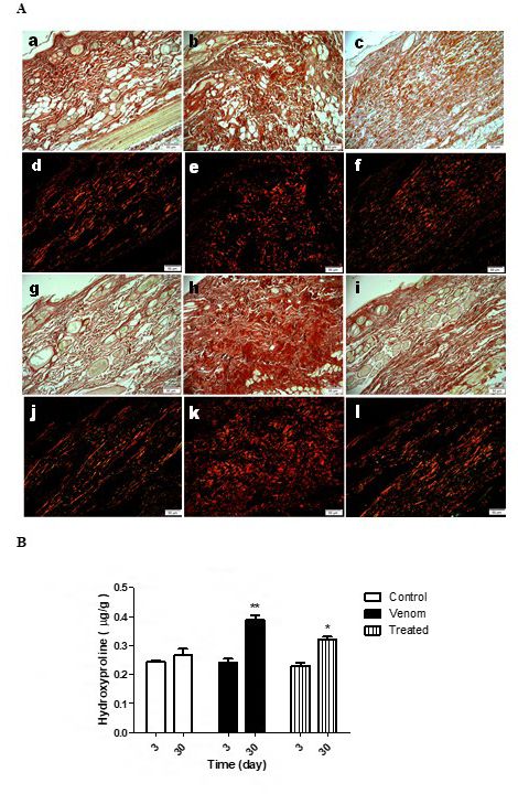
Ribeiro et al. Journal of Venomous Animals and Toxins including Tropical Diseases (2019) 25:e149318                                              Page 2 of 11

Background                                                                              also in northeastern Brazil. Moreover, it is also exported owing
The venom of spiders belonging to the genus Loxosceles produces                         to its broad indications for such diseases as cystitis, bronchitis,
a characteristic set of symptoms known as loxoscelism. Currently,                       chronic diarrhea, rheumatism, and psoriasis [23–26].
loxoscelism has been hypothesized to be a multifactorial                                   Sesquiterpene hydrocarbons are the major compounds
process involving a direct action of venom on the inflammatory                          derived from Copaifera oleoresins, the most dominant one
response [1–6]. The main characteristic of envenomation is                              being β-caryophyllene, which accounts for more than 90% of
dermonecrosis at the bite site that is manifested initially by                          the total composition [27]. Sesquiterpenes are responsible for
direct and degenerative effects of the venom components on the                          many of the pharmacological activities of copaiba oleoresins.
cell membrane, basement membrane, and extracellular matrix                              However, some studies correlate increased anti-inflammatory
resulting in drastic tissue damage [7].                                                 activity with the presence of high levels of diterpenes [28, 29].
   The early phase of loxoscelism is highlighted by local edema                            No study exists on the potential use of plants with healing
and erythema within 6 hours of the spider bite. Subsequently,                           effects as therapeutic tools for local manifestations induced
the lesion evolves into an ecchymotic area about 24 to 36 hours                         by the venom of Loxosceles sp. In this context, any scientific
after the accident. After 5 to 7 days, the necrotic lesion reaches                      evidence describing the healing effects of plant derivatives
maximum area and develops a dry crust; it is occasionally                               on skin lesions by loxoscelism would pave the way for the
associated with secondary infection. The necrotic scab falls off                        development of topical medicines with wide applicability
2 to 3 weeks after the accident, leaving an ulcer. The terminal                         and benefits for those injured by Loxosceles spiders. Thus, the
evolution of loxoscelism can induce severe intravascular hemolysis                      present study evaluated the healing effects of copaiba oil on
associated with acute anemia, jaundice, and hemoglobinuria of                           dermonecrosis induced by the venom of L. intermedia, also
varying degrees. Fatalities are rare, but frequently correlated                         known as the brown spider.
with acute renal failure [8, 9].
   Treatment of loxoscelism is still a controversial subject. The                       Methods
effectiveness of the antivenom serum in neutralizing local effects
                                                                                        L. intermedia Venom
varies in different therapeutic approaches, especially for the skin
lesion [10]. Therefore, there is no consensus as to its efficacy in                     The venom of L. intermedia was obtained from the Laboratory
reversing the local effects, while the ideal interval between the                       of Arthropods of Instituto Vital Brazil from adult specimens of
bite and its management that results in good recuperation from                          L. intermedia. The spiders were made to fast for a week followed
the injury. However, antivenom serum is indicated at any time                           by extraction of the crude venom.
in the case of hemolysis [11, 12].
   The low efficacy of the treatment has been attributed to poor                        Animals
understanding of action mechanisms of Loxosceles venom [13].                            Adult male albino rabbits (Oryctolagus cuniculus), weighing 2.5
Several treatment protocols that have been proposed and tested                          ± 3.0 kg, were used for in vivo tests. These were obtained from
for bites by L. intermedia include dapsone, corticosteroids,                            the animal colony at the Vital Brazil Institute. All rabbits were
antibiotics, and antivenom. Dapsone limits neutrophil migration                         fed food and water ad libitum under a 12-hour light–dark cycle
and infiltration at the site of the bite [14]; corticosteroids produce                  at 22 ± 2°C throughout the experiment. Venom of L. intermedia
a potent anti-inflammatory effect [15]; and antibiotics prevent                         was injected intradermally into the back of rabbits to induce
secondary infections [16]. However, this polytherapy is not                             cutaneous lesions.
completely effective in reducing skin lesions and restoring the
affected tissue. In many situations, dermonecrosis is so extensive                      Plant Material
that it requires skin grafts [9].
                                                                                        The Copaifera spp. oil (copaiba oil) was acquired from a pool of
   The strategies already employed to treat skin lesions include
phytotherapy and popular medicinal plant products. Indeed,                              individuals from eastern Amazon (2º 08’ 14’’–2º 12’ 26’’ S and
the effectiveness of some herbal agents used in traditional                             48º 47’ 34’’–48º 14’ W, at an elevation of 16 m above sea level),
medicine has been evaluated and confirmed by researchers                                state of Pará, Brazil. The oil was extracted using the method
worldwide [17, 18]. For instance, specialists in wound healing                          followed by the local population: Drilling of the trunk of the
have shown great interest in the use of copaiba oil, a popular                          Copaifera tree and insertion of a PVC cannula, through which the
medicine in Brazil, extracted from trees of the genus Copaifera                         oil flows. After the extraction is finished, the hole is sealed with
(Leguminosae-Caesalpinioideae family) to treat scarring [19, 20,                        the use of clay. A total of 1 mL of copaiba oil was administered
21]. Its property of healing wounds and ulcers is attributed to                         topically at the site of injury.
the presence of medicinally important active components [22].
Copaiba oil is popularly used, especially in the Amazon, as an                          Gas Chromatography–Mass Spectrometry Analysis
anti-inflammatory, healing, and antiseptic product that can be                          of Sesquiterpenes in Copaifera spp. Oleoresin
administered orally, topically, or vaginally [19]. This oil stands out                  The sesquiterpenes present in the Copaifera spp. oleoresin were
for its therapeutic properties not only in the Amazon region, but                       analyzed in a gas chromatograph coupled to a mass spectrometer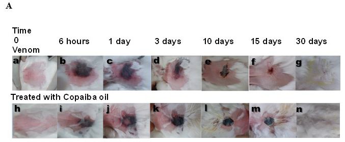
Ribeiro et al. Journal of Venomous Animals and Toxins including Tropical Diseases (2019) 25:e149318                                               Page 3 of 11

    (GC-MS, QP2010SE; Shimadizu). The following chromatographic                             Macroscopic Lesion Analysis
    conditions were used: Rtx-5 capillary column (30 m × 0.25                               Macroscopic lesions were analyzed by measuring the wound
    mm × 0.25 µM MMID); helium as carrier gas at a flow rate                                area. For this, photomicrographs of the lesions were obtained
    of 1.5 mL/minute, oven temperature maintained at 120°C for                              at 0 and 6 hours, and 1, 3, 10, 15, and 30 days after venom
    2 minutes followed by an increase of 3°C/minute to 160°C                                inoculation to monitor the healing process. These images were
    for 2 minutes. It was then increased by 8°C/minute to a final                           generated using a digital camera (Sony Cyber-Shot DSC-W350,
    temperature of 290°C for 5 minutes; injector temperature of                             14.1 mega pixels), which was kept at a constant distance from the
    270°C; and detector temperature of 290°C, operating at 40 to                            tripod base. The injury area was evaluated using the software
    400 m/z scan mode with electron impact of 70 eV. A volume of                            Image-Pro (unpaid).
    1.0 µL with 1:20 split ratio was injected into the column. The
    results obtained from analysis of retention times of peaks in                           Histopathological Analysis and Skin Processing
    the sesquiterpene chromatograms were compared to the data
                                                                                            Animal skins in the control group and the venom group,
    reported in the literature. We also compared the fragmentation
                                                                                            containing the venom-induced lesion area, were removed and
    spectra of sesquiterpenes with similarity over 90% to those
                                                                                            processed for histopathological analysis. All samples were fixed
    contained in the NIST Library 05.
                                                                                            in 10% paraformaldehyde in a phosphate buffer (pH 7.4) for 48
                                                                                            hours and subsequently dehydrated in increasing concentrations
    Minimum Necrotic Dose Determination                                                     of ethanol. These were next embedded in paraffin, sectioned
    To determine the minimum necrotic dose (MND), rabbits were                              and placed on slides (5 microns) where they were stained with
    divided into three groups, each consisting of four rabbits. The                         hematoxylin-eosin and picrosirius red.
    backs of these rabbits were first shaved, following which they
    were inoculated with increasing doses of venom (1.2, 2.4, and                           Total Collagen Determination
    12.0 μg/kg. Cutaneous lesions were observed at 6, 24, and 72
                                                                                            To quantify the total collagen present in the samples, hydroxyproline
    hours after injection. The MND was defined as the lowest dose
                                                                                            was measured using the adapted methodology recommended
    of venom capable of inducing a necrotic area of at least 1 cm2
                                                                                            by the Association of Analytical Communities (AOAC). The
    in 72 hours in 100 % of animals.


                                                                                            skin was macerated and then hydrolyzed with 1 mL of 6 M
                                                                                            hydrochloric acid per 0.01 g of skin (maximum 0.08 g) for 4
    Experimental Groups                                                                     hours at 130°C. In a separate tube, 1 mL of chloramine T and
    The animals were separated into three groups (n = 4 per group)                          5 μL of skin hydrolysate were mixed and maintained at room
    as follows: control, venom, and venom plus topical copaiba oil                          temperature for 20 minutes. Then, 1 mL of perchloric aldehyde
    treatment. Each group underwent the same routine observation                            (15 g of dimethylaminobenzaldehyde, 60 mL of n-propanol, 26
    adopted for calculating the MND. The control group received                             mL of 60 % perchloric acid, and n-propanol to complete the
    an intradermal injection of 200 µL of physiologic saline solution                       volume to 100 mL) was added and stored at 60 °C for 15 minutes.
    (PSS). All other groups received intradermal injection of L.                            Chloramine T is oxidized by hydroxyproline to pyrrole, which,
    intermedia venom at twice the MND determined in a final                                 in turn, reacts with perchloric aldehyde to form a red-purple
    volume of 200 µL. Topical application of copaiba oil was carried                        complex, the absorbance of which could be measured at 550 nm.
    out 6 hours after venom injection and repeated daily for 30 days.                       The quantification of samples was performed by interpolating the
    Each application using 1 mL of the product completely covered                           absorbance results obtained using the linear regression equation
    the wound area. During other days of treatment, the remaining                           of the standard curve (y = 0.5972x + 0.0142/R = 0.99).
    product was removed with saline jets, without compromising
    the newly formed tissue. After washing with 0.9% saline, the                            Statistical Analysis
    product was applied as described above. On days 3, 10, and 30
                                                                                            Data are expressed as mean ± standard error. The Student’s t
    after the spider venom inoculation and respective treatments,
                                                                                            test was employed to analyze data from the two groups. For
    animals were euthanized in a CO2 chamber, and skin samples                              analysis of the various groups and temporal procedures, we used
    were obtained from the lesioned sites for histopathological                             ANOVA, followed by Bonferroni’s post-test. Linear regression
    analysis according to the procedures described below.                                   was employed to design curves. P-values < 0.05 were considered
                                                                                            statistically significant.
    Blood Analysis
    At the beginning of the experiments, blood was collected from
    rabbits’ ear vein (time 0), prior to inoculation of venom or PSS.
                                                                                            Results
    After 24 hours, a fresh blood sample was collected from all animals                     Analysis of Sesquiterpenes and Diterpenes Present
    to measure hematological parameters, such as total leukocytes and                       in Copaiba Oil
    platelets, using an automatic counter (CC530–CELM). Differential                        The qualitative and quantitative analysis of oleoresin led to the
    counting was performed on hematological glass slides.                                   identification of 24 peaks of sesquiterpenes and diterpenes that
Ribeiro et al. Journal of Venomous Animals and Toxins including Tropical Diseases (2019) 25:e149318 Page 4 of 11
represented slightly more than 50 % of the total composition oleoresins in the range of 1 to 5% included copaene (1.61 %),
(Table 1). The major compounds found in the sample were the α-curcumene (4.62 %), and β-humulene (3.05 %). The major
sesquiterpenes α-bergamotene (7.04 %) and β-caryophyllene diterpenes found in the copaiba oil were kaur-16-ene (1.4 %),
(11.48 %). Other compounds frequently present in copaiba kaurenoic acid (2.0 %), and cativic acid (2.25 %) (Table 1).
Table 1. Sesquiterpenes and Diterpenes identified in the Copaifera oleoresin, compared with the literature and fragmentation
spectrum. It was possible to identify peaks of 24 compounds that represent little more than 50 % of the total composition. The major compounds found were
the sesquiterpenes α-bergamotene (7.04 %) and β-caryophyllene (11.48 %). Some compounds frequently presents in copaiba oleoresins were found at 1 to 5 %,
including Copaene (1.61 %), α-curcumene (4.62 %) and β-humulene (3.05 %). The major diterpenes found in the copaiba oil were Kaur-16-ene (1.4 %), kaurenoic
acid (2 %) and Cativic acid (2.25 %).
Compounds Retention Index Composition (%)
Cyclosativene 1125 0,82
Copaene 1221 1,61
α-cedrene 1403 4,18
α-zingiberene 1451 0,40
α-cubebene 1344 1,56
δ-selinene 1481 0,47
β-Caryophyllene 1494 11,48
α-bergamotene 1430 7,04
α-guaiene 1490 1,32
β-farnesene 1440 0,75
α-caryophyllene 1579 2,48
α-curcumene 1524 4,62
β-humulene 1574 3,05
γ-cadinene 1435 0,31
δ -cadinene 1469 1,39
Bergamotol 1673 0,58
α-bisabolene 1625 0,83
Guaiol 1614 0,27
Aromadendrene 1380 0,42
α-caryophyllene 1579 0,74
β-bisabolene 1619 0,3
Kaur-16-ene 1789 1,4
Cativic acid 2016 2,25
Kaurenoic acid 2050 2
Total of identified substances 50,27
Minimum Necrotic Dose Determination (MDN) as compared with the control group (Figure 1A). Although
The MDN was defined as 2.4 μg/kg, because this was the lowest the number of total leukocytes did not differ significantly
venom dose capable of inducing a necrotic area of at least 1 cm2 between the groups (Figure 1B), a notable reduction in the
in 72 hours in 100 % of animals (data not shown). number of heterophils in the blood of the animals inoculated
with L. intermedia venom was reported. Treatment of animals
L. intermedia Venom Reduced Platelet and with copaiba oil curbed the reduction in heterophil count
Heterophil Count induced by spider venom (Figure 1C). No significant difference
A significant reduction in the number of platelets was observed was observed between the groups analyzed with respect to
in all groups inoculated with L. intermedia venom (4.8 μg/kg) lymphocytes (Figure 1D).Ribeiro et al. Journal of Venomous Animals and Toxins including Tropical Diseases (2019) 25:e149318 Page 5 of 11
Figure 1. Effect of treatment with copaiba oil after Loxosceles intermedia venom injection on blood cells. Platelets (a) and heterophils (c)
count showed significant reduction after venom injection (4,8 μg/kg). The treatment was efficient in inhibiting the decrease of heterophils in the blood 24 hours
after venom inoculation *pRibeiro et al. Journal of Venomous Animals and Toxins including Tropical Diseases (2019) 25:e149318 Page 6 of 11
Figure 2. Evolution of the lesion 30 days after Loxosceles intermedia venom injection. Macroscopic comparison of animals that received only the
venom, compared to those treated with copaiba oil after venom inoculation showed lesions with smaller area and with mild erythema. The copaiba oil induced
a formation of thicker crust more delimited, without the presence of scarring on the skin (a). Ten days after venom injection the lesion area of the group that
received the treatment with copaiba oil was larger than the untreated group (b). n = 4 animals per experimental group.
animals (Figure 4G, I). Type I and type III collagen fibers were Discussion
frequently observed in the control group (Figure 4J). The venom- The oleoresin of Copaifera is widely used in Brazilian traditional
induced significant changes in the dermis and type I collagen
medicine as an anti-inflammatory and healing agent [20].
fibers were predominantly noted in this group (Figure 4K).
However, evidence on its effectiveness and protective roles in
Copaiba oil treatment restored the mixed profile of collagen
treating injuries caused by Loxosceles spider bite is scarce. The
fibers, such that both red and green stains were observed, similar
results of the present study demonstrated the efficacy of topical
to control groups (Figure 4L).
Total collagen was measured by quantification of application of copaiba oil in healing of dermonecrosis caused
hydroxyproline. All animals showed similar hydroxyproline by injection of brown spider venom. We suggest that copaiba
levels on day 3 after venom exposure and respective treatments. oil exerts its therapeutic effects on skin lesions induced by L.
However, 30 days after venom injection, hydroxyproline levels intermedia venom through various morphological changes,
were significantly increased in comparison with the control consequently leading to repair of damaged skin.
group. Treatment with copaiba oil at this stage did reduce Distinct compounds identified in copaiba oleoresin are
the hydroxyproline levels in comparison to the venom group potentially responsible for some of the pharmacological
although these were still elevated when compared with the properties, for example, β-caryophyllene, characterized by
control group (Figure 4M). anti-inflammatory, antibacterial, anti-edema, antifungal, andRibeiro et al. Journal of Venomous Animals and Toxins including Tropical Diseases (2019) 25:e149318 Page 7 of 11
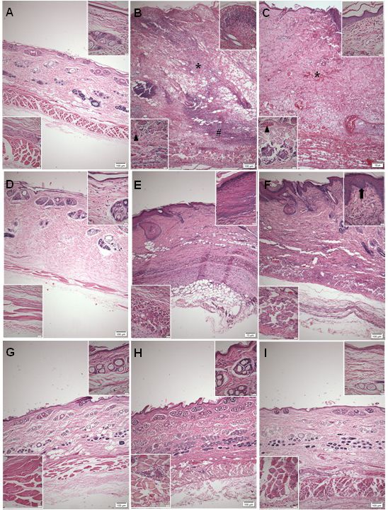
Figure 3. Photomicrographs of the skin of rabbits 3, 10 and 30 days after L. intermedia venom inoculation in the back. The control group
presented integrity of the skin layers (a/d/g). In the animals that received venom injection (4.8 µg/kg) heavy bleeding in the dermis (*), epidermal thickening
(upper insert - b), the presence of intense inflammatory infiltrate in muscle (lower insert - b) and hypodermis tissue (#) and blood vessels with clot ( ) were
observed 3 days after inoculation (b). In animals inoculated with venom and treated with copaiba oil, bleeding (*), injury of the epidermis (upper insert - c) and
inflammatory infiltrates in muscle tissue (lower insert - c) were observed 3 days after venom inoculation (c). After 10 days, the dermal thickness was reduced in
venom group (e), but the epidermis remained thick (upper insert - e); on the other hand, in the treated group, dermal thickness was reduced (f), but redefinition
was detected between papillary and reticular dermis (upper insert - f). After 30 days of regeneration of the dermis, epidermis (upper insert - h) and muscle
tissue, marked by the presence of central nuclei (inferior insert - h) after 30 days in the venom group (h); the treated group also showed regeneration of dermis,
epidermis (upper insert - i) and muscle tissue (lower insert - i), marked by an increase in the number of hair follicles (i). n = 4 animals per experimental group.Ribeiro et al. Journal of Venomous Animals and Toxins including Tropical Diseases (2019) 25:e149318 Page 8 of 11
Figure 4. Photomicrographs of collagen on skin of rabbits 3 and 30 days after inoculation of L. intermedia venom in the back. Picrosirius
red staining under optical light reveals total collagen as red, polarized light collagen type I as red fibers and collagen type III as green fibers. The control animals
presented normal distribution of collagen types on the skin at 3 (a - a/d) and 30 days (a - g/j), and did not differ in total collagen quantification by hydroxyproline
(b). The animals inoculated with venom at 4.8 µg/kg did not differ at day 3 post-inoculation compared to the control group (a - b/e) but showed increase of
collagen deposition at 30 days (a - h/k), quantified by hydroxyproline (b *pRibeiro et al. Journal of Venomous Animals and Toxins including Tropical Diseases (2019) 25:e149318 Page 9 of 11
the venom reaches the systemic circulation. In severe cases, fibers near the epidermis — the last events observed during the
it may progress to death resulting from kidney failure [33]. development of dermonecrotic injury in rabbits [5].
The abrupt reduction in platelet count observed in venom- Wound healing is a complex process. Major shortcomings
inoculated groups could not be countered by the application of in this process may occur in the early stages, producing severe
copaiba oil after 24 hours. Reduced platelet count and increased edema, reducing vascular proliferation, and decreasing cellular
fibrinogen synthesis are the first systemic responses of rabbits elements, such as leukocytes, macrophages, and fibroblasts [38].
to injection of Loxosceles spider venom, approximately 12 Studies on the healing property of copaiba oil have concentrated
hours after envenomation [12], a response very similar to that on the resin phase owing to the presence of diterpenes, but cellular
noted in humans. and molecular mechanisms of its potential therapeutic actions
Cellular changes in the bone marrow and peripheral blood are poorly understood. In the present study, no acceleration of
of rabbits after exposure to L. intermedia venom have been the healing process was observed in any animal group. However,
reported. Thrombocytopenia is an important clinical sign the venom group demonstrated an adherent crust that broke
during the diagnosis of L. intermedia spider envenomation away before 30 days of monitoring. This feature was not observed
[11]. Thrombocytopenia and a high number of heterophils were when animals received copaiba oil; in contrast, these animals
detected in the bloodstream of venom-inoculated rabbits, which showed a larger area of injury, at the 10th day, which may plausibly
were directly related to histopathological findings obtained account for the scar formation observed in the venom group.
from skin biopsies [34]. Evaluation of leukocyte mobilization The need for precision in tissue healing is impaired by the
and biochemical parameters in the blood of rabbits revealed speed of repairing the tissue damage, without aggravating it.
that platelet functions and blood coagulation showed a time- In this context, what occurs most often is the formation of
dependent trend at 3, 24, 48, 72, and 120 hours after Loxosceles scars, where the tissue will lose its function [39]. Thus, the
envenomation. These levels were associated with initial ideal treatment of wounds should also value the quality of the
leukopenia and thrombocytopenia, posterior leukocytosis, healing process, for which the deposition of collagen is vital
platelet aggregation, elevation of fibrinogen levels, and reduction for recovering the lost cell mass in the lesion, thus refilling the
of coagulation factor VII [35]. Increase in leukocytes and damaged tissue. However, exaggerated deposition of collagen
heterophils occurred at 72 hours, the same time at which red can cause fibrosis and impair the formation of functional tissue
blood cells declined [12]. [40]. After 30 days, the animals treated with copaiba oil presented
On the other hand, in the current study, blood collected 24 a distribution of collagen fibers more similar to the control
hours after inoculation of spider venom from L. intermedia group, which demonstrates a higher quality of healing. If the
did not show significant differences in the cellularity of total distribution of collagen is closer to that in the control, it is
leukocytes in any group. A significant reduction of heterophils indicative that the skin is more resistant to possible lesions, thus
was shown after venom injection but not after treatment with exerting its function of protecting the organism. Our results
copaiba oil. Diminution in the number of circulating heterophils demonstrated that copaiba oil promoted the regeneration of type
after 24 hours may be related to an acute inflammatory response, III collagen fibers, with a collagen-fiber distribution similar to
considering that these cells are found at the injury site within the normal skin of the animal. It can be inferred that copaiba
a few hours after venom injection and are reduced in blood oil may improve the process of skin regeneration, but further
circulation after 24 hours, as also observed in mice [1]. Together, studies should be done to evaluate the structure of this tissue.
these results indicated a lesser migration of heterophils to the
skin and possible protective effect associated with reduced
heterophilic response after venom injection and treatment with Conclusions
copaiba oil. These results imply that copaiba oil has the potential The increasing number of accidents caused by brown spider bites
to partially control the inflammatory response, once heterophil in recent years has become a major public health concern in
levels in blood circulation decrease. Brazil. Thus, the study of the actions of both venom and drugs has
Histopathological analysis of the skin obtained from gained extreme importance to better manage envenomation and
venom-inoculated rabbits demonstrated swelling of dermal to provide proper treatment. The present work demonstrated the
endothelial cells [36], followed by deposition of intravascular potential of copaiba oil to interfere in and curb the progression
fibrin, endothelial thickening, vasodilation and inflammatory of spider-bite-induced dermonecrosis. The results described
cell infiltration, predominantly by polymorphonuclear cells herein suggest that treatment with copaiba oil may modify
[37]. Moreover, 24 hours after venom administration, a massive scarring via deposition of collagen, thereby stimulating the
infiltration of leukocytes and platelets, bleeding, and thrombus growth of hair follicles and regeneration of muscle tissues. In
formation at the site of venom inoculation were observed [35]. addition, we now report that copaiba oil efficiently inhibits the
Long-term exposure to venom induces necrosis of myofibrils and heterophil reduction in the blood after venom injection. In
infiltration of leukocytes, damaging the skeletal muscle. This, light of the broad-spectrum applications of copaiba oil and its
in turn, causes severe destruction of the epidermal integrity economic importance in Brazil, the present study encourages
and necrosis of the connective tissue enriched with collagen further research to elucidate its effects on wound healing.Ribeiro et al. Journal of Venomous Animals and Toxins including Tropical Diseases (2019) 25:e149318 Page 10 of 11
Acknowledgements 2. Corrêa MA, Okamoto CK, Goncalves-de-Andrade RM, van den Berg
CW, Tambourgi DV. Sphingomyelinase D from Loxosceles laeta venom
The author acknowledges the support by Universidade Federal induces the expression of MMP7 in human keratinocytes: contribution
Fluminense and Instituto Vital Brazil. to dermonecrosis. PLoS One. 2016;11(4):e0153090.
3. Tambourgi DV, Paixao-Cavalcante D, Goncalves de Andrade RM,
Fernandes-Pedrosa Mde F, Magnoli FC, Paul Morgan B, et al. Loxosceles
Abbreviations sphingomyelinase induces complement-dependent dermonecrosis,
Not applicable. neutrophil infiltration, and endogenous gelatinase expression. J Invest
Dermatol. 2005;124(4):725-31.
4. Nowatzki J, de Sene RV, Paludo KS, Veiga SS, Oliver C, Jamur MC, et al.
Availability of data and material Brown spider venom toxins interact with cell surface and are endocytosed
All data generated or analyzed during this study are included by rabbit endothelial cells. Toxicon. 2010;56(4):535-43.
5. Gremski LH, Trevisan-Silva D, Ferrer VP, Matsubara FH, Meissner GO,
in this published article. Wille AC, et al. Recent advances in the understanding of brown spider
venoms: from the biology of spiders to the molecular mechanisms of
Funding toxins. Toxicon. 2014;83:91-120.
6. Chaves-Moreira D, Senff-Ribeiro A, Wille ACM, Gremski LH, Chaim OM,
This research was supported by grants from FAPERJ. Moreover, Veiga SS. Highlights in the knowledge of brown spider toxins. J Venom
this publication was supported in part by the Coordination Anim Toxins incl Trop Dis. 2017;23:6. doi: 10.1186/s40409-017-0097-8.
for the Improvement of Higher Education Personnel (CAPES) 7. Futrell JM. Loxoscelism. Am J Med Sci. 1992;304(4):261-7.
through “Programa Editoração CAPES” – call No. 3/2016, grant 8. Cacy J, Mold JW. The clinical characteristics of brown recluse spider
bites treated by family physicians: an OKPRN Study. Oklahoma Physicians
No. 0722/2017, record No. 88881.142062/2017-01 and by the Research Network. J Fam Pract. 1999;48(7):536-42.
National Council for Scientific and Technological Development 9. Delasotta LA, Orozco F, Ong A, Sheikh E. Surgical treatment of a brown
(CNPq) and Coordination for the Improvement of Higher recluse spider bite: a case study and literature review. J Foot Ankle Surg.
Education Personnel (CAPES) through “Programa Editorial 2014;53(3):320-3.
10. Isbister GK, Fan HW. Spider bite. Lancet. 2011;378(9808):2039-47.
CNPq/CAPES” call No. 18/2018, grant No. 404770/2018-5.
11. Pauli I, Puka J, Gubert IC, Minozzo JC. The efficacy of antivenom in
loxoscelism treatment. Toxicon. 2006;48(2):123-37.
Competing interests 12. Pauli I, Minozzo JC, da Silva PH, Chaim OM, Veiga SS. Analysis of
therapeutic benefits of antivenin at different time intervals after
The authors declare that they have no competing interests. experimental envenomation in rabbits by venom of the brown spider
(Loxosceles intermedia). Toxicon. 2009;53(6):660-71.
Authors’ contributions 13. Barbaro KC, Lira MS, Araujo CA, Pareja-Santos A, Távora BC, Prezotto-
Neto JP, et al. Inflammatory mediators generated at the site of inoculation
MFR carried out the conception and design of the study, the of Loxosceles gaucho spider venom. Toxicon. 2010;56(6):972-9.
acquisition, analysis and interpretation of the data and the 14. Swanson DL, Vetter RS. Bites of brown recluse spiders and suspected
drafting of the manuscript. FLO carried out analysis and necrotic arachnidism. N Engl J Med. 2005;352(7):700-7.
interpretation of data and helped to draft the manuscript. PFC, 15. da Silva PH, da Silveira RB, Appel MH, Mangili OC, Gremski W, Veiga SS.
Brown spiders and loxoscelism. Toxicon. 2004;44(7):693-709.
APS and ASN participated in acquisition of data, histological
16. Paixao-Cavalcante D, van den Berg CW, Goncalves-de-Andrade RM,
analysis and interpretation of data. AMS and TBM participated Fernandes-Pedrosa Mde F, Okamoto CK, Tambourgi DV. Tetracycline
in experimental design and helped draft the manuscript. protects against dermonecrosis induced by Loxosceles spider venom. J
CMVS conceived the study, and participated in its design and Invest Dermatol. 2007;127(6):1410-8.
17. Strauch MA, Tomaz MA, Monteiro-Machado M, Ricardo HD, Cons BL,
coordination. SCE conceived the study, and participated in its Fernandes FF, et al. Antiophidic activity of the extract of the Amazon plant
design and coordination and helped to draft the manuscript. Humirianthera ampla and constituents. J Ethnopharmacol. 2013;145(1):50-8.
All authors read and approved the final manuscript. 18. De Oliveira EC, Fernandes CP, Sanchez EF, Rocha L, Fuly AL. Inhibitory
effect of plant Manilkara subsericea against biological activities of Lachesis
muta snake venom. Biomed Res Int. 2014;2014:408068.
Ethics approval 19. Cascon V, Gilbert B. Characterization of the chemical composition of
All animal procedures were performed in accordance with oleoresins of Copaifera guianensis Desf., Copaifera duckei Dwyer and
protocols approved by the Ethics Committee on Animal Use Copaifera multijuga Hayne. Phytochemistry. 2000;55(7):773-8.
20. Cavalcanti Neto AT, Theodora TPA , Pereira SLS, Turatt E.
of the Vital Brazil Institute (protocol no. 01/2013). Comparative evaluation between copaiba oil-resin and chlorhexidine
digluconate on wound healing. Histological study in rats. Rev Odontol
Consent for publication UNESP. 2005;34(2):107-12.
21. Ricardo LM, Dias BM, Müggea FLB, Leite VV, Brandão MGL. Evidence
Not applicable. of traditionality of Brazilian medicinal plants: The case studies of
Stryphnodendron adstringens (Mart.) Coville (barbatimão) barks and
Copaifera spp. (copaíba) oleoresin in wound healing. J Ethnopharmacol.
References 2018;219:319-36.
1. Ribeiro MF, Oliveira FL, Monteiro-Machado M, Cardoso PF, Guilarducci- 22. Veiga Jr VF, Pinto AC. O gênero Copaifera L. Quím Nova. 2002;25(2):273-
Ferraz VV, Melo PA, et al. Pattern of inflammatory response to Loxosceles 86.
intermedia venom in distinct mouse strains: a key element to understand 23. Gelmini F, Beretta G, Anselmi C, Centini M, Magni P, Ruscica M, et al.
skin lesions and dermonecrosis by poisoning. Toxicon. 2015;96:10-23. GC-MS profiling of the phytochemical constituents of the oleoresinRibeiro et al. Journal of Venomous Animals and Toxins including Tropical Diseases (2019) 25:e149318 Page 11 of 11
from Copaifera langsdorffii Desf. and a preliminary in vivo evaluation of its 32. Zanetti VC, da Silveira RB, Dreyfuss JL, Haoach J, Mangili OC, Veiga SS,
antipsoriatic effect. Int J Pharm. 2013;440(2):170-8. et al. Morphological and biochemical evidence of blood vessel damage
24. Campos C, de Castro AL, Tavares AM, Fernandes RO, Ortiz VD, and fibrinogenolysis triggered by brown spider venom. Blood Coagul
Barboza TE, et al. Effect of free and nanoencapsulated copaiba oil on Fibrinolysis. 2002;13(2):135-48.
monocrotaline-induced pulmonary arterial hypertension. J Cardiovasc 33. Abdulkader RC, Barbaro KC, Barros EJ, Burdmann EA. Nephrotoxicity
Pharmacol. 2016;69(2):79-85. of insect and spider venoms in Latin America. Semin Nephrol.
25. Otaguiri ES, Morguette AEB, Biasi-Garbin RP, Morey AT, Lancheros 2008;28(4):373-82.
CAC, Kian D, et al. Antibacterial combination of oleoresin from Copaifera 34. da Silva PH, Hashimoto Y, dos Santos FA, Mangili OC, Gremski W, Veiga
multijuga Hayne and biogenic silver nanoparticles towards Streptococcus SS. Hematological cell findings in bone marrow and peripheral blood
agalactiae. Curr Pharm Biotechnol. 2016;18(2):177-90. of rabbits after experimental acute exposure to Loxosceles intermedia
26. Motta EV, Lemos M, Costa JC, Banderó-Filho VC, Sasse A, Sheridan (brown spider) venom. Toxicon. 2003;42(2):155-61.
H, et al. Galloylquinic acid derivatives from Copaifera langsdorffii leaves 35. Tavares FL, Sousa-e-Silva MC, Santoro ML, Barbaro KC, Rebecchi IM,
display gastroprotective activity. Chem Biol Interact. 2017;261:145-55. Sano-Martins IS. Changes in hematological, hemostatic and biochemical
27. da Trindade R, da Silva JK, Setzer WN. Copaifera of the Neotropics: parameters induced experimentally in rabbits by Loxosceles gaucho spider
A Review of the Phytochemistry and Pharmacology. Int J Mol Sci. venom. Hum Exp Toxicol. 2004;23(10):477-86.
2018;19(5):pii: E1511. 36. Veiga SS, Zanetti VC, Franco CR, Trindade ES, Porcionatto MA, Mangili
28. Leandro LM, Vargas Fde S, Barbosa PC, Neves JK, da Silva JA, da Veiga- OC, et al. In vivo and in vitro cytotoxicity of brown spider venom for blood
Junior VF. Chemistry and biological activities of terpenoids from copaiba vessel endothelial cells. Thromb Res. 2001;102(3):229-37.
(Copaifera spp.) oleoresins. Molecules. 2012;17(4):3866-89. 37. Smith CW, Micks DW. The role of polymorphonuclear leukocytes in
29. de S Vargas F, D O de Almeida P, Aranha ES, de A Boleti AP, Newton P, de the lesion caused by the venom of the brown spider, Loxosceles reclusa.
Vasconcellos MC, et al. Biological activities and cytotoxicity of diterpenes Lab Invest. 1970;22(1):90-3.
from Copaifera spp. Oleoresins. Molecules. 2015;20(4):6194-210. 38. Han G, Ceilley R. Chronic wound healing: a review of current management
30. Pieri FA, Mussi MCM, Fiorini JE, Moreira MAS, Schneedorf JM. Bacteriostatic and treatments. Adv Ther. 2017;34(3):599-610.
effect of copaiba oil (Copaifera officinalis) against Streptococcus mutans. 39. Gurtner GC, Werner S, Barrandon Y, Longaker MT. Wound repair and
Braz Dent J. 2012;23(1):36-8. regeneration. Nature. 2008;453(7193):314-21.
31. Santos AO, Ueda-Nakamura T, Dias Filho BP, Veiga Junior VF, Pinto AC, 40. Mori HM, Kawanami H, Kawahata H, Aoki M. Wound healing potential
Nakamura CV. Antimicrobial activity of Brazilian copaiba oils obtained of lavender oil by acceleration of granulation and wound contraction
from different species of the Copaifera genus. Mem Inst Oswaldo Cruz. through induction of TGF-β in a rat model. BMC Complement Altern
2008;103(3):277-81. Med. 2016;16:144.
You can also read