Influences of advanced glycosylation end products on the inner blood-retinal barrier in a co-culture cell model in vitro
←
→
Page content transcription
If your browser does not render page correctly, please read the page content below
Open Life Sciences 2020; 15: 619–628
Research Article
Chen Yuan, Ya Mo, Jie Yang, Mei Zhang*, Xuejun Xie*
Influences of advanced glycosylation end
products on the inner blood–retinal barrier in a
co-culture cell model in vitro
https://doi.org/10.1515/biol-2020-0067 dose- and time-dependent manner. AGEs increased VEGF
received March 20, 2020; accepted July 06, 2020 but lowered PEDF in a dose- and time-dependent manner.
Abstract: Advanced glycosylation end products (AGEs) are The intervention with AGEs led to the change of the
harmful factors that can damage the inner blood–retinal transendothelial resistance of the RMEC layer likely
barrier (iBRB). Rat retinal microvascular endothelial cells caused by the increased ratio of VEGF/PEDF.
(RMECs) were isolated and cultured, and identified by Keywords: advanced glycation end products, blood–
anti-CD31 and von Willebrand factor polyclonal antibo- retinal barrier, vascular endothelial growth factor, pigment
dies. Similarly, rat retinal Müller glial cells (RMGCs) were epithelium-derived factor
identified by H&E staining and with antibodies of glial
fibrillary acidic protein and glutamine synthetase. The
transepithelial electrical resistance (TEER) value was
measured with a Millicell electrical resistance system to 1 Introduction
observe the leakage of the barrier. Transwell cell plates for
co-culturing RMECs with RMGCs were used to construct an The blood–retinal barrier (BRB) is a special structure in
iBRB model, which was then tested with the addition of the retina that regulates the exchange of substances inside
AGEs at final concentrations of 50 and 100 mg/L for 24, 48, and outside the blood vessels of the retina. Humans and
and 72 h. AGEs in the in vitro iBRB model constructed by certain species of animals have dual blood supply
RMEC and RMGC co-culture led to the imbalance of the systems: retinal blood vessels and choroidal blood vessels.
vascular endothelial growth factor (VEGF) and pigment The BRBs corresponding to the two vascular systems are
epithelial derivative factor (PEDF), and the permeability of the inner blood–retinal barrier (iBRB) and the outer
the RMEC layer increased because the TEER decreased in a blood–retinal barrier. The iBRB is composed of retinal
capillary endothelial cells, pericytes, which are tightly
associated with the adjacent basement membrane sur-
rounded by astrocytes, and Müller cells as a structural
* Corresponding author: Mei Zhang, School of Pharmacy, Chengdu scaffolding for the iBRB [1]. This barrier prevents the free
University of Traditional Chinese Medicine & Key Laboratory of
diffusion between the circulating blood and the neural
Standardization of Chinese Herbal Medicines of Ministry of
Education & State Key Laboratory Breeding Base of Systematic
retina, but it provides nutrition to the retina and removes
Research, Development and Utilization of Chinese Medicine endogenous organisms and foreign objects from the
Resources, Chengdu, Sichuan Province, People's Republic of China, retina, such as inflammatory lymphocytes, blood-borne
e-mail: zhangmei63@cdutcm.edu.cn, tel: +86-28-61-80-0231 pathogens, excessive enzymes, and other toxic com-
* Corresponding author: Xuejun Xie, Department of Ophthalmology, pounds. Due to the presence of the BRB, the drugs
Hospital of Chengdu University of Traditional Chinese Medicine,
entering the retina are limited, and such a barrier has
Chengdu, 610072, Sichuan Province, People's Republic of China,
e-mail: xxj8847@163.com, tel: +86-28-87-76-7332 evolved well enough to provide protection for the retinal
Chen Yuan: Eye School, Chengdu University of Traditional Chinese microenvironment, and it restricts 98% of clinically
Medicine, Chengdu, Sichuan Province, People's Republic of China validated low molecular weight drugs [2]. Therefore, using
Ya Mo: Department of Ophthalmology, Hospital of Chengdu the iBRB model would help us to understand more the
University of Traditional Chinese Medicine, Chengdu, 610072,
retinal function. As a matter of fact, in vivo experiments
Sichuan Province, People's Republic of China
Jie Yang: Department of Neurology, Sichuan Academy of Medical
can maintain the complete structure of the iBRB, and the
Sciences & Sichuan Provincial People’s Hospital, Chengdu, Sichuan information obtained is closer to reality, but the factors
Province, People's Republic of China affecting the experiment are not easy to control, and the
Open Access. © 2020 Chen Yuan et al., published by De Gruyter. This work is licensed under the Creative Commons Attribution 4.0 Public
License.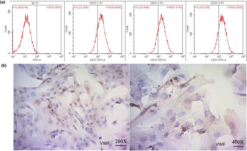
620 Chen Yuan et al.
operation is tedious, time-consuming, and economically models of rat RMEC plus RMGC co-culture in vitro need to
expensive, and hence is not suitable for large-scale drug be investigated through the transepithelial electrical
screening [3]. In recent years, more attention has been resistance (TEER), which is a good indicator of the
paid to the development of convenient, fast, economical, integrity of the endothelial tissue barrier and provides a
and more corresponding in vitro models [4]. To accurately certain experimental basis for in vivo research on human
predict how a drug will behave in vivo through an in vitro DR and other retinal vascular diseases [13].
model, one must have as many iBRB features as possible.
With an ideal in vitro model, the results or conclusions
may be closer to the situation in vivo, and the predictions
may have more practical significance [5]. 2 Materials and methods
Retina is a complex structure composed of many
types of cells, including retinal microvascular endothe-
2.1 Rat RMEC isolation, culture, and
lial cells (RMECs), retinal pigment epithelial cells,
neurons, retinal Müller glial cells (RMGCs), pericytes, identification
and other types of cells, and an in vitro model should
simulate the retinal structure and function; thus, the co- A single newborn 7-day-old Sprague Dawley (SD) rat (from
culture of different types of retinal cells with RMECs has the Laboratory Animal Center, Third Military Medical
advantages over culturing RMECs alone. The glial cells in University, Chongqing, China) was sacrificed by cervical
the vertebrate retina, mainly made of RMGCs, play dislocation. The eyeballs were subsequently removed
important roles in maintaining the normal structure, under aseptic conditions, soaked in 75% ethanol for
metabolism, and function of the retina. These glial cells 15 min, rinsed with phosphate-buffered saline containing
contact the vitreous cavity through the enlarged basal penicillin/streptomycin three times, and placed in an
and the subretinal space by the microvilli and act as a appropriate serum-free DMEM solution containing peni-
skeletal support in the formation of the BRB [6], and a cillin/streptomycin (Gibco, USA), and the retinas were
variety of cytokines regulate the permeability of the collected under a stereo microscope. The retinas were cut
retinal barrier [7]. However, most of the in vitro studies of into small pieces in a Petri dish, digested with 0.1% type I
the iBRB have been based on RMEC culture models. collagenase at 37°C for 30 min, and then filtered through a
RMECs are the sites of the early onset of diabetic 200-mesh stainless steel cell sieve. The filtrates were
retinopathy (DR), and the damage of RMECs can lead centrifuged at 1,000 rpm for 5 min and resuspended in
to the collapse of the iBRB, leading to the progress of DR. DMEM (Gibco, USA) containing 10% fetal bovine serum
Therefore, RMEC culture is often used as a model for (FBS). CD31 antibody-coated immunomagnetic beads
studying human pathogenesis of DR and evaluating the (eBioscience, USA) were added and incubated at 4°C for
effects of drugs [8]. 30 min. After centrifugation at 1,000 rpm for 5 min, the
Advanced glycosylation end products (AGEs) from cells were resuspended in 4 mL of DMEM and transferred
chronic hyperglycemia are involved in the occurrence to a test tube, which was then placed in a magnet for 2 min
and development of chronic complications of diabetes. to collect the cells. The cells were seeded into poly-L-
Once AGEs are formed, they are not easily degraded. lysine-coated plates and cultured at 37°C in a 5% CO2
Under the conditions of hyperglycemia, AGEs cause incubator (Sanyo, Japan) after washing the magnetic
endothelial dysfunction by inducing abnormal cross- bead-bound cells four times with 10% FBS DMEM. The
linking of extracellular matrix proteins and interacting culture medium was changed every other day, and the
with their receptors, triggering intracellular signaling cells were passaged with 0.25% trypsin (Gibco, USA) and
cascades [9–11]. Hyperglycemia induces oxidative stress, 0.02% EDTA (1:1) digestion solution when cell fusion
hypoxia, white blood cell arrest, vasoconstriction, reached 70–80%. The RMECs out of rat retinal vascular
inflammation, and angiogenesis in the retina, further endothelial cells (RRVECs) were recovered after identifica-
leading to vascular stiffness and dysfunction, pericyte tion by CD31/PE antibody and mouse IgG (eBioscience,
apoptosis, and intraretinal destruction [12]. The destruc- USA) flow cytometry and by using the Von Willebrand
tion of the iBRB is the early and main pathophysiological factor (VWF) antibody (dilution ratio 1:400, Abcam, USA)
basis of retinal vascular disease caused by AGEs. The with HRP-labeled secondary antibody at a dilution of 1:50
mechanism of iBRB injury is closely related to vascular (Beyotime, China). The immunostained cells were ob-
endothelial growth factor (VEGF) and pigment epithelial served with 3,3'-Diaminobenzidine (DAB) and hematox-
derivative factor (PEDF). The effects of AGEs on iBRB ylin staining under a microscope (Olympus, Japan).Influences of AGEs on the iBRB in a co-culture cell model in vitro 621
Ethical approval: The research related to animal use has RMECs were seeded at the bottom of the Transwell
been complied with all the relevant national regulations chamber as the upper compartment at a density of 2 ×
and institutional policies for the care and use of animals 104/cm2. The model was evaluated by recording the
and has been approved by the Ethics Committee of TEER of the RMEC layer over 3–13 days (Figure 3a).
Chengdu University of Traditional Chinese Medicine.
2.4 TEER measurement of the RMEC layer in
the iBRB model
2.2 Rat RMGC isolation, culture, and
identification The TEER was measured on the RMEC layer of the iBRB
cell co-culture in vitro using a MilliCell® ERS-2 voltohm-
A similar procedure was used until the retina was meter. The measurement of each well’s resistance took
gently collected under a stereo microscope. The retina place at three different points selected at random. The
was blown repeatedly to form a chyle-like mixture, average value Rt was recorded, and the background
which was subsequently inoculated into a gelatin- resistance formed in the cell-free culture pool as the
coated culture flask, and the culture medium was blank value R0 was used to calculate the resistance value
changed after 72 h. After the 0.25% trypsin digestion of the entire endothelial cell, using the formula TEER =
was terminated with DMEM containing FBS, the cells (Rt − R0) × S, where S is the effective surface area of the
were collected by centrifugation at 1,000 rpm for film. Transwells with TEER values greater than 90 Ω cm2
10 min and gently pipetted to make the mixture even, were selected for further use. The TEER value was
and a drop of the cell suspension was taken to count measured every other day. A decreased value of TEER
the cells to adjust the amount of culture medium indicates that the permeability is increased.
appropriately to a cell density of 1 × 10⁶/mL. The cells
were aliquoted and routinely cultured in a 3 mL flask in
a 5% CO2, 37°C incubator. When the cell fusion reached
80%, the cells were fixed with 4% paraformaldehyde 2.5 Enzyme-linked immunosorbent assay
and subjected to hematoxylin and eosin (H&E) (ELISA)
staining, and RMGCs were screened and identified by
glial fibrillary acidic protein (GFAP) or glutamine An ELISA was used to determine the levels of VEGF and
synthetase (GS) antibody (Abcam, USA) immunocyto- PEDF in the presence of AGEs (Bioss Co., Beijing, China)
chemical staining. at final concentrations of 50 and 100 mg/L for 24, 48,
and 72 h. After AGE treatment in each experimental
group, the cell supernatant was collected following
2.3 Construction of the in vitro iBRB model centrifugation at 3,000 rpm for 20 min and used for the
ELISA of VEGF and PEDF (BioTek, USA) according to the
RMECs and RMGCs were used to simulate iBRB models in manufacturer’s instructions. The absorbance was mea-
vitro after the characterization. The third generation cells sured at a wavelength of 450 nm, and the concentrations
of primary rat RMECs were collected at 70% fusion and of VEGF and PEDF were calculated according to the
cultured in a serum-free medium for 24 h. The second linear regression equation of the standard curve and the
passage cells of rat RMGCs were collected at close to 70% dilution factors.
fusion and cultured in a normal RMEC medium (DMEM
containing 20% FBS and 100 U/mL penicillin + 100 U/mL
streptomycin) for 24 h. In the meantime, a microporous
membrane was pre-coated with 1% gelatin in a 2.6 Statistical analysis
Transwell chamber (Corning, USA), and a single-cell
suspension of RMGCs was seeded at a density of 1 × 10⁴/ SPSS 21.0 (IBM Corp., Armonk, NY, USA) was used for
cm2 at the bottom of the Transwell chamber (lower statistical analysis. All data were expressed as mean ±
compartment) and incubated for 2 h at 37°C. The standard deviation. Comparisons between groups were
Transwell chamber was turned over in a six-well plate analyzed using one-way analysis of variance and post
after most of the RMGCs attached to the wall. Then, hoc tests. Univariate analysis of variance was used for622 Chen Yuan et al.
Figure 1: Isolation and identification of RMECs from RRVECs. RRVECs were isolated and cultured from the retina of a 7-day-old neonatal SD
rat, and RMECs were verified after three passages and flow cytometry. (a) Verification of RMECs using flow cytometry with CD31 antibody.
Kb - the blank control without antibody; P1 - the cell content after removal of cell debris; V1L - the content of the negative cells which were
not labeled by CD31; V1R - the content of the CD31 positive cells, RMECs > 80% among RRVECs. (b) Immunological verification of RMECs
with a polyclonal antibody against the VWF.
comparison of the same index among multiple groups; cells, and this protein is not present in smooth muscle cells
the least significant difference test was used for pairwise and fibroblasts, which ensured that the isolated RMECs
comparison between multiple groups. All experiments have the characteristics of pericytes [14].
for statistical analysis were repeated six times. P < 0.05
indicated that the difference was statistically significant.
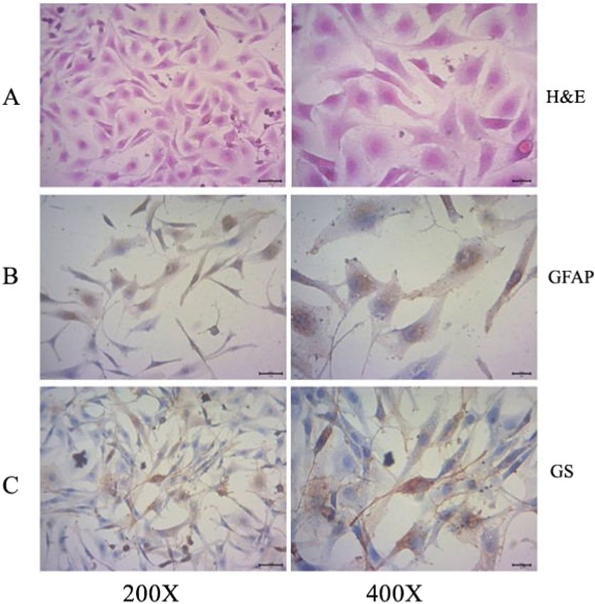
3.2 Isolation and identification of rat
RMGCs
3 Results
RMGCs were isolated and cultured from a newborn SD rat.
H&E staining showed that the cell shape was narrow and
3.1 Isolation and identification of rat RMECs
long, the cytoplasm was abundant, the membrane was
clear, and a deep red, large, and elliptical nucleus was
RMECs were isolated and cultured from a newborn SD rat. located in the center of the cell in a mononucleated or
As measured by CD31 flow cytometry, the purity of RMECs multinucleated form (Figure 2a). More than 85% of the
reached 81% (Figure 1a). The cells were also identified glial cells were GFAP positive, and brown-yellow silk-like
using VWF polyclonal antibody immunohistochemistry structures were visible in the cytoplasm (Figure 2b), and
(Figure 1b). These results showed that RMECs were more than 80% of the cells were positive for GS staining,
successfully isolated and cultured because CD31 is a unique especially the nucleus and the surrounding cytosol after 5
biomarker for RMECs. Similar to CD31, VWF is currently a days of culture (Figure 2c). The results showed that
commonly used vascular endothelial cell marker. However, RMGCs were successfully characterized and cultured
VWF is only abundantly expressed in vascular endothelial because GFAP and GS are biomarkers for RMGCs.Influences of AGEs on the iBRB in a co-culture cell model in vitro 623
Figure 2: Isolation and identification of RMGCs from rat retinal Müller cells (RRMCs). RRMCs were isolated and cultured from the retina of a
7-day-old newborn SD rat, and RMGCs were verified using (a) H&E staining, (b) GFAP, and (c) GS immunocytochemical staining after two
passages. RMGCs among RRMCs were indicated with GFAP and GS positive staining.
3.3 Changes of TEER in the iBRB model after nutrients and structural support and maintain the extra-
treatment with AGEs cellular environment. The co-culture model of RMECs and
RMGCs showed that the TEER of the RMEC layer was stable
The cell layers in the in vitro iBRB model made of RMECs until 13 days, indicating that the model was successfully
were established in the upper compartment (Figure 3b) established, which was then treated with AGEs at final
because RMECs form the peripheral boundary membrane concentrations of 50 and 100 mg/L for 24, 48, and 72 h, and
through interacting with RMGCs, where RMGCs provide the TEER values of the RMEC layer were measured.624 Chen Yuan et al.
Figure 3: TEER changes in the iBRB model caused by AGEs. (a) Diagram illustrating the iBRB model in vitro. (b) Transwell assay; RMEC and
RMGC layers (100×) of in vitro co-culture. (c) TEER measurement of the iBRB model in vitro conducted after treatment with indicated AGEs
for 24, 48, and 72 h.
Compared with the normal control group, the permeability concentrations between the different experimental groups in
of the RMEC layer of the model was increased after the same period was significant (P < 0.05). In the low-AGE
treatment with AGEs. The intervention with AGEs led to a group and the high-AGE group, the VEGF levels at different
decrease of TEER across the RMEC layer in a dose- and periods were compared, and P < 0.05. The results showed
time-dependent manner (Figure 3c). that treatment with AGEs resulted in a significant increase in
VEGF levels in the RMEC–RMGC co-culture model in a dose-
and time-dependent manner (Figure 4a).
3.4 Changes of VEGF induced by AGEs in
iBRB models in vitro 3.5 Changes of PEDF induced by AGEs in the
iBRB model in vitro
VEGF was measured by ELISA after treating the RMEC–RMGC
cell co-culture with AGEs at final concentrations of 50 and Similarly, ELISA was used to determine the level of PEDF
100 mg/L for 24, 48, and 72 h. The difference in VEGF in the RMEC–RMGC cell co-culture models treated withInfluences of AGEs on the iBRB in a co-culture cell model in vitro 625
and PEDF were in a relatively balanced state. However,
for both the low-AGE group and the high-AGE group,
they were higher than those in the normal control group.
The ratio for the low-AGE group and the high-AGE group
increased at 48 h and continued to increase at 72 h. The
results showed that compared with the normal control
group, the treatment of AGEs caused a proportional
increase of VEGF in the co-culture, which means it led to
the inhibition of PEDF in a time-dependent manner
(Figure 5). It is worthy to mention that the measurement
of VEGF and PEDF had been carried out in one
experiment for measuring VEGF, PEDF, and VEGF/
PEDF at the same time, and also in separate experi-
ments, which all were included in statistical analysis.
4 Discussion
It has been reported that Müller cells induce a barrier in
vascular endothelial cells [15], and Müller cells play an
Figure 4: Changes of VEGF and PEDF in the iBRB-Müller cell co- important role in the formation of the retinal vascular
culture due to AGEs. (a) VEGF in the media of the in vitro iBRB- barrier. Therefore, RMGCs play an active role in the
Müller cell co-culture model determined using ELISA after treatment
induction and maintenance of RMEC transition from
with indicated AGEs for 24, 48, and 72 h. (b) PEDF in the media of
the in vitro iBRB-Müller cell co-culture model determined using non-barrier cells to barrier cells [16,17]. Studies have
ELISA after treatment with indicated AGEs for 24, 48, and 72 h. *P < shown that the most important pathological basis and
0.05 compared with the vehicle control group; #P < 0.05 compared morphological changes in early DR start in the iBRB, and
with the low-dose group at 24 h earlier; ‡P < 0.05 compared with the structure of the iBRB will be damaged if it is directly
the high-dose group, at the same time point.
exposed to hyperglycemic conditions [18]. A large
number of studies have shown that AGEs are widely
present in various eyeball tissues such as the cornea,
AGEs at final concentrations of 50 and 100 mg/L for 24,
retina, vitreous body, lens, Bruch’s membrane, sclera,
48 and 72 h. The results showed that the difference in
and optic nerve, directly or indirectly leading to a series
PEDF concentrations between control and experimental
of eye diseases including retinopathy [19]. AGEs not only
groups at any time period was statistically significant,
cause platelet activation and aggregation but also
P < 0.05. The results showed that compared with the
stimulate prothrombin activity by increasing the expres-
normal control group, treatment with AGEs resulted
sion of tissue factors, which lead to thrombosis [20]. In
in a significant reduction in PEDF levels in the co-
the meantime, AGEs have been shown to inhibit the
culture model in a dose- and time-dependent manner
production of prostacyclin and induce the production of
(Figure 4b).
plasminogen activator inhibitor-1 in endothelial cells
through interaction with the receptor of AGEs [21].
Therefore, AGEs may cause platelet aggregation and
3.6 Changes of the ratio of VEGF/PEDF in fibrin stabilization leading to thrombosis, thereby pro-
vitro induced by AGEs moting vascular damage in diabetes. We demonstrated
that co-cultured RMECs and RMGCs were stabilized
VEGF and PEDF protein concentrations were simulta- because the TEER remained higher than 90 Ω cm2 after
neously measured with ELISA after treating RMEC– the 11th day, which indicated that the barrier was
RMGC co-cultivation models with AGEs at final concen- successfully established. With the established iBRB
trations of 50 and 100 mg/L for 24, 48, and 72 h. model, the addition of AGEs within the effective
The ratios of the normal control group at 24, 48, and concentration range of 50–100 mg/L led to iBRB damage
72 h were almost the same, and the amounts of VEGF [22], and this was indicated by the reduced TEER value.626 Chen Yuan et al.
reduced, and this situation is more pronounced in
patients with proliferative DR. Both cell culture and
animal experiments have confirmed that AGEs can
significantly improve their functions after adding exo-
genous PEDF. However, the accumulation of AGEs in
diabetes mellitus significantly inhibits the expression of
PEDF mRNA, and the transcription and expression of
PEDF are reduced [25]. The oxidative stress aggravates
injury in a high-glucose environment, promotes damage
to the iBRB structure, and thereby accelerates retinal
edema and the formation of new blood vessels [26]. In
this experiment, the PEDF concentrations between
control and experimental groups in the same period
were significantly different, and the difference in PEDF
Figure 5: VEGF/PEDF in the RMEC layer of the in vitro iBRB-Müller
cell co-culture after AGE treatment. VEGF and PEDF in the media of concentrations in low-AGE and high-AGE groups at
the in vitro iBRB-Müller cell co-culture were determined simulta- different periods was also significant. This shows that
neously using ELISA after treatment with indicated AGEs for 24, 48, higher AGE concentration and a longer time lowered the
and 72 h, and the ratios of the average values of the two cytokines concentration of PEDF.
were plotted against time points.
The mechanism of iBRB injury under diabetic
conditions is related to VEGF and PEDF. VEGF promotes
Due to the large accumulation of AGEs, the iBRB can early iBRB injury in DR and increases the leakage of
be easily destroyed, and DR aggravates resulting in retinopathy vessels; on the other hand, PEDF has the
vascular edema and nerve tissue damage, eventually function of inhibiting vascular leakage of DR with a
leading to vision loss [23]. Overexpression of VEGF under protective effect on the iBRB. Under physiological
pathological conditions causes increased vascular perme- conditions, there is a balanced relationship between
ability and neovascularization. At present, a large number PEDF and VEGF. PEDF inhibits the increase of VEGF’s
of clinical and animal experiments have shown that high vascular permeability and angiogenic potential [26]. In
amounts of glucose in blood and tissues stimulate the this experiment, we showed that the ratios of the normal
large-scale production of VEGF, which is one of the control group at different time points were almost the
mechanisms of DR development, and high levels of VEGF same, indicating that the levels of VEGF and PEDF
cause retinal iBRB damage and subsequently retinal proteins were in a relatively balanced state. But the
exudation, hemorrhage, edema, neovascularization, and ratios of VEGF/PEDF changed in the low-AGE group and
early DR. VEGF and PEDF are a group of major cytokines the high-AGE group at different time points, both
that are most closely related to the occurrence and increased at 48 h and continued to increase at 72 h.
development of DR. Therefore, the cytokines VEGF and This showed that the balance of VEGF and PEDF was lost
PEDF were tested separately and combinedly analyzed. In after the addition of AGEs, and the expression ratio in
a normal eye, Müller cells, pericytes, pigment epithelial vitro revealed a similar expression pattern to that in vivo.
cells, and endothelial cells of the retina secrete VEGF, but Our results also support the early finding that PEDF
the expression levels are low. The low secretion is inhibits the VEGF-mediated angiogenesis [28].
conducive to the maintenance of normal blood vessel
function. In this experiment, the VEGF concentration in
experimental groups in the same period was increased
from the low-AGE group to the high-AGE group at different 5 Conclusions
periods (P < 0.05), and our observation is consistent with
previous related studies: the higher the AGE concentra- In summary, this experiment successfully established an
tion, the higher the VEGF in the tested range [24]. iBRB cell co-culture model in vitro with permeability
PEDF is a polypeptide mainly from retinal pigment changes of the iBRB observed by indirect measurement
epithelium and retinal Müller cells, and it inhibits of the TEER, and the model was treated with AGEs to
vascular leakage and vascular regeneration. A large simulate iBRB damage and to detect changes of VEGF
number of studies have shown that the expression of and PEDF. Our findings showed that VEGF increased and
PEDF in the eyes of patients with DR is significantly PEDF decreased, both in a dose- and time-dependentInfluences of AGEs on the iBRB in a co-culture cell model in vitro 627
manner, and the balance of VEGF and PEDF was lost in endothelial cells under different oxygen conditions. Graefes
the presence of AGEs in the co-culture model. The Arch Clin Exp Ophthalmol. 2011;249(6):871–7.
increase of permeability shown by the TEER was [9] Tagami M, Yamagata K, Fujino H, Kubota A, Nara Y, Yamori Y.
Morphological differentiation of endothelial ceIls co-cultured
dependent on the dose and time of AGE intervention,
with astrocytes on type-I or type-IV collagen. Cell Tissue Res.
so the imbalance of VEGF and PEDF may be one of the 1992;268(2):225–32.
important reasons for the change of permeability of the [10] Igarashi Y, Chiba H, Utsumi H, Miyajima H, Ishizaki T, Gotoh T,
RMEC layer. Thus, the in vitro iBRB model constructed by et al. Expression of receptors for glial cell line-derived
RMEC and RMGC co-culture can be used for studying the neurotrophic factor (GDNF) and neurturin in the inner blood-
retinaI barrier of rats. Cell Struct Funct. 2000;25(4):237–41.
pathogenesis of retinal vascular diseases such as DR for
[11] Calderon GD, Juarez OH, Hernandez GE, Punzo SM, De, la
evaluating the impact of candidate drugs, even though Cruz ZD. Oxidative stress and diabetic retinopathy: develop-
the relevant assays in vivo remain necessary. ment and treatment. Eye. 2017;31(8):1122–30.
[12] Arden GB, Sivaprasad S. Hypoxia and oxidative stress in the
Acknowledgments: This work was supported by grants causation of diabetic retinopathy. Curr Diabetes Rev.
2011;7(5):291–304.
from the National Natural Science Foundation of China
[13] Poenar DP, Yang G, Wan WK, Feng S. Low-cost method and
(81473735 and 81774202).
biochip for measuring the trans-epithelial electrical resistance
(TEER) of esophageal epithelium. Materials.
Conflict of interest: The authors state no conflict of 2020;13(10):2354.
interest. [14] Mbagwu SI, Filgueira L. Differential expression of CD31 and
Von Willebrand Factor on endothelial cells in different regions
of the human brain: potential implications for cerebral malaria
Data availability statement: The datasets generated
pathogenesis. Brain Sci. 2020 Jan 6;10(1):31.
during and/or analyzed during the current study are [15] Wisniewska-Kruk J, Klaassen I, Vogels IM, Magno AL, Lai CM,
available from the corresponding author on reasonable Van Noorden CJ, et al. Molecular analysis of blood-retinal
request. barrier loss in the Akimba mouse, a model of advanced
diabetic retinopathy. Exp Eye Res. 2014;122:123–31.
[16] Tout S, Chan-Ling T, Hollander H, Stone J. The role of Müller
cells in the formation of the blood-retinal barrier.
Neuroscience. 1993;55(1):291–301.
References [17] Matsubara TA, Murata TA, Wu GS, Barron EA, Rao NA. Isolation
and culture of rat retinal microvessel endothelial cells using
magnetic beads coated with antibodies to PECAM-1. Curr Eye
[1] Hosoya K, Tachikawa M. The inner blood-retinal barrier:
Res. 2000;20(1):1–7.
molecular structure and transport biology. Adv Exp Med Biol.
[18] Tretiach M, Madigan MC, Wen L, Gillies MC. Effect of Müller
2012;763:85–104.
cell co-culture on in vitro permeability of bovine retinal
[2] Hudson N, Campbell M. Inner blood-retinal barrier
vascular endothelium in normoxic and hypoxic conditions.
regulation in retinopathies. Adv Exp Med Biol.
Neurosci Lett. 2005;378(3):160–5.
2019;1185:329–33.
[19] Romero-Aroca P, Baget-Bernaldiz M, Pareja-Rios A, Lopez-
[3] Tervonen A, Vainio I, Nymark S, Hyttinen J. Prediction of
Galvez M, Navarro-Gil R, Verges R. Diabetic macular edema
passive drug permeability across the blood-retinal barrier.
pathophysiology: vasogenic versus inflammatory. J Diabetes
Pharm Res. 2014;31(9):2297–311.
Res. 2016;2016:2156273.
[4] Rizzolo LJ, Peng S, Luo Y, Xiao W. Integration of tight junctions
[20] Yamagishi S, Fujimori H, Yonekura H, Yamamoto Y,
and claudins with the barrier functions of the retinal pigment
Yamamoto H. Advanced glycation endproducts inhibit pros-
epithelium. Prog Retin Eye Res. 2011;30:296–323.
tacyclin production and induce plasminogen activator inhi-
[5] Hanrahan F, Campbell M, Nguyen AT, Suzuki M, Kiang AS,
bitor-1 in human microvascular endothelial cells.
Tam LC, et al. On further development of barrier modulation as a
Diabetologia. 1998;41(12):1435–41.
technique for systemic ocular drug delivery. Adv Exp Med Biol.
[21] Chilelli NC, Burlina S, Lapolla A. AGEs, rather than hypergly-
2012;723:155–9.
cemia, are responsible for microvascular complications in
[6] Hirrlinger PG, Pannicke T, Winkler U, Claudepierre T,
diabetes: a “glycoxidation-centric” point of view. Nutr Metab
Varshney S, Schulze C, et al. Genetic deletion of laminin
Cardiovasc Dis. 2013;23(10):913–9.
isoforms β2 and γ3 induces a reduction in Kir4.1 and
[22] Li C, Chang Y, Li Y, Chen S, Chen Y, Ye N, et al. Advanced
aquaporin-4 expression and function in the retina. PLoS One.
glycation end products promote the proliferation and migra-
2011;6(1):e16106.
tion of primary rat vascular smooth muscle cells via the
[7] Distler C, Dreher Z. Glia cells of the monkey retina-II. Müller
upregulation of BAG3. Int J Mol Med. 2017;39(5):1242–54.
cells. Vis Res. 1996;36(16):2381–94.
[23] Cameron NE, Eaton SE, Cotter MA, Tesfaye S. Vascular factors
[8] Ma X, Bi H, Qu Y, Xie X, Li J. The contrasting effect of estrogen
and metabolic interactions in the pathogenesis of diabetic
on mRNA expression of VEGF in bovine retinal vascular
neuropathy. Diabetologia. 2001;44(11):1973–88.628 Chen Yuan et al.
[24] Diaz-Coranguez M, Ramos C, Antonetti DA. The inner blood-retinal [27] Gao G, Li Y, Zhang D, Gees S, Crosson C, Ma J.
barrier: cellular basis and development. Vis Res. 2017;139:123–37. Unbalanced expression of VEGF and PEDF in
[25] Yamagishi S, Matsui T, Nakamura K, Yoshida T, Takeuchi M, ischemia-induced retinal neovascularization. FEBS Lett.
Inoue H, et al. Pigment-epithelium-derived factor suppresses 2001;489(2–3):270–6.
expression of receptor for advanced glycation end products in [28] Yamagishi S, Nakamura K, Matsui T, Inagaki Y, Takenaka K,
the eye of diabetic rats. Ophthalmic Res. 2007;39:92–7. Jinnouchi Y, et al. Pigment epithelium-derived factor inhibits
[26] Klaassen I, Van Noorden CJ, Schlingemann RO. Molecular advanced glycation end product-induced retinal vascular
basis of the inner blood-retinal barrier and its breakdown in hyperpermeability by blocking reactive oxygen species-
diabetic macular edema and other pathological conditions. mediated vascular endothelial growth factor expression. J Biol
Prog Retin Eye Res. 2013;34:19–48. Chem. 2006;281(29):20213–20.You can also read