Polyneuropathy in POEMS syndrome: role of angiogenic factors in the pathogenesis
←
→
Page content transcription
If your browser does not render page correctly, please read the page content below
doi:10.1093/brain/awh519                                                                                                                     Brain (2005), 128, 1911–1920

Polyneuropathy in POEMS syndrome: role of
angiogenic factors in the pathogenesis
Marina Scarlato,1,2 Stefano Carlo Previtali,2 Marinella Carpo,1 Davide Pareyson,3 Chiara Briani,4
Roberto Del Bo,1 Eduardo Nobile-Orazio,1 Angelo Quattrini2 and Giacomo Pietro Comi1
1
Department of Neurological Sciences, Centro Dino Ferrari, University of Milan, I.R.C.C.S. Ospedale Maggiore Policlinico,
2
Department of Neurology, San Raffaele Scientific Institute, 3‘C. Besta’ National Neurological Institute,
Milan and 4Department of Neurosciences, University of Padova, Padova, Italy
Correspondence: Marina Scarlato, MD, PhD, Department of Neurological Sciences, Padiglione Ponti, Centro Dino Ferrari,
I.R.C.C.S. Ospedale Maggiore Policlinico, Via F. Sforza 35, 20122 Milan, Italy
E-mail: mamasca@hotmail.com

In order to clarify the role of angiogenic factors in polyneuropathy of POEMS (polyneuropathy, organomegaly,
endocrinopathy, monoclonal gammopathy, skin changes) syndrome, we measured the serum concentrations of
vascular endothelial growth factor (VEGF) and erythropoietin (EPO) in 11 patients and correlated these with
VEGF and EPO peripheral nerve expression and the degree of endoneurial vessel involvement. We found that
POEMS syndrome was associated with high levels of serum VEGF and, conversely, low levels of serum EPO.
Similarly, in POEMS nerves VEGF was highly expressed in blood vessels and some non-myelin-forming Schwann

                                                                                                                                                                                             Downloaded from by guest on March 5, 2015
cells. In contrast, the expression of VEGF receptor 2 was down-regulated compared with that in normal nerves.
Both EPO and EPO receptor were localized to the nerve vasculature and were expressed to similar extents in
normal and POEMS nerves. The inverse correlation between VEGF and EPO serum levels was maintained
during the clinical course; however, both levels returned to normal when there was a response to therapy. High
serum VEGF, low serum EPO and high peripheral nerve VEGF were all associated with more severe endoneur-
ial vessel involvement and nerve damage. Light microscopy showed an increased thickness of the basal lamina
and a narrowing of the lumina of endoneurial vessels in POEMS samples, while proliferation of endothelial cells
and opening of tight junctions were observed by electron microscopy. The present data support the role of
angiogenic factors as diagnostic and prognostic markers of POEMS syndrome. They also suggest that VEGF and
EPO are involved in the pathogenesis of polyneuropathy. In conclusion, establishing the role of angiogenic
factors in polyneuropathy may lead to a better understanding of the effects of VEGF and EPO on microan-
giopathy and Schwann cell function.

Keywords: endoneurial vessels; EPO; nerve; POEMS; VEGF

Abbreviations: EPO = erythropoietin; EPO-R = EPO receptor; HIF-1 = hypoxia-inducible factor; IVIg = intravenous
immunoglobulins; MGUS = monoclonal gammopathy of undetermined significance; POEMS = polyneuropathy, organomegaly,
endocrinopathy, monoclonal gammopathy, skin changes; VEGF = vascular endothelial growth factor; VEGFR2 =
VEGF receptor 2
Received September 21, 2004. Revised March 3, 2005. Accepted March 18, 2005. Advance Access publication June 23, 2005

Introduction
POEMS (polyneuropathy, organomegaly, endocrinopathy,                                            syndrome is quite broad, and since not all the typical features
monoclonal gammopathy, skin changes) syndrome is a rare                                         may be present at onset, it is crucial to establish minimal
multisystemic disease that occurs in the setting of plasma                                      criteria for diagnosis. Peripheral nerves are one of the
cell dyscrasia and is characterized by an elevation of serum                                    principal targets in POEMS syndrome (Vital et al., 2003).
vascular endothelial growth factor (VEGF) levels (Miralles                                      In fact, the presence of a chronic progressive, distal, sensori-
et al., 1992; Watanabe et al., 1996; Hashiguchi et al., 2000;                                   motor polyneuropathy is mandatory in establishing dia-
Michizono et al., 2001). The clinical spectrum of POEMS                                         gnosis along with monoclonal plasmaproliferative disorder

#   The Author (2005). Published by Oxford University Press on behalf of the Guarantors of Brain. All rights reserved. For Permissions, please email: journals.permissions@oupjournals.org
1912 Brain (2005), 128, 1911–1920 M. Scarlato et al.
(Dispenzieri et al., 2003). There is currently no evidence to levels of VEGF and sometimes EPO, inducing a paraneo-
suggest that any of the characteristics of the disease (axonal plastic erythrocytosis.
degeneration, segmental demyelination or subperineurial In order to provide new insights into the pathogenetic role
oedema) are the result of immune-mediated damage to the of angiogenic factors in POEMS syndrome, we evaluated
nerve components. Instead, an alteration of the blood–nerve longitudinally VEGF and EPO serum levels in 11 POEMS
barrier and abnormalities of serum coagulation factors, owing patients and correlated these with the clinical course, response
to the elevation of VEGF, have been proposed as being patho- to therapy and neuropathological findings. We also investig-
genetic in the nerve damage (Saida et al., 1997; Watanabe et al., ated the expression and localization of HIF-1a, VEGF,
1998). Despite it being unclear which cell type is responsible VEGFR2, EPO and EPO-R in peripheral nerves.
for the increased VEGF production, almost all of the symp- Our data suggest that polyneuropathy in POEMS syn-
toms of POEMS syndrome have been related to changes in drome might be caused by the direct or indirect effect of
VEGF serum levels. angiogenic factors. VEGF and EPO serum changes therefore
VEGF and erythropoietin (EPO) were first characterized, have diagnostic, prognostic and pathogenetic implications.
respectively, as angiogenic and haematopoietic growth fac-
tors. VEGF acts as a potent, multifunctional cytokine indu-
cing angiogenesis and microvascular hyperpermeability via Material and methods
action on endothelial cells, while EPO regulates proliferation Patients: clinical and laboratory data
and differentiation of red blood cells. However, growing evid-
Between 1990 and 2003, we recruited 11 patients (10 males and one
ence suggests that they have other biological roles. The pro- female) affected with POEMS syndrome, from three hospitals in
duction of VEGF and EPO is under the control of a labile Northern Italy (Table 1). All the patients initially presented with a
transcription factor called hypoxia-inducible factor (HIF-1). chronic progressive sensorimotor polyneuropathy that began
HIF-1 is a heterodimer consisting of an inducible a- and a 8–14 months before the diagnosis was made. Deep sensation was
constitutive b-subunit and acts as a DNA-binding factor. generally affected more than cutaneous sensation and the sensory
Downloaded from by guest on March 5, 2015
Under hypoxic conditions the HIF-1a subunit is induced disturbances were reported as being as severe as the motor symp-
and interacts with the hypoxia response elements (HREs) toms. Duration of follow-up, defined as the interval between the time
of target genes to determine transcriptional activity (Pugh of the first clinical examination, and collection of the first serum
and Ratcliffe, 2003). The human VEGF gene gives rise to sample, and the last visit, ranged from 3 to 50 months. The presence
of polyneuropathy was diagnosed on the basis of clinical symptoms
six different protein isoforms (Ferrara et al., 2003). A number
and signs (distal sensory disturbances at onset with progressive weak-
of polymorphisms localized in the promoter region of the
ness in the lower and, less extensively, in the upper limbs, with
VEGF gene, outside the HRE site, have been described and reduced or absent deep tendon reflexes) and electrophysiological
associated with several diseases (Del Bo et al., 2005) and findings (slowing of motor and sensory nerve conduction velocities
modification of transduction levels. VEGF exerts its action with reduction of compound muscle action and sensory nerve action
via high-affinity binding to two types of phosphotyrosine potentials) (data not shown). For serial evaluation of neurological
kinase receptors: VEGF receptor 1 (VEGFR1; also known impairment, we used the Rankin scale score, which ranges from 0
as FLT-1) and VEGF receptor 2 (VEGFR2; also known as (no deficit) to 5 (maximal deficit). CSF protein levels were elevated in
KDR and FLK-1). Both receptors are essential for the devel- all 11 subjects. Seven patients underwent sural nerve biopsy, after
opment and organization of endothelial cells and, interest- giving informed consent. Regarding the systemic features, the most
ingly, VEGFR2 has been shown to be present on neurons and frequent endocrinopathies present were asymptomatic reduction of
thyroid function (six out of 11) and hypogonadism (seven out of 11)
Schwann cells in mice (Sondell et al., 1999). Similarly, EPO
with or without gynecomastia. Three patients (cases 2, 5 and 11) also
receptor (EPO-R), a class 1 cytokine receptor, has been iden-
had Castelman’s disease diagnosed by lymph node biopsy. Bone
tified in several different tissue types including neurons, lesions of a sclerotic type were present in four patients. Kidney
dorsal root ganglia and Schwann cells. function was normal in all of the patients.
Recent studies have revealed that VEGF and EPO are Standard approaches to the treatment of POEMS syndrome
involved in neuroprotection and have neurotrophic activity (Dispenzieri and Gertz, 2004), including different combinations of
(Sakanaka et al., 1998; Rosenstein and Krum, 2004). In fact, corticosteroid therapy, plasma exchange, intravenous immuno-
EPO has been shown to prevent and even reverse diabetic globulins (IVIg), radiation, alkylator-based chemotherapy and peri-
neuropathy in rats (Bianchi et al., 2004). A lack of VEGF pheral blood stem cell transplant, were provided. All patients
activity has been implicated in many diseases, including received at least one combined treatment. A positive response was
amyotrophic lateral sclerosis (ALS) (Lambrechts et al., defined as improvement in the Rankin score during follow-up. Six
patients died of complications of congestive stroke, cardiorespiratory
2003) and diabetes (Kakizawa et al., 2004), and a lack
or renal failure.
of EPO activity has been described in some renal disorders
As controls for VEGF and EPO measurements, we selected
(Hassan et al., 2003). In contrast, there are few conditions 16 patients with monoclonal gammopathy of undetermined signi-
associated with increased levels of VEGF and EPO; these are ficance (MGUS), eight of whom had a neuropathy, as well as sixty
usually related to inherited (Krieg et al., 1998) or sporadic healthy controls.
tumours. In fact, tumour cells under active replication are This retrospective and ongoing study was approved by the local
frequently hypoxic and, accordingly, this can lead to increased ethics committee.VEGF and EPO in POEMS syndrome Brain (2005), 128, 1911–1920 1913
Table 1 Clinical features of patients with POEMS syndrome
Therapy responders Therapy non-responders
Case 1 Case 5 Case 7 Case 9 Case 2 Case 3 Case 4 Case 6 Case 8 Case 10 Case 11
Age at onset (years)/sex 56/M 33/M 33/F 45/M 54/M 60/M 61/M 56/M 55/M 64/M 58/M
Polyneuropathy s>m m>s s>m sm sm sm sm m>s sm sm sm
Rankin score pre-therapy 2 2 2 2 3 3 3 3 3 3 3
Organomegaly + + + + + + + + + + +
Endocrinology + + + + + + + + + + +
M-protein type G-l/M-l G-l A-l G-l G-l/M-l A-l G-l A-l A-k A-l G-l/A-l
Bone lesions – S – S – – S – S – –
Hyperpigmentation + + + + + + + + + + +
Hypertrichosis + – – + – + + + + – –
Oedema/ascites + + – + 6 6 6 + – + +
Papilloedema – – + + NE NE NE + – – +
First VEGR (pg/ml)* 1487.2 1216.7 2085.5 1252.4 3179.39 2092.5 2845.82 2489.12 2230.6 2929.09 2983.08
First EPO (mIU/ml)* 1.06 1.42 0.79 3.13 2.23 1.35 3.04 1.57 1.2 1.28 1.75
Therapy treatment† I III+IVIg IV III I I III II III II III
Rankin score post-therapy 1 0 0 0 4 4 4 4 4 4 4
Last VEGR (pg/ml)* 781.35 576.9 217.77 21.88 2900.93 1823 2793.08 2840.85 2942 2482.12 /
Last EPO (mIU/ml)* 5.8 5.4 6.93 1.7 2.9 1.62 2.27 4.52 0.79 0.4 /
Follow-up (months) 36 32 50 48 16 7 10 23 3 3 /
Deceased + + + + + +
Platelet count (·103/dl)‡ 270 230 622 463 718 431 443 431 483 352 482
Red blood cells count (·106/dl)‡ 4.21 5.17 4.84 4.29 5.2 4.93 4.8 5.17 5.15 4.39 4.91
Haemoglobin (g/dl)‡ 13.8 14.9 14.3 12 14.3 15.08 13.9 15.7 15 11.7 12.7
Downloaded from by guest on March 5, 2015
Creatinine (mg/dl)‡ NV NV NV NV NV NV NV NV NV NV NV
Sural nerve biopsy + – – – + + + + + + –
*VEGF and EPO measurements were done on the same serum samples. †Therapy combination: I, corticosteroids and alkylators;
II, corticosteroids, plasma exchange and IVIg; III, corticosteroids, plasma exchange and radiation; IV, corticosteroids, alkylators and
peripheral stem cell transplant. ‡Platelet and red blood cells count, haemoglobin and creatinine serum level were those pre-therapy.
M = male; F = female; NE = not examined; NV = normal value; S = sclerotic bone lesion.
Measurement of serum VEGF and EPO light microscopy. Ultra-thin sections were stained with uran acetate
concentrations and lead citrate and examined by electron microscopy.
To detect serum VEGF and EPO concentrations, blood samples were For quantitative analysis of endoneurial microvessels, three
allowed to clot at room temperature for 2 h, then centrifuged for POEMS nerves (cases 1, 6 and 8) and a nerve biopsy of a
5 min at 2500 rpm. Aliquoted serum was stored at 20 C until ana- 54-year-old patient with ALS were studied. Photographs of the
lysis. VEGF and EPO serum levels were assessed in duplicate by an nerve sections were enlarged to 4500· and measurements were
enzyme-linked immunosorbent assay (ELISA) kit (Quantikine ; made using the Image Analyzer (Leica QWin Software; Leica,
R&D Systems, Minneapolis, MN, USA) according to the manu- Germany). Vessels were considered obstructed, narrowed or opened
facturer’s protocol. VEGF and EPO concentrations were determined based on the difference between the vessel diameter (the diameter of
by linear regression from a standard curve using the VEGF and EPO a circle of equivalent area), excluding the basement membrane, and
standards supplied with the kit. One-way analysis of variance and the luminal diameter (the diameter of a circle of equivalent area).
Pearson’s correlation test were used where appropriate. In all stat- Vessels were considered closed when the lumina was completed
istical tests, a P value of1914 Brain (2005), 128, 1911–1920 M. Scarlato et al.
(mouse/ESEE122) and CD31 (PECAM-1) (mouse/ab8166) from 2000), we compared serum VEGF concentrations and platelet
Novus Biologicals (Littleton, CO, USA); GFAP (mouse/G-A-5) counts (PLT) among the patients. There was a positive cor-
from Chemicon (Temecula, CA, USA); GFAP (rabbit) from relation (r = 0.53) but it did not reach significance (P = 0.092).
Sigma; and MBP (rat) was a gift from V. Lee. Fluorescein isothiocy- In fact, the three patients with VEGF levels 1500 pg/ml (mean
Birmingham, AL, USA). Ten micrometre thick sections of nerve were 2604.4 pg/ml) [PLT, mean 321 (·103/dl) 6 124.5 SD versus
placed on poly-L-lysine coated glass slides, fixed in cold acetone, 495 (·103/dl) 6 118 SD].
blocked with 10% (w/v) normal goat serum, and then incubated
with primary antibody. Double staining was performed by consec-
utively applying on the same section antibodies raised in different Localization of VEGF, VEGFR2, EPO,
species. Positive staining was revealed with FITC- and/or TRITC- EPO-R and HIF-1a in the nerve
conjugated secondary antibodies and examined with an Olympus To verify whether the VEGF/EPO balance was also altered in
BX 51 microscope or a Bio-Rad confocal microscope. Control experi- the peripheral nerve of POEMS patients, we used immuno-
ments included the omission of the primary antiserum. histochemistry to compare the expression and localization of
Immunofluorescence and electron microscopy sections were
VEGF, EPO, their receptors and the modulatory molecule
observed by blinded evaluators (M.S., S.C.P. and A.Q.).
HIF-1a in the sural nerve of four patients affected by
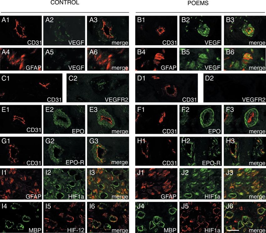
POEMS and two control samples (Fig. 2). We observed
VEGF staining in the epineurial and endoneurial blood vessels
Results
in both controls and POEMS sural nerves. Interestingly, this
Inverse correlation between VEGF and immunoreactivity was consistently higher in blood vessels of
EPO at the time of diagnosis the POEMS specimens when compared with controls. In
Serum levels of VEGF were analysed in 11 patients (Table 1) POEMS nerves, VEGF was also expressed in some non-
Downloaded from by guest on March 5, 2015
with POEMS syndrome at the time of diagnosis, in 16 patients myelin-forming Schwann cells identified as GFAP-positive
with MGUS and in 60 healthy controls. In POEMS patients, cells (Fig. 2A and B).
levels of serum VEGF (median 2230.62 pg/ml; range Conversely, VEGFR2 staining was only detectable in some
1216.65–3179.4) were higher than in patients affected by endoneurial blood vessels of control patients, whereas it was
MGUS (median 299.59 pg/ml; range 91.58–576.94; P < 0.01) completely absent in POEMS nerves (Fig. 2C and D).
or healthy subjects (median 395.14 pg/ml; range 64.76–858.28; We then investigated the expression of EPO end EPO-R in
P < 0.01) (Fig. 1A). EPO serum levels were measured in the the contiguous sections. EPO staining was equally expressed
same 11 POEMS patients, in 16 patients with MGUS and in the epineurial and endoneurial blood vessels in both
in 24 healthy controls. Median EPO serum concentration in normal and POEMS sural nerves, as was the EPO receptor
POEMS patients at the time of the diagnosis (1.42 mIU/ml; staining, although this latter one was mainly restricted to
range 0.79–3.13) was lower than in patients with MGUS endothelial cells (Fig. 2E–H).
(3.72 mIU/ml; range 1.87–8.91; P < 0.05) or healthy subjects HIF-1a staining showed a similar distribution of immuno-
(4.29 mIU/ml; range 2.17–6.65; P < 0.05) (Fig. 1B). reactivity in both POEMS and control nerves. HIF-1a was
Since VEGF is released from platelets under physiological expressed in myelin-forming Schwann cells, generally with
conditions, and serum levels of VEGF are markedly higher ad-axonal localization as identified by an external ring to
than the corresponding plasma levels (Hashiguchi et al., MBP staining (Fig. 2, I4-6 and J4-6). Similarly, HIF-1a
A * * B
3500 10 * *
Serum concentration (mIU/mL)
VEGF
Serum concentration (pg/mL)
3000 EPO
8
2500
2000 6
1500
4
1000
500
2
0
0
CTR POEMS MGUS CTR POEMS MGUS
Fig. 1 Box plots showing VEGF and EPO serum levels at the time of the diagnosis. (A) Serum VEGF in 60 controls (CTR), 11 POEMS
and 16 MGUS patients. (B) Serum EPO in 24 controls (CTR), 11 POEMS and 16 MGUS patients.VEGF and EPO in POEMS syndrome Brain (2005), 128, 1911–1920 1915
Downloaded from by guest on March 5, 2015
Fig. 2 Expression of VEGF, VEGFR2, EPO, EPO-R and HIF-1a in POEMS nerves. Cryosections of sural nerve biopsies from control patients
(A, C, E, G, I) and two POEMS patients, cases 10 (B and D) and 3 (F, H, J). The VEGF expression was very mild in blood vessels
(identified by CD31 staining) of control nerves (A1–A3), whereas non-myelin-forming Schwann cells (identified by GFAP) were unstained
(A4–A6). In POEMS patients, the VEGF expression was very high in the blood vessel wall (B1–B3) and presented also in
non-myelin-forming Schwann cells (B4–B6). The VEGFR2 expression was mild in blood vessels (identified on a consecutive section by
CD31) of control nerves (C1 and C2), but always absent in the blood vessels of POEMS nerves (D1 and D2). The EPO expression was
similar and restricted to the blood vessel wall in control and POEMS nerves (E1–E3 and F1–F3, respectively). Regarding the EPO-R
expression, there was no difference between controls and POEMS nerves (G and H, respectively) and the staining was restricted to the
blood vessels, especially the endothelium (G1–G3 and H1–H3, respectively). The presence of HIF-1a was diffuse and identical in both
control and POEMS nerves (I and J), including non-myelin-forming Schwann cells (identified by GFAP, respectively I1-I3 and J1-J3) and
myelin-forming Schwann cells (identified by MBP, respectively, I4–I6 and J4–J6). Scale bar = 7 mm in all panels except I1–I6 and
J1–J6, where scale bar = 20 mm.
was present in non-myelin-forming Schwann cells, identified the severity of endoneurial vessel involvement, seven sural
as GFAP-positive cells (Fig. 2, I1-3 and J1-3) and in endoneur- nerve biopsies of POEMS patients were analysed. Light micro-
ial vessels (data not shown). When observed by confocal scopic examination of paraffin-embedded sections did not
microscopy using identical settings, the staining appeared reveal any inflammatory infiltrate. Endoneurial oedema was
higher in POEMS than in control nerves. reported in two cases. In transverse semi-thin sections,
myelinated nerve fibre density was variably diminished and
all patients had a variable degree of endoneurial fibrosis.
Neuropathology of POEMS: endoneurial Myelin ovoids, indicating acute axonal degeneration, were
vessel involvement observed in cases 2, 3 and 8. Clusters of thinly myelinated
To evaluate whether the VEGF and EPO relationship and the fibres, a sign of regeneration after axon degeneration, were
high VEGF expression in peripheral nerve was correlated with observed in case 1. In case 8, some myelinated fibres were1916 Brain (2005), 128, 1911–1920 M. Scarlato et al.
Table 2 Quantitative study of endoneurial microvessels
Case Vessel density No. vessels Vessel Luminal Lumina
(No./mm2) diameter (mm) diameter (mm)
No. open (%) No. narrowed (%) No. closed (%)
1 44 9 12.71 6 3.2 5.54 6 1.86* 2 (22.3) 6 (66.6) 1 (11.04)
6 37 12 10.18 6 2.14 2.63 6 1.96* 1 (8.3) 6 (50) 5 (41.7)
8 47 8 10.08 6 2.8 3.8 6 2.11* 2 (25) 4 (50) 2 (25)
Control 49 9 10.98 6 3.92 6.79 6 2.91 8 (88.9) 1 (11.1) 0
Vessels in the endoneurium were measured. Cases 1, 6 and 8 were POEMS nerves; control, ALS patient’s nerve. Vessel diameter (basement
membrane was not included) = mean 6 SD. Lumen diameter = mean 6 SD. *Statistically significant data as determined by two-tailed
paired t-test.
demyelinated. We observed a trend of correlation between groups of patients, responders and non-responders, differed
VEGF serum levels and the degree of endoneurial vessels in pre-treatment VEGF levels; in particular, three patients out
damage, confirmed by quantitative study (Table 2). In fact, of four who improved after therapy had a pre-therapy VEGF
the luminal diameters of POEMS nerves were significantly level 2500 pg/ml presented more com- responses to therapy and grades of endoneurial vessel involve-
promised endoneurial vessels, with a higher percentage of ment are briefly summarized here.
narrowed or closed vessels (cases 6 and 8; Table 2 ) compared
with a POEMS patient with VEGF levelVEGF and EPO in POEMS syndrome Brain (2005), 128, 1911–1920 1917
A B
3500 8
Serum concentration (pg/mL)
Serum concentration (mIU/mL)
7
3000 EPO
6
2500
5
2000
4
1500
3
1000 2
500 1
VEGF
0 0
Before therapy After therapy Before therapy After therapy
C 3500
Serum level (pg/mL)
3000
VEGF
2500 *
2000
1500
1000
500
0
T0 T+3 T+6 T+11 T+17 T+20 T+22 T+26 T+28 T+31
30
Serum level (mIU/mL)
EPO
Downloaded from by guest on March 5, 2015
25
20
15
10
5
0
T0 T+3 T+6 T+11 T+17 T+20 T+22 T+26 T+28 T+31
Time (months of the follow-up)
D 3500
Serum level (pg/mL)
3000 *
2500
2000
1500
1000
500 VEGF
0
T0 +5 T T+15 T+18 T+20 T+22
Serum level (mIU/mL)
30
25 EPO
20
15
10
5
0
T0 T+5 T+15 T+18 T+20 T+22
Time (months of the follow-up)
Fig. 3 (A and B) VEGF and EPO serum levels before and after therapy; for each patient, first and last sample of the follow-up. Dashed lines
indicate patients that did not respond to therapy while continuous lines indicate responders to therapy. (A) VEGF serum concentration in
11 POEMS patients. (B) EPO serum concentration in 11 POEMS patients. (C) Longitudinal evaluation of VEGF (top) and EPO (bottom)
serum concentrations of case 1. Time points refer to the follow-up period (months). The mark indicates when the sural nerve biopsy was
done, the arrow when the therapy was started. (D) Longitudinal evaluation of VEGF (top) and EPO (bottom) serum concentrations of case 6.
Time points refer to the follow-up period (months). The mark indicates when the sural nerve biopsy was done, the arrow when the therapy
was started.1918 Brain (2005), 128, 1911–1920 M. Scarlato et al.
Downloaded from by guest on March 5, 2015
Fig. 4 Transverse semithin sections of POEMS sural nerves: (A) case 1; (B and C) case 6. (A) Moderate thickening of vessel wall due to
basal lamina and cellular proliferation. (B) Representative areas of vessels showing abnormally thickened walls and endothelial proliferation,
resulting in luminal narrowing. (D and E) Electron micrograph sections of POEMS sural nerve (case 6). (D) Endothelial cytoplasmic
enlargement and gap between endothelial cells and microfilaments within an endothelial cell are evident. (E) Inclusion, microfilaments and
many pinocytic vesicles adjacent to the cell membranes are present. Scale bar = 15 mm in A–C; 1 mm in D; 1.5 mm in E.
therapy was started with no benefit. The general conditions of of the skin changes, improvement of the neuropathic disturb-
the patient worsened (grade 4 according to Rankin scale) ances and reduction all of the features assumed to be related to
despite 4 months of therapy with corticosteroids and plasma increased permeability, such as papilloedema and organo-
exchange. The patient is now undergoing autologous stem- megaly. These data, while confirming VEGF as a diagnostic
cell bone marrow transplant. VEGF serum levels remained marker of the disease, also support VEGF as a prognostic
constantly high (>2500 pg/ml) and EPO serum levels low marker for POEMS syndrome, strongly predictive of response
(median 1.83 mIU/ml; range 1.1–4.52) (Fig. 3D). to therapy. Conversely, EPO levels pre-therapy do not differ
significantly between responders and non-responders to treat-
ment. The efficacy of the therapies might well be related to
Discussion their ability to interfere with VEGF production and action.
We have shown that the angiogenic factors VEGF and EPO are In fact, according to our experience, IVIg are not effective in
inversely correlated during the clinical course of POEMS syn- modifying the disease course, plasma exchange provides only
drome and in terms of their nerve expression. VEGF is already temporary benefit, while radiation therapy and alkylators are
known to be a useful marker of the disease, but it has not more successful, probably because they affect the source of
previously been shown how well it correlates with the clinical VEGF. In fact, VEGF production has been shown to be stimu-
course, the response to therapy and the severity of endoneur- lated by oestrogen hormones and improvement of POEMS
ial vessel involvement. We have also demonstrated for the first syndrome symptoms has been reported after treatment
time that a decrease in EPO serum concentration, with a with tamoxifen (Enevoldson and Harding, 1992), a drug
normal red blood cell count and kidney function, is related with strong anti-oestrogen action. In addition, thalidomide
to POEMS syndrome. (Sinisalo et al., 2004), a drug with anti-angiogenic action, has
In all patients, polyneuropathy was the presenting symp- been reported to be effective in treating the symptoms.
tom and reason for consultation. In our population the The correlation between VEGF serum concentration
neuropathic symptoms improved in four out of 11 patients and severity of POEMS syndrome suggests that VEGF may
treated with standard combined therapy (from Rankin score 2 be a causative agent in the disease. Moreover, the neuropatho-
to 0–1). We observed a better prognosis in patients with VEGF logical data of POEMS nerve biopsies, showing involvement
serum levelsVEGF and EPO in POEMS syndrome Brain (2005), 128, 1911–1920 1919
previously reported vascular abnormalities in the nerve of the immunofluorescence studies were not quantitative.
POEMS patients giving rise to the possibility of a chronically It has been reported that EPO effects include prevention
accelerated coagulation due to alterations in the serum coagu- of neuronal apoptosis and that it has protective effects against
lation factors. In fact, VEGF can also act as activator of the diabetic neuropathy in rats (Bianchi et al., 2004). Interest-
coagulation pathway (Senger et al., 1996). Adams and Said ingly, diabetic neuropathy is characterized by a microan-
(1998) instead suggested a direct role of the M component in giopathy and a direct correlation between hyperglycaemia
the lesion of nerve fibres having observed M deposit in the and VEGF has been found (Kakizawa et al., 2004). It is there-
endoneurium. However, it could be a consequence of fore conceivable that the reduction of EPO could be respons-
microvascular hyperpermeability induced by VEGF with a ible for the increased susceptibility of endothelial cells to
secondary opening of the blood–nerve barrier, which is less VEGF-induced damage. An imbalance between these two
tight than the blood–brain barrier (Kanda et al., 2000). No angiogenic factors in the local setting of the nerve might
deposition of Ig or amyloid was detected in our cases. On the alter their neurotrophic activities.
contrary, we found a considerable involvement of the nerve The absence of different distribution of the four poly-
vasculature and a direct correlation with the VEGF serum morphisms or macrodeletions in the VEGF gene promoter
levels. We propose that the endothelial injury is directly or region analysed in five POEMS patients (unpublished obser-
indirectly caused by an abnormal activation of endothelial vations) leaves open the question about the cause of VEGF
cells by VEGF, which is overexpressed in the nerves of over-production. Identification of the source and mechanism
patients with POEMS syndrome. Elevated systemic levels of behind this increase will lead to a more effective therapeutic
VEGF probably determine hypertrophy and proliferation approach.
of the endothelial cells with a secondary microangiopathy.
The consequent reduction of oxygen supply induces a
Acknowledgements
robust expression of HIF-1a by all of the constituents of
Authors are indebted to Dr G. Dina, Dr G. Galizia and
the nerve, with a secondary increase in local VEGF expression
Alembic for technical assistance, to Dr J. Beesley for revision
Downloaded from by guest on March 5, 2015
causing a self-perpetuating VEGF toxic gain of function.
of the English and to Dr M. C. Malaguti for morphometric
At the same time, since physiological activities of VEGF
analysis. The Telethon bank (GTF02008), the Eurobiobank
include induction of platelet aggregation and promotion
Network (QLTR-2001-02769) and R.F.02 187 are gratefully
of vascular permeability, VEGF also indirectly sustains
acknowledged. The research was supported by Telethon
its release and induction. The biological activity of
(GGP030193) and a FISM grant to S.C.P.
VEGF can also be modulated indirectly, as happens with
receptor transcription. The absence of VEGFR2 staining in
POEMS nerve biopsies could, in fact, be due to receptor References
down-regulation. Adams D, Said G. Ultrastructural characterisation of the M protein in nerve
Microvascular changes have been reported in vasa ner- biopsy of patients with POEMS syndrome. J Neurol Neurosurg Psychiatry
1998; 64: 809–12.
vorum of patients with peripheral neuropathy associated Bianchi R, Buyukakilli B, Brines M, Savino C, Cavaletti G, Oggioni N, et al.
with dysglobulinaemia (Powell et al., 1984). In a series of Erythropoietin both protects from and reverses experimental diabetic
11 nerve biopsies, eight with MGUS, two with polyclonal neuropathy. Proc Natl Acad Sci USA 2004; 101: 823–8.
gammopathy and one with Waldenström’s macroglobu- Del Bo R, Scarlato M, Ghezzi S, Martinelli Boneschi F, Fenoglio C, Galbiati S,
et al. VEGF gene variability is associated with increased risk of Alzheimer’s
linaemia, electron microscopy showed abnormal accumula-
disease. Ann Neurol 2005; 57: 373–80.
tion of masses of intracytoplasmic filaments accompanied Dispenzieri A, Gertz MA. Treatment of POEMS syndrome. Curr Treat
by endothelial proliferation. Our ultrastuctural analysis of Options Oncol 2004; 5: 249–57.
POEMS nerves revealed instead endothelial cytoplasmic Dispenzieri A, Kyle RA, Lacy MQ, Rajkumar SV, Therneau TM, Larson DR,
enlargement, opening of the tight juctions between endotheli- et al. POEMS syndrome: definitions and long-term outcome. Blood 2003;
als cells and presence of many pinocytic vesicles adjacent to 101: 2496–506.
Enevoldson TP, Harding AE. Improvement in the POEMS syndrome after
the cell membranes, suggesting a permeability alteration of administration of tamoxifen. J Neurol Neurosurg Psychiatry 1992; 55: 71–2.
endoneurial vessels. Ferrara N, Gerber HP, LeCouter J. The biology of VEGF and its receptors.
The low EPO concentration in POEMS serum may reflect a Nat Med 2003; 9: 669–76.
compensatory consequence of the high levels of VEGF, since Giannini C, Dyck PJ. Ultrastructural morphometric features of human sural
they share some angiogenic properties. Low EPO may also be nerve endoneurial microvessels. J Neuropathol Exp Neurol 1993; 52: 361–9.
Hashiguchi T, Arimura K, Matsumuro K, Otsuka R, Watanabe O,
due to renal dysfunction. Although blood urea and creatinine Jonosono M, et al. Highly concentrated vascular endothelial growth factor
levels in the patients were within the normal range, we cannot in platelets in Crow–Fukase syndrome. Muscle Nerve 2000; 23: 1051–6.
exclude involvement of the kidney as the result of high VEGF Hassan K, Simri W, Rubenchik I, Manelis J, Gross B, Shasha SM, et al.
circulating levels, especially in patients that did not respond to Effect of erythropoietin therapy on polyneuropathy in predialytic patients.
J Nephrol 2003; 16: 121–5.
therapy.
Kakizawa H, Itoh M, Itoh Y, Imamura S, Ishiwata Y, Matsumoto T, et al.
The role of EPO in nerve damage during POEMS The relationship between glycemic control and plasma vascular endothelial
syndrome is more difficult to define. We found similar growth factor and endothelin-1 concentration in diabetic patients. Meta-
EPO expression in POEMS and control nerves, although bolism 2004; 53: 550–5.1920 Brain (2005), 128, 1911–1920 M. Scarlato et al.
Kanda T, Iwasaki T, Yamawaki M, Tai T, Mizusawa H. Anti-GM1 antibody Saida K, Kawakami H, Ohta M, Iwamura K. Coagulation and vascular
facilitates leakage in an in vitro blood–nerve barrier model. Neurology abnormalities in Crow–Fukase syndrome. Muscle Nerve 1997; 20: 486–92.
2000; 55: 585–7. Sakanaka M, Wen TC, Matsuda S, Masuda S, Morishita E, Nagao M, et al.
Krieg M, Marti HH, Plate KH. Coexpression of erythropoietin and vascular In vivo evidence that erythropoietin protects neurons from ischemic
endothelial growth factor in nervous system tumors associated with von damage. Proc Natl Acad Sci USA 1998; 95: 4635–40.
Hippel–Lindau tumor suppressor gene loss of function. Blood 1998; 92: Senger DR, Ledbetter SR, Claffey KP, Papadopoulos-Sergiou A, Peruzzi CA,
3388–93. Detmar M. Stimulation of endothelial cell migration by vascular permeab-
Lambrechts D, Storkebaum E, Morimoto M, Del-Favero J, Desmet F, ility factor/vascular endothelial growth factor through cooperative mech-
Marklund SL, et al. VEGF is a modifier of amyotrophic lateral sclerosis anisms involving the alphavbeta3 integrin, osteopontin, and thrombin.
in mice and humans and protects motoneurons against ischemic death. Am J Pathol 1996; 149: 293–305.
Nat Genet 2003; 34: 383–94. Sinisalo M, Hietaharju A, Sauranen J, Wirta O. Thalidomide in POEMS
Michizono K, Umehara F, Hashiguchi T, Arimura K, Matsuura E, syndrome: case report. Am J Hematol 2004; 76: 66–8.
Watanabe O, et al. Circulating levels of MMP-1, -2, -3, -9, and TIMP-1 Sondell M, Lundborg G, Kanje M. Vascular endothelial growth factor has
are increased in POEMS syndrome. Neurology 2001; 56: 807–10. neurotrophic activity and stimulates axonal outgrowth, enhancing cell
Miralles GD, O’Fallon JR, Talley NJ. Plasma-cell dyscrasia with poly- survival and Schwann cell proliferation in the peripheral nervous system.
neuropathy. The spectrum of POEMS syndrome. N Engl J Med 1992; J Neurosci 1999; 19: 5731–40.
327: 1919–23. Vital C, Vital A, Ferrer X, Viallard JF, Pellegrin JL, Bouillot S, et al.
Powell HC, Rodriguez M, Hughes RA. Microangiopathy of vasa nervorum in Crow–Fukase (POEMS) syndrome: a study of peripheral nerve biopsy
dysglobulinemic neuropathy. Ann Neurol 1984; 15: 386–94. in five new cases. J Peripher Nerv Syst 2003; 8: 136–44.
Pugh CW, Ratcliffe PJ. Regulation of angiogenesis by hypoxia: role of the HIF Watanabe O, Arimura K, Kitajima I, Osame M, Maruyama I. Greatly raised
system. Nat Med 2003; 9: 677–84. vascular endothelial growth factor (VEGF) in POEMS syndrome. Lancet
Quattrini A, Previtali S, Feltri ML, Canal N, Nemni R, Wrabetz L. Beta 4 1996; 347: 702.
integrin and other Schwann cell markers in axonal neuropathy. Glia 1996; Watanabe O, Maruyama I, Arimura K, Kitajima I, Arimura H, Hanatani M,
17: 294–306. et al. Overproduction of vascular endothelial growth factor/vascular per-
Rosenstein JM, Krum JM. New roles for VEGF in nervous tissue—beyond meability factor is causative in Crow–Fukase (POEMS) syndrome. Muscle
blood vessels. Exp Neurol 2004; 187: 246–53. Nerve 1998; 21: 1390–7.
Downloaded from by guest on March 5, 2015You can also read