HAND/PERIPHERAL NERVE - BINASSS
←
→
Page content transcription
If your browser does not render page correctly, please read the page content below
HAND/PERIPHERAL NERVE
  Nerve Pain after Burn Injury: A Proposed
  Etiology-Based Classification
         Kevin M. Klifto, Pharm.D.
   C. Scott Hultman, M.D., M.B.A.
                                            Background: Understanding the mechanism of nerve injury may facilitate man-
        A. Lee Dellon, M.D., Ph.D.          aging burn-related nerve pain. This proposed classification, based on cause of
                                            nerve injury, was developed to enhance the understanding and management of
    Philadelphia, Pa., and Baltimore, Md.   burn-related nerve pain.
                                            Methods: This retrospective investigation included patients aged 15 years or
                                            older admitted to the burn center from 2014 to 2019. Burn-related nerve pain
                                            was patient-reported and clinically assessed as pain 6 months or more after
                                            burn injury, unrelated to preexisting illnesses/medications. The pain classifica-
                                            tion consisted of direct nerve injury, nerve compression, electrical injury, and
                                            nerve dysfunction secondary to systemic injury. The four categories were statis-
                                            tically analyzed between groups, using 52 variables.
                                            Results: Of the 1880 consecutive burn patients, 113 developed burn-related
                                            nerve pain and were eligible for validation of the classification: direct nerve
                                            injury, n = 47; nerve compression, n = 12; electrical injury, n = 7; and nerve dys-
                                            function secondary to systemic injury, n = 47. Factors, significantly increased,
                                            that distinguished one category from another were as follows: for direct nerve
                                            injury, continuous symptoms (p < 0.001), refractory nerve release response
                                            (p < 0.001), nerve repair (p < 0.001), and pruritus (p < 0.001); for nerve compres-
                                            sion, Tinel signs (p < 0.001), shooting pain (p < 0.001), numbness (p = 0.003),
                                            intermittent symptoms (p < 0.001), increased percentage total body surface
                                            area burned (p = 0.019), surgical procedures (p < 0.001), and nerve release
                                            (p < 0.001); and for electrical injury, Tinel sign (p < 0.001), intermittent symptoms
                                            (p = 0.002), amputations (p = 0.002), fasciotomies (p < 0.001), and nerve release
                                            (p < 0.001). Nerve dysfunction secondary to systemic injury was distinguished
                                            by significantly less Tinel signs (p < 0.001), shooting pain (p < 0.001), numb-
                                            ness and tingling (p < 0.001), pruritus (p < 0.001), fascial excision (p = 0.004),
                                            skin grafts (p < 0.001), amputation (p = 0.004), nerve releases (p < 0.001),
                                            and third-degree burns (p = 0.002).
                                            Conclusion: A classification consisting of direct nerve injury, nerve compres-
                                            sion, electrical injury, and nerve dysfunction secondary to systemic injury is
                                            presented that may guide patient management and research methods, with the
                                            goal of improving pain outcomes in burn-related nerve pain. (Plast. Reconstr.
                                            Surg. 147: 635, 2021.)

  I
    t seems axiomatic that a patient with a burn will                  Less well perceived, and less well understood, are
    have acute pain related to the injury itself, acute                the mechanisms involved with the burn patient who
    pain related to the débridement and grafting,                      has chronic, neuropathic pain at the time of dis-
  and perhaps pain during the healing phases. Burn                     charge from the burn unit. This chronic pain can
  physicians are well versed in the appropriate phar-                  affect the reconstruction process. Recently, chronic
  macologic treatment of the acute forms of pain.                      nerve compression and painful neuromas in burn
                                                                       patients have been described.1,2

   From the Division of Plastic Surgery, University of
   Pennsylvania School of Medicine, and the Departments of
   Plastic and Reconstructive Surgery and Neurosurgery, The              Disclosure: The authors have no financial interest
   Johns Hopkins University School of Medicine.                          to declare in relation to the content of this article.
   Received for publication November 29, 2019; accepted July             This research did not receive any specific grant from
   1, 2020.                                                              funding agencies in the public, commercial, or not-
   Copyright © 2021 by the American Society of Plastic Surgeons          for-profit sectors.
   DOI: 10.1097/PRS.0000000000007639

                                                       www.PRSJournal.com                                                    635
Copyright © 2021 American Society of Plastic Surgeons. Unauthorized reproduction of this article is prohibited.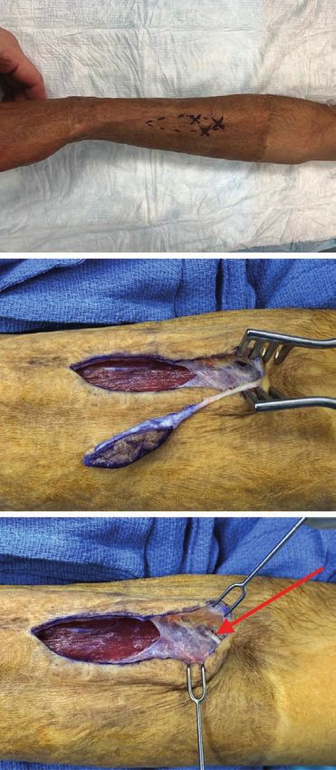
Plastic and Reconstructive Surgery • March 2021

      The prevalence of chronic pain after burn            “pruritus” is not typically considered as neu-
  has been reported to range from a low of 7 per-          ropathic pain, we included it because patients
  cent to a high of 82 percent.3–12 To understand          verbalized this symptom. Neuropathic pain was
  the mechanisms that can involve the peripheral           evaluated by a minimum of two health care pro-
  nerve as a source of pain in this group of patients,     viders. A trial of one or more “neuropathic” med-
  a classification of nerve injury in the burn patient     ications was attempted for all patients, typically
  must be created. The understanding derived from          gabapentin (Neurontin; Pfizer, New York, N.Y.)
  this classification will permit awareness during the     or pregabalin (Lyrica; Pfizer). Patients were
  inpatient care of the burn patient, and possibly         stratified into each nerve injury category based
  create diagnostic and therapeutic approaches to          on the symptoms that contributed most to their
  manage these patients in the acute setting to pre-       morbidity on follow-up for simplicity and clarity.
  vent the creation of chronic pain. In the outpa-         It is likely that patients had overlapping causes in
  tient setting, after discharge from the burn unit,       different anatomical locations. All patients pre-
  this classification has the potential to create diag-    sented for follow-up visits after being discharged
  nostic and therapeutic approaches to manage              from the burn center.
  these patients to relieve their chronic pain.
                                                           Variables Analyzed
           PATIENTS AND METHODS                                A total of 52 variables were measured for each
                                                           of the four categories. These variables consisted
  Study Design                                             of patient demographics, characteristics of pain
      We performed a retrospective, medical record         rated on a 0- to 10-point scale, long-term medi-
  review approved by The Johns Hopkins Medicine            cations, surgical and nonsurgical treatments, and
  Institutional Review Board (IRB00213320) to col-         complications. Variables were searched manually
  lect a cohort of patients admitted to The Johns          for each patient by notes from all health care pro-
  Hopkins Burn Center from January 1, 2014, through        viders/staff in electronic medical records. A vari-
  January 1, 2019. The Strengthening the Reporting         able was considered if evaluated and documented
  of Observational Studies in Epidemiology guide-          by a minimum of a physician and physical thera-
  lines were adhered to throughout the observa-            pist or occupational therapist for each patient.
  tional component of the review.13 We were unable
  to identify an appropriate Enhancing the Quality         Statistical Analysis
  and Transparency of Health Research network                  Descriptive statistics were used to compare
  guideline to adhere to for reporting an evidence-        medians, interquartile ranges, odds ratios, 95 per-
  based classification model.14                            cent confidence intervals, areas under the curve,
                                                           frequencies, and percentages between demo-
  Study Population                                         graphic and clinical variables based on the non-
      Patients included were consecutive, older            parametric distribution of population data and
  than 15 years, sustained a burn injury, and were         small sample sizes. Statistical analyses were per-
  admitted to the burn center. The burn center             formed to compare differences among the four
  consisted of the burn wound unit and the burn            nerve injury categories, followed by differences
  intensive care unit. Patients were excluded if           within each category. Dichotomous variables were
  they had preexisting neuropathic pain caused             assessed using Fisher’s exact cross-tabulation tests.
  by an underlying medical illness or medication,          After a significant value for the Fisher’s exact
  or less than a 6-month multidisciplinary coordi-         test was obtained, a post hoc test was run using
  nated follow-up.                                         a Bonferroni test with α of 0.006 from eight cells
      We stratified patients into four categories          in a 2 × 4 contingency table to determine which
  for comparison by type of nerve injury follow-           groups were different.15 Continuous variables were
  ing their burn. Those categories were as follows:        assessed using the Kruskal-Wallis test followed by
  direct nerve injury, nerve compression, electri-         the Dunn post hoc test. Univariate analyses were
  cal injury, and nerve dysfunction secondary to           followed by multivariate stepwise logistic regres-
  systemic injury. All patients had neuropathic            sions adjusting for age, race, body mass index,
  pain lasting greater than 6 months following             and percentage total body surface area burned.
  burn. Pain was self-described clinically as shoot-       Analyses outcomes were two-tailed, with a signifi-
  ing, stabbing, sharp, burning, tingling, pruritus,       cance level set at α of 0.05. All analyses were per-
  throbbing, numbness, and intermittent and/               formed with IBM SPSS Version 25.0 (IBM Corp.,
  or continuous dysesthetic sensations. Although           Armonk, N.Y.).

  636
Copyright © 2021 American Society of Plastic Surgeons. Unauthorized reproduction of this article is prohibited.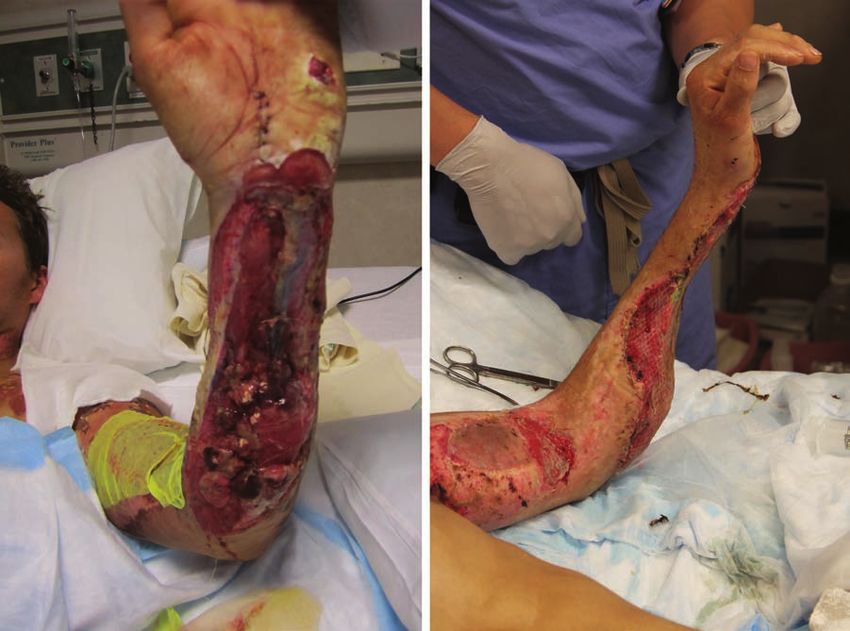
Volume 147, Number 3 • Nerve Pain after Burn Injury

                            RESULTS                                        Direct Nerve Injury
      Of the 1880 consecutive burn patients, 113                                Direct nerve injury was significantly associated
  developed chronic nerve pain (prevalence, 6                              with characteristics of continuous pain symptoms
  percent) after burn injury and were eligible                             (p < 0.001) and pruritus (p < 0.001) (Table 3).
  for validation of the proposed classification                            Attempts at treating direct nerve pain with nerve
  model. There were statistically significant differ-                      release were not significantly successful (p < 0.001);
  ences among all four categories of nerve injury                          however, nerve repair was significantly associated with
  (p < 0.001) (Table 1). Of the 113 patients with                          the resolution of chronic pain (p < 0.001) (Table 4).
  burn-related nerve injury, 47 were categorized as                        Median follow-up was 24 months (range, 8 to 52
  having direct nerve injury, 12 were categorized                          months). Multivariate analyses resulted in decreased
  as having nerve compression, seven were catego-                          odds of direct nerve injury with the absence of pruri-
  rized as having electrical injury, and 47 were cat-                      tus (OR,
Plastic and Reconstructive Surgery • March 2021 Table 3. Pain Characteristics Characteristic DNI (%) NC (%) EI (%) NDSSI (%) p Tinel sign 6 (13) 11 (92)* 5 (71)* 0 (0)*
Volume 147, Number 3 • Nerve Pain after Burn Injury

  Table 6. Long-Term Patient Medications to Manage Pain
  Medication                                  DNI                     NC                      EI                   NDSSI                     p
  Gabapentin/pregabalin                     32 (68)                 10 (83)                 3 (43)                33 (70)                 0.374
  SNRI                                       2 (4)                   5 (42)*                1 (14)                 4 (9)                  0.006
  TCA                                        2 (4)                   2 (17)                 0 (0)                  5 (11)                 0.331
  Antiepileptic                              0 (0)                   0 (0)                  0 (0)                  2 (4)                  0.651
  Lidocaine                                  0 (0)                   1 (8)                  1 (14)                 1 (2)                  0.090
  NSAID                                     13 (28)                  4 (33)                 3 (43)                13 (28)                 0.793
  Acetaminophen                             16 (34)                  0 (0)                  0 (0)                 17 (36)                 0.014
  Ascorbic acid                              2 (4)                   0 (0)                  1 (14)                 5 (11)                 0.358
  Opioid                                    16 (34)                  9 (75)*                3 (43)                10 (21)                 0.005
  Tramadol                                   1 (2)                   0 (0)                  0 (0)                  2 (4)                  1.000
  DNI, direct nerve injury; NC, nerve compression; EI, electrical injury; NDSSI, nerve dysfunction secondary to systemic injury; SNRI, serotonin-
  norepinephrine reuptake inhibitor; TCA, tricyclic antidepressant; NSAID, nonsteroidal antiinflammatory drug.
  *Statistically significant value.

  Table 7. Complications
  Complication                                DNI (%)                NC (%)                EI (%)               NDSSI (%)                    p
  Hospital infection                            9 (19)               3 (38)                 2 (29)                 5 (11)                 0.167
  Ventilator-associated events                  1 (2)                0 (0)                  0 (0)                  0 (0)                  0.131
  Pressure sores                                0 (0)                0 (0)                  0 (0)                  0 (0)                  0.168
  VTE                                           1 (2)                1 (8)                  1 (14)                 0 (0)                  0.073
  Other complication                            2 (15)               4 (38)                 0 (0)                  4 (44)                   —
  Overall complications                        13 (28)               8 (67)*                3 (43)                 9 (19)                 0.011
  DNI, direct nerve injury; NC, nerve compression; EI, electrical injury; NDSSI, nerve dysfunction secondary to systemic injury; VTE, venous
  thromboembolism.
  *Statistically significant value.

  intermittent symptoms (p = 0.002) compared                                systemic injury compared to other categories of
  with other categories (Table 3). Amputations                              pain (Table 5). Significantly less surgical and non-
  (p = 0.002) and fasciotomies (p < 0.001) were sig-                        surgical treatments for nerve pain were required
  nificantly higher in patients suffering electrical                        in systemic injury compared to other categories
  injury. Compression, followed by nerve release, was                       of pain [laser (p = 0.002) and physical therapy/
  significantly higher for treating pain (p < 0.001)                        occupational therapy (p < 0.001)]. There were
  in this category (Table 4). Median follow-up was                          no nerve releases (p < 0.001) for patients with sys-
  12 months (range, 8 to 36 months). Multivariate                           temic injury compared to other categories of pain
  analyses resulted in decreased odds of electrical                         (Table 4). Median follow-up was 26 months (range,
  injury with the absence of amputation (OR, 0.02;                          8 to 52 months). Multivariate analyses resulted in
  95 percent CI, 0.001 to 0.26; area under the curve,                       increased odds of nerve dysfunction secondary
  0.716; p = 0.004) and fasciotomy (OR,
Plastic and Reconstructive Surgery • March 2021

                     Fig. 1. (Left) Direct nerve injury to ulnar nerve and cutaneous sensory nerves after
                     excision. (Right) Cutaneous sensory nerves are buried into remaining muscle to pre-
                     vent neuroma formation. Autologous skin was meshed to provide skin coverage after
                     addressing the nerve injuries.

  complication. For example, with direct nerve                   to surgery on the burn sites themselves, donor
  injury, if during débridement of a wound a cuta-               sites can sustain direct injury to free nerve end-
  neous nerve is divided, this should be recognized              ings during autologous skin grafting. Wound heal-
  and the proximal end of that nerve implanted                   ing by secondary intention will form hypertrophic
  into a subjacent muscle proximal to the burn or                scar contractures. These scars have altered sen-
  repaired with a graft or allograft (Fig. 1).16 If a tan-       sory function compared to uninjured tissues.24
  gential excision of a distal radial forearm wound              Direct injury to a peripheral nerve releases neu-
  required the division of the radial sensory and/               rotransmitters as the wound heals, causing pain
  or the lateral antebrachial cutaneous nerve, an                and pruritus.25 Abnormal cutaneous innervation
  incision could be made in the proximal forearm,                of injured nerve fibers can manifest in a chronic
  away from the burn, and the proximal nerve ends                state of pain.24,25 Forceful physical therapy dur-
  implanted into the brachioradialis muscle.17 If a              ing rehabilitation may also result in direct nerve
  proximal anterior thigh wound was débrided and                 injury from overstretching.26
  branches of the lateral cutaneous nerve of the                      With nerve compression, there will be some
  thigh were resected, this nerve could be identified            patients who have had a preexisting carpal tunnel
  at the inguinal ligament level and the proximal                syndrome or subclinical median nerve compres-
  end of the damaged nerve divided and relocated                 sion at the wrist. During the resuscitation phase
  into the pelvis.18 This concept not only permits a             of treatment, the swelling in the extremity makes
  proactive approach to prevention of chronic pain               that nerve compression symptomatic. In the over-
  but also gives structure to a prospective analysis of          all treatment of the patient’s burn, that relatively
  this approach in the burn unit.19                              “unimportant” tingling in the fingers may not be
      Peripheral nerve injury, not repairable by                 recognized, leading to chronic pain in the extrem-
  physiologic remyelination, collateral sprouting,               ity on discharge.27 Compartment syndrome in an
  and axon regrowth can be surgically treated to                 extremity can clearly cause nerve compression,
  reestablish continuity.20 Nerve repair following               which may persist if the fasciotomies alone do not
  direct injury was significantly associated with less           release the nerve sufficiently. This recognition
  chronic pain. This repair can be achieved by a                 can lead the burn team to institute regular physi-
  direct repair or nerve connection or bridging with             cal examinations for nerve compression at known
  nerve transfers, conduits, or grafts.21–23 In addition         sites of anatomical narrowing using the Tinel

  640
Copyright © 2021 American Society of Plastic Surgeons. Unauthorized reproduction of this article is prohibited.
Volume 147, Number 3 • Nerve Pain after Burn Injury
sign, and using a noninvasive and nonpainful tun-
ing fork for the evaluation of sensibility.28,29 Tight
dressings, compression garments, and incorrect
splinting or positioning in their bed or on the
operating room table can result in nerve compres-
sion unrelated to the burn itself.26 Compression
injuries should be recognized and prevented dur-
ing the recovery period (Fig. 2).
Patients that did not undergo surgical nerve
release were more likely to require opioids and
serotonin-norepinephrine reuptake inhibitors to
manage pain. Neuroma excision without proper
management of the transected nerve is likely to
result in the conversion of a nerve compression
into a direct nerve injury. If the nerve is transected
during neuroma excision, reestablishing continu-
ity or burying the nerve in the muscle can reduce
chronic pain symptoms. Improper initial diag-
nosis and management may increase long-term
patient morbidity by overprescription of opioid
and antidepressant medications to manage their
pain instead of surgical treatment of the nerve
injury.
Nerve compression was significantly associ-
ated with patients suffering from overall compli-
cations. For example, there were four patients
with heterotopic ossification, two with cellulitis
infections, one with necrotizing fasciitis infection,
and one with a deep vein thrombosis. Pseudomonas
aeruginosa was identified in blood cultures from
two patients who had chronic pain related to
nerve compression.
Electrical injury is known to cause the highest
incidence of nerve injury among burn patients.30
Electrical energy prefers to travel through nerve
rather than tendon or bone, and creates a heat
“sink” at a joint. The heat generated from electri-
cal energy can directly injure a nerve; encase the
nerve in fibrous tissue; or indirectly create damage
through local inflammation, edema, and vascular
damage.31–38 Therefore, a peripheral nerve can be
directly thermally injured and have to be recon-
structed with a graft, or entrapped and decom-
Fig. 2. (Above) A 26-year-old man with intermittent shooting
pressed. Acute and delayed timings of surgical
right forearm pain (4 of 10) over previously grafted skin. Physical
decompression have both been shown to be effi-
examination of the right upper extremity revealed three positive
cacious in reducing pain symptoms in the upper
Tinel signs in the distribution of the lateral antebrachial cutaneous
and lower extremities.5,8,19,32,39–41 Amputations
nerve. (Center) A neuroma was identified causing compression in
and fasciotomies were more common after elec-
the lateral antebrachial cutaneous nerve. (Below) The neuroma was
trical energy (Fig. 3). These surgical procedures
excised and the proximal end of the lateral antebrachial cutaneous
can directly injure peripheral nerves, creating a
nerve was implanted into the brachioradialis muscle (arrow).
direct nerve injury. Symptoms may present with
immediate onset, delayed onset, or a gradual pro-
gression.32 These damaging effects often occur Nerve dysfunction secondary to systemic
without noticeable cutaneous involvement and injury remains poorly understood etiologically
may lead to neuropsychiatric morbidity.32,42 but seems to be related to released inflammatory
641
Copyright © 2021 American Society of Plastic Surgeons. Unauthorized reproduction of this article is prohibited.Plastic and Reconstructive Surgery • March 2021
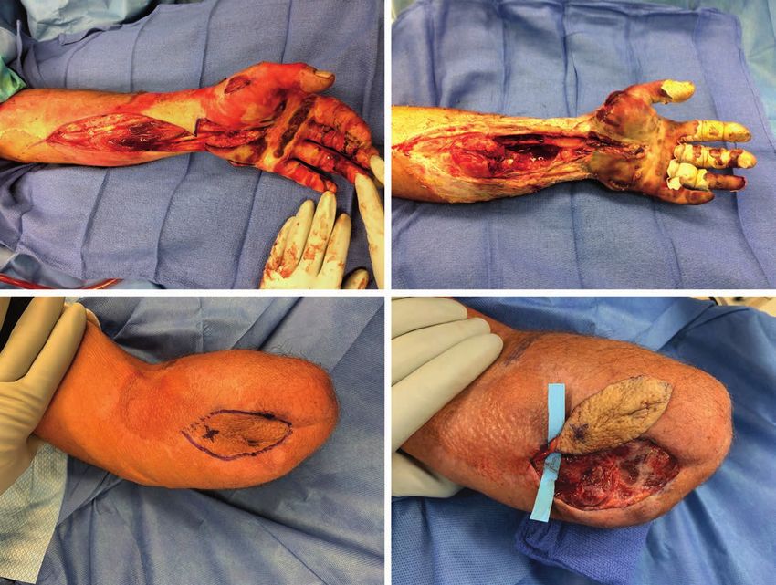
Fig. 3. (Above, left) A 45-year-old man with 30 percent total body surface area electrical injury from 7000 V, with a compart-
ment syndrome of his left upper extremity, underwent fasciotomy. (Above, right) Subsequent necrosis resulted in amputa-
tion of the extremity. (Below, left) On follow-up, a positive Tinel sign was located at the amputation stump. (Below, right)
An amputation revision and neuroma excision were performed to reduce pain.
mediators that can affect peripheral nerve func- performed to treat the nerve pain that followed.
tion.10,26,43,44 Greater percentage total body surface Fascial excision was associated with more nerve
area burns result in greater systemic responses by compression and less nerve dysfunction second-
releasing larger quantities inflammatory media- ary to systemic injury. Skin grafting was associated
tors into systemic circulation. Several inflamma- with less nerve dysfunction secondary to systemic
tory mediators identified that can cause nerve injury. Amputation was associated with more elec-
damage are cyclic adenosine monophosphate, trical injury and less nerve dysfunction secondary
cyclic guanosine monophosphate, prostaglandin to systemic injury. Fasciotomy was associated with
E2, insulin-like growth factor–binding protein 3, more electrical injury. Surgical interventions to
and tumor necrosis factor-alpha.45 When released treat nerve pain were used once pharmacologic
systemically, these mediators can cause dam- interventions were optimized or unsuccessful
age to nerves at locations distal from the sites of for treating the symptoms of pain. Less laser was
injury.45,46 In addition, nociceptive fibers may up- used for nerve dysfunction secondary to systemic
regulate and increase fiber density in both burned injury. More nerve repairs were performed for
and unburned tissues with concurrent central direct nerve injury with successful results. More
plasticity.25 This reemphasizes that local burns nerve releases were performed for nerve compres-
can manifest systemically in unaffected areas from sion and electrical injury, and less nerve releases
central and peripheral nerve changes in chronic, were performed for direct nerve injury and
neuropathic pain.46,47 nerve dysfunction secondary to systemic injury.
Surgical interventions were categorized into Fewer patients required physical therapy/occu-
those performed for the primary burn and those pational therapy for nerve dysfunction secondary
642
Copyright © 2021 American Society of Plastic Surgeons. Unauthorized reproduction of this article is prohibited.Volume 147, Number 3 • Nerve Pain after Burn Injury
to systemic injury. Nerve dysfunction secondary removed from our study. The rigidity of our meth-
to systemic injury had the greatest reductions in odology may explain the lower prevalence of 6 per-
pain scores at final follow-up. This category of cent observed in our population compared to the
nerve pain was self-limiting for many, and did not literature. A misconception that has reoccurred in
require long-term pharmacologic therapy or sur- the literature is the use of electrodiagnostic studies
gical interventions. Reductions in pain scores at to diagnose pain. Pain is subjective, and electrodiag-
final follow-up were achieved through combina- nostic studies are not capable of differentiating pain
tions of surgical and pharmacologic therapies. from no pain. This has resulted in higher reported
With prospective studies, we hope to further clar- rates of chronic pain in other studies. We diagnosed
ify specific responses to interventions and corre- nerve pain clinically in our patient population.
sponding reductions in pain scores.
Limitations of our study relate to the dispro-
portionate sizes of categories and retrospective
CONCLUSION
design. The sample size did not allow for a nor- In burn patients, direct nerve injury, nerve
mal distribution; therefore, we used the Fisher’s compression, electrical injury, and nerve dysfunc-
exact test and Kruskal-Wallis test, and imple- tion secondary to systemic illness were categorized
mented posttest analyses to prevent overestimat- into a comprehensive etiology-based classification
ing our results and a type II error. Although we to guide patient management and research meth-
were able to compare different categories of burn ods to improve patient pain outcomes.
nerve injury, the small, unequal sizes of our sam- C. Scott Hultman, M.D., M.B.A.
ples require prospective validation. Patients were Department of Plastic and Reconstructive Surgery
stratified into each nerve injury category based on The Johns Hopkins Burn Center
the symptoms that contributed most to their mor- 4940 Eastern Avenue
Baltimore, Md. 21224
bidity on follow-up for simplicity and clarity. It is chultma1@jhmi.edu
likely patients have overlapping causes in differ- Facebook: Charles Scott Hultman
ent anatomical locations, which we plan to inves-
tigate. Long-term follow-up care information was
difficult to obtain in international patients, home- ACKNOWLEDGMENTS
less patients, and patients with substance abuse The authors thank Carrie Cox, M.S., R.N., and
or advanced psychiatric illness. Of the 2024 pos- Vidhi Javia, B.S., for assistance with coordinating clini-
sible consecutive burned patients evaluated over cal research at The Johns Hopkins Burn Center. Without
the 5 years, 1880 met eligibility criteria and were their assistance, the authors’ work would not be possible.
included. All 113 of the 1880 patients included in
the study had multidisciplinary coordinated fol-
low-up to confirm a clinical diagnosis of chronic REFERENCES
pain beyond 6 months. The 144 burned patients 1. Ono T, Matsunaga W. Traumatic neuroma: Multiple lesions
in the fingers occurring after deep burns. J Dermatol.
that were excluded consisted of the difficult long-
1990;17:760–763.
term follow-up population and those with preex- 2. Rapolti M, Wu C, Schuth OA, Hultman CS. Under pres-
isting neuropathic pain caused by an underlying sure: Applying practice-based learning and improvement to
medical illness or medication. We analyzed data the treatment of chronic neuropathic pain in patients with
from a single burn center. Although categories of burns. Clin Plast Surg. 2017;44:925–934.
3. Schneider JC, Harris NL, El Shami A, et al. A descriptive
nerve injury would remain the same, our findings
review of neuropathic-like pain after burn injury. J Burn Care
should be generalized with caution until further Res. 2006;27:524–528.
validation is performed through a multicenter 4. Choinière M, Melzack R, Papillon J. Pain and paresthesia
study, and in the pediatric population. in patients with healed burns: An exploratory study. J Pain
Strengths of our study relate to the rigidity of Symptom Manage. 1991;6:437–444.
our methods in the largest known study performed 5. Gabriel V, Kowalske KJ, Holavanahalli RK. Assessment of
recovery from burn-related neuropathy by electrodiagnostic
to date assessing a proposed evidence-based clas- testing. J Burn Care Res. 2009;30:668–674.
sification model for nerve pain after burn injury. 6. Browne AL, Andrews R, Schug SA, Wood F. Persistent pain
We performed multidisciplinary evaluations by dif- outcomes and patient satisfaction with pain management
ferent clinicians and used consistent definitions after burn injury. Clin J Pain 2011;27:136–145.
for nerve pain and chronicity. Patients with neuro- 7. Dauber A, Osgood PF, Breslau AJ, Vernon HL, Carr DB.
Chronic persistent pain after severe burns: A survey of 358
pathic pain attributed to an underlying medical ill- burn survivors. Pain Med. 2002;3:6–17.
ness and/or medication, and signs and symptoms 8. Khedr EM, Khedr T, el-Oteify MA, Hassan HA. Peripheral
of neurologic impairment before burn injury, were neuropathy in burn patients. Burns 1997;23:579–583.
643
Copyright © 2021 American Society of Plastic Surgeons. Unauthorized reproduction of this article is prohibited.Plastic and Reconstructive Surgery • March 2021
9. Malenfant A, Forget R, Papillon J, Amsel R, Frigon JY, 28. Dellon AL. Patient evaluation and management consider-
Choinière M. Prevalence and characteristics of chronic sen- ations in nerve compression. Hand Clin. 1992;8:229–239.
sory problems in burn patients. Pain 1996;67:493–500. 29. Dellon AL. Clinical use of vibratory stimuli to evaluate
10. Margherita AJ, Robinson LR, Heimbach DM, Fishfader VL, peripheral nerve injury and compression neuropathy. Plast
Schneider VA, Jones D. Burn-associated peripheral polyneu- Reconstr Surg. 1980;65:466–476.
ropathy: A search for causative factors. Am J Phys Med Rehabil. 30. Kowalske K, Holavanahalli R, Helm P. Neuropathy after burn
1995;74:28–32. injury. J Burn Care Rehabil. 2001;22:353–357; discussion 352.
11. Tamam Y, Tamam C, Tamam B, Ustundag M, Orak M, 31. Rosenberg DB. Neurologic sequelae of minor electric burns.
Tasdemir N. Peripheral neuropathy after burn injury. Eur Arch Phys Med Rehabil. 1989;70:914–915.
Rev Med Pharmacol Sci. 2013;17(Suppl 1):107–111. 32. Smith MA, Muehlberger T, Dellon AL. Peripheral nerve
12. Ward RS, Saffle JR, Schnebly WA, Hayes-Lundy C, Reddy R. compression associated with low-voltage electrical injury
Sensory loss over grafted areas in patients with burns. J Burn without associated significant cutaneous burn. Plast Reconstr
Care Rehabil. 1989;10:536–538. Surg. 2002;109:137–144.
13. von Elm E, Altman DG, Egger M, Pocock SJ, Gøtzsche PC, 33. Dendooven AM, Lissens M, Bruyninckx F, Vanhecke J.
Vandenbroucke JP; STROBE Initiative. The Strengthening Electrical injuries to peripheral nerves. Acta Belg Med Phys.
the Reporting of Observational Studies in Epidemiology 1990;13:161–165.
(STROBE) statement: Guidelines for reporting observa- 34. Kinnunen E, Ojala M, Taskinen H, Matikainen E. Peripheral
tional studies. Int J Surg. 2014;12:1495–1499. nerve injury and Raynaud’s syndrome following electric
14. Simera I, Moher D, Hoey J, Schulz KF, Altman DG. The shock. Scand J Work Environ Health 1988;14:332–333.
EQUATOR Network and reporting guidelines: Helping to 35. McCreery DB, Agnew WF, Yuen TG, Bullara LA. Damage
achieve high standards in reporting health research studies. in peripheral nerve from continuous electrical stimulation:
Maturitas 2009;63:4–6. Comparison of two stimulus waveforms. Med Biol Eng Comput.
15. Beasley MT, Schumacker RE. Multiple regression approach 1992;30:109–114.
to analyzing contingency tables: Post hoc and planned com- 36. Parano E, Uncini A, Incorpora G, Pavone V, Trifiletti RR.
parison procedures. J Exp Educ. 1995;64:79–93. Delayed bilateral median nerve injury due to low-tension
16. Dellon AL, Mackinnon SE, Pestronk A. Implantation of electric current. Neuropediatrics 1996;27:105–107.
sensory nerve into muscle: Preliminary clinical and experi- 37. Skoog T. Electrical injuries. J Trauma 1970;10:816–830.
mental observations on neuroma formation. Ann Plast Surg. 38. Ashraf A, Mohammadi A, Roshanzamir S, Ayaz M, Tolide-ie
1984;12:30–40. H, Ghasempoor MZ. Sympathetic skin response in electrical
17. Mackinnon SE, Dellon AL. Results of treatment of recurrent burn injury. Burns 2012;38:232–235.
dorsoradial wrist neuromas. Ann Plast Surg. 1987;19:54–61. 39. Marquez S, Turley JJ, Peters WJ. Neuropathy in burn patients.
18. Dellon AL, Mont M, Ducic I. Involvement of the lateral fem- Brain 1993;116:471–483.
oral cutaneous nerve as source of persistent pain after total 40. Chang Z, Shen Z, Sun Y, Wang N, Cao D. Early repair
hip arthroplasty. J Arthroplasty 2008;23:480–485. treatment of electrical burns and recovery of tendons
19. Wu C, Calvert CT, Cairns BA, Hultman CS. Lower extrem- and nerves: Report of 194 operations. Ann N Y Acad Sci.
ity nerve decompression in burn patients. Ann Plast Surg. 1999;888:327–333.
2013;70:563–567. 41. Engrav LH, Gottlieb JR, Walkinshaw MD, Heimbach DM,
20. Mitchell SW. Injuries of Nerves and Their Consequences. Miami, Trumble TE, Grube BJ. Outcome and treatment of electrical
Fla: HardPress Publishing; 1872. injury with immediate median and ulnar nerve palsy at the
21. Oberlin C, Beal D, Leechavengvongs S, Salon A, Dauge MC, wrist: A retrospective review and a survey of members of the
Sarcy JJ. Nerve transfer to biceps muscle using a part of ulnar American Burn Association. Ann Plast Surg. 1990;25:166–168.
nerve for C5-C6 avulsion of the brachial plexus: Anatomical 42. Chudasama S, Goverman J, Donaldson JH, van Aalst J, Cairns
study and report of four cases. J Hand Surg Am. 1994;19:232–237. BA, Hultman CS. Does voltage predict return to work and
22. Millesi H. Bridging defects: Autologous nerve grafts. Acta neuropsychiatric sequelae following electrical burn injury?
Neurochir Suppl. 2007;100:37–38. Ann Plast Surg. 2010;64:522–525.
23. Learmonth J. A technique for transplanting the ulnar nerve. 43. Carver N, Logan A. Critically ill polyneuropathy associated
Surg Gynecol Obstet. 1942;75:792–793. with burns: A case report. Burns 1989;15:179–180.
24. Henderson J, Terenghi G, McGrouther DA, Ferguson 44. Chan Q, Ng K, Vandervord J. Critical illness polyneuropathy
MW. The reinnervation pattern of wounds and scars may in patients with major burn injuries. Eplasty 2010;10:e68.
explain their sensory symptoms. J Plast Reconstr Aesthet Surg. 45. Coert JH. Pathophysiology of nerve regeneration and nerve
2006;59:942–950. reconstruction in burned patients. Burns 2010;36:593–598.
25. Hamed K, Giles N, Anderson J, et al. Changes in cutaneous 46. Sepulchre C, Moati F, Miskulin M, et al. Biochemical and
innervation in patients with chronic pain after burns. Burns pharmacological properties of a neurotoxic protein isolated
2011;37:631–637. from the blood serum of heavily burned patients. J Pathol.
26. Anastakis DJ, Peters WJ, Lee KC. Severe peripheral burn poly- 1979;127:137–145.
neuropathy: A case report. Burns Incl Therm Inj. 1987;13:232–235. 47. Huang SH, Wu SH, Lee SS, et al. Fat grafting in burn scar
27. Dellon AL, Kallman CH. Evaluation of functional sensation alleviates neuropathic pain via anti-inflammation effect in
in the hand. J Hand Surg Am. 1983;8:865–870. scar and spinal cord. PLoS One 2015;10:e0137563.
644
Copyright © 2021 American Society of Plastic Surgeons. Unauthorized reproduction of this article is prohibited.You can also read