Neural Correlates of Emotional Ambiguity in Patients with Schizophrenia - Relationship with Expressive Deficits
←
→
Page content transcription
If your browser does not render page correctly, please read the page content below
Research

Neural Correlates of Emotional Ambiguity in Patients with
Schizophrenia – Relationship with Expressive Deficits
Jozarni J Dlabac-de Lange1,2,3,†, Leonie Bais3,4, Remco J Renken3, Henderikus Knegtering1,2,3,4, Edith J Liemburg1,2,3,
André Aleman3,5

Abstract
Objective
Negative symptoms can be grouped into two factors, expressive deficits and social-emotional withdrawal. We
aimed to examine the neural correlates of the two negative symptom factors during a social cognition task, which
measures emotional ambiguity in a social context by presenting an array of faces with varying degrees of consistency
in emotional expressions.
Methods
Patients with schizophrenia (N=38) and healthy controls (N=20) performed a social cognition task during fMRI that
probed both affect and ambiguity. Differences in brain activation between the healthy controls and patients were
non-parametrically tested. Subsequently, the expressive deficits and social-emotional withdrawal factors were
regressed against task-related brain activation.
Results
Severity of expressive deficits was negatively correlated with activation of the ventromedial prefrontal cortex when
comparing ambiguous emotional decisions to ambiguous gender decisions. During emotional ambiguity, severity of
expressive deficits was negatively correlated with activation in thalamic, prefrontal, precentral, parietal and temporal
brain areas. No associations between social-emotional withdrawal and brain activation were observed.
Conclusion
Hypoactivation of the fronto-thalamic circuitry during ambiguous social appraisal may imply a reduced action
readiness in social situations, underlying expressive deficits but not social-emotional withdrawal. The findings
provide further evidence for different neurobiological bases of the two factors of negative symptoms.
Keywords
Schizophrenia, Negative symptoms, Imaging, Social cognition, Expressive deficits

Introduction                                                           component of social cognition. It can be described
                                                                       as the ability to infer emotional information
In many patients with schizophrenia, substantial                       from facial expressions, vocal inflections or a
and persistent deficits in social functioning and                      combination of these [3]. In schizophrenia,
social cognition have been reported [1,2]. There                       there is substantial evidence for deficits in facial
is evidence for a relationship between impaired                        emotion perception, which may negatively
social cognition and aspects of functional                             affect psychosocial functioning and quality of
outcome in patients with schizophrenia, such                           life [4]. Furthermore, negative symptoms of
as community functioning, social skills, and                           schizophrenia may be associated with these
social behavior [3]. Emotion perception is a key                       deficits [5,6]. On the one hand, social cognitive
1
    University of Groningen, University Medical Center Groningen, Department of Psychiatry, The Netherlands
2
    University of Groningen, University Medical Center Groningen, Rob Giel Research Centrum, The Netherlands
University of Groningen, University Medical Center Groningen, Department of Neuroscience and BCN Neuroimaging Center, The
3

Netherlands
4
    Lentis Psychiatric Institute, Lentis Research, The Netherlands
5
    University of Groningen, University Medical Center Groningen, Department of Psychology, The Netherlands
†
 Author for correspondence: Jozarni J Dlabac-de Lange, University Medical Center Groningen, Department of Psychiatry, Postbox
30.001, 9700 RB Groningen, The Netherlands, email: j.j.l.a.s.n.dlabac@umcg.nl

10.4172/Neuropsychiatry.1000357 © 2018                               Neuropsychiatry (London) (2018) 8(1), 360–371              p- ISSN 1758-2008   360
                                                                                                                                e- ISSN 1758-2016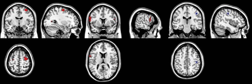
Research Jozarni J Dlabac-de Lange
deficits could contribute to negative symptoms, in recent years, investigating the effects of
as they hamper effective social interaction and treatment with antipsychotics (EUDRA-CT:
may enhance the likelihood of abstaining from 2007-002748-79) or transcranial magnetic
social interaction. On the other hand, negative stimulation (Dutch Trial Registry: NTR1261)
symptoms could enhance social cognitive on negative symptoms of schizophrenia [15,19].
deficits, as social withdrawal may negatively The fMRI data on the Wall of Faces task have
affect one’s proficiency in understanding others. not been published as yet. Patients (N=38) were
recruited from three regional mental health
Regarding the neurocircuitry of negative
care institutions (Lentis, GGz Drenthe and
symptoms in patients with schizophrenia,
GGz Friesland) and the University Medical
neuroimaging studies on reward and motivation
Center Groningen (UMCG). All patients that
found abnormalities in information processing
were included in the current study were 18
of the prefrontal cortex (PFC), the anterior
years or older and met the DSM-IV criteria
cingulate cortex (ACC) and the striatum [7-
for schizophrenia, which was confirmed by a
9]. Five parallel fronto-subcortical circuits link
Schedules for Clinical Assessment (SCAN 2.1)
regions of the frontal cortex to the striatum,
trained rater [20]. Severities of symptoms were
globus pallidus/substantia nigra, and thalamus.
assessed with the Positive and Negative Syndrome
These circuits mediate motivation, social
Scale (PANSS) [21] and the Montgomery
behavior, executive functions, motor and
Åsberg Depression Rating Scale (MADRS) [22].
oculomotor functions [10]. Thus, alterations in
Exclusion criteria were age 60 years,
the fronto-striato-thalamo-cortical circuitry may
rTMS and MRI contraindications, neurological
underlie or contribute to the development of
disorders, head injury with loss of consciousness in
negative symptoms of schizophrenia.
the past, substance dependency within the previous
Recently, there has been a shift in the approach 6 months, previous treatment with rTMS, severe
of negative symptoms. Originally, negative behavioral disorders, inability to provide informed
symptoms were thought to constitute one consent and pregnancy. Participants gave oral and
dimension. However, recent studies have found written consent after the procedure had been fully
two or more dimensions of negative symptoms explained. The studies were executed in accordance
[11-13]. Most of these studies have found two with the declaration of Helsinki and approved by a
factors of negative symptoms, namely expressive licensed local medical ethical committee (METC-
deficits and avolition/apathy/social emotional UMCG).
withdrawal [11,13,14]. These two factors
may have different underlying neural working Healthy controls (N=20) were matched to the
mechanisms [15-17]. These findings emphasize patients based on age, gender, education level
the importance of acknowledging the two factors and handedness. Level of education was defined
of negative symptoms. according to the scoring system of Verhage [23].
The healthy controls did not have a psychiatric
In this study, differences in brain activation history or any current psychiatric problems as
between patients with schizophrenia and measured by the mini-SCAN [24]. Differences
healthy controls during a social cognition in demographic characteristics between patients
task were explored. Furthermore, we explored and controls were tested with independent t-tests,
the differential correlates of the two negative except for gender and handedness, which were
symptom dimensions, namely expressive
tested with a Chi-square test for independence.
deficits and social-emotional withdrawal, with
brain activation during the task. The aim of Task design
this paper was to examine possible differences The Wall of Faces (WoF) task examines the neural
in the underlying neural working mechanisms substrates underlying emotional ambiguity [18].
of expressive deficits and social-emotional During this task, a group of faces is presented
withdrawal during a social cognition task that at once, and the dominant emotion or gender
measures emotional ambiguity [18]. has to be identified, in both ambiguous and
unambiguous situations. In healthy subjects,
Materials and Methods ambiguous emotional trials relative to ambiguous
gender trials have been shown to activate the
Subjects
ventromedial prefrontal cortex (VMPFC) and
Our analyses are performed with the baseline the ventral ACC [18]. Ambiguous relative to
data from two trials conducted at our center unambiguous trials activated the dorsal part
361 Neuropsychiatry (London) (2018) 8(1)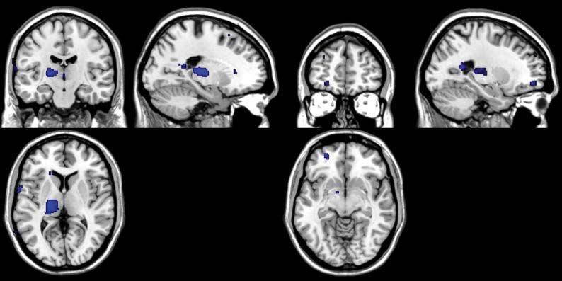
Neural Correlates of Emotional Ambiguity in Patients with Schizophrenia – Relationship with Expressive Research
Deficits
of the ACC, the dorsolateral prefrontal cortex negative symptoms [13]. The factor expressive
(DLPFC), the visual cortex and the posterior deficits consisted of PANSS items Flat affect
parietal cortex (PPC) [18]. (N1), Poor rapport (N3), Lack of spontaneity
Figure 1 shows a display screen of The Wall of (N6), Mannerisms and posturing (G5), Motor
Faces task. In each trial, an array of 32 emotional retardation (G7), and Avolition (G13). The
faces (i.e., angry or happy) was presented to social-emotional withdrawal factor consisted
a subject. The ratio of angry to happy faces of PANSS items Emotional withdrawal (N2),
(emotional trials, experimental condition) and Passive/apathetic social withdrawal (N4), and
male to female faces (gender trials, control Active social avoidance (G16) [13].
condition) varied and could be equal (ambiguous, Task performance was measured by comparing
16:16) or unequal (unambiguous, 26:6 or 6:26). reaction times (in seconds) and accuracy of the
In each trial, the array of faces was presented unambiguous trials (% correct). As there were
for the duration of 3 s with an additional 1.5 no correct responses in the ambiguous trials,
s response time. Participants were asked to since the distribution of faces were equal (i.e.,
identify the predominant emotion (experimental 16 versus 16), the response percentage of male
condition) or the predominant gender (control faces for the gender trials and angry faces for the
condition) intuitively, and not by counting of emotional trials were measured. Missing entries
the number of faces. During face presentation were not included in the analysis. Performance
and additional response time, the options “Angry was compared between patients and controls
- Happy” or “Female - Male” were displayed on using an independent samples t-test.
the screen. Blocks of 8 trials (48 s) started with
an instruction (“emotion” or “gender”) and Within the patient group, expressive deficits and
were interleaved with a rest condition (24 s). social-emotional withdrawal were correlated with
Emotion and gender blocks alternated. The task performance, controlling for level of education.
was presented using E-prime 1.2, which logged Also, correlation coefficients between expressive
timing of the task and responses of the subjects. deficits and social-emotional withdrawal on the
Subjects responded by button presses on an one hand and education level, MADRS, PANSS
MR-compatible button box using the index and positive subscale and percentage of estimated D2
middle finger of their right hand. receptor occupancy [25] on the other hand were
calculated.
Behavioral measures
Image acquisition
The PANSS was used to assess the two factors
of negative symptoms, expressive deficits and MRI scans were acquired with a Philips 3
social-emotional withdrawal. These two factors Tesla MRI scanner (Achieva Intera, Best, The
are based on an extensive factor analysis of the Netherlands), equipped with an 8-channel
PANSS, which revealed a two-factor structure of SENSE head coil. Movement was restricted
A B
Figure 1: Display screen of The Wall of Faces task. (a) In the experimental condition, subjects were asked to indicate whether there are more angry or happy
faces (emotion) (b) In the baseline condition, subjects were asked to indicate whether there are more male or female faces (gender).
362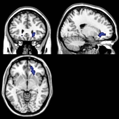
Research Jozarni J Dlabac-de Lange
by foam pads fixating the head, and noise was with nuisance factors was done separately for
reduced by earplugs and head phones. The task control and labeled images. After this, labeled
was presented on a screen visible via a mirror on images were subtracted from control images
top of the head coil. using spline interpolation of subsequent scans
in both image types separately [27,28]. The
During the task, a pseudo-continuous Arterial
subtracted ASL images were entered in a first
Spin Labeling (PCASL) sequence was acquired.
level analysis. The four task conditions and
As mentioned earlier, baseline data of two trials
an instruction condition (notifying task and
were used in this study. ASL was used because in
resting blocks) were modeled in a block design
these two trials a second scan was made after
convolved with a Hemodynamic Response
several weeks to assess treatment effect, and
Function. Implicit masking and high-pass
ASL is more suitable to compare measurements
filtering were not applied in the first-level
when they are repeated over time. In addition,
analysis, which are standard procedures for
ASL provides reliable absolute quantification
BOLD fMRI, but not for ASL. Instead, an
of cerebral blood flow, and it has higher spatial
explicit mask was used consisting of the gray
and temporal resolution than other techniques
and white matter of the segmented brain.
[26]. In comparison to a BOLD sequence,
Since the ASL images contained artifacts in the
ASL can examine baseline activity of the
highest and lowest planes (i.e., in the cranium,
brain instead of relative changes as measured
not in the brain tissue), these parts were
with BOLD. Furthermore, ASL has lower
excluded from the explicit mask. Contrasts
inter-subject variability and allows superior
were created of the emotional versus the gender
functional localization [27,28]. Control and
trials, the ambiguous versus the unambiguous
labeled scans (4 s; 127 of both) were alternated.
trials, for emotion-gender in ambiguity,
Labeling time was 1650 ms, delay time 1525
gender ambiguity, emotional ambiguity and
ms and acquisition time was 825 ms. Further
emotion-gender in unambiguity.
parameters: flip angle 90º, 14 slices, FOV (ap,
fh, rl)=224 × 98 × 224 mm, voxel size 1.75 × Since the ASL sequence changed during the
1.75 × 7 mm. study due to a scanner upgrade, the histogram
(i.e., image intensity range) of the contrast
Data analysis
images was found to be different. Equalization
Data were analyzed using in-home scripts of the intensity distribution of contrast-images
based on Statistical Parametric Mapping was applied by taking the 25% and 75% values
(SPM8; FIL Wellcome Department of Imaging of the cumulative histogram of the baseline beta-
Neuroscience, London, UK) routines and image (last column design matrix) and using
functions. First, raw PAR files were converted to these values for histogram normalization of
NIFTI format. Next, labeled and control images the contrast images. Fourteen controls and 16
were realigned separately, because intensity patients were scanned before the update and 6
differences between both image modalities may controls and 22 patients were scanned after the
cause spurious motion correction. Mean images update. The mean control image created during
of both realignments were created. The mean realignment was co-registered to the anatomy,
labeled image was co-registered to the mean and the anatomy and histogram-normalized
control image and the same parameters were contrast-images were normalized to the T1
applied to all labeled images. Images were then template of SPM.
smoothed with an 8 mm FWHM Gaussian
The normalized contrast images were entered
isotropic kernel.
in a second level analysis using Statistical non-
Due of the low signal-to-noise ratio of ASL data, Parametric Mapping (SnPM) [29]. Non-
nuisance factors were filtered from the data by parametric analyses were used since ASL data has
regression, including the motion parameters, a non-normal distribution. Analyses were done
white matter (WM) signals, and cerebrospinal with a variance smoothing of 8 mm FWHM,
fluid (CSF) signal. For the CSF and WM 5000 iterations, and no additional scaling. Out
signal, masks were created by co-registering the of brain voxels were removed by masking with
anatomy to the mean control image, which was the brain mask of SPM. Resulting pseudo-T
then segmented. The first principle components maps were inspected at a threshold of p20, pseudo-T>3 (pseudo-T threshold to
the functional image series by using these WM control for type-I errors), as is used in other ASL
and CSF masks. Regression of the ASL data functional MRI studies [15,30]. This pseudo-T
363 Neuropsychiatry (London) (2018) 8(1)Neural Correlates of Emotional Ambiguity in Patients with Schizophrenia – Relationship with Expressive Research
Deficits
threshold was used to correct for multiple in the patient group was larger, but again these
comparison, which has been suggested in case differences were not statistically significant. The
of non-homogeneous smoothness of the data percentage missing data for the unambiguous
(Tom Nichols, SPM mailing list Item #7573 (26 emotion trials was 2.6% for the control group
Nov 2001 17:56) - “Re: Cluster level statistics and 5.9% for the patient group (p=0.08), for
in SnPM, https://www.jiscmail.ac.uk/cgi-bin/ the ambiguous emotion trial 10.5% for the
webadmin?A0=spm)”. Contrasts of interest control group and 13.7% for the patient
group (p=0.35), for the unambiguous
in the final analyses of the Wall of Faces tasks
gender trial 2.3% for the control group and
were: 1) ambiguous emotion relative to the 6.4% for the patient group (p=0.23) and
unambiguous emotion, 2) ambiguous emotion for the ambiguous gender trial 4.9% for
relative to ambiguous gender and 3) ambiguous the control group and 11% for the patient
relative to unambiguous regardless of valence. group (p=0.13). Table 2 shows accuracy
for unambiguous trials and the response
First, activity of all contrasts was investigated in selection for ambiguous trials.
the healthy control sample and patient sample
using the one sample T-test option of SnPM.
Next, patients and controls were compared using Table 1: Demographic and baseline clinical characteristics.
the two sample T-test of SnPM. The effect Patients with Schizophrenia Healthy Controls P-value
of the two factors of negative symptoms on
Age (years) 32.42 (11.05) 31.10 (11.69) 0.67
brain activation during emotional appraisal in
patients with schizophrenia was investigated Gender (% male) 87 70 0.12
by regression of the PANSS expressive Handedness (% right)* 97 90 0.32
deficits and the PANSS social-emotional Education (Verhage) 4.89 (1.87) 5.7 (1.34) 0.09
withdrawal [13] to all contrasts of interest, Illness duration (years) 9.02 (9.57)
using the “MultiSub: Simple Regression; Current antipsychotics (%)
1 covariate of interest” function of SnPM. None 18.42
As negative symptoms may be secondary to Antipsychotic polypharmacy 26.32
depressive symptoms, positive symptoms Aripiprazole 15.79
and medication side effects, we repeated the
Bromperidol 2.63
analyses with the PANSS positive symptom
Clozapine 23.68
scale, the estimated percentage of D2 receptor
occupancy [25] and the Montgomery Åsberg Flupentixol 5.26
Depression Rating Scale (MADRS) total Haloperidol 2.63
score to control for potential confounding Olanzapine 28.95
variables. The MADRS data of one patient Paliperidone 2.63
was missing. Quetiapine 5.26
Risperidone 13.16
Results Zuclopentixole 7.89
PANSS scores
Demographics
Positive subscale 14.03 (4.77)
Data from 38 patients and 20 control subjects Negative Subscale 18.66 (4.35)
were included in the analyses. Demographic General psychopathology subscale 31.68 (7.99)
characteristics are shown in Table 1. The subject
Depression subscale (items G1, G2,
groups did not differ significantly in age, gender, G3, G6)
9.63 (4.08)
handedness or education level.
Expressive deficits (items N1, N3,
Behavioral results 14.34 (4.04)
N6, G5, G7 and G13)¹
In general, patients were less accurate and needed Social-emotional withdrawal
8.34 (2.99)
more time to respond, but these differences were (items N2, N4, G16)¹
not statistically significant, except for accuracy MADRS score 17 (9.92)
on gender unambiguous trials (p=0.02, effect Data are means (+/- SD) or percentages; PANSS, Positive and Negative Syndrome Scale; MADRS,
size Cohen’s d=0.61). Additionally, a larger Montgomery Åsberg Depression Rating Scale. *some data is missing.
percentage of patients did not press the button 1) Liemburg, E., Castelein, S., Stewart, R., van der Gaag, M., Aleman, A., Knegtering, H., et al. (2013)
box after a face presentation as compared to Two subdomains of negative symptoms in psychotic disorders: established and confirmed in
two large cohorts. Journal of psychiatric research, 47, 718-725.
controls and thus the percentage of missing data
364Research Jozarni J Dlabac-de Lange
Table 2: Accuracy for unambiguous trials, response selection for ambiguous trials of both patients and healthy controls
during the task.
Groups Mean SD P
Accuracy in percentage for emotion unambiguous trials (6 angry Healthy controls 92.5 8.3
0.64
and 26 happy or 6 happy and 26 angry faces) Patients 91.4 8.0
Accuracy in percentage for gender unambiguous trials (6 male and Healthy controls 98.0 4.3
0.02
26 female or 6 female and 26 male faces) Patients 93.4 9.6
Percentage angry faces for emotion ambiguous trials (16 angry and Healthy controls 52.7 8.5
0.66
16 happy faces) Patients 51.0 15.3
Percentage male faces for gender ambiguous trials (16 male and 16 Healthy controls 46.3 13.0
0.43
female faces) Patients 43.2 14.5
Data are means (+/- SD), presented for the two groups.
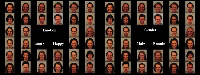
There were significant positive correlations Brain activation in patients with
between expressive deficits and reaction time schizophrenia as compared to healthy
on all trials; gender ambiguous trials (r=0.4, controls
p=0.016), gender unambiguous trials (r=0.39,
Patients with schizophrenia as compared to
p=0.018), emotional ambiguous trials (r=0.36,
controls showed higher activation in the right
p=0.027) and emotional unambiguous trials
DLPFC and the left precentral gyrus, and lower
(r=0.37, p=0.023). Also, there were significant
activation in the right postcentral gyrus of the
positive correlations between expressive deficits
parietal lobe during the ambiguous relative to
and accuracy on both unambiguous gender
unambiguous contrast, see Table 3 and Figure 2.
(r=0.36, p=0.031) and unambiguous emotional
The other contrasts did not result in significant
(r=0.34, p=0.038) trials. There was no significant
differences.
association between expressive deficits and level
of education. Regression analysis with the PANSS
expressive deficits
There were no significant correlations between
social-emotional withdrawal on the one hand For the ambiguous emotion relative to
and performance measures or level of education unambiguous emotion contrast, severity of
on the other hand. expressive deficits was negatively correlated with
brain activation in the left thalamus, the bilateral
precentral gyrus, the bilateral precuneus, the
Imaging results
right superior temporal gyrus and the left middle
One sample t-test control group frontal gyrus. For the same contrast, there was a
positive correlation between levels of expressive
Ambiguous emotion relative to unambiguous
deficits and brain activation in the right
emotion showed more brain activation in the left
postcentral gyrus. Figure 3 illustrates the effect
DLPFC, the left postcentral gyrus and the right of expressive deficits on brain activation during
middle occipital gyrus. Ambiguous emotion the emotional ambiguity contrast.
relative to ambiguous gender showed lower
brain activation in the left middle occipital In the ambiguous emotion relative to the
gyrus. The ambiguous relative to unambiguous ambiguous gender contrast, severity of expressive
contrast (across gender and emotion decision deficits was negatively correlated with activation
trials) demonstrated more brain activation in of the VMPFC (ventral anterior cingulate
the left cuneus and the right middle occipital and the medial prefrontal gyrus) and the left
gyrus. middle and superior temporal gyrus. Figure 4
illustrates the effect of expressive deficits on brain
One sample t-test patient group activation during the ambiguous emotion versus
The ambiguous relative to unambiguous ambiguous gender contrast.
contrast, regardless of valence, demonstrated Regression analysis of the ambiguous versus
higher levels of brain activation in the right unambiguous contrast revealed a negative
superior frontal gyrus. The other contrasts did correlation between expressive deficits and
not show significant differences in activation. brain activation in the right superior temporal
365 Neuropsychiatry (London) (2018) 8(1)Neural Correlates of Emotional Ambiguity in Patients with Schizophrenia – Relationship with Expressive Research
Deficits
Table 3: Areas (MNI coordinates) that show significant differences in activation between patients with schizophrenia and healthy
controls (p20, pseudo-T>3).
Contrast K Pseudo-T X Y Z Area
Ambiguous versus unambiguous 143 3,78 30 -8 62 right middle frontal gyrus
159 3,59 -60 8 14 left precentral gyrus
28 3,36 44 -22 46 right postcentral gyrus
Figure 2: Difference in brain activation between the healthy control group and the schizophrenia patient group during the ambiguous versus unambiguous
trails, red=patients-controls, blue=controls-patients, neurological format.
Figure 3: Relationship between expressive deficits and brain activation for the emotional ambiguity contrast, blue=negative association, neurological format.
gyrus and left cuneus, and a positive correlation withdrawal with brain activation was found.
between expressive deficits and brain activation
Depressive symptoms, positive
in the right postcentral gyrus. For an overview of
symptoms and D2 receptor occupancy
the MNI coordinates and areas see Table 4.
There was no significant correlation between
Regression analysis with the PANSS
the MADRS and the PANSS expressive deficits
social-emotional withdrawal
(r=0.05, p=0.76), the PANSS positive subscale
No significant association of social-emotional and the PANSS expressive deficits (r=0.084,
366Research Jozarni J Dlabac-de Lange
Figure 4: Relationship between expressive deficits and brain activation during the ambiguous emotion versus ambiguous gender contrast,
blue=negative association, neurological format.
Table 4: Areas (MNI coordinates) showing a significant association with expressive deficits of schizophrenia (p20,
pseudo-T>3).
Contrast K pseudo-t x y z Area
Ambiguous versus unambiguous 45 3,73 54 -20 58 Right Postcentral Gyrus
28 3,64 46 -46 16 Right Superior Temporal Gyrus
73 3,33 -4 -98 12 Left Cuneus
Affect versus gender in ambiguity 189 4,36 22 32 -4 Ventromedial Prefrontal Cortex
36 3,58 -56 -68 28 Left Middle Temporal Gyrus
20 3,57 -62 4 6 Left Superior Temporal Gyrus
Ambiguous emotion-unambiguous emotion 62 4,59 -62 4 8 Left Precentral Gyrus
490 4,21 -20 -16 6 Left Thalamus
193 3,83 -36 -74 42 Left Precuneus
61 3,82 46 -48 14 Right Superior Temporal Gyrus
26 3,73 -26 52 -8 Left Middle Frontal Gyrus
29 3,53 68 2 12 Right Precentral Gyrus
114 3,4 6 -74 46 Right Precuneus
26 3,61 52 -18 58 Right Postcentral Gyrus
p=0.62) and the estimated D2 receptor estimated D2 receptor occupancy and the PANSS
occupancy and the PANSS expressive deficits social-emotional withdrawal (r=0.18, p=0.29).
(r=0.092, p=0.58).
No significant association on the MADRS
There was a significant correlation between was found for the ambiguous emotion relative
the MADRS and the PANSS social-emotional to unambiguous emotion contrast. In the
withdrawal items (r=0.54, p=0.001) and the ambiguous emotion relative to ambiguous gender
PANSS positive subscale and the PANSS social- contrast, regression analysis showed a positive
emotional withdrawal items (r=0.34, p=0.037). relation between level of depressive symptoms
There was no significant association between the and brain activation in the right anterior
367 Neuropsychiatry (London) (2018) 8(1)Neural Correlates of Emotional Ambiguity in Patients with Schizophrenia – Relationship with Expressive Research
Deficits
cingulate, and a negative association between was associated with hypoactivation of the fronto-
depressive symptoms and brain activation in thalamic pathway for the emotional ambiguity
the left precuneus, the left posterior cingulate contrast. In addition, severity of expressive
and the right superior frontal gyrus. Regression deficits was also related to hypoactivation of
analysis of the ambiguous versus unambiguous the VMPFC, including the ventral ACC, in the
contrast found a negative association between ambiguous emotion versus ambiguous gender
depressive symptoms and brain activation in the contrast. Moreover, these findings appear to be
right caudate. independent of severity of depressive symptoms,
positive symptoms and percentage of estimated
Regression analysis of percentage of estimated
D2 receptor occupancy, as the regression analysis
D2 receptor occupancy found a negative relation
of the MADRS, PANSS positive subscale and
between levels of D2 receptor occupancy and
percentage of estimated D2 receptor occupancy
brain activation in the right supramarginal
showed a distinctive pattern of brain activation
gyrus of parietal lobe and right inferior frontal
that differed from the areas found for expressive
gyrus during the ambiguous emotion relative to
deficits. Furthermore, there was no significant
unambiguous emotion contrast. No significant
correlation between the MADRS, PANSS
association was found for the ambiguous emotion
positive subscale, the estimated D2 receptor
relative to ambiguous gender contrast. For the
occupancy on the one hand and the PANSS
ambiguous versus unambiguous contrast a negative
expressive deficits items on the other hand.
relation between estimated D2 receptor occupancy
and brain activation was found in the right inferior A key finding of our study concerns the
frontal lobe and the right temporal lobe. association of expressive deficits, but not social-
emotional withdrawal, with lower prefrontal and
Regression analysis of the PANSS positive
thalamic activation during ambiguous emotion
subscale revealed a positive relation between
perception. The association between higher
higher levels of positive symptoms and brain
levels of expressive deficits and hypoactivation
activation in the right middle frontal gyrus and
of the PFC is in line with earlier studies that
right posterior cingulate during the ambiguous
report on dysfunctioning of the PFC in patients
emotion relative to unambiguous emotion
with negative symptoms of schizophrenia [7].
contrast. In the ambiguous emotion relative to
Interestingly, a large study on patients with
ambiguous gender contrast, regression analysis
neurodegenerative diseases, such as Alzheimer
found a negative relation between positive
and frontotemporal dementia, found a poorer
symptoms and brain activation in the right
performance on an emotion expression tasks
superior temporal gyrus. Finally, the ambiguous
to be correlated with volume loss in prefrontal
versus unambiguous contrast revealed a positive
and thalamic regions in the brain [31].
relation between positive symptoms and brain
Hypoactivation of the fronto-thalamic circuit
activation in the left superior temporal gyrus.
during ambiguous social appraisal may imply
a reduced action readiness in social situations,
Discussion underlying expressive deficits. Indeed, behavioral
In this study, we investigated whether the two data showed a significant positive correlation
factors of negative symptoms of schizophrenia, between expressive deficits and reaction times,
namely expressive deficits and social-emotional implicating that patients with more expressive
withdrawal, were related to abnormalities in the deficits needed more time to respond than
neurocircuitry of emotion appraisal, using a task patients with lower levels of expressive deficits.
that probed emotional ambiguity in a group of In contrast, no significant correlations between
different faces. To our knowledge, this is the the behavioral data and levels of social-
first study to investigate the neural correlates of emotional withdrawal were found. Earlier
emotional decision making under uncertainty in studies have found social-emotional withdrawal
schizophrenia. We compared brain activation to be associated with reduced frontoparietal
of healthy controls with that of patients with activation during a planning task [15] and
schizophrenia, and related levels of expressive apathy/avolition/emotional withdrawal but not
deficits and social-emotional withdrawal with expressive deficits to be associated with reduced
brain activation. Ambiguity, regardless of valence, ventral striatal activation during a monetary
activated the fronto-parietal circuitry differently reward task [32]. It is possible that social-
in patients with schizophrenia as compared to emotional withdrawal is related to anticipatory
healthy controls. Severity of expressive deficits pleasure deficits [33], which cannot be detected
368Research Jozarni J Dlabac-de Lange
with the Wall of Faces task, as it is not designed in patients with more expressive deficits may
for this purpose. In conclusion, the present task negatively affect interactions.
may be more sensitive for brain circuits involved
A limitation of this study could be the artificial
in expressive deficits, which further supports
nature of the stimulus display (thirty-two faces on
the neuroanatomical differentiation of the two
one screen), as people are not presented with an
factors of negative symptoms.
array of individual pictures of faces in daily life.
A previous study conducted among healthy Indeed, impairments may become more apparent
volunteers found that ambiguous emotion when using stimuli that better approximate real-
relative to ambiguous gender contrast activated world contexts [38]. Future studies could use
brain areas that seem to be involved in emotional more dynamic and realistic images of real-life
processing and emotion recognition, such as social situations to further elucidate the neural
the VMPFC (ventral ACC and the ventral circuitry of emotion processing in social contexts.
medial PFC), the right superior temporal Another limitation includes the chronic use of
gyrus and the right supramarginal gyrus [18]. antipsychotic medication in the patient group
We did not replicate these findings in our as compared to controls. Antipsychotics alter
healthy control group. This may imply that the neuronal function, and up to date it is unclear
contrast does not reliably isolate brain regions how chronic use of antipsychotic medication
associated with emotional processing, maybe influences brain function [39]. A final limitation
because both contrasts contain human faces is the use of ASL, which is not used as frequently
that contain social-emotional cues or because as BOLD fMRI, and thus replication of these
the task instruction was geared towards a findings may be relatively more difficult.
group-level judgment. Alternatively, statistical
power could play a role as well as signal loss in Prefronto-striato-thalamic functional
ventral prefrontal areas. However, regression dysconnectivity seems to be implicated in the
analysis of the same contrast in the patient group pathophysiology of schizophrenia [40]. However,
revealed higher levels of expressive deficits to be there may be intercultural differences in social
associated with hypoactivation of the VMPFC. processes and it would be interesting to examine
Thus, impairment in the neurocircuitry of these intercultural differences in social cognitive
emotional processing may specifically emerge in processes in patients with schizophrenia. Also,
association with impairments in social cognition environmental factors [41,42], dietary influence
in schizophrenia with expressive deficits. [43,44] or stress-induced lifestyle condition may
influence brain function and future studies may
Interestingly, the correlation analysis within the
want to investigate this.
patient group found patients with higher levels of
expressive deficits to have greater reaction times, In conclusion, fronto-thalamic dysfunctioning
but also greater accuracy. This is in line with a during an ambiguous emotional appraisal
previous study that found severity of symptoms task was associated with expressive deficits but
in schizophrenia contributed relatively more not with social-emotional withdrawal. Thus,
to the variance in speed of emotion processing the two factors of negative symptoms seem to
than to the variance in accuracy [34]. Indeed, have different underlying neural mechanisms.
emotional expressions bring forth quick The alterations found could contribute to
responses and often this includes imitation of the increase our understanding of the neural basis
emotion in the observed face [35]. Earlier studies of social dysfunctioning as a characteristic of
have found impairments in emotional face expressive deficits, typically seen in patients with
imitation in patients with schizophrenia [36,37] schizophrenia.
and this impairment was not accompanied by
impairment in the identification of emotional
expressions [37]. In other words, impairment Acknowledgement
in emotional processing may not necessarily be We would like to thank all the participants of this
accompanied by impairment in accuracy during study.
identification of emotional faces. The ability
to quickly process social stimuli is essential for
Funding
social interactions, and it has been suggested that
decreased speed in processing social stimuli may The study on treatment of negative symptoms
lead to deficits in social functioning [3]. Thus, of schizophrenia with transcranial magnetic
the compromised emotional processing speed stimulation (Dutch Trial Registry: NTR1261)
369 Neuropsychiatry (London) (2018) 8(1)Neural Correlates of Emotional Ambiguity in Patients with Schizophrenia – Relationship with Expressive Research
Deficits
was supported by means of an unconditional Compliance with Ethical Standards
research grant from AstraZeneca and an
Ethical approval: All procedures performed in
unconditional research grant from Stichting
studies involving human participants were in
Roos. Author A.A. was supported by a VICI
accordance with the ethical standards of the
grant from N.W.O., grant number 435-11-004
institutional and/or national research committee
and with the 1964 Helsinki declaration and
Conflict of Interest its later amendments or comparable ethical
Henderikus Knegtering, MD, PhD, is on the standards. This article does not contain any
speakers’ list of and/or has received unconditional studies with animals performed by any of the
grants from Janssen, Eli Lilly, Bristol Meyers authors.
Squibb and Astra Zeneca. André Aleman received Informed consent: Informed consent was
speaker fees from Lundbeck. All other authors obtained from all individual participants
declare that there are no conflicts of interests. included in the study.
References 11. Blanchard JJ, Cohen AS. The structure of 20. Giel R, Nienhuis FJ. SCAN-2.1: Schedules for
negative symptoms within schizophrenia: clinical assessment in neuropsychiatry (in
1. Penn DL, Sanna LJ, Roberts DL. Social cogni- Implications for assessment. Schizophr. Bull dutch). Geneva/Groningen: WHO (1996).
tion in schizophrenia: An overview. Schizophr. 32(2), 238-245 (2006).
Bull 34(3): 408-411 (2008). 21. Kay SR, Fiszbein A, Opler LA. The positive and
12. Peralta V, Cuesta MJ. Negative symptoms in negative syndrome scale (PANSS) for schizo-
2. Aleman A, Kahn RS. Strange feelings: Do schizophrenia: A confirmatory factor anal- phrenia. Schizophr. Bull 13(2):261-276 (1987).
amygdala abnormalities dysregulate the ysis of competing models. Am. J. Psychiatry
emotional brain in schizophrenia? Prog. Neu- 152(10), 1450-1457 (1995). 22. Montgomery SA, Asberg M. A new depression
robiol 77(5), 283-298 (2005). scale designed to be sensitive to change. Br. J.
13. Liemburg E, Castelein S, Stewart R, et al. Two Psychiatry 134(1), 382-389 (1979).
3. Couture SM, Penn DL, Roberts DL. The subdomains of negative symptoms in psy-
functional significance of social cognition in chotic disorders: Established and confirmed 23. Verhage F. Revised scoring method. (1983).
schizophrenia: A review. Schizophr. Bull 32 in two large cohorts. J. Psychiatr. Res 47(6), 24. Nienhuis FJ, van de Willige G, Rijnders CA,
Suppl 1: S44-63 (2006). 718-725 (2013). et al. Validity of a short clinical interview for
4. Kohler CG, Walker JB, Martin EA, et al. Facial 14. Messinger JW, Tremeau F, Antonius D, et al. psychiatric diagnosis: The mini-SCAN. Br. J.
emotion perception in schizophrenia: A Avolition and expressive deficits capture neg- Psychiatry 196(1), 64-68 (2010).
meta-analytic review. Schizophr. Bull 36(5), ative symptom phenomenology: Implications 25. Lako IM, van den Heuvel ER, Knegtering H,
1009-1019 (2010). for DSM-5 and schizophrenia research. Clin. et al. Estimating dopamine D(2) receptor
5. Gur RE, Loughead J, Kohler CG, et al. Limbic Psychol. Rev 31(1), 161-168 (2011). occupancy for doses of 8 antipsychotics: A
activation associated with misidentification of 15. Liemburg EJ, Dlabac-De Lange JJ, Bais L, et al. meta-analysis. J. Clin. Psychopharmacol 33(5),
fearful faces and flat affect in schizophrenia. Neural correlates of planning performance 675-681 (2013).
Arch. Gen. Psychiatry 64(12),1356-1366 (2007). in patients with schizophrenia - relationship 26. Borogovac A, Asllani I. Arterial spin labeling
6. Ventura J, Wood RC, Jimenez AM, et al. with apathy. Schizophr. Res 161(2-3), 367-375 (ASL) fMRI: Advantages, theoretical constrains
Neurocognition and symptoms identify links (2015). and experimental challenges in neuroscienc-
between facial recognition and emotion 16. Kirschner M, Hager OM, Bischof M, et al. es. Int. J. Biomed. Imaging 2012(1), 818456
processing in schizophrenia: Meta-analytic Ventral striatal hypoactivation is associated (2012).
findings. Schizophr. Res 151(1-3), 78-84 (2013). with apathy but not diminished expression 27. Liu TT, Wong EC. A signal processing model
7. Goghari VM, Sponheim SR, MacDonald AW. in patients with schizophrenia. J. Psychiatry. for arterial spin labeling functional MRI. Neu-
The functional neuroanatomy of symptom di- Neurosci 40(5), 140383 (2015). roimage 24(1), 207-215 (2005).
mensions in schizophrenia: A qualitative and 17. Mucci A, Dima D, Soricelli A, et al. Is avolition 28. Wang Z, Aguirre GK, Rao H, et al. Empirical
quantitative review of a persistent question. in schizophrenia associated with a deficit of optimization of ASL data analysis using an
Neurosci. Biobehav. Rev 34(3), 468-486 (2010). dorsal caudate activity? A functional magnet- ASL data processing toolbox: ASLtbx. Magn.
8. Harvey PO, Armony J, Malla A, et al. Function- ic resonance imaging study during reward Reson. Imaging 26(2), 261-269 (2008).
al neural substrates of self-reported physical anticipation and feedback. Psychol. Med 45(8),
1765-1778 (2015). 29. Nichols TE, Holmes AP. Nonparametric per-
anhedonia in non-clinical individuals and in
mutation tests for functional neuroimaging:
patients with schizophrenia. J. Psychiatr. Res 18. Simmons A, Stein MB, Matthews SC, et al. A primer with examples. Hum. Brain. Mapp
44(11), 707-716 (2010). Affective ambiguity for a group recruits 15(1), 1-25 (2002).
9. Juckel G, Schlagenhauf F, Koslowski M, et al. ventromedial prefrontal cortex. Neuroimage
29(2), 655-661 (2006). 30. Henriksen OM, Jensen LT, Krabbe K, et al. Rest-
Dysfunction of ventral striatal reward pre-
ing brain perfusion and selected vascular risk
diction in schizophrenia. Neuroimage 29(2), 19. Dlabac-de Lange JJ, Bais L, van Es FD, et al. factors in healthy elderly subjects. PLoS. One
409-416 (2006). Efficacy of bilateral repetitive transcranial 9(5), e97363 (2014).
10. Tekin S, Cummings JL. Frontal-subcortical magnetic stimulation for negative symptoms
of schizophrenia: Results of a multicenter 31. Gola KA, Shany-Ur T, Pressman P, et al. A neu-
neuronal circuits and clinical neuropsychiatry:
double-blind randomized controlled trial. ral network underlying intentional emotional
An update. J Psychosom. Res 53(2), 647-654
Psychol. Med 1-13 (2014). facial expression in neurodegenerative dis-
(2002).
ease. Neuroimage. Clin 14(1), 672-678 (2017).
370Research Jozarni J Dlabac-de Lange
32. Hartmann MN, Hager OM, Reimann AV, et phrenia. Psychiatry. Res 145(2-3), 87-94 41. Mucci A, Galderisi S, Green MF, et al. Fa-
al. Apathy but not diminished expression in (2006). milial aggregation of MATRICS consensus
schizophrenia is associated with discount- cognitive battery scores in a large sample
ing of monetary rewards by physical effort. 37. Park S, Matthews N, Gibson C. Imitation, of outpatients with schizophrenia and their
Schizophr. Bull 41(2), 503-512 (2015). simulation and schizophrenia. Schizophr. unaffected relatives. Psychol. Med 1-10
Bull 34(4),698-707 (2008). (2017).
33. Gard DE, Kring AM, Gard MG, et al. Anhedo-
nia in schizophrenia: Distinctions between 38. Sasson NJ, Pinkham AE, Weittenhiller LP, et 42. Martins I. Increased risk for obesity and
anticipatory and consummatory pleasure. al. Context effects on facial affect recogni- diabetes with neurodegeneration in
Schizophr. Res 93(1-3), 253-260 (2007). tion in schizophrenia and autism: Behav- developing countries. J. Mol. Genet. Med S1,
ioral and eye-tracking evidence. Schizophr. 001 (2013).
34. Barkhof E, de Sonneville LM, Meijer CJ, et al. Bull (2015).
Processing of facial and nonsocial informa- 43. Alam R, Abdolmaleky HM, Zhou JR. Microbi-
tion is differentially associated with severity 39. Handley R, Zelaya FO, Reinders AA, et al. ome, inflammation, epigenetic alterations
of symptoms in patients with multiepisode Acute effects of single-dose aripiprazole and mental diseases. Am. J. Med. Genet.
schizophrenia. J. Nerv. Ment. Dis 203(2), and haloperidol on resting cerebral blood B. Neuropsychiatr. Genet 174(6), 651-660
112-119 (2015). flow (rCBF) in the human brain. Hum. Brain. (2017).
Mapp 34(2), 272-282 (2013).
35. Frith C. Role of facial expressions in social 44. Martins IJ. Magnesium therapy prevents
interactions. Philos. Trans. R. Soc. Lond. B. 40. Vandevelde A, Leroux E, Delcroix N, et al. senescence with the reversal of diabetes
Biol. Sci 364(1535), 3453-3458 (2009). Fronto-subcortical functional connectivity and Alzheimer’s disease. Health 8, 694-710
in patients with schizophrenia and bipolar (2016).
36. Schwartz BL, Mastropaolo J, Rosse RB, et al. disorder during a verbal fluency task. World.
Imitation of facial expressions in schizo- J. Biol. Psychiatry 2017(1):1-9.
371 Neuropsychiatry (London) (2018) 8(1)You can also read