Sandfly-Borne Viruses of Demonstrated/Relevant Medical Importance - IntechOpen
←
→
Page content transcription
If your browser does not render page correctly, please read the page content below
Chapter

Sandfly-Borne Viruses of
Demonstrated/Relevant Medical
Importance
Nazli Ayhan and Remi N. Charrel

Abstract

    Sandflies show distribution in a vast geographical area from Europe to Asia,
Africa, Australia, and Central and South America where they can transmit a large
number of viruses. Between these viruses, the most important are grouped into
the Phlebovirus genus (family Phenuiviridae). Among them, several sandfly-borne
phleboviruses cause self-limiting febrile disease (sandfly fever) or central and
peripheral nervous system infections. Data concerning the geographic distribution of
these phleboviruses has drastically increased during the last decade in both the new
and the old worlds. The current situation depicts a high viral diversity with taxonomic
groups containing human pathogenic and non-pathogenic viruses. This merits to
provide insight to address the question of medical and veterinary public health impact
of all these viruses, which are poorly studied. To do so, integrated and translational
approaches must use ecological, epidemiological, serological and direct clinical evi-
dence. Beside, other viruses transmitted by sandflies and belonging to Rhabdoviridae
and Reoviridae families can also be of veterinary and public health importance. The
chapter aims to provide a comprehensive view of the sandfly-borne viral pathogens of
the public health impact on humans and other vertebrates in the old and new worlds.

Keywords: sandfly-borne phleboviruses, sandfly fever, phlebovirus, Toscana
virus, Sandfly fever Naples virus, Sandfly fever Sicilian virus, Punta Toro virus,
Vesiculovirus, Chandipura virus, Changuinola virus

1. Introduction

    Sandflies are present in tropical and subtropical, arid and semi-arid areas
and temperate zones including southern Europe, Asia, Africa, Australia, Central
and South America. Phlebotomine sandflies are tiny diptera insects grouped in
the family Psychodidae, subfamily Phlebotominae. To date, over 800 species are
estimated to exist in different regions of the world [1]. Two genera (Phlebotomus
and Sergentomyia) of Phlebotominae are mostly recorded in the old world (OW)
and the other genus Lutzomyia exists in the new world (NW) [2]. Only females are
hematophagous and require a blood meal to develop their eggs. Sandflies take blood
from a wide range of animals such as cold-blooded vertebrates, mammals and birds;
trophic preferences vary depending on the sandfly species.
    Of the 800 sandfly species, at least 98 are proven or suspected vectors of micro-
organisms capable to cause parasitic, viral or bacterial diseases in vertebrates [1].

1
Vectors and Vector-Borne Zoonotic Diseases
This chapter will focus essentially on sandfly-borne viruses, which have been
proven agents of diseases in humans.
The arthropod-borne diseases including sandfly-borne viral diseases affect
urban, peri-urban, and rural population but mostly the communities with poor
living conditions. Economic, social and ecological conditions have a huge impact
on sandfly-borne viral diseases [3, 4]. The factors that described as associated with
arthropod-borne diseases emergence or invasion are (i) competent vector and ver-
tebrate host population repeatedly in contact within an appropriate environment,
(ii) vertebrate or vector host species composition changes, (iii) environmental or
niche changes and (iv) genetic changes [5].
Although sandflies can transmit a number of arthropod-borne viruses within
the families Phenuiviridae, Reoviridae and Rhabdoviridae, they remain neglected
vectors of viral diseases in contrast with a high interest for parasitic diseases such
as leishmaniasis. The three virus families contain human/animal pathogens. In the
Rhabdoviridae family, attention will be given to Chandipura virus; in the Reoviridae
family, we will focus on Changuinola virus [2, 6–8]. In the Phenuiviridae family, we
will focus on Sandfly fever Sicilian virus, Sandfly fever Naples virus, Toscana virus,
Adria virus and Punta Toro virus (PTV).
2. Sandfly-borne phleboviruses
Phleboviruses are enveloped viruses with single-stranded trisegmented
RNA. They contain three genomics segments: L (Large) segment encodes the viral
RNA polymerase (RdRp), M (medium) segment encodes envelope glycoproteins
(Gn and Gc) and non-structural protein m (NSm) and S (small) segment encodes
nucleocapsid protein (N) and non-structural protein s (NSs) [9].
Currently, 10 species within the genus Phlebovirus are recognized by the
International Committee on Taxonomy of Viruses (ICTV): Sandfly fever Naples
virus, Salehabad virus, Rift Valley fever virus, Uukuniemi virus, Bujaru virus, Candiru
virus, Chilibre virus, Frijoles virus, Punta Toro virus, and Severe fever with thrombocy-
topenia syndrome virus. Of interest, almost 40 phleboviruses are still listed as tenta-
tive species for which the ICTV has not officially ruled; interestingly, Sandfly fever
Sicilian virus still belongs to this pending group although discovered in 1943 [10].
Phleboviruses can be detected and isolated from blood-sucking female sandflies
and from non-blood-sucking males in equal proportions [11–14]. This suggests that
alternative transmission pathways (other than blood-borne from vertebrate reservoir)
such as transovarial transmission (female to offsprings) and/or venereal transmission
play an important role in the natural cycle [2]. Experimental results done with colo-
nized P. papatasi sandflies proved venereal virus transmission and transovarial virus
transmission [15]. Viral maintenance during the diapausing period of Phlebotomus
perniciosus larvae was proved and was not affected by transstadial transmission in
laboratory [15]. These routes of virus transmission suggest that phleboviruses can be
sustainably transmitted from one generation of sandflies to the next generation. It
also raises the question of whether a vertebrate host acting as reservoir is required or
not for virus perpetuation. To date, these experiments have been performed primar-
ily with high passage colonies and should be taken with caution because laboratory
reared sandflies may behave differently from wild populations. In addition, they
have been performed with few species of phlebovirus and with P. papatasi and P.
perfiliewi only. Elucidation of phlebovirus maintenance and transmission is crucial to
understand better the natural history of these viruses and to develop adapted method
to combat those which are human pathogens [15–17]. Although several sandfly-borne
phlebovirus species were isolated from humans, bats and sandflies [18–23], there is no
2Sandfly-Borne Viruses of Demonstrated/Relevant Medical Importance
DOI: http://dx.doi.org/10.5772/intechopen.81023
undisputable evidence that vertebrates play an important role in the natural history of
sandfly-borne phleboviruses other that as dead-end hosts.
With recently discovered novel viruses, the geographic distribution of phleboviruses
has drastically increased in both the new and the old worlds. The current situation
depicts a high viral diversity with taxonomic groups containing pathogenic and non-
pathogenic viruses. This merits to provide insight to address the question of medical and
veterinary public health impact of all these viruses, which are poorly studied.
2.1 Sandfly-borne phleboviruses in the old world
In the old world (OW), the risk for the infection with sandfly-borne phlebovi-
ruses is high depending upon the presence and the density of vectors [24].
Historic and recent epidemics have been caused by sandfly-borne phleboviruses
in the OW. In 1937, a massive outbreak occurred in, Athens, Greece [25, 26]. During
World War II (WWII), outbreaks were described among out-comer soldiers in the
Mediterranean basin and Middle East (the Austrian Commission in Balkan coun-
tries, British and German troops in the Mediterranean area) [17, 26, 27].
After WWII, sandfly fever epidemics were reported in Belgrade, Serbia, where
thousands were sick [28], with subsequent spread into other regions of the Balkans
[29–32]. More recently, large epidemics were recorded in Cyprus, Iraq, Turkey and
Ethiopia [33–36].
In addition, during the last two decades, an impressive number of novel phlebo-
viruses was either isolated or detected by molecular techniques in France, Italy,
Portugal, Greece, Albania, Croatia, Bosnia Herzegovina, Turkey, Iran, Tunisia,
Algeria and Morocco [11–14, 23, 37–43]. Accordingly, the Mediterranean area
witnesses a very high diversity of phleboviruses transmitted by sandflies [44]. This
situation has raised the public health concerns in southern Europe, North Africa
and in the Middle East [11, 13, 14, 39, 41, 45, 46].
OW sandfly-borne phleboviruses can be classified into three serological com-
plexes, which are also regarded as taxonomic species, Salehabad species, Sandfly
fever Naples species and Sandfly fever Sicilian tentative species.
Sandfly fever Sicilian and Sandfly fever Naples viruses cause fever, also known as
“sandfly fever”, “Pappataci fever” or “three-day fever”. It is not possible to distin-
guish Sandfly fever Sicilian virus infection from Sandfly fever Naples virus infection
based on clinical signs, which are virtually identical. They both cause abrupt illness
with fever, headache, malaise, photophobia, myalgia and retro-orbital pain usually
lasting 2–3 days after 3–5 day incubation [47].
Toscana virus, which belongs to the Sandfly fever Naples species, is so far the
most pathogenic sandfly-borne phlebovirus due to its propensity to affect the
central nervous system (CNS) and cause meningitis and meningoencephalitis [45].
Recently, Adria virus, identified in a case of meningitis, is the first virus belonging
to the Salehabad species to display human pathogenesis [38].
2.1.1 Sandfly fever
Before WWII, the knowledge on sandfly fever was limited to clinical and
epidemiological grounds. It was known that the fever caused by a filterable agent
and transmitted by Phlebotomus papatasi sandflies [48, 49]. Early assumptions
claimed that sandfly fever might be caused by distinct agents or viruses, despite it
was impossible to distinguish them from the clinical symptoms [47, 50].
Between 1934 and 1939, human sera samples from sandfly fever virus-infected
individuals (presumably containing the infectious agent) were inoculated into
rhesus monkeys which presented with febrile illness [51]. Inoculation of infectious
3Vectors and Vector-Borne Zoonotic Diseases
human serum (i) into chick embryos showed lesions on the chorioallantoic mem-
brane, whereas (ii) no clinical sign were noticed after inoculation to guinea pigs,
rabbits or dogs [47]. Three out of four human volunteers without a previous sandfly
fever history developed the typical symptoms and fever after inoculation of 1 ml
of the pool of acute sandfly fever serum [47]. Subsequently, Phlebotomus papatasi,
Culex pipiens and Pulex irritans fed on sandfly fever acutely infected volunteers,
only P. papatasi was able to transmit the disease to naïve volunteers [47]. In 1937, a
massive outbreak occurred in Athens, Greece [26]. However, most of the outbreaks
occurred in non-native persons having entered the endemic area for the first time
recently such as soldiers [13]. During WWII, several outbreaks of sandfly fever
knocked down battalions of soldiers in both the Allied and Axis troops which were
stationed in the Middle East, the Mediterranean and North Africa [17, 52, 53]. The
suspected variety of causing agents was shown through isolation of two different
viruses names Naples and Sicilian virus from sick soldiers in southern Italy [47, 53].
The antigenic differences between Naples virus and Sicilian virus were confirmed
by human cross-immunity test, neutralization and complement fixation test [54].
2.1.2 Sandfly fever Naples virus
Sandfly fever Naples virus was first isolated from blood of a febrile soldier who
became ill when stationed in Naples, Italy in 1944 [47]. Afterward, Naples virus was
isolated again (i) from febrile patients in Egypt, Turkmenia, Pakistan, Italy, Cyprus
and India [55–61] (ii) and from sandflies in Egypt (P. papatasi), in Italy (P. perniciosus)
and Serbia (P. perfiliewi) [19, 59, 62]. This was the first clue that Naples virus could
be transmitted by distinct vector species. A large and seminal neutralization-based
seroprevalence study, performed by Tesh et al. in 1976, showed that Naples virus was
likely to have a much wider distribution than initially believed from virus isolation
reports [24]; indeed, neutralizing antibodies were detected in human populations from
Bangladesh, Ethiopia, Greece, Iraq, Morocco, Saudi Arabia, Sudan, Djibouti, Turkey
and former Yugoslavia [24]. Highest rates (55–62%) were observed in Egypt, former
Yugoslavia (now Croatia), and Turkey. Another study reported neutralizing antibodies
in populations living in Turkmenia, Tajikistan, Uzbekistan, and Moldavia [63]. Clearly,
Naples virus circulation has drastically decreased after the 1980s, and the absence of
virus isolation or PCR detection, despite an increasing number of studies conducted in
previously endemic areas, question whether Naples virus has gone extinct or not [58].
2.1.2.1 Toscana virus
Toscana virus (TOSV) was first isolated from P. perniciosus in central Italy in
1971 [17]. It has taken 12 years to recognize that TOSV was capable to infect humans
and was able to cause not only sandfly fever, but also more severe infections char-
acterized by central nervous system (CNS) manifestations such as meningitis and
encephalitis. The first cases pointing out that Toscana virus causes CNS infections
came from two travelers returning from Italy and Portugal to the United States and
Sweden, respectively [64, 65]. This underlines the importance of travel-related
medicine in the surveillance of infectious diseases, particularly vector-borne infec-
tious diseases. Most of the Toscana virus case records are coming from important
or autochthonous human cases from the Mediterranean basin countries [45, 70].
Autochthonous cases in humans have been reported in Italy [71], Greece [72],
Cyprus [73], Croatia [74], Turkey [75, 76], Portugal [77] and France [78]. However,
these cases account for a minimal proportion of literature-described cases. In
most countries, where TOSV is endemic, it is not a notifiable disease; this together
with the absence of pathognomonic clinical sign, and the very limited number of
4Sandfly-Borne Viruses of Demonstrated/Relevant Medical Importance
DOI: http://dx.doi.org/10.5772/intechopen.81023
commercially available diagnostic assay may explain why autochthonous cases are
drastically under detected, and that most of reported cases have affected travel-
ers, the diagnosis of which is done when returning to their homeland. TOSV cases
in travelers have been reported from Italy [66], France [67], Spain [68, 69] and
Portugal [64], but also from the Mediterranean islands such as Cyprus [73], Elba
[79, 80], Sicily [81] and Sardinia [82].
Seroepidemiological studies showed the presence of neutralizing antibodies
against Toscana virus in several Mediterranean countries, however, the rates vary
depending on the region Mediterranean basin considered as endemic region of
Toscana virus [45, 71, 74, 78, 83–88].
Special attention must also be brought to the technique used for serology, since
results can greatly vary due to different levels of cross-reactivity depending on
the assay; for instance, the most stringent technique is based on neutralization
assays whereas ELISA or immunofluorescence techniques are more prone to cross-
reactivity between phleboviruses within the same antigenic group, but also between
antigenically distinct phleboviruses [88–90].
The geographic distribution of sandfly-borne phleboviruses can also be mea-
sured by surveillance of non-human vertebrates such as domestic animals: such
studies have demonstrated that TOSV was actively circulating in Portugal, Greece,
Cyprus and Algeria [83–85] from the study of dog sera, and in Kosovo from study-
ing cow and sheep sera [86]. TOSV was also isolated and/or detected in different
phlebotomine species such as P. perniciosus, P. perfiliewi, P. longicuspis, P. sergenti,
P. neglectus and Sergentomyia minuta in Italy, France, Spain, Croatia, Morrocco,
Tunisia, Algeria and Corsica [13, 18, 82, 87, 91–96]. These results tend to suggest
that TOSV can be transmitted by species other that P. perniciosus and P. perfiliewi.
Such data are compatible with the fact that TOSV might be more widely dispersed
than believed from the early studies. Of course, this merit to be further investi-
gated through experimental studies addressing competence of these species for
TOSV. TOSV belongs to the Sandfly fever Naples species.
To date, three genetic groups of TOSV have been recognized, and they are called
lineages A, B and C. Although only one lineage has been identified in a given coun-
try, the co-circulation of two lineages has been shown in France, in Turkey, and in
Croatia. It is possible that different lineages are transmitted by the same sandfly spe-
cies and that sympatry may be frequent [91, 93, 97]. Recent Toscana virus antibody
characterization assay performed with 41 patients diagnosed with Toscana virus
meningitis of meningoencephalitis found that specific IgM titers were high during
acute infection up to day 30, the presence of IgM antibodies lasts up to 6 months
after acute infection in 71% of cases, however IgG antibodies against Toscana virus
persisted at least 2 years in the patients, which gets in line with the fact that TOSV
infection is associated with long-term, maybe lifelong immunity [88]. There is accu-
mulating evidence that TOSV is one important cause of meningitis and encephalitis
during the warm season and that it should be included in the panel of microorgan-
isms to be systematically tested in clinical microbiology laboratory for patients
presenting with febrile illness, CNS and peripheral nervous system manifestations.
2.1.3 Sandfly fever Sicilian virus
Sandfly fever Sicilian virus (SFSV) was first isolated, characterized and named
Sicilian virus, from the serum of a US soldier, presenting with sandfly fever when he
was stationed in Palerma (Sicily) after the landing of the Allied army forces in Italy,
in 1943 during WWII [47]. Almost simultaneously, it was also described in sick US
soldiers stationed in Egypt. Subsequent studies allowed isolation of SFSV in Egypt,
India, Iran, Pakistan and Afghanistan [56, 98–100].
5Vectors and Vector-Borne Zoonotic Diseases
Accumulating direct (virus isolation or molecular detection) and indirect (sero-
prevalence studies) data allowed to list the following countries as areas where SFSV
was circulating: Bangladesh, Greece, Cyprus, Iraq, Morocco, Saudi Arabia, Somalia,
Ethiopia, Sudan, Tunisia, Turkey, Turkmenia, Tajikistan, Uzbekistan, Azerbaijan,
Moldavia, Croatia, Kosovo, France and Portugal [24, 33, 34, 61, 63, 83, 86, 101–103].
Beside the outbreaks described in the Allied and Axis forces during WWII, more
recent epidemics were reported in Cyprus, in Turkey and in Ethiopia caused by
genetic variants [29–36, 104]. Recent seroprevalence studies provided evidence
that SFSV and its genetic variants were still actively circulating in Greece, Cyprus,
Portugal and Kosovo [83, 84, 86]. Although Phlebotomus papatasi, Phlebotomus
ariasi and P. major complex were indisputably identified as SFSV vectors, transmis-
sion might also be done by phlebotomies belonging to other species [20, 100].
2.1.4 Adria virus
Adria virus was first detected in 2005 from field-collected sandflies in Albania
[39]. Genetic data consisting of partial sequence in the polymerase gene showed that
Adria virus is much closely related with viruses belonging to the Salehabad species
than with other phleboviruses belonging to the Sandfly fever Naples or to the Sandfly
fever Sicilian species. In 2009, a 30-month-old patient was admitted to hospital in
Greece for fever and seizure during summertime; his blood was tested positive for
the presence of phlebovirus RNA, whose sequence was most closely related with
Adria virus sequence [23]. Adria virus is the first, and so far the only member of the
Salehabad species to be associated with human disease. Interestingly, the number
of viruses identified in this species has drastically increased during the last decade.
Thus efforts should now be deployed to investigate to what extent, Adria virus in
particular but also other newly recognized Salehabad viruses have a medical impact.
2.1.5 Other phleboviruses
The last decade has been marked by discovery of an unprecedented number
of sandfly-borne phleboviruses in old world phlebotomies. Although most of the
remains to be classified or listed by the ICTV, they each belong to one of the three
species aforementioned: Sandfly fever Naples, Sandfly fever Sicilian or Salehabad.
Accordingly, they have drastically increased the genetic diversity within each of these
species. Since several of these viruses were discovered in sandflies trapped in coun-
tries where phleboviruses had never been described before, the geographic range of
circulation of the phleboviruses transmitted by sandflies has dramatically expanded.
Sandfly fever Naples species shows an important genetic diversity which has
motivated a proposed subdelineation into four groups [42]: subgroup I includes
Tehran virus (Iran), Zerdali virus (Turkey) and Sandfly fever Naples virus strain
YU 8–76 (Serbia); subgroup II contains the three genotypes of Toscana virus; sub-
group III includes Sandfly fever Naples virus and subgroup IV comprises Massilia
virus (France), Arrabiata virus (Portugal), Granada virus (Spain) and Punique
virus (Tunisia) [11, 18, 37, 42, 46, 47, 105]. Whether viruses belonging to subgroup I
and IV can infect humans and may cause disease is currently unknown.
Genetic and phylogenetic analyses show that viruses that can be grouped into
the Sandfly fever Sicilian/Corfou virus group or tentative species can be subdivided
into two clusters: (i) lineage I contains Sandfly fever Sicilian viruses together with
the newly isolated Dashli virus [43] and (ii) lineage II includes Corfou virus together
with Toros virus which were isolated from Greece and Turkey, respectively [42, 89].
During the last decade, the Salehabad virus species which contained initially only
Salehabad and Arbia viruses has greatly increased by addition of newly discovered
6Sandfly-Borne Viruses of Demonstrated/Relevant Medical Importance
DOI: http://dx.doi.org/10.5772/intechopen.81023
viruses such as Adana virus, Alcube virus and Medjerda Valley virus, respectively,
isolated from sandflies collected in Turkey, Portugal and Tunisia [37–40, 106].
Several other viruses were not isolated but discovered through sequencing a part of
their genome such as Adria virus (Albania and Greece), Edirne virus (Turkey) and
Olbia virus (France) [23, 39, 107, 108]. To date, Adria virus is the only virus belong-
ing to the Salehabad species that was associated with a case of human disease.
Although a large number of these viruses have not been associated with cases of
human or veterinarian diseases, it must be remembered that 12 years have passed
between the discovery of Toscana virus and the first evidence that it was pathogenic
for humans. It is, therefore, crucial to address the public health impacts of these
newly described phleboviruses via seroprevalence studies and molecular virologi-
cal investigations of clinical cases of fever of unknown origin and infections of the
central nervous system during summer.
2.2 Sandfly-borne phleboviruses in the new world
2.2.1 Punta Toro virus
Medically speaking, it is the most important phlebovirus in the Americas. Punta
Toro virus (PTV) was first identified in the blood of a febrile soldier who participated
in military training in the jungle of the Panama Canal Zone, in 1966 [109]. PTV was
isolated for the second time in the blood of an entomologist who was doing field
collection of insects in the forested area of Darien Province in Panama [108]. Fever,
headache, weakness, back, and retro-orbital pain were the common symptoms in
both cases with 3–4 days duration. Several Punta Toro virus strains were isolated
from sandflies and wild sentinel hamsters in Bayano district of Panama between
1975 and 1976 [109]. To date, PTV has been described only in Central America where
several strains of the virus isolated from Lutzomyia (Nyssomyia) trapidoi and L. (Ny.)
ylephiletor [16]. One strain was isolated from the blood of an apparently healthy wild-
caught sloth in central Panama [110]. In 1974, a seroprevalence study showed that
5% of the children under the age of 20 and 27–40% of adults in Panama had specific
antibodies [111]. In 2009, during the dengue surveillance programme in Panama,
dengue virus-negative human samples were found to contain PTV RNA strains. Of
the 201 tested sera from febrile patients, 27 (13.4%) were positive for PTV [112].
PTV has been used in several experimental studies [113–115]. Interestingly,
when Syrian golden hamsters are inoculated with the Adames strain (PTV-A), they
develop a fatal disease; in contrast, hamsters infected with the Baillet strain (PTV-
B) do develop a disease but all survive the challenge [113].
2.2.2 Other phleboviruses
A large number of phleboviruses have been isolated from sandflies in Brazil, Panama
and Peru [116, 117]. Several viruses have been classified into one the five following
groups or species: Punta Toro, Candiru, Bujaru, Tapara and Frijoles species. However,
those which were not classified were included in the tentative species category.
Cocle virus (Punta Toro species) was isolated from the serum of a febrile patient in
Cocle province, Panama in 2009 [109]. Although it appears that Cocle virus belongs
to a species which contains viruses transmitted by sandflies, the absence of entomo-
logical data does not allow to conclude about the vector involved in the natural cycle.
Oriximina, Turuna, and Ariquemes viruses (Candiru species) were isolated
from Lutzomyia sp. sandflies in Brazil and Nique virus was isolated from Lutzomyia
panamensis in Madre de Dios, Peru [116]. Although several viruses belonging to the
Candiru virus species were identified from febrile patients, there is limited knowl-
edge about the nature of the insect species that transmit these viruses.
7Vectors and Vector-Borne Zoonotic Diseases
3. Other pathogenic sandfly-borne viruses
3.1 Rhabdoviridae family
The Rhabdoviridae family includes 18 genera and 134 species with negative-
sense, single-stranded RNA genomes [118]. In this family, members of the
Vesiculovirus genus are able to infect at least 28 invertebrates and vertebrates
including human [27, 119]. They cause vesicular stomatitis in human and domestic
animals and they show a worldwide distribution both in the new and old worlds.
The disease manifests itself into two different forms in the United States; either as
sporadic outbreaks with a 10-year intervals in the southwestern states (New Mexico,
Arizona, Utah and Colorado) [120]. However, in some other states as Georgia,
Alabama, North and South Carolina, the disease occurred yearly with clinical signs in
cattle, pig and horses. Since 1970, viral activity has been focal and limited to isolated
wildlife populations. [120]. In addition, the virus is considered as endemic in Colombia,
Venezuela, Ecuador, Peru and Mexico, where outbreaks occur every year [121, 122].
In the old world, another vesiculovirus, Chandipura virus has recently emerged
and caused severe encephalitis in human in different parts of India [6, 123]. The
first isolation of Chandipura virus was from two patients with febrile illness in 1965
[6]. In 2003, the virus caused the first outbreak of acute encephalitis in children
with high fatality rate (183 deaths out of 329 cases, 55.6%) in Andhra Pradesh, India
[124]. The second outbreak has occurred in the eastern state of Gujarat with higher
fatality rate in 2004 (>75%) [123]. Recently, an outbreak of acute encephalitis
syndrome was recorded in Maharashtra, India with 43.6% fatality rate in children
younger than 15-year-old [125].
Chandipura virus has been isolated from field-collected Phlebotomus spp.
sandflies [7]. The virus was also detected in sandflies belonging to the genus
Sergentomyia in India [126]. This virus has not only been detected in India but also
in Senegal and Nigeria, respectively, from phlebotomine sandflies and hedgehog
(Atelerix spiculus) [127]. This suggests that Chandipura virus is widely distributed
and should be investigated in a more detailed manner.
3.2 Reoviridae family
Changuinola virus was first isolated from Lutzomyia sp. sandflies in 1960 in Panama
[128]. Since then 12 isolates were described from phlebotomine flies [129]. Another,
seven strains were isolated from 80 wild-trapped sloths (Bradypus variegatus and
Choloepus hoffmanni) from Central Panamá [109]. Neutralizing antibody were detected
in these two sloth species, despite they were virtually absent from other wild vertebrate
species tested. Several strains were associated with prolonged or recrudescent viremias
in slots [130]. Besides, one strain of Changuinola virus was identified from a febrile
patient [8]. Changuinola virus can replicate in mosquito cell lines (C6/36 [Aedes albopic-
tus cells]), Culicoides sonorensis KC and African green monkey kidney Vero cells [131].
4. Conclusions
Sandfly-borne viral pathogens are widespread in both old and new worlds
particularly in tropical/subtropical areas, and temperate zones including southern
Europe, Asia, Africa, Australia and Central and South America [24]. Due to vec-
tor sandfly species activity, the sandfly-borne viral diseases peaks during summer
which affect both urban, peri-urban and rural population, but mostly the commu-
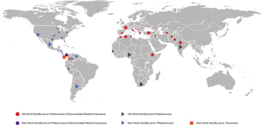
nities with poor living conditions [3, 4] (Figure 1, Table 1).
8Sandfly-Borne Viruses of Demonstrated/Relevant Medical Importance
DOI: http://dx.doi.org/10.5772/intechopen.81023
Figure 1.
Schematic overview of the sandfly-brone viruses, according to geographical regions.
Group Virus Virus origin Country
Sandfly-borne phleboviruses of demonstrated medical importance
Old World Sandfly-borne phleboviruses of demonstrated medical importance
Sandfly fever Sandfly fever Naples virus Blood sample Italy
Naples Species Sabin
Sandfly fever Naples virus Human sera Cyprus
R-3
Sandfly fever Naples virus Phlebotomus papatasi Egypt
Namru
Sandfly fever Naples virus Human Turkmenistan
Sandfly fever Naples virus Human Afghanistan
Sandfly fever Naples virus Phlebotomus longicuspis Algeria
Sandfly fever Naples virus Phlebotomus spp. and India
humans
Sandfly fever Naples virus Phlebotomus perfiliewi Serbia
YU 8-76
Toscana virus Phlebotomus perniciosus Italy
Toscana virus Human CSF Italy
Toscana virus Pipistrellus kuhli brain Italy
Toscana virus Phlebotomus spp. Italy
Toscana virus Phlebotomus perniciosus France
Toscana virus Sergentomyia minuta France
Toscana virus human CSF Croatia
Toscana virus Phlebotomus neglectus Croatia
Toscana virus Phlebotomus spp. Cyprus
Toscana virus Human sera, urine Turkey
Toscana virus Phoenicopterus roseus, Turkey
Pelecanus onocrotalus,
Ciconia nigra
Toscana virus Phlebotomus perniciosus Morocco
Toscana virus P. longicuspis, P. sergenti Morocco
9Vectors and Vector-Borne Zoonotic Diseases
Group Virus Virus origin Country
Toscana virus Phlebotomus spp. Algeria
Toscana virus Phlebotomus spp. Tunisia
Toscana virus Human CSF Greece
Sandfly Fever Sandfly fever Sicilian virus Human sera Italy
Sicillian Species Sabin
Sandfly fever Sicilian virus Phlebotomus spp. Iran
Sandfly fever Sicilian virus Human sera Cyprus
Sandfly fever Sicilian virus Phlebotomus papatasi Pakistan
Sandfly fever Sicilian virus Phlebotomus ariasi Algeria
Sandfly fever Sicilian virus Phlebotomus papatasi Algeria
Sandfly fever Sicilian virus Human Afghanistan
Sandfly fever Sicilian virus Phlebotomus spp. India
Sandfly fever Sicilian virus Human Ethiopia
Sandfly fever Cyprus virus Human sera Cyprus
Sandfly fever Turkey virus Human sera Turkey
Sandfly fever Turkey virus Phlebotomus major complex Turkey
Dashli virus Phlebotomus spp./ Iran
Sergentomyia spp.
Salehabad Adria virus Human blood Greece
Species
Adria virus Phlebotomus spp. Albania
New World Sandfly-borne phleboviruses of demonstrated medical importance
Punta Toro Punta Toro virus Adames Human Panama
Species
Punta Toro virus Balliet Human Panama
Punta Toro virus Human Panama
Punta Toro virus Human Panama
Punta Toro virus Sentinel hamster Panama
Punta Toro virus Sentinel hamster Panama
Punta Toro virus Lutzomyia spp. Panama
Punta Toro virus Human Panama
Punta Toro virus Human Panama
Punta Toro virus Human Panama
Punta Toro virus Human Panama
Sandfly-borne Rhabdoviruses
Vesiculovirus Vesiculovirus Horse South Africa
Species
Vesiculovirus Bovine Indiana, USA
Vesiculovirus Bovine, equine New Jersey
Vesiculovirus Cattle, horse Wisconsin,
Minnesota, Dakota
Vesiculovirus Cattle, horse Argentine
Vesiculovirus Cow, horse, pig Venezuela
Vesiculovirus Horse Texas, Louisiana
Vesiculovirus Horse Kansas
Vesiculovirus Horse Colorado
Vesiculovirus Swine Colombia
10Sandfly-Borne Viruses of Demonstrated/Relevant Medical Importance
DOI: http://dx.doi.org/10.5772/intechopen.81023
Group Virus Virus origin Country
Vesiculovirus Swine Venezuela
Vesiculovirus Swine Missouri
Vesiculovirus Swine Colorado
Vesiculovirus Cattle California
Vesiculovirus Horse Arizona
Vesiculovirus Cattle Mexico
Vesiculovirus Horse Alabama
Vesiculovirus Horse Mississippi,
Georgia, Tennessee,
Florida
Vesiculovirus Bovine, porcine Guatemala
Vesiculovirus Equine Belize
Vesiculovirus Bovine Honduras
Vesiculovirus Bovine El Salvador
Vesiculovirus Bovine, porcine Nicaragua
Vesiculovirus Bovine Costa Rica
Vesiculovirus Bovine Peru
Chandipura virus Human India
Chandipura virus Sandfly India
Chandipura virus Sandfly Senegal
Chandipura virus Sandfly Nigeria
Sandfly-borne Reoviruses
Changuinola Changuinola virus Lutzomyia sp. Panama
virus Species
Changuinola virus Rice rat, armadillo, sloth Panama
Changuinola virus Human Panama
Changuinola virus Lutzomyia sp. Colombia
Changuinola virus Bradypus variegatus, Panama
Choloepus hoffmanni
Table 1.
Features of the medically important sandfly‐borne viruses.
Both molecular characterization and seroepidemiological studies demon-
strated broad distribution of sandfly-borne phleboviruses in the old world in the
Mediterranean region, in the African continent, in the Indian subcontinent, in the
Middle East and in Central Asia. However, the pathogen sandfly-borne phlebovi-
ruses were recorded in the limited geographical area (Panama) in the new world
with sporadic human cases. This must be due to (i) limited investigations in the new
world; (ii) vector competence of phlebovirus in the new world; (iii) small-sized
human population and (iv) lack of case report.
Acknowledgements
This work was supported in part by the European Virus Archive goes Global
(EVAg) project that has received funding from the European Union’s Horizon 2020
research and innovation programme under grant agreement No 653316.
Nazli Ayhan is a Post-Doctoral fellow supported by a IRD grant.
11Vectors and Vector-Borne Zoonotic Diseases Author details Nazli Ayhan1* and Remi N. Charrel1,2 1 Unite des Virus Emergents (Aix-Marseille Univ – IRD 190 – Inserm 1207 – IHU Mediterranee Infection), Marseille, France 2 Emerging Pathogens Institute, University of Florida, Gainesville, Florida, USA *Address all correspondence to: nazliayhann@gmail.com © 2018 The Author(s). Licensee IntechOpen. This chapter is distributed under the terms of the Creative Commons Attribution License (http://creativecommons.org/licenses/ by/3.0), which permits unrestricted use, distribution, and reproduction in any medium, provided the original work is properly cited. 12
Sandfly-Borne Viruses of Demonstrated/Relevant Medical Importance
DOI: http://dx.doi.org/10.5772/intechopen.81023

References

[1] Killick-Kendrick R. The biology            [9] Elliott RM, Schmaljohn CS,
and control of Phlebotomine sand               Collett MS. Bunyaviridae Genome
flies. Clinics in Dermatology.                 Structure and Gene Expression. Berlin,
1999;17:279-289. DOI: 10.1016/                 Heidelberg: Springer; 1991. pp. 91-141.
S0738-081X(99)00046-2                          DOI: 10.1007/978-3-642-76018-1_4

[2] Tesh RB. The genus phlebovirus             [10] Adams MJ, Lefkowitz EJ, King
and its vectors. Annual Review of              AMQ, et al. Changes to taxonomy
Entomology. 1988;33:169-181                    and the international code of virus
                                               classification and nomenclature
[3] Teutsch SM, Churchill RE, editors.         ratified by the international committee
Principles and Practice of Public Health       on taxonomy of viruses. Archives of
Surveillance. USA: Oxford University           Virology. 2017;62:2505-2538. DOI:
Press; 2000                                    10.1007/s00705-017-3358-5

[4] Lemon SM, Sparling PF, Hamburg             [11] Zhioua E, Moureau G, Chelbi I,
MA, Relmn DA, Choffnes ER,                     Ninove L, Bichaud L, Derbali M, et al.
Marck A. Vector-borne diseases:                Punique virus, a novel phlebovirus,
Understanding the environmental,               related to sandfly fever Naples virus,
human health, and ecological                   isolated from sandflies collected
connections. Workshop summary. In:             in Tunisia. The Journal of General
Vector-Borne Dis Underst Environ               Virology. 2010;91:1275-1283. DOI:
Hum Heal Ecol Connect Work Summ.               10.1099/vir.0.019240-0
Washington: National Academies
Press; 2008                                    [12] Peyrefitte CN, Grandadam M,
                                               Bessaud M, Andry PE, Fouque F, Caro
[5] Weaver SC, Reisen WK. Present              V, et al. Diversity of Phlebotomus
and future arboviral threats. Antiviral        perniciosus in Provence, Southeastern
Research. 2010;85:328-345                      France: Detection of two putative
                                               new Phlebovirus sequences. Vector
[6] Bhatt PN, Rodrigues FM.                    Borne and Zoonotic Diseases.
Chandipura: A new arbovirus isolated in        2013;13(9):630-636. DOI: 10.1089/
India from patients with febrile illness.      vbz.2012.1169
The Indian Journal of Medical Research.
1967;55:12                                     [13] Remoli ME, Fortuna C, Marchi A,
                                               Bucci P, Argentini C, Bongiorno G, et al.
[7] Dhanda V, Rodrigues FM, Ghosh              Viral isolates of a novel putative phlebovirus
SN. Isolation of Chandipura virus              in the Marche region of Italy. The American
from sandflies in Aurangabad. The              Journal of Tropical Medicine and Hygiene.
Indian Journal of Medical Research.            2014;90(4):760-763. DOI: 10.4269/
1970;58:179-180                                ajtmh.13-0457

[8] Travassos da Rosa AP, Tesh                 [14] Alkan C, Alwassouf S, Piorkowski
RB, Pinheiro FP, Travassos da                  G, Bichaud L, Tezcan S, Dincer E, et al.
Rosa JP, Peralta PH, Knudson                   Isolation, genetic characterization, and
DL. Characterization of the                    seroprevalence of Adana virus, a novel
Changuinola serogroup viruses                  phlebovirus belonging to the salehabad
(Reoviridae: Orbivirus).                       virus complex, in Turkey. Journal of
Intervirology. 1984;21:38-49. DOI:             Virology. 2015:03027-03014. DOI:
10.1159/000149501                              10.1128/JVI.03027-14

13
Vectors and Vector-Borne Zoonotic Diseases
[15] Tesh RB, Lubroth J, Guzman H. Emerging Infectious Diseases.
Simulation of arbovirus overwintering: 2010;16:1498-1500. DOI: 10.3201/
Survival of Toscana virus (Bunyaviridae: eid1609.100505
Phlebovirus) in its natural sand fly
vector philebotomus perniciosus. The [22] Epelboin L, Hausfater P,
American Journal of Tropical Medicine Schuffenecker I, Riou B, Zeller H,
and Hygiene. 1992;47:574-581. DOI: Bricaire F, et al. Meningoencephalitis
10.4269/ajtmh.1992.47.574 due to Toscana virus in a French traveler
returning from Central Italy. Journal of
[16] Depaquit J, Grandadam M, Fouque Travel Medicine. 2008;15:361-363. DOI:
F, Andry PE, Peyrefitte C. 10.1111/j.1708-8305.2008.00221.x
Arthropod-borne viruses transmitted
by Phlebotomine sandflies in [23] Anagnostou V, Pardalos G,
Europe: A review. Eurosurveillance. Athanasiou-Metaxa M, Papa A.
2010;15(10):19507 Novel phlebovirus in febrile child,
Greece. Emerging Infectious
[17] Alkan C, Bichaud L, De Lamballerie Diseases. 2011;17:940. DOI: 10.3201/
X, Alten B, Gould EA, Charrel RN. eid1705.101958
Sandfly-borne phleboviruses of
Eurasia and Africa: Epidemiology, [24] Tesh RB, Saidi S, Gajdamovic SJ,
genetic diversity, geographic range, Rodhain F, Vesenjak-Hirjan J. Serological
control measures. Antiviral Research. studies on the epidemiology of sandfly
2013;100(1):54-74. DOI: 10.1016/j. fever in the Old World. Bulletin of
antiviral.2013.07.005 the World Health Organization.
1976;54:663-674
[18] Verani P, Ciufolini MG, Nicoletti L,
Balducci M, Sabatinelli G, Coluzzi M, et al. [25] Alivisatos GP. The Outbreak
Ecological and epidemiological studies of Three-Day Fever at Athens
of Toscana virus, an arbovirus isolated in 1935. Bulletin de l'Office
from Phlebotomus. Annali dell'Istituto International d'Hygiene Publique.
Superiore di Sanità. 1982;18:397-399 1936;28(11):2146-2166.
[19] Coluzzi M, Sabatinelli G, Paci P, [26] Tesh RB, Papaevangelou G. Effect of
Renzi A, Amaducci L, Ciufolini MG, insecticide spraying for malaria control
et al. Ecology of viruses isolated from on the incidence of sandfly fever in
sand flies in Italy and characterization Athens, Greece. The American Journal
of a new phlebovirus (Arbia virus). The of Tropical Medicine and Hygiene.
American Journal of Tropical Medicine 1977;26:163-166. DOI: 10.4269/
and Hygiene. 1988;38:433-439 ajtmh.1977.26.163
[20] Hacioglu S, Dincer E, Isler CT, [27] Maroli M, Feliciangeli MD,
Karapinar Z, Ataseven VS, Ozkul A, Bichaud L, Charrel RN, Gradoni
et al. A snapshot avian surveillance L. Phlebotomine sandflies and the
reveals west nile virus and evidence of spreading of leishmaniases and
wild birds participating in Toscana cirus other diseases of public health
circulation. Vector Borne and Zoonotic concern. Medical and Veterinary
Diseases. 2017;17:698-708. DOI: Entomology. 2013;27(2):123-147. DOI:
10.1089/vbz.2017.2138 10.1111/j.1365-2915.2012.01034.x
[21] Kay MK, Gibney KB, Riedo FX, [28] Karakašević BO. Prvoj epidemiji
Kosoy OL, Lanciotti RS, Lambert AJ. papatačijeve groznice na teritoriji
Toscana virus infection in American NR Srbije. Vojnosanitetski Pregled.
traveler returning from Sicily, 2009. 1947;4:224-228
14Sandfly-Borne Viruses of Demonstrated/Relevant Medical Importance
DOI: http://dx.doi.org/10.5772/intechopen.81023
[29] Vesenjak-Hirjan J, Punda-Polić V, [37] Amaro F, Hanke D, Zé-Zé L, Alves
Dobe M. Geographical distribution of MJ, Becker SC, Höper D. Genetic
arboviruses in Yugoslavia. Journal of characterization of Arrabida virus,
Hygiene, Epidemiology, Microbiology, a novel phlebovirus isolated in
and Immunology. 1991;35:129-140 South Portugal. Virus Research.
2016;214:19-25. DOI: 10.1016/J.
[30] Hukić M, Numanović F, Šiširak VIRUSRES.2016.01.004
M, Moro A, Dervović E, Jakovac S,
et al. Surveillance of wildlife zoonotic [38] Amaro F, Zé-Zé L, Alves MJ,
diseases in the Balkans region. Alves MJ, Börstler J, Clos J, et al.
Medicinski Glasnik. 2010;7(2) Co-circulation of a novel phlebovirus
and Massilia virus in sandflies, Portugal.
[31] Hukić M, Bešić IS. Sandfly- Virology Journal. 2011;17(4):585-587.
Pappataci fever in Bosnia and DOI: 10.1186/s12985-015-0407-0
Herzegovina: The new-old disease.
Bosnian Journal of Basic Medical [39] Papa A, Velo E, Bino S. A novel
Sciences. 2009;9(1):39 phlebovirus in Albanian sandflies.
Clinical Microbiology and Infection.
[32] Simic C. O posleratnoj pojavi 2011;17:585-587
papatacˇijeve groznice u Srbiji i
Banatu. Glas Srpske akademije nauka [40] Ayhan N, Velo E, De Lamballerie
CCIV. Odjeljenje Medicinskih Nauka. X, Kota M, Kadriaj P, Ozbel Y, et al.
1951;4:143-152 Detection of Leishmania infantum and
a novel Phlebovirus (Balkan virus) from
[33] Ahmed A, Abera NA, Cao S, sand flies in Albania. Vector Borne and
Omballa V, Wang D, Montgomery JM, Zoonotic Diseases. 2016;16(12):802-
et al. An outbreak of acute febrile illness 806. DOI: 10.1089/vbz.2016.2002
caused by sandfly fever Sicilian virus in
the Afar region of Ethiopia, 2011. The [41] Ayhan N, Alten B, Ivovic V, Dvořák
American Journal of Tropical Medicine V, Martinkovic F, Omeragic J, et al.
and Hygiene. 2014;91:1250-1253. DOI: Direct evidence for an expanded
10.4269/ajtmh.14-0299 circulation area of the recently
identified Balkan virus (Sandfly
[34] Papa A, Konstantinou G, fever Naples virus species) in several
Pavlidou V, Antoniadis A. Sandfly countries of the Balkan archipelago.
fever virus outbreak in Cyprus. Parasites & Vectors. 2017;10(1). DOI:
Clinical Microbiology and 10.1186/s13071-017-2334-y
Infection. 2006;12:192-194. DOI:
10.1111/j.1469-0691.2005.01330.x [42] Alkan C, Erisoz Kasap O,
Alten B, De Lamballerie X, Charrel
[35] Ellis SB, Appenzeller G, Lee H, RN. Sandfly-borne phlebovirus
Mullen K, Swenness R, Pimentel G, isolations from Turkey: New insight
et al. Outbreak of sandFly fever in into the sandfly fever sicilian
Central Iraq, September 2007. Military and sandfly fever Naples species.
Medicine. 2008;173:949-953. DOI: PLoS Neglected Tropical Diseases.
10.7205/MILMED.173.10.949 2016;10:e0004519. DOI: 10.1371/
journal.pntd.0004519
[36] Guler S, Guler E, Caglayik DY,
Kokoglu OF, Ucmak H, Bayrakdar F, [43] Alkan C, Moin Vaziri V, Ayhan N,
et al. A sandfly fever virus outbreak Badakhshan M, Bichaud L, Rahbarian
in the East Mediterranean region N, et al. Isolation and sequencing
of Turkey. International Journal of of Dashli virus, a novel Sicilian-like
Infectious Diseases. 2012;16:e244-e246. virus in sandflies from Iran; genetic
DOI: 10.1016/j.ijid.2011.12.001 and phylogenetic evidence for the
15Vectors and Vector-Borne Zoonotic Diseases
creation of one novel species within the Indian Journal of Medical Research.
Phlebovirus genus in the Bunyaviridae 1934;21:775-788
family. PLoS Neglected Tropical
Diseases. 2017;11(12). DOI: 10.1371/ [52] Anderson WME. Clinical
journal.pntd.0005978 observations on sandfly fever in the
Peshawar district. Journal of the Royal
[44] Aspöck H, Gerersdorfer T, Army Medical Corps. 1941;77:225-239
Formayer H, Walochnik J. Sandflies
and sandfly-borne infections of [53] Maroli M, Feliciangeli MD,
humans in Central Europe in the light Bichaud L, Charrel RN, Gradoni
of climate change. Wiener Klinische L. Phlebotomine sandflies and the
Wochenschrift. 2008;120(4):24-29. spreading of leishmaniases and
DOI: 10.1007/s00508-008-1072-8 other diseases of public health
concern. Medical and Veterinary
[45] Charrel RN, Gallian P, Navarro-Marí Entomology. 2013;27:123-147. DOI:
JM, Nicoletti L, Papa A, Sánchez-Seco 10.1111/j.1365-2915.2012.01034.x
MP, et al. Emergence of Toscana virus in
Europe. Emerging Infectious Diseases. [54] American Society of Tropical
2005;11(11):1657. DOI: 10.3201/ Medicine and Hygiene. AB. The
eid1111.050869 American Journal of Tropical Medicine
and Hygiene. Allen Press; 1955
[46] Charrel RN, Moureau G,
Temmam S, Izri A, Marty P, Parola [55] Československá Akademie Věd SY,
P, et al. Massilia virus, a novel Kurakhmedova SA, Melnikova EE. Acta
Phlebovirus (Bunyaviridae) isolated Virologica. Academia, Pub. House of
from sandflies in the Mediterranean. the Czechoslovak Academy of Sciences;
Vector Borne and Zoonotic Diseases. 1974
2009;9(5):519-530. DOI: 10.1089/
vbz.2008.0131 [56] Goverdhan MK, Dhanda V, Modi
GB, Bhatt PN, Bhagwat RB, Dandawate
[47] Sabin AB. Experimental studies CN, et al. Isolation of phlebotomus
on phlebotomus (pappataci, sandfly) (sandfly) fever virus from sandflies
fever during world war II. Archiv für die and humans during the same season in
Gesamte Virusforschung. 1951;4(4):367- Aurangabad District, Maharashtra state,
410. DOI: 10.1007/BF01241161 India. The Indian Journal of Medical
Research. 1976;64:57-63
[48] Doerr R, Franz KTS. Das Papatatsi
Fieber. Franz Deuticke, Leipzig- Wien; [57] Gaidamovich SY, Khutoretskaya
1909 NV, Asyamov YV, Tsyupa VI,
Melnikova EE. Sandfly fever in
[49] Taussig S. Die Hundskrankheit, Central Asia and Afghanistan. In:
endemischer Magenkatarrh in der Hemorrhagic Fever with Renal
Herzegowina. Wiener klinische Syndrome, Tick- and Mosquito-
Wochenschrift. 1905;50:164 Borne Viruses. Vienna: Springer
Vienna; 1990. pp. 287-293. DOI:
[50] Livschitz J. Studies on Pappataci 10.1007/978-3-7091-9091-3_32
fever. Medical Parasitology and Parasitic
Diseases (Moscow). 1937;6:938-943 [58] Ennis WH, Peters CJ, Feinsod FM,
et al. Sand fly fever-Naples infection
[51] Shortt HE, Poole LT, Stephens in Egypt. The American Journal of
ED. Sandfly fever on the Indian Tropical Medicine and Hygiene.
frontier. A preliminary note on 1987;37:193-196. DOI: 10.4269/
some laboratory investigations. The ajtmh.1987.37.193
16Sandfly-Borne Viruses of Demonstrated/Relevant Medical Importance
DOI: http://dx.doi.org/10.5772/intechopen.81023
[59] Schmidt JR, Schmidt ML, Said infection. Lancet (London, England).
MI, Soliman AK, Farrag IH, El Said S, 1993;342:803-804
et al. Phlebotomus fever in Egypt. The
American Journal of Tropical Medicine [67] Dobler G, Treib J, Haass A, Frösner
and Hygiene. 1971;20:483-490. DOI: G, Woesner R, Schimrigk K. Toscana
10.4269/ajtmh.1971.20.483 virus infection in German travellers
returning from the Mediterranean.
[60] Darwish MA, Feinsod FM, Scott Infection. 1997;25(5):325-325
RM, Ksiazek TG, Botros BAM,
Farrag IH, et al. Arboviral causes of [68] Eitrem R, Niklasson B, Weiland
non-specific fever and myalgia in a fever O. Sandfly fever among Swedish
hospital patient population in Cairo, tourists. Scandinavian Journal of
Egypt. Transactions of the Royal Society Infectious Diseases. 1991;23:451-457
of Tropical Medicine and Hygiene.
1987;81:1001-1003 [69] Veater J, Mehedi F, Cheung CK,
Nabarro L, Osborne J, Wong N, et al.
[61] Eitrem R, Vene S, Niklasson B. Toscana virus meningo-encephalitis:
Incidence of sand fly fever among An important differential diagnosis
Swedish United Nations soldiers on for elderly travellers returning from
Cyprus during 1985. The American Mediterranean countries. BMC
Journal of Tropical Medicine and Geriatrics. 2017;17:193. DOI: 10.1186/
Hygiene. 1990;43:207-211. DOI: s12877-017-0593-2
10.1016/0035-9203(87)90378-6
[70] Arden KE, Heney C, Shaban B,
[62] Gligić A, Mišcević Z, Tesh Nimmo GR, Nissen MD, Sloots TP,
RB, Travassos da Rosa AZV. First et al. Detection of Toscana virus from
isolations of Naples sandfly fever an adult traveler returning to Australia
virus in Yugoslavia. Mikrobiologija. with encephalitis. Journal of Medical
1982;19:167-175 Virology. 2017;89:1861-1864. DOI:
10.1002/jmv.24839
[63] Gaidamovich SI, Obukhova VR,
Sveshnikova NA, Cherednichenko IN, [71] Renzi A, Caciolli S, Nicoletti L,
Kostiukov MA. Natural foci of viruses Bartolozzi D, Balducci M, Traini E, et al.
borne by Phlebotomus papatasi in the Central nervous system involvement
USSR according to a serologic study of during infection by Phlebovirus Toscana
the population. Voprosy Virusologii. of residents in natural foci in Central
1978:556-560 Italy (1977-1988). The American Journal
of Tropical Medicine and Hygiene.
[64] Ehrnst A, Peters CJ, Niklasson B, 1991;45:429-434. DOI: 10.4269/
Svedmyr A, Holmgren B. Neurovirulent ajtmh.1991.45.429
Toscana virus (a sandfly fever
virus) in Swedish man after visit to [72] Papa A, Paraforou T,
Portugal. Lancet (London, England). Papakonstantinou I, Pagdatoglou K,
1985;1:1212-1213 Kontana A, Koukoubani T. Severe
encephalitis caused by Toscana virus,
[65] Calisher CH, Weinberg AN, Muth Greece. Emerging Infectious Diseases.
DJ, Lazuick JS. Toscana virus infection 2014;20:1417-1419. DOI: 10.3201/
in United States citizen returning eid2008.140248
from Italy. Lancet (London, England).
1987;1:165-166 [73] Eitrem R, Stylianou M, Niklasson
B. High prevalence rates of antibody
[66] Schwarz TF, Gilch S, Jäger G. to three sandfly fever viruses
Travel-related Toscana virus (Sicilian, Naples and Toscana) among
17Vectors and Vector-Borne Zoonotic Diseases
Cypriots. Epidemiology and Infection. returning from Sicily, 2009. Emerging
1991;107:685-691. DOI: 10.1017/ Infectious Diseases. 2010;16(9):1498. DOI:
S0950268800049384 10.3201/eid1609.100505
[74] Punda-Polić V, Mohar B, Duh D, [82] Dupouey J, Bichaud L, Ninove
Bradarić N, Korva M, Fajs L, et al. L, Zandotti C, Thirion-Perrier L,
Evidence of an autochthonous Toscana De Lamballerie X, et al. Toscana
virus strain in Croatia. Journal of virus infections: A case series from
Clinical Virology. 2012;55:4-7. DOI: France. The Journal of Infection.
10.1016/j.jcv.2012.06.006 2014;68(3):290-295. DOI: 10.1016/j.
jinf.2013.11.006
[75] Kuşcu F, Menemenlioğlu D, Oztürk
DB, Korukluoğlu G, Uyar Y. Acute [83] Alwassouf S, Maia C, Ayhan N,
Toscana virus infection in an anti-HIV Coimbra M, Cristovao JM, Richet
positive patient. Mikrobiyoloji Bülteni. H, et al. Neutralization-based
2014;48:168-173 seroprevalence of Toscana virus and
sandfly fever sicilian virus in dogs and
[76] Ocal M, Orsten S, Inkaya AC, Yetim cats from Portugal. The Journal of
E, Acar NP, Alp S, et al. Ongoing activity General Virology. 2016;97(11):2816-
of Toscana virus genotype a and West 2823. DOI: 10.1099/jgv.0.000592
Nile virus lineage 1 strains in Turkey: A
clinical and field survey. Zoonoses and [84] Alwassouf S, Christodoulou V,
Public Health. 2014;61:480-491. DOI: Bichaud L, et al. Seroprevalence of
10.1111/zph.12096 sandfly-borne Phleboviruses
belonging to three serocomplexes
[77] Santos L, Simões J, Costa R, Martins (sandfly fever Naples, sandfly fever
S, Lecour H. Toscana virus meningitis in sicilian and salehabad) in dogs f
Portugal, 2002-2005. Euro Surveillance. rom Greece and Cyprus using
2007;2:E3-E4 neutralization test. PLoS Neglected
Tropical Diseases. 2016;10:
[78] Peyrefitte CN, Devetakov I, e0005063. DOI: 10.1371/journal.
Pastorino B, et al. Toscana virus and pntd.0005063
acute meningitis, France. Emerging
Infectious Diseases. 2005;11:778-780.
[85] Tahir D, Alwassouf S, Loudahi A,
DOI: 10.3201/EID1105.041122
Davoust B, Charrel RN. Seroprevalence
of Toscana virus in dogs from Kabylia
[79] Karunaratne K, Nicholas D. Toscana
(Algeria). Clinical Microbiology and
virus meningitis following a holiday in
Infection. 2016;22(3):e16-e17. DOI:
Elba, Italy. British Journal of Hospital
10.1016/j.cmi.2015.10.029
Medicine (London, England: 2005).
2018;79(5):292. DOI: 10.12968/
hmed.2018.79.5.292 [86] Ayhan N, Sherifi K, Taraku A,
Bërxholi K, Charrel RN. High rates
[80] Gabriel M, Resch C, Günther S, of neutralizing antibodies to Toscana
Schmidt-Chanasit J. Toscana virus and sandfly fever Sicilian viruses in
infection imported from Elba into livestock, Kosovo. Emerging Infectious
Switzerland. Emerging Infectious Diseases. 2017;23:989-992. DOI:
Diseases. 2010;16(6):1034. DOI: 10.3201/eid2306.161929
10.3201/eid1606.091763
[87] Alkan C, Allal-Ikhlef AB,
[81] Kay MK, Gibney KB, Riedo FX, Kosoy Alwassouf S, Baklouti A, Piorkowski
OL, Lanciotti RS, Lambert AJ. Toscana G, de Lamballerie X, et al. Virus
virus infection in American traveler isolation, genetic characterization
18Sandfly-Borne Viruses of Demonstrated/Relevant Medical Importance
DOI: http://dx.doi.org/10.5772/intechopen.81023
and seroprevalence of Toscana virus Parasites & Vectors. 2015;8:205. DOI:
in Algeria. Clinical Microbiology and 10.1186/s13071-015-0826-1
Infection. 2015;21:1040.e1-1040.e9.
DOI: 10.1016/j.cmi.2015.07.012 [95] Bichaud L, Dachraoui K, Piorkowski
G, Chelbi I, Moureau G, Cherni S,
[88] Pierro A, Ficarelli S, Ayhan N, et al. Toscana virus isolated from
Morini S, Raumer L, Bartoletti M, et al. sandflies, Tunisia. Emerging Infectious
Characterization of antibody response Diseases. 2013;19:322. DOI: 10.3201/
in neuroinvasive infection caused by EID1902.121463
Toscana virus. Clinical Microbiology
and Infection. 2017;23(11):868-873. [96] Bichaud L, Izri A, de Lamballerie
DOI: 10.1016/j.cmi.2017.03.017 X, Moureau G, Charrel RN. First
detection of Toscana virus in Corsica,
[89] Rodhain F, Madulo-Leblond G, France. Clinical Microbiology and
Hannoun C, Tesh RB. Le virus Corfou: Infection. 2014;20(2):101-104. DOI:
Un nouveau phlebovirus isolé de 10.1111/1469-0691.12347
phlébotomes en Grèce. Annales
de l'Institut Pasteur Virologie. [97] Dincer E, Ozkul A, Gargari
1985;136:161-166. DOI: 10.1016/ S, Ergunay K. Potential animal
S0769-2617(85)80042-3 reservoirs of Toscana virus and
coinfections with Leishmania
[90] Ergünay K, Litzba N, Lo MM, infantum in Turkey. The American
Aydoğan S, Saygan MB, Us D, et al. Journal of Tropical Medicine and
Performance of various commercial Hygiene. 2015;92:690-697. DOI:
assays for the detection of Toscana virus 10.4269/ajtmh.14-0322
antibodies. Vector Borne and Zoonotic
Diseases. 2011;11(6):781-787. DOI: [98] Gaĭdamovich SI, Khutoretskaia
10.1089/vbz.2010.0224 NV, Aziamov IV, Tsiupa VI, Melnikova
EE. Virological study of cases of
[91] Charrel RN, Izri A, Temmam S, sandfly fever in Afghanistan. Voprosy
Delaunay P, Toga I, Dumon H, et al. Virusologii. 1990;35:45-47
Cocirculation of 2 genotypes of Toscana
virus, southeastern France. Emerging [99] Javadian E, Saidi S, Tesh R, Nadim
Infectious Diseases. 2007;13:465. DOI: A. Studies on the epidemiology of
10.3201/eid1303.061086 Sandfly fever in Iran. The American
Journal of Tropical Medicine and
[92] Charrel RN, Izri A, Temmam S, Hygiene. 1977;26:282-287. DOI: 10.4269/
De Lamballerie X, Parola P. Toscana ajtmh.1977.26.282
virus RNA in Sergentomyia minuta
files. Emerging Infectious Diseases. [100] George JE. Isolation of
2006;12. DOI: 1299. DOI:10.3201/ phlebotomus fever virus from
eid1208.060345 Phlebotomus Papatasi and
determination of the host ranges of
[93] Ayhan N, Alten B, Ivovic V, sandflies (Diptera: Psychodidae) in
Martinkovic F, Kasap OE, Ozbel Y, west Pakistan1. Journal of Medical
et al. Cocirculation of two lineages of Entomology. 1970;7:670-676. DOI:
Toscana virus in Croatia. Frontiers in 10.1093/jmedent/7.6.670
Public Health. 2017;5:336. DOI: 10.3389/
fpubh.2017.00336 [101] Carhan A, Uyar Y, Ozkaya E,
Ertek M, Dobler G, Dilcher M, et al.
[94] Es-sette N, Ajaoud M, Anga L, Characterization of a sandfly fever
Mellouki F, Lemrani M. Toscana virus Sicilian virus isolated during a sandfly
isolated from sandflies, Morocco. fever epidemic in Turkey. Journal of
19You can also read