Involvement of Hepcidin in Cognitive Damage Induced by Chronic Intermittent Hypoxia in Mice
←
→
Page content transcription
If your browser does not render page correctly, please read the page content below
Hindawi
Oxidative Medicine and Cellular Longevity
Volume 2021, Article ID 8520967, 14 pages
https://doi.org/10.1155/2021/8520967

Research Article
Involvement of Hepcidin in Cognitive Damage Induced by
Chronic Intermittent Hypoxia in Mice

 Ya-Shuo Zhao ,1,2 Miao Tan ,1 Ji-Xian Song ,1 Ji-Ren An ,1 Xin-Yue Yang ,1
 Wen-Ya Li ,1 Ya-Jing Guo ,1 and En-Sheng Ji 1
 1
 Department of Physiology, Institute of Basic Medicine, Hebei University of Chinese Medicine, Shijiazhuang 050200, China
 2
 Hebei Technology Innovation Center of TCM Formula Preparations, Shijiazhuang 050200, China

 Correspondence should be addressed to En-Sheng Ji; jesphy@126.com

 Received 7 April 2021; Revised 3 July 2021; Accepted 17 July 2021; Published 4 August 2021

 Academic Editor: Massimo Collino

 Copyright © 2021 Ya-Shuo Zhao et al. This is an open access article distributed under the Creative Commons Attribution License,
 which permits unrestricted use, distribution, and reproduction in any medium, provided the original work is properly cited.

 Obstructive sleep apnea (OSA) patients exhibit different degrees of cognitive impairment, which is related to the activation of
 reactive oxygen species (ROS) production by chronic intermittent hypoxia (CIH) and the deposition of iron in the brain. As a
 central regulator of iron homeostasis, whether hepcidin is involved in OSA-induced cognitive impairment has not been clarified.
 In order to simulate OSA, we established the mouse model by reducing the percentage of inspired O2 (FiO2) from 21% to 5%,
 20 times/h for 8 h/day. We found hepcidin was rising during CIH, along with increasing iron levels and neuron loss. Then, we
 constructed a mouse with astrocyte-specific knockdown hepcidin gene (shHamp). During CIH exposure, the shHamp mice
 showed a lower level of total iron and neuronal iron in the hippocampus, via stabilizing ferroportin 1 (FPN1) and decreasing L-
 ferritin (FTL) levels, when compared with wild-type (WT) mice. Furthermore, the shHamp mice showed a decrease of ROS by
 downregulating the elevated NADPH oxidase (NOX2) and 4-hydroxynonenal (4-HNE) levels mediated by CIH. In addition, the
 shHamp mice presented improved cognitive deficit by improving synaptic plasticity and BDNF expression in the hippocampus
 when subjected to CIH. Therefore, our data revealed that highly expressed hepcidin might promote the degradation of FPN1,
 resulting in neuronal iron deposition, oxidative stress damage, reduced synaptic plasticity, and impaired cognitive performance
 during CIH exposure.

1. Introduction early stage. Therefore, it is particularly necessary to explore
 further mechanisms involved in the pathogenesis and con-
Obstructive sleep apnea (OSA) is a prevalent sleep breathing duct appropriate intervention and treatment.
disorder. Clinically, the OSA patient is characterized by the Similar to ischemia-reperfusion, the CIH-induced
repeated cycle of the upper airway obstruction and arousal hypoxia/reoxygenation is the most important pathological
during sleep, which leads to chronic intermittent hypoxia feature of OSA, which could cause the formation of a large
(CIH), hypercapnia, hypoxemia, and sleep fragmentation, number of reactive oxygen species (ROS) [6]. The levels of
resulting in daytime sleepiness and memory decline and deg- oxidative stress markers in serum of OSA patients are
radation of reactivity [1]. Epidemiological study has demon- increased, which is related to the severity of OSA [7]. The ele-
strated that the number of OSA patients worldwide has vated ROS could cause endoplasmic reticulum stress, induce
exceeded 9.36 million, and it is becoming a serious global the changes of mitochondrial membrane potential, and acti-
problem [2]. OSA has been identified as an early risk factor vate the MAPK signaling pathway, resulting in neuronal cell
for developing neurodegenerative disease, such as Alzhei- apoptosis in the hippocampus of rodents [8]. The excessive
mer’s disease (AD) [1, 3]. Clinical studies have shown that ROS could also induce lipid peroxidation in the hippocam-
OSA could accelerate the progression of AD by increasing pus to promote structural damage of neurons, increase the
the formation and accumulation of Aβ42 in the cerebrospinal synaptic space, and reduce the long-term enhancement effect
fluid (CSF) [4, 5]. OSA is often overlooked by patients in the involved in memory impairment [9, 10].
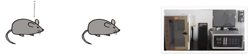
2 Oxidative Medicine and Cellular Longevity

 Hepcidin, predominantly produced and secreted by the “ACCGGGCAGACATTGCGATACCAATTCTCGAGAATT
liver, is the master regulator of systemic iron availability and GGTATCGCAATGTCTGCTTTTTGAATTC” (Figure S1A).
plays a role by controlling ferroportin1 (FPN1), the sole iron The 4 μl LV-U6-shHamp plasmid (virus titer ≥ 1 × 10⁸ TU/ml)
export protein [11]. Hepcidin has also been shown to be was injected into the lateral ventricle of mice. The WT mice
expressed in the brain of glial cells, predominantly in astro- were given the same volume LV-U6-Scramble-shHamp with
cytes, rather than mature neurons [12, 13]. Hepcidin appears the astrocyte promoters followed by Hamp gene control
to control iron entry from plasma into the brain and the shRNA sequence. Two weeks later, the hepcidin gene was
transfer in different neural cells [13]. Circulating iron is trans- identified by RT-PCR. When the hepcidin gene expression
ported via Tf/TfR1 to cross the blood-brain barrier (BBB). was decreased off about 50%, the shHamp mice were
Subsequently, the complexity of Fe-Tf/TfR1 undergoes endo- considered to be successful. Then, the WT and shHamp,
cytosis, which releases iron into intracellular by divalent metal respectively, were exposed to CIH for 21 days to evaluate the
transporter 1 (DMT1). The cytoplasmic iron is used to pro- changes of behavioristics, pathology, and molecular biology
duce heme and is excreted by via FPN1 or stored in ferritin (Figure 2(a)).
[13–15]. The excess free iron could generate free radicals by
Fenton reaction, resulting in oxidative damage in neural cells, 2.2. Reagents and Antibodies. Reagents include lentivirus
which has demonstrated in AD [16], Parkinson’s disease [17], plasmid (Cyagen Biosciences), potassium ferrocyanide
and ischemic stroke [18, 19]. (Sigma-Aldrich), DAPI (2 mg/ml, Servicebio), DHE (Cayman
 Clinically, OSA patients exhibit a high level of hepcidin, Chemical), protease inhibitors (Thermo Fisher), and phospha-
lower iron, and transferrin saturation (TSA) in serum [20– tase inhibitors (Servicebio). The TUNEL kit was purchased
22]. Correlation analysis clarifies that serum hepcidin level from Vazyme Biotech. The BCA protein assay kit was pur-
is positively correlated with sleep-disordered breathing index chased from CoWin Biosciences. RNA extraction kit was pur-
and disease severity [20, 23]. Our previous study revealed chased from Tiangen Biotech.
that CIH exposure could induce the mobilization and Antibodies used were as follows: hepcidin (1 : 100, Affinity),
absorption of iron, leading to iron deposition in the hippo- TfR1 (1 : 10000, Invitrogen), FPN1 (1 : 5000 for WB, 1 : 200 for
campal CA1, CA3, and dentate gyrus (DG) [24]. However, IF, Alpha Diagnostic International), FTL (1 : 5000 for WB,
whether hepcidin is involved in CIH-induced cognitive 1 : 200 for IF, Abcam), GFAP (1 : 100, Servicebio), NeuN
deficit remains unclear. Therefore, we established the mouse (1 : 200, Abcam), Bcl-2 (1 : 2000, ImmunoWay), Bax (1 : 1000,
model of CIH to simulate OSA and explored the hepcidin Servicebio), p-JNK (1 : 1000, Cell Signaling Technology), JNK
expression at different CIH simulating times. In addition, we (1 : 1000, Arigo Biolaboratories), NOX2 (1 : 2000, GeneTex),
constructed genotypic mice with specific knockdown of hepci- 4-HNE (1 : 200 for IF, 1 : 1000 for WB, Arigo Biolaboratories),
din in astrocytes (shHamp), so as to study the relationship of BDNF (1 : 1000, Servicebio), β-actin (1 : 1000, Cwbiotech),
hepcidin and CIH-induced neurocognitive impairment. and β-tubulin (1 : 1000, Servicebio).

2. Materials and Methods 2.3. Morris Water Maze. The Morris water maze (MWM)
2.1. Experimental Animals and Grouping. The SPF was used to assess memory function as previously described
C57BL/6N mice (male, 20 g ± 2 g) were purchased from Bei- [16]. As shown in Figure 3(a), in the first two days, the visible
jing Vital River Laboratory Animal Technology Co., Ltd. platform (the platform above the water surface) was used for
(Beijing, China). All mice were adapted to their living condi- training. The hidden platform (the platform below the water
tions for at least 7 days before the experiment. All animal surface) was used for training in the next five days. Each
experimental procedures were carried out in strict accor- mouse was released into a quadrant facing the wall of the
dance with the National Institutes of Health Guide for the water tank. The time spent to find the hidden platform was
Care and Use of Laboratory Animals and approved by the referred as latency time, and latency route and distance were
Animal Care and Use Committee of Medical Ethics of Hebei also recorded. The latency time and route from the opposite
University of Chinese Medicine (No. DWLL2018006). side of the platform to the platform are counted and calcu-
 We established a murine CIH model to simulate OSA. lated. On the 6th day, the hidden platform was removed for
The mice were placed in a chamber in which the fraction of the probe trial, and then, the times crossing the platform of
inspired oxygen (FiO2) was decreased from 21% to 5% and original position were recorded within 2 min.
then gradually returned to 21%. The exposure cycle was
repeated every 3 min, 20 times/h for 8 h/day (Figure 1(a)). 2.4. Perls’ Staining. The iron distribution in the hippocampus
The mice in the control group (Con) received normal air was evaluated through Perls’ staining. After dewaxed, the sec-
(21% O2) in the identical chambers. The C57BL/6N mice tions were dipped in PBS (0.01 M) and treated with 3% H2O2
(n = 24) were randomly assigned to the four CIH exposure for 20 min to remove endogenous peroxidase. The sections
groups, and there was a washout period of 7 days between were immersed in fresh Perls’ solution containing 1% potas-
CIH exposures. sium ferrocyanide and 1% hydrochloric acid for 10 h. After
 Next, we prepared the wild-type (WT) and shHamp mice washed thoroughly in PBS, the sections were strengthened
(n = 10 for each group) and exposed to CIH for 21 days. Firstly, with DAB kits, dehydrated, and finally covered with neutral
we constructed a lentivirus plasmid LV-U6-shHamp with the balsam. The mean density was calculated by Image-Pro Plus
astrocyte promoters followed by Hamp gene shRNA sequence 6.0 software.
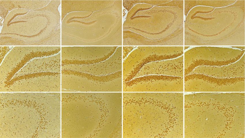
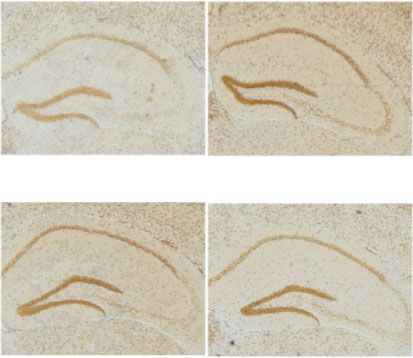
Oxidative Medicine and Cellular Longevity 3

 FiO2 2.5
 ⁎⁎

 Hepcidin mRNA
 21% 2.0
 ⁎⁎ ⁎⁎
 1.5
 1.0
 5%
 0.5
 0 1.5 3.0 4.5 6.0 (min) 0.0
 Con 7 d 14 d 21 d
 (a) (b)
 Con 7d 0.4

 The mean density of
 hepcidin protein
 0.3 ⁎⁎ ⁎⁎ ⁎⁎

 0.2
 14 d 21 d
 0.1

 0.0

 Con

 14 d

 21 d
 7d
 (c) (d)
 Con 7d 14 d 21 d 0.6 DG CA3
 ⁎

 density of Fe content
 0.4 ⁎

 The mean
 0.2

 0.0

 Con

 Con
 14 d
 21 d

 14 d
 21 d
 7d

 7d
 (e) (f)
 Con 7d 14 d 21 d

 DG CA3
 number of neuronal cells

 400
 350
 300 ⁎⁎
 250
 The

 100 ⁎
 50
 0
 Con

 Con
 14 d
 21 d

 14 d
 21 d
 7d

 7d

 (g) (h)

Figure 1: The elevated hepcidin and Fe content in the hippocampus with different exposed times. (a) The flow chart of CIH exposure
procedure. (b) Hepcidin mRNA level determined by q-PCR in the hippocampus of mice subjected to CIH for 7 d, 14 d, and 21 d (n = 6).
(c) The hepcidin protein expression detected by immunohistochemistry (scale bar = 100 μm). (d) The mean density of hepcidin protein
expression in the whole hippocampus as shown in panel (c) (n = 3). (e) Perls’ staining of the dentate gyrus (DG) and hippocampal CA3
(scale bar = 25 μm). (f) The mean density of Fe content as shown in panel (e) (n = 3). (g) The Nissl staining of the hippocampus subjected
to CIH for 7 d, 14 d, and 21 d (scale bar = 100 or 25 μm). (h) The number of neuronal cells in the dentate gyrus (DG) and hippocampal
CA3 as shown in panel (g) (n = 3). The data are shown as the means ± SEM. ∗ p < 0:05 and ∗∗ p < 0:01 vs. Con.
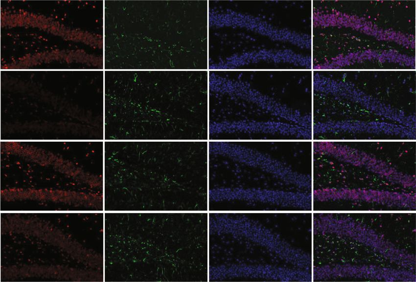
4 Oxidative Medicine and Cellular Longevity

 14 days later CIH exposure

 Hepcidin gene
 identified by RT-PCR CIH for 21 days

 (a)
 $ Hepcidin GFAP DAPI Merge
 3 ##
 ⁎⁎
 Hepcidin mRNA

 WT
 2

 1
 **
 0
 WT+CIH
 WT

 WT+CIH

 (b) (c)
 WT WT+CIH $
 0.5
 #
 The mean density of

 0.4 ⁎⁎
 Fe content

 0.3
 0.2
 0.1
 0.0
 DG

 shHamp

 shHamp+CIH
 WT

 WT+CIH
 CA3

 (d) (e)

Figure 2: The preparation and identification of shHamp mice and iron level. (a) The prepared pattern diagram of shHamp mice of astrocyte-
specific knockdown hepcidin. (b) Hepcidin mRNA level determined by q-PCR in the hippocampus of WT and shHamp mice exposed to CIH
for 21 days (n = 6). (c) Double immunofluorescence staining in the hippocampal dentate gyrus (DG). The sections were labeled for hepcidin
(red), GFAP (green), and DAPI (blue) (scale bar = 25 μm, n = 3). (d) Perls’ staining of the dentate gyrus (DG) and hippocampal CA3
(scale bar = 100 μm or 25 μm). (e) The mean density of Fe content as shown in panel (d) (n = 3). The data are shown as the means ± SEM.
∗
 p < 0:05 and ∗∗ p < 0:01 vs. WT. #p < 0:05 vs. shHamp. $p < 0:05 vs. WT+CIH.
Oxidative Medicine and Cellular Longevity 5

 CIH exposure

 Day 1 -13 Day 14-15 Day 16-20 Day 21

 Visible platform Hidden platform training Probe trial
 training
 (a)
 60 WT shHamp 12
 lantency time (sec)

 10 $

 Passing times
 The escape

 40 $ 8
 # $ 2 1 2 1
 ⁎ ⁎ # 3 4 3 4 ⁎
 ⁎ 6
 20 4
 WT+CIH shHamp+CIH 2
 0 0

 WT

 WT+CIH
 1 2 3 4 5

 shHamp

 shHamp+CIH
 2 1 2 1
 3 4 3 4
 WT WT+CIH

 (b) (c) (d)

Figure 3: Morris water maze of the WT and shHamp mice treated with CIH for 21 days. (a) The flow chart of Morris water maze. (b) The
escape latency time was performed on days 16-20. (c) The escape route distance on day 20. (d) The number of passing times of probe trial on
day 21. The data are shown as the means ± SEM (n = 10). ∗ p < 0:05 vs. WT. #p < 0:05 vs. shHamp. $p < 0:05 vs. WT+CIH.

2.5. TUNEL. The TUNEL staining was in accordance to the The double immunofluorescence was performed as fol-
instruction as previously described [24]. The frozen sections lows. The brain sections were incubated with the mouse
were cleaned with PBS and incubated with equilibration anti-GFAP or NeuN antibody and rabbit antihepcidin or
buffer (EB) to remove endogenous peroxidase. The newly FPN1 or FTL antibody overnight at 4°C. On the following
configured reaction mixture of Bright Green Labeling Mix day, the secondary antibodies, DyLight 549 goat anti-rabbit
and Recombinant TdT Enzyme was added into the sections IgG and DyLight 488 goat anti-mouse IgG, were added to
at 37°C for 1 h. After washing, the sections were incubated incubate the brain sections and were incubated at 37°C for
with DAPI at room temperature. The antifluorescence 60 min. After washing, the sections were sealed with an anti-
quencher was used to seal the sections. FITC and DAPI fluo- fluorescence quencher and visualized with a fluorescence
rescence were detected at 488 nm and 460 nm, respectively. microscope.
The number of total apoptotic cells was calculated by Photo-
shop CS 5.0 software. 2.8. Transmission Electron Microscopy. The ultrastructural
 change of synapses was visualized using transmission elec-
2.6. DHE Staining. The dihydroethidium (DHE) staining was tron microscopy (TEM). After deep anesthesia, the hippo-
adopted to measure the ROS level in the hippocampus. The campal dentate gyrus (DG) slices were removed and fixed
frozen sections were cleaned with PBS and added self- using osmium tetroxide. Then, the sample was prepared
fluorescence quenching agent for 5 min, then incubated with according to the standard procedures. The ultrathin sections
5 μM of DHE at 37°C for 30 min in dark. The sections were (80 nm) were collected and stained with uranyl acetate and
rewashed and sealed with an antifluorescence quencher. lead nitrate. The sections were observed under a Hitachi
The red fluorescence was detected at 549 nm. The mean den- HT7800/HT7700 electron microscope.
sity was calculated by Image-Pro Plus 6.0 software.
 2.9. Western Blot Analysis. Western blot was performed to
2.7. Immunohistochemistry. The frozen sections were washed detect protein expression. First, the brain tissues were
with PBS and incubated with 3% H2O2 to block endogenous homogenized in 4°C RIPA lysis buffer, which contained pro-
peroxidase. The antigen retrieval was applied with the boiling tease inhibitors and phosphatase inhibitors. After centrifuga-
citrate (10 mM, pH 6.0) method. After the washing stage, the tion, the supernatants were collected, and the total protein
sections were incubated with 10% goat serum for 60 min at concentrations were determined with BCA protein assay
room temperature (RT). kit. The proteins were separated by SDS-PAGE and trans-
 Immunohistochemistry was performed as follows. The ferred into PVDF membranes. The membranes were blocked
brain sections were incubated with primary antibodies with 5% skim milk powder for 2 h at RT and incubated with
hepcidin and 4-HNE overnight at 4°C. On the following primary antibodies: TfR1, FPN1, FTL, NOX2, 4-HNE, Bcl-2,
day, the slides were incubated with the HRP-conjugated Bax, p-JNK, JNK, BDNF, β-actin, and β-tubulin at 4°C over-
second antibody at 37°C for 60 min. Subsequently, DAB night. On the following day, the blots were washed with
staining, dehydration, hyalinization, and mounting were TBST and incubated with HRP-conjugated secondary anti-
performed successively. bodies for 1 h at RT. The immunoreactive protein bands were
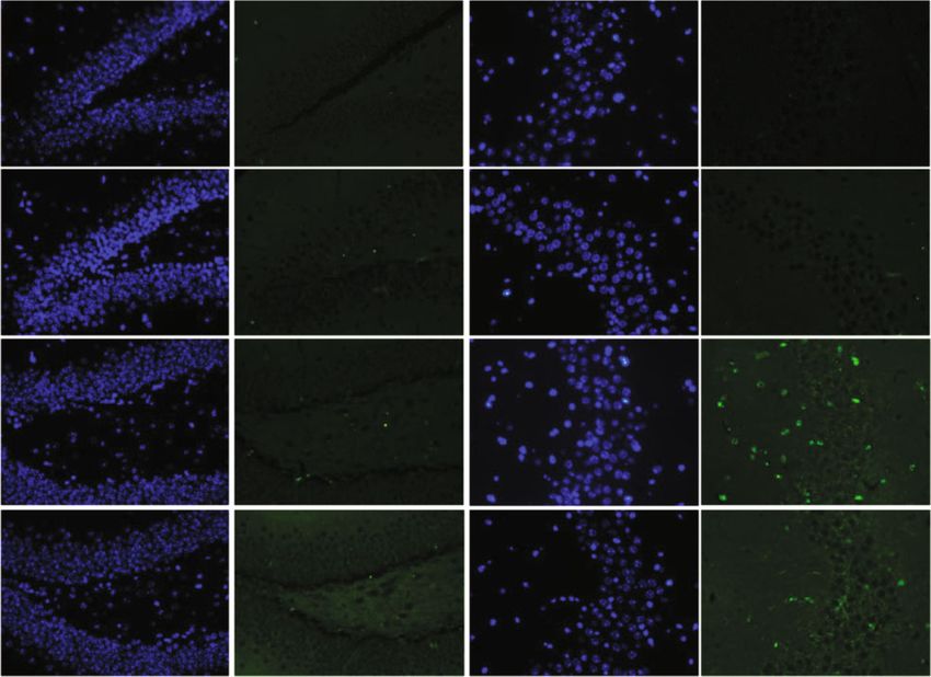
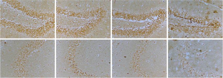
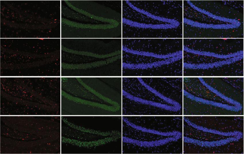
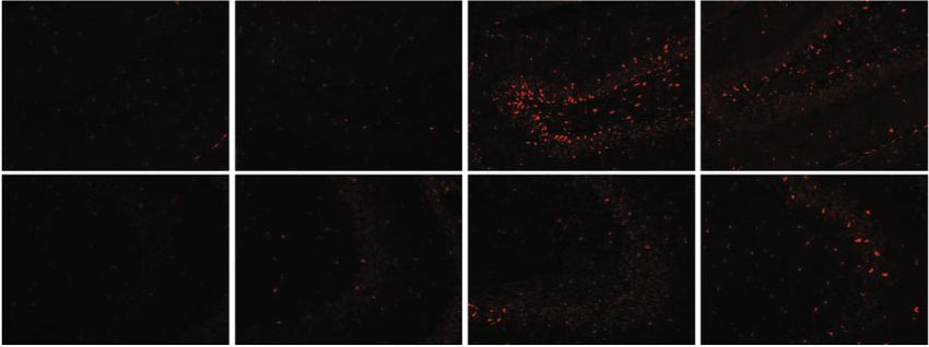
6 Oxidative Medicine and Cellular Longevity

imaged using chemiluminescence method. The mean density were lower in the shHamp group when exposed to normal
of the bands was calculated by ImageJ software. air or CIH (Figure 2(b)). Meanwhile, the double immunoflu-
 orescence showed repression of hepcidin expression secreted
2.10. PCR. The total RNA was extracted from the hippocam- by astrocyte in shHamp mice; the hepcidin protein level was
pus using RNA extraction kit following the manufacturer’s still lower than the normal even though it was elevated by
instructions. 1 μg total RNA was reversely transcribed into CIH exposure (Figure 2(c)). Compared with the WT mice,
cDNA. RT-PCR was used to identify the expression of hepci- the iron-positive cells were decreased in the hippocampus
din mRNA (primer 1) of shHamp mouse. The quantitated of shHamp mice during CIH exposure (Figures 2(d)
hepcidin mRNA (primer 2) in WT mouse was used as q- and 2(e)).
PCR with the Bio-Rad CFX Connect system. The primer However, how did the iron levels rise? For this purpose,
sequences used were as follows: β-actin: forward: AGACAT we detected the related proteins involved in iron metabolism.
TGCGATACCAATGCA, reverse: GCAACAGATACCAC The western blot results showed that the expression of TfR1,
ACTGGGAA; hepcidin primer 1: forward: TATCTCCGG iron intake of protein, was increased when subjected to CIH
CAACAGACGAG, reverse: TGTCTCGCTTCCTTCGCTT in the WT and shHamp mice (Figure 4(a)). During CIH
C; and hepcidin primer 2: forward: AGGCCCAGAGCAAG exposure, the shHamp exhibited a lower level of TfR1 com-
AGAGGTA, reverse: TCTCCATGTCGTCCCAGTTG. pared with WT mice (Figure 4(a)). Hepcidin regulated iron
 levels in neural cells mainly by binding to the iron-releasing
2.11. Statistical Analyses. The results are presented as the
 protein FPN1. We further found that the expression of
mean ± SEM. The statistical analysis was performed using
 FPN1 protein elevated in the hippocampal tissues and neu-
one-way ANOVA followed by the LSD post hoc test or
 ron of shHamp mice (Figures 4(b) and 4(d)). Compared with
t-test. Two-way ANOVA was used to analyze the results of
 the WT mice, the FPN1 protein levels in shHamp mice
the behavioral tests. The significance level was regarded as
 increased after CIH exposure (Figure 4(b)). Besides, we
p < 0:05.
 examined the FTL protein expression in the hippocampal
 and neurons to indirectly determine the iron levels. Similar
3. Results to our previous studies, the FTL protein levels rose in the hip-
3.1. Hepcidin Expression in the Hippocampus with Different pocampal tissues and neuron during CIH exposure
Exposed Times. The expression of hepcidin gene and protein (Figures 4(c) and 4(e)). The shHamp mice showed a lower
in the hippocampus was measured with different exposed level of FTL when exposed to CIH (Figure 4(c)). These results
times. As shown in Figure 1(b), hepcidin mRNA level was suggest that the knockdown of hepcidin might decrease the
increased after CIH exposure, and the rise persisted till to CIH-induced iron deposition, especially in neurons.
the day 21 of the experiment. Consistent with gene expres-
sion, immunohistochemical analysis showed that the expres- 3.3. Decreased ROS Levels in shHamp Mice Attenuated CIH-
sion of hepcidin protein in the hippocampus tissue was Induced Oxidative Stress. An excess of iron contributes to
increased (Figures 1(c) and 1(d)). Furthermore, the distrib- ROS production by Fenton reaction. Therefore, we detected
uted iron in the hippocampus was evaluated by Perls’ stain- the ROS levels and oxidative damage induced by CIH. We
ing. The images presented that the exposure of CIH could firstly found with the help of DHE probes that compared
significantly lead to the iron deposition in the mouse hippo- with WT mice, the increased mean fluorescence intensity in
campus, especially lasted to 21 days (Figures 1(e) and 1(f)). shHamp mice was significantly reduced during CIH
In addition, Nissl staining revealed that the Nissl body stain- exposure (Figures 5(a) and 5(b)). NADPH oxidases (NOXs)
ing became shallow and small when CIH was exposed to 21 could participate in ROS production in the mitochondria.
days, indicating the loss of neurons (Figures 1(g) and 1(h)). Compared with WT mice, western blot results revealed that
Therefore, we chose 21 days as the time point of CIH expo- the high level of NOX2 proteins was receded in the shHamp
sure in the subsequent experiment. These results suggested mice when subjected to CIH (Figure 5(c)). 4-Hydroxynonenal
that the high level of hepcidin might contribute to iron depo- (4-HNE), one of lipid peroxidation products, was increased in
sition and neuron loss in the hippocampus. the WT mice with CIH exposure (Figures 5(d)–5(f)). However,
 the higher 4-HNE level was weakened in the mice of the
3.2. Astrocyte-Specific Knockdown Hepcidin Decreased the shHamp+CIH group (Figures 5(d)–5(f)). These results reveal
Elevation of Iron Induced by CIH. To further characterize that hepcidin deficiency could reduce CIH-induced oxidative
the role of hepcidin in CIH-induced cognitive deficit, the stress damage.
mice of astrocyte-specific knockdown hepcidin were estab-
lished. Firstly, we compared hepcidin gene and protein 3.4. Effects of Hepcidin Deficiency on the Apoptosis in the
expression between shHamp and WT mice. RT-PCR Hippocampus. Oxidative stress is an initial factor of neuronal
revealed that hepcidin mRNA gene was decreased off 54% apoptosis when subjected to CIH exposure. We used TUNEL
after lateral ventricular injection of LV-U6-shHamp staining to observe the loss of neurons in the hippocampus
(Figure S1B). Therefore, we chose 14 days as the time point induced by CIH. As shown in Figures 6(a) and 6(b), a consid-
of shHamp mouse preparation. erable number of apoptosis bodies existed in the hippocam-
 Then, hepcidin mRNA levels were determined by q-PCR pus of WT mice subjected to CIH. However, the number of
in the hippocampus of shHamp mice exposed to CIH for 21 apoptosis bodies of the shHamp+CIH group was lower than
days. Compared to the WT group, hepcidin mRNA levels that of the WT+CIH group. Meanwhile, we found that the
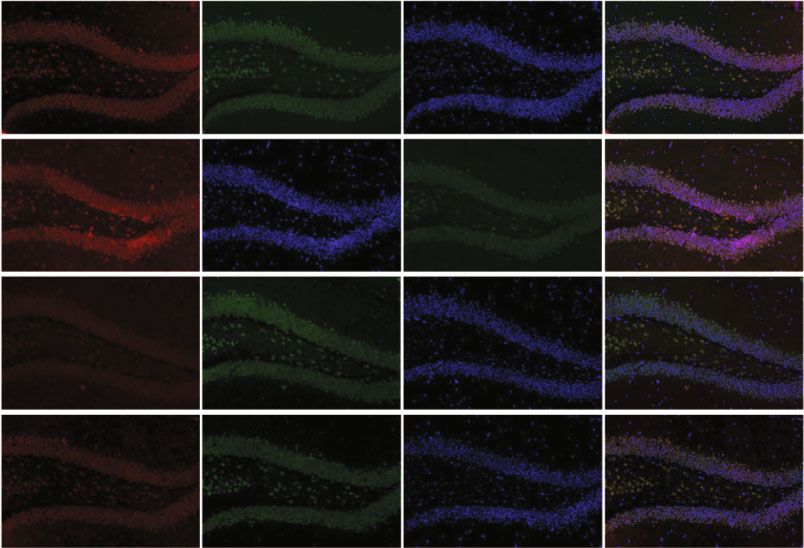
Oxidative Medicine and Cellular Longevity 7

 WT+CIH

 WT+CIH

 WT+CIH
 WT

 WT

 WT
 TfR1 92 kD FPN1 65 kD FTL 17 kD
 -Actin 43 kD -Actin 43 kD
 -Actin 43 kD

 $
 $
 0.8 # 1.0 1.0
 ⁎⁎ $ ⁎

 FPN1 protein level
 TfR1 protein level

 FTL protein level
 0.8 0.8
 0.6
 ⁎⁎
 0.6 0.6
 0.4
 0.4 0.4
 0.2 0.2 0.2
 0.0 0.0 0.0
 WT

 WT+CIH

 WT

 WT+CIH

 WT

 WT+CIH
 (a) (b) (c)
 FPN1 NeuN DAPI Merge

 WT

 WT+CIH

 (d)
 FTL NeuN DAPI Merge

 WT

 shHamp

 WT+CIH

 shHamp+CIH

 (e)

Figure 4: The expression of proteins related to iron metabolism in the hippocampus tissue and neurons. (a–c) The expression of TfR1, FPN1,
and FTL proteins measured by western blot (n = 6). (d, e) Double immunofluorescence staining in the hippocampal dentate gyrus (DG). The
sections were labeled for FPN1 or FTL (red), NeuN (green), and DAPI (blue) (scale bar = 25 μm, n = 3). The data are shown as the means
 ± SEM. ∗ p < 0:05 and ∗∗ p < 0:01 vs. WT. #p < 0:05 vs. shHamp. $p < 0:05 vs. WT+CIH.

decreased ratio of Bcl-2/Bax induced by CIH was increased in pared with the WT+CIH group, the ratio of p-JNK/JNK
the shHamp mice (Figures 6(c) and 6(d)). The ratio of p- was decreased in the shHamp+CIH group. These results sug-
JNK/JNK showed elevation in the WT and shHamp mice gest that hepcidin deficiency reduces apoptosis induced by
exposed to CIH, respectively (Figures 6(c) and 6(e)). Com- CIH exposure.
8 Oxidative Medicine and Cellular Longevity

 WT WT+CIH $

 fluorescence density
 1.0 ##
 ⁎⁎
 0.8 $

 The mean
 DG
 0.6 #
 ⁎⁎
 0.4
 0.2
 0.0
 CA3

 1 2
 WT

 WT+CIH

 (a) (b)
 WT WT+CIH
 WT+CIH
 WT

 NOX2 64 kD
 -Actin 43 kD
 DG

 $
 2.0
 NOX2 protein level

 #
 ⁎⁎
 1.5
 CA3

 1.0
 0.5
 0.0
 WT

 WT+CIH

 (c) (d)
 WT+CIH
 WT

 4-HNE 65kd
 -tubulin 55kd

 $ $$
 4-HNE protein level

 2.0
 0.3 ⁎⁎
 of 4-HNE protein
 The mean density

 #
 ⁎ 1.5
 0.2
 1.0
 0.1 0.5

 0.0 0.0
 WT

 WT+CIH
 WT

 WT+CIH

 (e) (f)

Figure 5: The oxidative stress levels in the WT and shHamp mice treated with CIH. (a) The DHE staining of the hippocampal dentate gyrus
(DG) and hippocampal CA3 (scale bar = 25 μm, n = 3). (b) The mean fluorescence intensity as shown in panel (a). (c) The expression and
statistics of NOX2 protein (n = 6). (d) The immunohistochemical staining of 4-HNE protein in the hippocampal dentate gyrus (DG) and
hippocampal CA3 (scale bar = 25 μm, n = 3). (e) The mean density of 4-HNE protein as shown in panel (d). (f) The expression and
statistics of 4-HNE protein measured by western blot (n = 4). The data are shown as the means ± SEM. ∗ p < 0:05 and ∗∗ p < 0:01 vs. WT.
#
 p < 0:05 and ##p < 0:01 vs. shHamp. $p < 0:05 and $$p < 0:01 vs. WT+CIH.
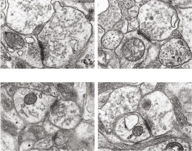
Oxidative Medicine and Cellular Longevity 9

 DAPI TUNEL DAPI TUNEL

 WT

 shHamp

 WT+CIH

 shHamp+CIH

 DG CA3
 (a)

 WT+CIH
 ##
 WT

 70 $$
 The total number of

 60 ⁎⁎
 Bcl-2 22 kD
 apoptotic cells

 50 Bax 28 kD
 54 kD
 30 p-JNK
 46 kD
 JNK 54 kD
 46 kD
 0
 WT

 WT+CIH

 (b) (c)
 $
 1.5 1.5 #
 #
 Ratio of p-JNK/JNK

 ⁎⁎
 Ratio of Bcl-2/Bax

 1.0 1.0
 ⁎⁎

 0.5 0.5

 0.0 0.0
 WT

 WT+CIH
 WT

 WT+CIH

 (d) (e)

Figure 6: The apoptosis in the hippocampus of WT and shHamp mice exposed to CIH. (a) The TUNEL staining of the hippocampal dentate
gyrus (DG) and hippocampal CA3 (scale bar = 25 μm, n = 3). (b) The total number of apoptotic bodies as shown in panel (a). (c–e) The ratio
of Bcl-2/Bax (n = 6) and p-JNK/JNK (n = 4) proteins. The data are shown as the means ± SEM. ∗∗ p < 0:01 vs. WT. # p < 0:05 and ##p < 0:01 vs.
shHamp. $p < 0:05 and $$p < 0:01 vs. WT+CIH.

3.5. Synaptic Plasticity and Recognition Memory Improvement Figure 7, the TEM images showed that the thickness and
in shHamp Mice Exposed to CIH. Studies have been reported length of postsynaptic density (PSD) were smaller and the
that the decrease of synaptic plasticity was related to memory width of synaptic cleft was wider in the CIH group compared
loss induced by CIH. We try to clarify hepcidin might be to that in the WT group. This indicated that the synaptic
involved in memory loss during CIH exposure. As shown in structure was damaged induced by CIH exposure.
10 Oxidative Medicine and Cellular Longevity

 WT 60

 post synaptic density (nm)
 ⁎

 The tickness of
 40

 20
 500 nm 500 nm
 WT+CIH
 0

 WT

 WT+CIH
 500 nm 500 nm
 (a) (b)

 The width of synaptic cleft (nm)
 400 30 $
 post synaptic density (nm)

 ⁎
 $
 300
 The length of

 20
 ⁎
 200
 10
 100

 0 0
 WT

 WT+CIH
 WT

 WT+CIH

 (c) (d)
 0.8
 $$
 shHamp+CIH

 BDNF protein level

 0.6
 ⁎⁎
 WT+CIH
 shHamp

 0.4
 WT

 BDNF 18 kD 0.2
 -Actin 43 kD
 0.0
 WT

 WT+CIH

 (e) (f)

Figure 7: The change of synaptic structure and BDNF expression in the WT and shHamp mice exposed to CIH. (a) The TEM of synaptic
structure of the mouse hippocampus in four different groups (n = 3). (b) The thickness of postsynaptic density. (c) The length of
postsynaptic density. (d) The width of synaptic cleft. (e, f) The expression of BDNF protein detected by western blot (n = 6). The data are
shown as the means ± SEM. ∗ p < 0:05 and ∗∗ p < 0:01 vs. WT. $p < 0:05 and $$p < 0:01 vs. WT+CIH.

Furthermore, the injury of synapse morphology was partially Furthermore, we studied the behavioral changes in WT
rescued in shHamp mice when compared with the WT+CIH and shHamp mice during CIH. The MWM was carried out
group (Figures 7(a), 7(c), and 7(d)). In addition, the expres- as shown in Figure 3(a). After exposure to CIH, the escape
sion of BDNF was significantly reduced in the WT mice latency time on days 18-20 was increased in the WT+CIH
treated with CIH (Figure 7(e)). The BDNF level was increased group (Figure 3(b)). Compared with the WT+CIH group,
in the shHamp+CIH group compared with that in the WT the escape latency time on days 19-20 was decreased in the
+CIH group. These results showed that the synaptic plasticity shHamp+CIH group. As shown in Figure 3(c), the escape
was improved in shHamp mice to resist to CIH-induced neu- route distance on day 20 was decreased in the shHamp+CIH
rological impairment. group compared with that in the WT+CIH group. On day 21,
Oxidative Medicine and Cellular Longevity 11

the CIH mice exhibited a decrease of number of passing times of neurons, which means that the neuron is vulnerable to
during the probe trial (Figure 3(d)). The decreased platform the toxicity of iron [15]. Therefore, lower level of FPN1 and
crosses were elevated in the shHamp mice. These results indi- higher level of FTL indicated an intracellular iron accumula-
cate that the gene loss of hepcidin could improve the cognitive tion status of neuron exposing to CIH (Figure 4). The data
dysfunction when exposed to CIH. indicates that unbalance of hepcidin-FPN1 might be the
 main contributor to the iron overload in the hippocampus
4. Discussion of mice exposed to CIH.
 In addition, the iron-responsive element (IRE)/iron regu-
OSA is an independent risk factor for the development of latory proteins (IRPs) are also important posttranscription-
cognitive impairment. Our previous studies have proved that ally regulatory switch during different iron states [32]. FTL
it is mainly attributed to oxidative stress injury and iron and TfR1 mRNA contain IRE in their 5 ′ UTR and 3 ′ UTR,
deposition in the hippocampus of mice exposed to CIH. respectively [32]. During CIH exposure, iron mobilization
However, the mechanisms underlying how the different neu- plays an indispensable role in meeting the oxygen and energy
ral cells are involved in regulating iron remain largely demand of the brain, and thus, the expression of TfR1 is
unknown. In this study, we revealed an underappreciated upregulated to improve iron uptake by promoting the combi-
role of hepcidin in the cognitive impairment induced by nation of IRE and IRPs [24]. The activity of IRE-binding
CIH. During three weeks of CIH exposure, with 8 hours IRPs is diminished in states of iron overload and thereby
per day of 5% O2 saturation, hepcidin was induced in the hip- plays a role in inhibiting TfR1 expression and promoting
pocampus. The high expression of hepcidin secreted by FTL expression [33, 34]. Similarly, the expression of TfR1
astrocyte was associated with excessive iron in neurons in was consistent with the iron level in the shHamp+CIH group
the CIH mouse model. Specific deficiency of hepcidin gene compared to that in the WT+CIH group. Concordantly, we
could alleviate the toxic damage of iron induced by CIH. found a decreased expression of FTL in the shHamp+CIH
 Hepcidin plays a fundamental role in maintaining sys- group, which may be attributed to the decrease in iron levels
temic iron by inhibiting iron absorption from the duodenum
 without the need for excessive FTL for binding. These results
and releasing from macrophages via degradation of FPN1
 inferred that the hepcidin deficiency influenced iron uptake
[25, 26]. The abnormally elevated expression of hepcidin is
 and decreased brain iron overload, thereby protecting against
associated with various chronic conditions, which may lead
 CIH-induced injury.
to iron excess [11]. However, deletion of hepcidin gene is also
 However, the role of hepcidin in the development of cogni-
a key factor to induce iron overload, which shares the similar
 tive impairment-related diseases remains controversial. The
pathological phenotypes as hereditary hemochromatosis
 AD mice show a decrease of hepcidin in the CSF or hippocam-
(HH) [11, 27, 28]. Evidence from animal studies suggests that
 pus [16, 35]. If the hepcidin is overexpressed in astrocytes,
young Hamp knockout mice present dramatically systemic
iron overload reflected in elevated serum and hepatic iron, amyloid-β induced the cognitive impairment and toxic iron
owing to the failure of hepcidin-FPN1 axis [28]. The iron damage in neurons could be improved [34]. Nevertheless, the
deposition of Hamp knockout mice presents a time- hepcidin has a reverse expression level in the stroke animal
dependent trend in peripheral organs [28], rather than in models. Adding hepcidin could aggravate brain injury and iron
the brain [29]. It has been reported that inhibition of hepci- overload in the rats subjected to subarachnoid hemorrhage
din expression in the brain could weaken iron accumulation [36]. Specific inhibition of the expression of hepcidin has been
and oxidative injury in the rodent model of ischemia- demonstrated to attenuate neurologic deficit and brain iron
reperfusion [18] or intracerebral hemorrhage [29], which deposition in the rodent models of subarachnoid hemorrhage
indicated that the brain iron metabolism was readily regu- [36], ischemic stroke [18, 19], and intracerebral hemorrhage
lated by the local hepcidin [25]. [29]. In our study, inhibition of hepcidin gene also improved
 As the most abundant cell type within the CNS, astro- the cognition ability of CIH mice. The reason may be attributed
cytes are the main type of glial cells and play essential roles rather to the similar pathological characteristics of CIH model
in iron transport of the brain, most particularly in maintain- to ischemia-reperfusion, which mainly manifests as repeated
ing the iron transport in neuronal cells [30]. Astrocytes are hypoxia and reoxygenation recycling [6].
also the main glia cells which produce and secrete hepcidin CIH exposure in OSA leads to increased ROS production;
[13, 30]. Studies have demonstrated that CIH exposure could excessive iron generates hydroxyl radicals via the Fenton
further induce the activation of astrocytes in APP/PS1 mice reaction and induces the overproduction of ROS, resulting
and exacerbate the pathogenesis of disease [3]. Coinciden- in neuronal damage consequently [37–39]. In vitro experi-
tally, results of the present study indicated the increased ment has confirmed that there is a positive correlation
astrocyte activation during CIH exposure, and the expression between iron level and ROS content in neurons [39]. Like-
level of hepcidin also increased concomitantly (Figure 1). wise, lower level of iron and ROS has been demonstrated in
Some studies have indicated that high expression of hepcidin the shHamp mice subjected to CIH exposure. NADPH oxi-
is able to promote the degradation of FPN1 and iron deposi- dase (NOX) enzymes could transport electrons across the
tion in neurons in the ischemic brain or LPS stimulus [18, plasma membrane and participate in the production of
31]. Our previous study found a decrease of FPN1 protein ROS in the mitochondria [40]. NOX2 is localized in synaptic
in the hippocampus when exposed to CIH for 21 days [24]. sites of neurons and plays a role in superoxide-dependent
In the brain, FPN1 is more expressed in the cell membranes long-term potentiation and memory function [41]. Similar
12 Oxidative Medicine and Cellular Longevity

to previous studies, NOX2 expression increases after CIH expo- Conflicts of Interest
sure in WT mice [24]; however, a decrease of NOX2 appears in
case of the deficiency of hepcidin genes. The polyunsaturated The authors declare no conflict of financial interest or
fatty acids (PUFAs) are readily be oxidized with excessive benefit.
ROS [42]. 4-HNE is one of the typical lipid peroxidation prod-
ucts and primary attack proteins, DNA, and membrane lipids Authors’ Contributions
[43]. CIH exposure significantly increases 4-HNE levels, indi-
cating that oxidative stress damage occurs in the hippocampus. Ya-Shuo Zhao, Miao Tan, and En-Sheng Ji carried out the
Study has demonstrated that the iron level is positively corre- experiments, analyzed the results, and wrote the manuscript.
lated with NOX2 [19] and 4-HNE content, respectively [34]. Ji-Xian Song and Ji-Ren An performed the CIH model and
These results further confirm that the CIH mice have a lower behavioral testing. Xin-Yue Yang, Wen-Ya Li, and Ya-Jing
iron level and a decrease of oxidative damage. Guo performed the immunofluorescence and western
 Studies have confirmed that CIH-induced OSA could blot. Ya-Shuo Zhao and Miao Tan contributed equally
lead to neuronal apoptosis and dysfunction in the hippocam- to this work.
pus [44] because of the production of ROS and spread of
oxidative stress [45]. Apoptosis is mainly controlled by two Acknowledgments
cascades, including kinase cascades and protease cascades
[46]. MAPK cascade is one of the most important members This work was supported by projects of the National Natural
of kinase cascade and could be activated in stress responses Science Foundation of China (grant number 82004127), the
like ROS [47]. Our previous studies showed that JNK- Natural Science Foundation of Hebei Province (grant num-
MAPK was activated in cardiac tissue when exposed to CIH ber C2019423117), and the Science and Technology Capabil-
[6]. The activated JNK declines the ratio of Bcl-2/Bax, result- ity Enhancement Project of Hebei University of Chinese
ing in mitochondrial dysfunction and apoptosis [19]. In our Medicine (grant number KTY2019060).
study, the activated JNK-MAPK was presented in the hippo-
campus of CIH mice, and the activation was suppressed in
shHamp mice. Furthermore, these data suggested the corre- Supplementary Materials
lation between JNK and iron levels during CIH exposure. Figure S1: the structure of lentivirus plasmid LV-U6-shHamp
 The synapse, a specialized structure of neurons, is a key and identification of shHamp mice. (A) The structure of lenti-
part of the information transmission between neurons [48]. virus plasmid LV-U6-shHamp with the astrocytes promoters
Synaptic plasticity is associated with learning and memory followed by Hamp gene shRNA sequence. (B) The expression
formation [49]. We observed the shortening of the active of hepcidin mRNA by RT-PCR in the hippocampus of
zone during CIH expose, which indicated that the synaptic shHamp mice. The data are shown as the means ± SEM.
transmission and plasticity were weakened [8]. BDNF has ∗
 p < 0:05 vs. WT (n = 6). (Supplementary Materials)
emerged as an important role in synaptic plasticity and neu-
ronal survival [50]. On the one hand, the loss of BDNF leads References
to damage of long-term memory and participates in neuro-
cognitive impairment in rodent of CIH [8, 24]. On the other [1] A. G. Andrade, O. M. Bubu, A. W. Varga, and R. S. Osorio,
hand, BDNF significantly prevents neuron damage and apo- “The relationship between obstructive sleep apnea and Alzhei-
ptosis induced by ROS [51]. Our results revealed that the mer’s disease,” Journal of Alzheimer's Disease, vol. 64, no. s1,
decrease of synaptic plasticity and BDNF level were all signif- pp. S255–S270, 2018.
icantly increased in the shHamp mice subjected to CIH. [2] A. V. Benjafield, N. T. Ayas, P. R. Eastwood et al., “Estimation
Therefore, these data confirm that hepcidin is involved in of the global prevalence and burden of obstructive sleep
the dysfunction of synaptic plasticity induced by CIH. apnoea: a literature-based analysis,” The Lancet Respiratory
 Medicine, vol. 7, no. 8, pp. 687–698, 2019.
 [3] T. Macheda, K. Roberts, D. N. Lyons et al., “Chronic Intermit-
5. Conclusion tent Hypoxia Induces Robust Astrogliosis in an Alzheimer's
In conclusion, our result demonstrates the role of hepcidin in Disease-Relevant Mouse Model,” Neuroscience, vol. 398,
 pp. 55–63, 2019.
CIH-induced cognitive impairment. First of all, hepcidin is
induced during CIH exposure, accelerating the iron overload [4] R. A. Sharma, A. W. Varga, O. M. Bubu et al., “Obstructive
 sleep apnea severity affects amyloid burden in cognitively nor-
in the hippocampus and cognitive impairment. Furthermore,
 mal elderly. A longitudinal study,” American Journal of Respi-
when the hepcidin gene is specifically knocked down in ratory and Critical Care Medicine, vol. 197, no. 7, pp. 933–943,
astrocyte, the excess of iron content in neuron and oxidative 2018.
stress are decreased, and neuronal apoptosis and synaptic [5] J. Attier-Zmudka, J. M. Serot, J. Valluy et al., “Sleep apnea syn-
plasticity are improved when subjected to CIH. drome in an elderly population admitted to a geriatric unit:
 prevalence and effect on cognitive function,” Frontiers in
Data Availability Aging Neuroscience, vol. 11, p. 361, 2020.
 [6] Y. S. Zhao, J. R. An, S. Yang et al., “Hydrogen and oxygen mix-
The data used to support the findings of this study are ture to improve cardiac dysfunction and myocardial patholog-
included within the article. ical changes induced by intermittent hypoxia in rats,”
Oxidative Medicine and Cellular Longevity 13

 Oxidative Medicine and Cellular Longevity, vol. 2019, Article [23] O. Abakay, A. Abakay, Y. Palanci et al., “Relationship between
 ID 7415212, 12 pages, 2019. hepcidin levels and periodic limb movement disorder in
 [7] T. DeMartino, R. E. Ghoul, L. Wang et al., “Oxidative stress patients with obstructive sleep apnea syndrome,” Sleep &
 and inflammation differentially elevated in objective versus Breathing, vol. 19, no. 2, pp. 459–466, 2015.
 habitual subjective reduced sleep duration in obstructive sleep [24] J. R. An, Y. S. Zhao, L. F. Luo, P. Guan, M. Tan, and E. S. Ji,
 apnea,” Sleep, vol. 39, no. 7, pp. 1361–1369, 2016. “Huperzine A, reduces brain iron overload and alleviates cog-
 [8] L. H. Xu, H. Xie, Z. H. Shi et al., “Critical role of endoplasmic nitive deficit in mice exposed to chronic intermittent hypoxia,”
 reticulum stress in chronic intermittent hypoxia-induced def- Life Sciences, vol. 250, article 117573, 2020.
 icits in synaptic plasticity and long-term memory,” Antioxi- [25] T. Ganz, “Hepcidin and iron regulation, 10 years later,” Blood,
 dants & Redox Signaling, vol. 23, no. 9, pp. 695–710, 2015. vol. 117, no. 17, pp. 4425–4433, 2011.
 [9] L. Zhou, P. Chen, Y. Peng, and R. Ouyang, “Role of oxidative [26] E. Nemeth and T. Ganz, “The role of hepcidin in iron metab-
 stress in the neurocognitive dysfunction of obstructive sleep olism,” Acta Haematologica, vol. 122, no. 2-3, pp. 78–86, 2009.
 apnea syndrome,” Oxidative Medicine and Cellular Longevity, [27] A. Piperno, D. Girelli, E. Nemeth et al., “Blunted hepcidin
 vol. 2016, Article ID 9626831, 15 pages, 2016. response to oral iron challenge in HFE-related hemochroma-
[10] M. A. Khuu, C. M. Pagan, T. Nallamothu et al., “Intermittent tosis,” Blood, vol. 110, no. 12, pp. 4096–4100, 2007.
 hypoxia disrupts adult neurogenesis and synaptic plasticity [28] L. Sun, W. Guo, C. Yin et al., “Hepcidin deficiency undermines
 in the dentate gyrus,” The Journal of Neuroscience, vol. 39, bone load-bearing capacity through inducing iron overload,”
 no. 7, pp. 1320–1331, 2019. Gene, vol. 543, no. 1, pp. 161–165, 2014.
[11] N. L. Blanchette, D. H. Manz, F. M. Torti, and S. V. Torti, [29] X. Y. Xiong, L. Liu, F. X. Wang et al., “Toll-like receptor
 “Modulation of hepcidin to treat iron deregulation: potential 4/MyD88-mediated signaling of hepcidin expression causing
 clinical applications,” Expert Review of Hematology, vol. 9, brain iron accumulation, oxidative injury, and cognitive
 no. 2, pp. 169–186, 2016. impairment after intracerebral hemorrhage,” Circulation,
[12] S. M. Wang, L. J. Fu, X. L. Duan et al., “Role of hepcidin in vol. 134, no. 14, pp. 1025–1038, 2016.
 murine brain iron metabolism,” Cellular and Molecular Life [30] D. Vela, “The dual role of hepcidin in brain iron load and
 Sciences, vol. 67, no. 1, pp. 123–133, 2010. inflammation,” Frontiers in Neuroscience, vol. 12, p. 740, 2018.
[13] D. Vela, “Hepcidin, an emerging and important player in brain [31] L. H. You, C. Z. Yan, B. J. Zheng et al., “Astrocyte hepcidin is a
 iron homeostasis,” Journal of Translational Medicine, vol. 16, key factor in LPS-induced neuronal apoptosis,” Cell Death &
 no. 1, p. 25, 2018. Disease, vol. 8, no. 3, article e2676, 2017.
[14] F. Codazzi, I. Pelizzoni, D. Zacchetti, and F. Grohovaz, “Iron [32] N. Wilkinson and K. Pantopoulos, “The IRP/IRE system
 entry in neurons and astrocytes: a link with synaptic activity,” in vivo: insights from mouse models,” Frontiers in Pharmacol-
 Frontiers in Molecular Neuroscience, vol. 8, p. 18, 2015. ogy, vol. 5, p. 176, 2014.
[15] G. M. Bishop, T. N. Dang, R. Dringen, and S. R. Robinson, [33] T. A. Rouault, “The role of iron regulatory proteins in mam-
 “Accumulation of non-transferrin-bound iron by neurons, malian iron homeostasis and disease,” Nature Chemical Biol-
 astrocytes, and microglia,” Neurotoxicity Research, vol. 19, ogy, vol. 2, no. 8, pp. 406–414, 2006.
 no. 3, pp. 443–451, 2011. [34] X. Zhang, Y. J. Gou, Y. Zhang et al., “Hepcidin overexpression
[16] Y. S. Zhao, L. H. Zhang, P. P. Yu et al., “Ceruloplasmin, a in astrocytes alters brain iron metabolism and protects against
 potential therapeutic agent for Alzheimer’s disease,” Antiox- amyloid-β induced brain damage in mice,” Cell Death Discov-
 idants & Redox Signaling, vol. 28, no. 14, pp. 1323–1337, ery, vol. 6, no. 1, p. 113, 2020.
 2018. [35] Z. M. Qian and Y. Ke, “Hepcidin and its therapeutic potential
[17] X. Guan, Y. Zhang, H. Wei et al., “Iron-related nigral degener- in neurodegenerative disorders,” Medicinal Research Reviews,
 ation influences functional topology mediated by striatal dys- vol. 40, no. 2, pp. 633–653, 2020.
 function in Parkinson's disease,” Neurobiology of Aging, [36] G. Tan, L. Liu, Z. He, J. Sun, W. Xing, and X. Sun, “Role of hep-
 vol. 75, pp. 83–97, 2019. cidin and its downstream proteins in early brain injury after
[18] H. Ding, C. Z. Yan, H. Shi et al., “Hepcidin is involved in iron experimental subarachnoid hemorrhage in rats,” Molecular
 regulation in the ischemic brain,” PLoS One, vol. 6, no. 9, arti- and Cellular Biochemistry, vol. 418, no. 1-2, pp. 31–38, 2016.
 cle e25324, 2011. [37] P. J. Urrutia, N. P. Mena, and M. T. Nunez, “The interplay
[19] Y. Zhao, Z. Xin, N. Li et al., “Nano-liposomes of lycopene between iron accumulation, mitochondrial dysfunction, and
 reduces ischemic brain damage in rodents by regulating iron inflammation during the execution step of neurodegenerative
 metabolism,” Free Radical Biology & Medicine, vol. 124, disorders,” Frontiers in Pharmacology, vol. 5, p. 38, 2014.
 pp. 1–11, 2018. [38] I. Pelizzoni, R. Macco, M. F. Morini, D. Zacchetti, F. Grohovaz,
[20] Y. Liu, Z. Yu, D. Hua, Y. Chen, S. Zheng, and L. Wang, “Asso- and F. Codazzi, “Iron handling in hippocampal neurons:
 ciation of serum hepcidin levels with the presence and severity activity-dependent iron entry and mitochondria-mediated
 of obstructive sleep apnea syndrome,” Medical Science Moni- neurotoxicity,” Aging Cell, vol. 10, no. 1, pp. 172–183, 2011.
 tor, vol. 21, pp. 27–31, 2015. [39] L. X. Tao, X. T. Huang, Y. T. Chen, X. C. Tang, and H. Y. Zhang,
[21] I. Baik, S. Lee, R. J. Thomas, and C. Shin, “Obstructive sleep “Acetylcholinesterase-independent protective effects of huper-
 apnea, low transferrin saturation levels, and male-pattern zine A against iron overload-induced oxidative damage and
 baldness,” International Journal of Dermatology, vol. 58, aberrant iron metabolism signaling in rat cortical neurons,” Acta
 no. 1, pp. 67–74, 2019. Pharmacologica Sinica, vol. 37, no. 11, pp. 1391–1400, 2016.
[22] L. M. O'Brien, J. Koo, L. Fan et al., “Iron stores, periodic leg [40] K. Bedard and K. H. Krause, “The NOX family of ROS-
 movements, and sleepiness in obstructive sleep apnea,” Jour- generating NADPH oxidases: physiology and pathophysiol-
 nal of Clinical Sleep Medicine, vol. 5, no. 6, pp. 525–531, 2009. ogy,” Physiological Reviews, vol. 87, no. 1, pp. 245–313, 2007.
14 Oxidative Medicine and Cellular Longevity

[41] M. V. Tejada-Simon, F. Serrano, L. E. Villasana et al., “Synap-
 tic localization of a functional NADPH oxidase in the mouse
 hippocampus,” Molecular and Cellular Neurosciences, vol. 29,
 no. 1, pp. 97–106, 2005.
[42] G. Barrera, S. Pizzimenti, E. S. Ciamporcero et al., “Role of 4-
 hydroxynonenal-protein adducts in human diseases,” Antiox-
 idants & Redox Signaling, vol. 22, no. 18, pp. 1681–1702, 2015.
[43] H. Zhong and H. Yin, “Role of lipid peroxidation derived 4-
 hydroxynonenal (4-HNE) in cancer: focusing on mitochon-
 dria,” Redox Biology, vol. 4, pp. 193–199, 2015.
[44] Q. Yang, Y. Wang, J. Feng, J. Cao, and B. Chen, “Intermittent
 hypoxia from obstructive sleep apnea may cause neuronal
 impairment and dysfunction in central nervous system: the
 potential roles played by microglia,” Neuropsychiatric Disease
 and Treatment, vol. 9, pp. 1077–1086, 2013.
[45] W. Xu, L. Chi, B. W. Row et al., “Increased oxidative stress is
 associated with chronic intermittent hypoxia- mediated brain
 cortical neuronal cell apoptosis in a mouse model of sleep
 apnea,” Neuroscience, vol. 126, no. 2, pp. 313–323, 2004.
[46] L. Chang and M. Karin, “Mammalian MAP kinase signalling
 cascades,” Nature, vol. 410, no. 6824, pp. 37–40, 2001.
[47] A. Minden and M. Karin, “Regulation and function of the JNK
 subgroup of MAP kinases,” Biochimica et Biophysica Acta,
 vol. 1333, no. 2, pp. F85–104, 1997.
[48] G. Neves, S. F. Cooke, and T. V. Bliss, “Synaptic plasticity,
 memory and the hippocampus: a neural network approach
 to causality,” Nature Reviews Neuroscience, vol. 9, no. 1,
 pp. 65–75, 2008.
[49] E. Tonnies and E. Trushina, “Oxidative stress, synaptic dys-
 function, and Alzheimer’s disease,” Journal of Alzheimer's Dis-
 ease, vol. 57, no. 4, pp. 1105–1121, 2017.
[50] Y. Lu, K. Christian, and B. Lu, “BDNF: a key regulator for pro-
 tein synthesis-dependent LTP and long-term memory?,” Neu-
 robiology of Learning and Memory, vol. 89, no. 3, pp. 312–323,
 2008.
[51] N. I. Boyadjieva and D. K. Sarkar, “Cyclic adenosine mono-
 phosphate and brain-derived neurotrophic factor decreased
 oxidative stress and apoptosis in developing hypothalamic
 neuronal cells: role of microglia,” Alcoholism, Clinical and
 Experimental Research, vol. 37, no. 8, pp. 1370–1379, 2013.
You can also read


