Hantaan Virus Infection Causes an Acute Neurological Disease That Is Fatal in Adult Laboratory Mice
←
→
Page content transcription
If your browser does not render page correctly, please read the page content below
JOURNAL OF VIROLOGY, Sept. 2002, p. 8890–8899                                                                                    Vol. 76, No. 17
0022-538X/02/$04.00⫹0 DOI: 10.1128/JVI.76.17.8890–8899.2002
Copyright © 2002, American Society for Microbiology. All Rights Reserved.

  Hantaan Virus Infection Causes an Acute Neurological Disease That
                  Is Fatal in Adult Laboratory Mice
    Dominic Wichmann,1 Hermann-Josef Gröne,2 Michael Frese,3† Jovan Pavlovic,4 Bärbel Anheier,1
                    Otto Haller,3 Hans-Dieter Klenk,1 and Heinz Feldmann5*
          Institut für Virologie, Philipps-Universität, Marburg,1 Department of Cellular and Molecular Pathology, Deutsches
             Krebsforschungszentrum, Heidelberg,2 and Institut für Medizinische Mikrobiologie und Hygiene, Universität
                Freiburg, Freiburg,3 Germany; Institut für Medizinische Virologie, Zürich, Switzerland4; and Canadian
                            Science Centre for Human and Animal Health, National Microbiology Laboratory,
                                              Health Canada, Winnipeg, Manitoba, Canada5

                                                                                                                                                   Downloaded from http://jvi.asm.org/ on December 29, 2020 by guest
                                            Received 26 December 2001/Accepted 21 May 2002

             Hantaan virus, the etiological agent of Korean hemorrhagic fever, is transmitted to humans from persis-
          tently infected mice (Apodemus agrarius), which serve as the primary reservoir. Here we demonstrate that
          several strains of adult Mus musculus domesticus (C57BL/6, BALB/c, AKR/J, and SJL/J) were susceptible to
          Hantaan virus infection when infected intraperitoneally. First clinical signs were loss of weight, ruffled fur, and
          reduced activity, which were followed by neurological symptoms, such as paralyses and convulsions. Within 2
          days of disease onset, the animals died of acute encephalitis. PCR analysis indicated a systemic infection with
          viral RNA present in all major organs. Immunohistochemical and in situ hybridization analyses of postmortem
          material detected viral antigen and RNA in the central nervous system (predominantly brain), liver, and
          spleen. In the central nervous system, viral antigen and RNA colocalized with perivascular infiltrations, the
          predominant pathological finding. To investigate the involvement of the interferon system in Hantaan virus
          pathogenesis, we infected alpha/beta interferon receptor knockout mice. These animals were more susceptible
          to Hantaan virus infection, indicating an important role of interferon-induced antiviral defense mechanisms
          in Hantaan virus pathogenesis. The present model may help to overcome shortcomings in the development of
          therapeutic and prophylactic measurements against hantavirus infections.

   The genus Hantavirus is one of five genera in the family                   virus (HTNV) infections. The cardinal manifestations of
Bunyaviridae. Hantaviruses possess a tripartite, single-strand-               HFRS are fever, hemorrhages, and renal impairment. Severe
ed, negative-sense RNA genome with segments designated                        HFRS, caused by HTNV, Dobrava virus, and sometimes Seoul
large (L), medium (M), and small (S) (2, 15, 31). In contrast to              virus, shows several phases (febrile, hypotensive, oliguric, poly-
other bunyaviruses, hantaviruses are rodent-borne human                       uric, and convalescent) with variable lengths and transitions
pathogens that persistently infect their rodent reservoirs with-              which are not always present in moderate or mild cases. Im-
out causing obvious clinical signs of illness. Infected rodents               portant laboratory findings are a rise in serum creatinine and
shed the virus in urine, saliva, and feces, and transmission to               blood urea nitrogen levels, thrombocytopenia, proteinuria, and
humans normally occurs through the aerosol route when small-                  changes in serum electrolytes (5, 27).
particle aerosols of contaminated excreta are inhaled. There-                    Hantaviruses primarily affect blood vessels and lead to vari-
fore, the worldwide appearance of human hantavirus disease is                 able degrees of generalized capillary dilatation and edema.
primarily determined by the geographic distribution of the                    Viral antigens are found within capillary endothelium of vari-
rodent reservoirs of the different hantaviruses (5, 30, 32).                  ous tissues. Despite the extensive accumulation of viral anti-
   Two clinical syndromes are currently known to be associated                gens, there is little ultrastructural evidence of a cytopathic
with hantavirus infections in humans: hemorrhagic fever with                  effect in endothelial cells. A hallmark of pathogenesis is in-
renal syndrome (HFRS) and hantavirus pulmonary syndrome                       creased vascular permeability that seems to be due to endo-
(HPS). These can be further distinguished by clinical symp-                   thelial cell dysfunction. By activating complement and by trig-
toms into severe, moderate, and mild HFRS, classical HPS,                     gering mediator release from platelets and immune effector
and a renal variant of HPS (5, 27, 30). HFRS especially con-                  cells, immune complexes may be involved in vascular injury.
tinues to cause significant numbers of human illnesses in Asia                Thus, many data are consistent with the hypothesis that han-
and Europe, with approximately 100,000 cases per year (16,                    tavirus-induced diseases are due to immunologically mediated
22). HFRS lethality ranges from ⬍1% for Puumala virus                         capillary leakage in target organs, which differ for HFRS and
(PUUV) infections to approximately 5 to 15% for Hantaan                       HPS. The systemic manifestation of capillary dysfunction leads
                                                                              to hypotension and shock, which may be influenced by addi-
                                                                              tional factors, such as virus distribution and virus-induced re-
  * Corresponding author. Mailing address: Canadian Science Centre            lease of cytokines and other inflammatory mediators. The on-
for Human and Animal Health, 1015 Arlington St., Winnipeg, Mani-              set of disease coincides with the appearance of a specific
toba R3E 3R2, Canada. Phone: (204) 789-6019. Fax: (204) 789-2140.
E-mail: Heinz_Feldmann@hc-sc.gc.ca.
                                                                              immune response, and immune complexes can be detected in
  † Present address: Department of Molecular Virology, University of          HFRS patients (5, 17, 22, 28, 41).
Heidelberg, Heidelberg, Germany.                                                 Hantavirus research has long been hampered by the lack of

                                                                       8890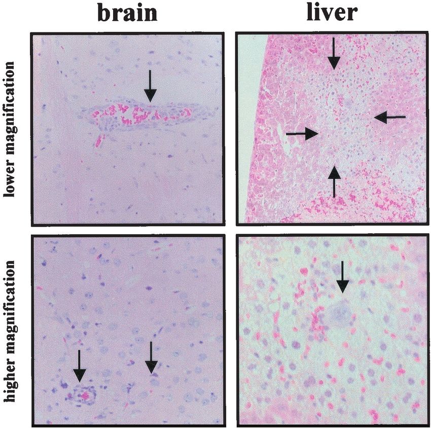
VOL. 76, 2002 LETHAL HTNV INFECTION IN MICE 8891
suitable animal models. However, cynomolgus macaques (Ma- for 30 min with rabbit anti-mouse immunoglobulin G (IgG) (Dako, Hamburg,
caca fascicularis) have been successfully used to mimic nephro- Germany) to block endogenous mouse IgG. After extensive phosphate-buffered
saline washes, antigen detection was performed by using anti-HTNV polyclonal
pathia epidemica, a mild form of HFRS caused by PUUV (8, mouse IgG followed by rabbit anti-mouse IgG conjugated to horseradish perox-
19). Laboratory mice (Mus musculus) and rats (Rattus norve- idase (Dako); subsequent staining was done with diaminobenzidine tetrahydro-
gicus) have been used to study hantavirus pathogenicity, but chloride (Dako). Anti-HTNV polyclonal mouse IgG was obtained from HTNV-
lethal disease could be induced only in newborn or immuno- infected C57BL/6 mice.
In situ hybridization. A fragment of 350 nucleotides from the S segment
deficient animals (13, 18, 21, 24, 39, 40). Most recently, an HPS
(positions 37 to 1345) of strain 76-118 of HTNV was amplified and cloned into
disease model with Syrian hamsters (Mesocricetus auratus) was plasmid pCI (Promega, Mannheim, Germany). 32P-labeled in vitro runoff tran-
reported for Andes virus (ANDV), one of the South American scripts were produced by using an SP6/T7 transcription kit (Boehringer Mann-
hantaviruses (12). This exciting model, which so far works only heim, Mannheim, Germany) and purified by using Nuc Trap probe purification
for ANDV, will be of great use for the development of vaccine columns (Stratagene, Heidelberg, Germany). Hybridization was performed as
described previously (3). Briefly, the sections were deparaffinized with xylol, fixed
candidates and antiviral substances. However, it is less suitable in 4% formaldehyde, and treated with 10% hydrochloric acid and 10 g of
for the characterization of host genetic factors involved in the proteinase K/ml (37°C for 20 min). Subsequently, the sections were hybridized
Downloaded from http://jvi.asm.org/ on December 29, 2020 by guest
pathogenesis of hantavirus infections due to the lack of proper with the radiolabeled probe, washed, and dehydrated by using increasing ethanol
genetic and immunological tools for studies with hamsters. concentrations (50, 70, 80, 90, and 100% for 5 min each). The tissue sections
were dipped in NTB2 photoemulsion (Kodak, Rochester, N.Y.) and stored in the
Here we report that HTNV infection is lethal for several
dark. After 7 days, the photoemulsion was developed, and the sections were
inbred strains of M. musculus domesticus. Knockout mice lack- counterstained with HE and examined by bright- and dark-field microscopy with
ing a functional receptor for alpha/beta interferon (IFN-␣/) an Axiolab microscope (Zeiss, Göttingen, Germany).
(25) are even more susceptible to the infection than wild-type RT-PCR. RNA was extracted by using an RNeasy kit (Qiagen, Hilden, Ger-
mice, indicating an important role of IFN-induced antiviral many), and RT-PCR was performed by using a Titan one-tube RT-PCR system
(Boehringer Mannheim) and a Perkin-Elmer model 2400 thermocycler. The RT
defense mechanisms in HTNV pathogenesis. Although clinical reaction was performed at 50°C for 30 s and was followed by 40 amplification
symptoms in HTNV-infected mice differ from those of HFRS cycles (94°C for 30 s and 68°C for 90 s). The following L segment-specific
in humans, the mouse model will be helpful for studying han- oligonucleotides were used: LH3forward (5⬘-ATG AAA CTC TGT GCC ATC
tavirus pathogenesis and genetics. Furthermore, it will be use- TTT GAC-3⬘; positions 1886 to 1910) and LH4reverse (5⬘-CCA CTT TGT AGC
ATC TGC ACT AAC-3; positions 2966 to 2941). All products were subsequently
ful for developing vaccines against HFRS.
gel purified and sequenced for confirmation.
(Dominic Wichmann performed this work in partial fulfill- Animal protection experiments. C57BL/6 mice were immunized by three con-
ment of the requirements for a Ph.D. degree from Philipps- secutive intraperitoneal and subcutaneous injections of either VVHTNV/GPC
Universität, Marburg, Germany.) (105 PFU) or VVT7 (105 PFU) (controls) administered 2 weeks apart. HTNV-
specific antibody titers were determined by using a previously published enzyme-
linked immunosorbent assay (ELISA) before infection with 100 50% lethal doses
MATERIALS AND METHODS (LD50) of strain 76-118 of HTNV (4, 20). Animals were monitored for clinical
Animals. C57BL/6, BALB/C, AKR/J, and SJL/J mice were purchased from symptoms twice daily, and survivors were kept for 1 month after the last of the
Charles River, Sülzfeld, Germany. Knockout mice lacking a functional receptor control animals had died.
for IFN-␣/ (IFNAR-1⫺/⫺ mice) (25) were bred locally. All experiments with
live animals were performed under the guidelines of local laws.
Viruses and cells. Strain 76-118 of HTNV was used for all infections. Virus RESULTS
stocks were grown in Vero E6 cells (ATCC 1887) (two passages) and determined
to yield titers of 2 ⫻ 106 PFU. Two recombinant vaccinia viruses were used for HTNV causes a lethal infection in adult laboratory mice. In
the protection experiments: a virus expressing the HTNV glycoprotein precursor an attempt to develop a small-animal model for hantaviruses,
(VVHTNV/GPC) (23) and a virus expressing the bacteriophage T7 RNA poly- we tested the susceptibilities of different laboratory mouse
merase (designated here VVT7) (kindly provided by B. Moss, National Institutes strains. Adult C57BL/6, SJL/J, BALB/c, and AKR/J mice (8
of Health, Bethesda, Md.). To exclude virus stock contamination, blood samples
(200 l) from three VVHTNV/GPC-vaccinated and HTNV-challenged C57BL/6
weeks old) were intraperitoneally infected with 105 PFU of
mice were taken 21 days postchallenge. The samples tested positive for hanta- HTNV strain 76-118. At day 5 or 6 postinfection, weight loss
viruses (HTNV) but negative for other common mouse pathogens in routine was registered in some animals and was quickly followed by
serological assays performed by Harlan Winkelmann (Borchen, Germany). reduced activity and ruffled fur. In the late stage of the disease,
Animal infection and preparation of samples. Animals either were transferred
the animals developed neurological symptoms, such as paral-
to HEPA filter isolator units (E. E. Roberts Isolators, Wistanswick, United
Kingdom) located in a biosafety level 2 (BSL2) animal facility (Institute of ysis and tonic-clonic convulsions, which could be provoked by
Virology, Marburg, Germany) or were housed in a BSL4 laboratory (Canadian tactile and acoustic stimuli. The quickly progressing disease led
Science Centre for Human and Animal Health, Winnipeg, Manitoba, Canada). to death in most animals within approximately 24 to 36 h after
Adult female mice were infected intraperitoneally with HTNV and examined for the onset of the first neurological signs (Fig. 1A). Except for
clinical symptoms twice daily. Moribund animals were euthanized, and parts of
the major organs (liver, kidneys, spleen, lungs, and brain) were sampled for
AKR/J mice (two of five survived; both animals serocon-
further analyses. For histological, immunohistochemical, and in situ hybridiza- verted), all other animals were susceptible to infection and
tion analyses, organs were fixed in 4% paraformaldehyde for 10 days. For reverse showed similar but sometimes delayed disease courses.
transcription (RT)-PCR, organ material was immediately placed into guani- C57BL/6 seemed to be the most susceptible mouse strain;
dinium isothyocyanate buffer. After proper inactivation, samples were removed
therefore, that strain was used to determine the LD50 of in-
from either the isolator units or the BSL4 laboratory by using standard operating
protocols. fection. Tenfold dilutions were prepared from the HTNV
Histological analysis. Organ samples were embedded in blocks of paraffin and stock, and four 8-week-old mice were intraperitoneally in-
cut with a microtome into 5-m sections. The sections were subsequently fected with each of the dilutions (Fig. 1B). The LD50, calcu-
mounted onto glass slides and stained with hematoxylin-eosin (HE), periodic lated by the method of Reed and Muench (29), was approxi-
acid-Schiff, and Trichrome stains.
Immunohistochemical analysis. Paraffin-embedded sections (see above) were
mately 60 PFU. In all further experiments, animals were
incubated with xylol to remove the paraffin, rehydrated by using decreasing challenged with an LD50 of 100.
concentrations of ethanol (100, 95, 80, and 70% for 5 min each), and incubated HTNV-infected mice benefit from the antiviral activity of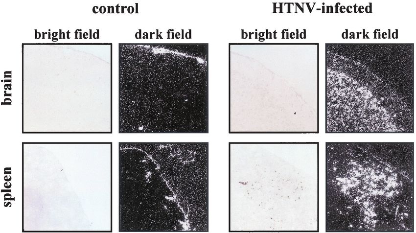
8892 WICHMANN ET AL. J. VIROL.
Downloaded from http://jvi.asm.org/ on December 29, 2020 by guest
FIG. 1. Laboratory mice are susceptible to HTNV infection. (A) Different strains of laboratory mice were intraperitoneally infected with 105
PFU of HTNV strain 76-118. Except for AKR/J mice, all other animals were susceptible to infection and showed similar but sometimes delayed
disease courses. (B) C57BL/6 mice were used to determine the LD50 by intraperitoneal infection with 10-fold dilutions of the virus stock. The LD50
was calculated to be approximately 60 PFU. (C) Knockout mice lacking a functional receptor for IFN-␣/ (IFNAR-1⫺/⫺ mice) were infected
intraperitoneally with 100 LD50 of HTNV. Weight loss was registered earlier for IFNAR-1⫺/⫺ mice than for C57BL/6 mice. The greater
susceptibility to infection may indicate a role of IFN-␣/ in antiviral defense. p.i., postinfection.
IFN-␣/. Knockout mice lacking a functional receptor for IFNAR-1⫺/⫺ mice, weight loss was registered much earlier (4
IFN-␣/ (IFNAR-1⫺/⫺ mice) were tested next for their sus- days postinfection). Once the animals became symptomatic,
ceptibility to HTNV. These animals lack the  subunit of the there was no difference in the disease courses between the two
IFN-␣/ receptor and thus are more sensitive to various virus study groups (Fig. 1C). This finding indicates an important role
infections (6, 10, 25). C57BL/6 control and IFNAR-1⫺/⫺ mice of IFN-␣/ in the defense against HTNV.
were infected intraperitoneally with 100 LD50 of HTNV. With HTNV appears to cause a systemic infection, with the cen-VOL. 76, 2002 LETHAL HTNV INFECTION IN MICE 8893
Downloaded from http://jvi.asm.org/ on December 29, 2020 by guest
FIG. 1—Continued.
tral nervous system being the primary target. In order to evidence of a systemic infection of the animals, with the most
better describe and understand the lethal neurological disease prominent lesions located in the central nervous system, par-
in laboratory mice, we performed RT-PCR, histological, im- ticularly the brain. Throughout the brain, distinct signs of vas-
munohistochemical, and in situ hybridization analyses on ma- culitis with perivascular edema and infiltration of mononuclear
terial of several organs taken from infected and uninfected cells were found (Fig. 3). Focally, neurons displayed eosino-
C57BL/6 mice. In the first step, RT-PCR performed with L philic cytoplasm and basophilic, often fragmented, nuclei a
segment-specific oligonucleotides generated a 1,080-bp specific sign of neuronal apoptosis. Based on the histological findings,
amplicon (Fig. 2). HTNV RNA was detected in the brain, which correlated well with the clinical symptoms, we concluded
lungs, liver, kidneys, and spleen, indicating a systemic infec- that the animals died of acute encephalitis. The liver showed
tion. Since sporadically nonspecific amplicons of minor sizes focal centrilobular necrosis with infiltration of mononuclear
were detected, all amplicons of the expected size were se- cells and large macrophage-like cells with phagocytic activity
quenced. The sequences were identical to those of the virus (Fig. 3). The histological findings in the spleen resembled those
stock that we used for intraperitoneal infection (data not of a viral infection, with hyperplastic germ centers and giant
shown). cell formation (data not shown). No significant alterations
Histological examination of organ samples provided further were found in the kidneys and lungs.
FIG. 2. HTNV causes a systemic infection in laboratory mice. RNA was isolated from different organs, and RT-PCR was performed with L
segment-specific oligonucleotides (see Material and Methods). The amplicons were analyzed on 1% agarose gels and visualized with ethidium
bromide. HTNV-specific amplions (1,080 bp) were detected in all organs tested. Lanes 1 and 10, DNA standards; lanes 2 and 9, negative PCR
control (H2O, no template); lane 3, negative control for RNA isolation (no template); lanes 4 to 8, samples from the liver, spleen, lungs, kidneys,
and brain, respectively.8894 WICHMANN ET AL. J. VIROL.
Downloaded from http://jvi.asm.org/ on December 29, 2020 by guest
FIG. 3. HTNV infection causes encephalitis in laboratory mice. Sections of different organs were HE stained. The brain sections show
perivascular edema (arrow in upper panel) with massive mononuclear cell infiltration in and around vessel walls and leukocytes which adhere to
endothelial cells. Strongly basophilic nuclear fragments indicate apoptosis (arrows in lower panel). The liver sections show focal large necrotic areas
(arrows in upper panel). A higher magnification of the sections shows giant cells with prominent nucleoli (arrow in lower panel) and a mononuclear
cell infiltrate consisting of lymphocytes and monocytes.
In order to associate the histological lesions with hantavirus C57BL/6 mice (8 weeks old) were infected with a recombinant
replication, we performed immunohistochemical and in situ vaccinia virus expressing the glycoprotein precursor of strain
hybridization analyses. As expected, we detected viral proteins 76-118 of HTNV (23). The virus was administered by three
in neurons near areas of necrosis in the brain (Fig. 4). In the consecutive intraperitoneal and subcutaneous injections of 105
spleen, positive signals were observed in lymphocytes; in the PFU at intervals of 2 weeks. Following the final immunization,
liver, viral antigen was found in perisinusoidal cells, such as antibody titers against HTNV were determined by using a
Kupffer cells, and central vein endothelial cells. Corresponding previously described ELISA (4, 20). All immunized animals
organ sections of noninfected animals remained negative (Fig. developed an antibody response (titers, 1:400 to 1:800), indi-
4, bottom panel). In situ hybridization was performed with an cating successful immunization. Control mice, which were im-
S segment-specific, 32P-labeled RNA probe. Signs of viral rep- munized with a recombinant vaccinia virus expressing the bac-
lication were detected, particularly in neurons (Fig. 5). Viral teriophage T7 RNA polymerase under the same scheme, did
RNA distribution in the spleen sections correlated with that of not react positively in the ELISA (Fig. 6A). Subsequently, both
viral antigen detected by immunohistochemical analysis (Fig. 4 groups of animals were challenged intraperitoneally with 100
and 5). Although the results of histological and immunohisto- LD50 of HTNV. While all control animals died, 60% of the
chemical analyses revealed strong evidence for virus infection immunized animals (three of five) were protected against the
of the liver, we failed to detect a positive signal for viral RNA lethal challenge (Fig. 6B). This result indicates that protective
in sections of this organ. immunity can be achieved and supports the notion that HTNV
Immunized mice are partially protected against a lethal was the causative agent of the lethal encephalitis in the ani-
HTNV challenge. The clinical course, distribution of viral an- mals.
tigen and RNA, and pathologic findings basically matched and
indicated that HTNV caused a systemic lethal infection in
DISCUSSION
adult immunocompetent mice. In order to provide further
proof that HTNV was the causative agent of the encephalitis in One of the priorities in hantavirus research is the develop-
the animals, we performed protection experiments. Adult ment of small-animal models. A disease model was recentlyDownloaded from http://jvi.asm.org/ on December 29, 2020 by guest
8895
LETHAL HTNV INFECTION IN MICE
FIG. 4. HTNV antigen can be detected in the brain, spleen, and liver. Sections from the brain, spleen, and liver were prepared for immunohistochemical analysis
(see Material and Methods). To detect viral antigen, the sections were incubated with a mouse polyclonal antiserum against HTNV (1:100 dilution) followed by rabbit
anti-mouse IgG conjugated to horseradish peroxidase (1:6,000). Viral antigen was detected in neurons (brain), endothelial cells (brain and liver), and lymphoid cells
VOL. 76, 2002
(spleen).8896 WICHMANN ET AL. J. VIROL.
Downloaded from http://jvi.asm.org/ on December 29, 2020 by guest
FIG. 5. HTNV nucleic acid can be detected in the brain and spleen. Sections from the brain and spleen were prepared for in situ hybridization
with an S segment-specific, 32P-labeled RNA probe that was 350 nucleotides long (see Material and Methods). Sections were analyzed by bright-
and dark-field microscopy. Viral nucleic acid was detected in neuronal cell layers of the brain and hyperplastic germ centers of the white pulp of
the spleen.
established for HPS by infection of Syrian hamsters with and more severe infections with hantaviruses in any host seem
ANDV, one of the South American New World hantaviruses to be systemic infections (for this model, see Fig. 2). A com-
(12). Although several species of rodents, rabbits, and nonhu- mon hallmark for all of these hantavirus infections seems to be
man primates have been infected with Old World hantaviruses, the disturbance of endothelial cell function (27, 41). A similar
only humans and eventually macaques (8, 19) have been shown observation was made with this model when histological find-
to present symptoms similar to those of HFRS. In the past, ings showed cerebral vasculitis (Fig. 3 and 4). A disturbance in
symptomatic or lethal disease courses were reported only fol- oxygen supply could therefore be a reason for neuronal apo-
lowing infection of newborn or immunodeficient laboratory ptosis, which was observed in many parts of the brain (Fig. 3
mice or rats (13, 18, 21, 24, 39, 40). Hamsters and bank voles and 4). These findings implicate endothelial cells as a general
have been used for challenge studies with HTNV (11) and target for hantaviruses independent of the host species.
PUUV (36). Again, however, all challenges resulted in persis- A distinct, unexpected clinical manifestation may imply the
tent infections with few or no clinical symptoms. Here we have possibility of a different disease-causing agent or substance.
shown that HTNV, the prototype of Old World hantaviruses, Our investigations demonstrated a good correlation among the
causes lethal encephalitis in adult immunocompetent labora- clinical symptoms, the cause of death, and the histological
tory mice. findings. At locations with histological alterations, hantavirus
The neurological disease associated with the HTNV infec- antigen and RNA were detected, indicating that the changes
tion of adult laboratory mice in this study is in contrast to were caused by hantavirus replication (Fig. 3 to 5). Further-
HFRS and HPS in humans. Interestingly, previous studies of more, the protection of animals following vaccination with a
hantavirus infections in newborn and immunodeficient rodents recombinant vaccinia virus expressing the HTNV glycoprotein
also described neurological symptoms, including cachexia and precursor strongly supported the involvement of HTNV in the
pareses (13, 18, 21, 24, 39, 40). In contrast to what was seen pathological process (Fig. 6). Finally, the negative microbio-
with the model described here, the outcome and severity of the logical test results excluded infections with other mouse patho-
disease in partially immunocompetent animals differed from gens which could have been directly or indirectly responsible
survival to delayed death for up to 30 or more days postchal- for the lethal disease.
lenge. A change in clinical symptoms as a consequence of a HTNV is sensitive to the antiviral action of human IFN-␣
different virus target organ is not a novel observation with (35), which is mediated by effector proteins, such as the MxA
hantaviruses. This finding was also noted with the discovery of protein (9). It was previously demonstrated that MxA inhibits
HPS in 1993 and actually delayed the laboratory diagnosis of HTNV replication by interacting with an early step of virus
this emerging disease (1, 20, 26). However, all symptomatic replication (7). The mouse genome also encodes Mx proteinsVOL. 76, 2002 LETHAL HTNV INFECTION IN MICE 8897
Downloaded from http://jvi.asm.org/ on December 29, 2020 by guest
FIG. 6. Immunized mice are partially protected against lethal HTNV challenge. Adult C57BL/6 mice were intraperitoneally and subcutaneously
immunized three times with 105 PFU of recombinant vaccinia viruses expressing either the glycoprotein precursor of HTNV (VVHTNV/GPC) or
the T7 RNA polymerase (VVT7). Prior to challenge with HTNV (100 LD50), antibody titers were determined by using an HTNV-specific ELISA.
In contrast to the control animals (VVT7), all immunized animals (VVHTNV/GPC) developed an antibody response, indicating successful
immunization (A). While all control animals died, protective immunity was achieved in 60% of the immunized animals (three of five) (B). OD,
optical density; p.i., postinfection.8898 WICHMANN ET AL. J. VIROL.
(Mx1 and Mx2) which possess antiviral activity (9). It is un- 2. Elliott, R. M. 1990. Molecular biology of Bunyaviridae. J. Gen. Virol. 71:
501–522.
likely that the nuclear Mx1 protein interferes with the replica- 3. Feldmann, A., M. K. Schaefer, W. Garten, and H. D. Klenk. 2000. Targeted
tion of HTNV, since hantaviruses are not known to have a infection of endothelial cells by avian influenza virus A/FPV/Rostock/
nuclear phase in their replication cycle. However, the cytoplas- 34(H7N1) in chicken embryos. J. Virol. 74:8018–8027.
4. Feldmann, H., A. Sanchez, S. Morzunov, C. F. Spiropoulou, P. E. Rollin,
mic Mx2 protein seems to inhibit hantavirus replication, as was T. G. Ksiazek, C. J. Peters, and S. T. Nichol. 1993. Utilization of autopsy
recently shown for HTNV and Seoul virus (14). Thus, it was RNA for the synthesis of the nucleoprotein antigen of a newly recognized
tempting to speculate that the IFN-induced expression of Mx2 virus associated with hantavirus pulmonary syndrome. Virus Res. 30:351–
367.
contributes to the prolonged incubation time observed in 5. Feldmann, H. March 2000, posting date. Hantaviruses, p. 1–8. In R. M.
HTNV-infected C57BL/6 control mice compared to knockout Atlas, M. M. Cox, S. F. Gilbert, J. W. Roberts, and B. A. Wood (ed.),
mice lacking a functional IFN-␣/ receptor (Fig. 1C). How- Encyclopedia of life sciences. [Online.] Nature Publishing Group, London,
England. http://www.els.net.
ever, this was not the case, because C57BL/6 mice, like most 6. Fiette, L., C. Aubert, U. Mueller, S. Huang, M. Aguet, M. Brahic, and J. F.
other inbred mice, have only nonfunctional Mx genes (33, 34). Bureau. 1995. Theiller’s virus infection in 129Sv mice that lack the interferon
Therefore, mice must possess additional IFN-induced defense alpha/beta or interferon gamma receptor. J. Exp. Med. 181:2069–2076.
7. Frese, M., G. Kochs, H. Feldmann, C. Hertkorn, and O. Haller. 1996.
Downloaded from http://jvi.asm.org/ on December 29, 2020 by guest
mechanisms which interfere with the replication of HTNV. Inhibition of bunyaviruses, phleboviruses, and hantaviruses by human MxA
Future experiments should examine whether the model will protein. J. Virol. 70:915–923.
8. Groen, J., M. Gerding, J. Koeman, P. Roholl, G. van Amerogen, H. Jordans,
work for other, particularly Old World, hantaviruses. Prelimi- H. Niesters, and A. Osterhaus. 1995. A macaque model for hantavirus.
nary attempts to infect C57BL/6 mice with PUUV failed to J. Infect. Dis. 172:38–44.
induce a symptomatic infection (H. Feldmann et al., unpub- 9. Haller, O., M. Frese, and G. Kochs. 1998. Mx proteins: mediators of innate
rsistance to RNA viruses. Rev. Sci. Tech. 17:220–230.
lished data). This result may be explained by the reservoir host 10. Hefti, H. P., M. Frese, H. Landis, C. Di Paolo, A. Aguzzi, O. Haller, and J.
for PUUV, which is Clethrionomys glareolus, belonging to the Pavlovic. 1999. Human MxA protein protects mice lacking a functional
subfamily Microtinae of the family Muridae (37, 38). C57BL/6 alpha/beta interferon system against La Crosse virus and other lethal viral
infections. J. Virol. 73:6984–6991.
mice, however, belong to the subfamily Murinae, as does 11. Hooper, J. W., K. I. Kamrud, F. Elgh, D. M. Custer, and C. S. Schmaljohn.
Apodemus agrarius, the primary reservoir host for HTNV. In- 1999. DNA vaccination with hantavirus M segment elicits neutralizing anti-
terestingly, in contrast to ANDV, the closely related Sin Nom- bodies and protects against Seoul virus infection. Virology 255:269–278.
12. Hooper, J. W., T. Larsen, D. M. Custer, and C. S. Schmaljohn, C. S. 2001.
bre virus does not cause any disease in Syrian hamsters (12); A lethal disease model for hantavirus pulmonary syndrome. Virology 289:
the reason is unknown. Therefore, the genetic background of 6–14.
the host species may determine the susceptibility to hantavi- 13. Huggins, J. W., G. R. Kim, O. M. Brand, and K. T McKee Jr. 1986. Ribavirin
therapy for Hantaan virus infection in suckling mice. J. Infect. Dis. 153:489–
ruses. At this point, we cannot exclude the possibility that the 497.
susceptibility of laboratory mice to HTNV is due to mutations. 14. Jin, H. K., K. Yoshimatsu, A. Takada, M. Ogino, A. Asano, J. Arikawa, and
Although this possibility seems unlikely, sequence determina- T. Watanabe. 2001. Mouse Mx2 protein inhibits hantavirus but not influenza
virus replication. Arch. Virol. 146:41–49.
tion for this particular HTNV has indeed resulted in a few S 15. Johnson, C. B., and C. S. Schmaljohn. 2001. Replication of hantaviruses.
(two nucleotide and two amino acid changes) and M (seven Curr. Top. Microbiol. Immunol. 256:15–32.
nucleotide and four amino acid changes) segment mutations 16. Johnson, K. M. 1999. Introduction, p. 1–6. In H. W. Lee, C. Calisher, and
C. S. Schmaljohn (ed.), Manual of hemorrhagic fever with renal syndrome
compared to the published sequence for strain 76-118 of and hantavirus pulmonary syndrome. W. H. O. Collaborating Center for
HTNV (B. Anheier and H. Finkemeier, unpublished data). Virus Reference and Research (Hantaviruses), Asan Institute for Life Sci-
ences, Seoul, Korea.
In conclusion, we have shown that strain 76-118 of HTNV 17. Kanerva, M., J. Mustonen, and A. Vaheri. 1998. Pathogenesis of Puumala
can cause lethal systemic infections in several inbred strains of and other hantavirus infections. Rev. Med. Virol. 8:67–86.
adult immunocompetent laboratory mice. The major target 18. Kim, G., and J. McKee. 1985. Pathogenesis of Hantaan virus infection in
suckling mice: clinical virologic, and serologic observations. Am. J. Trop.
organ is the central nervous system, and the cause of death is Med. Hyg. 34:388–395.
acute encephalitis. Even though this is not a disease model for 19. Klingstrom, J., A. Plyusnin, A. Vaheri, and A. Lundkvist. 2002. Wild-type
HFRS, it has the added advantage of incorporating the genetic Puumala hantavirus infection induces cytokines, C-reactive protein, creatine,
and nitric oxide in cynomolgus macaques. J. Virol. 76:444–449.
tools available for mice. This mouse model, as well as the 20. Ksiazek, T. G., C. J. Peters, P. E. Rollin, S. Zaki, S. T. Nichol, C. F.
Syrian hamster model for HPS, will be helpful in the develop- Spiropoulou, S. Morzunov, A. Sanchez, H. Feldmann, A. S. Khan, K. Wach-
ment of therapeutic and prophylactic measurements against smuth, and J. C. Butler. 1995. Identification of a new north American
hantavirus that causes acute pulmonary insufficiency. Am. J. Trop. Med.
hantavirus infections. Hyg. 52:117–123.
21. Kurata, T., T. Tsai, S. Bauer, and J. B. McCormick. 1983. Immunofluores-
ACKNOWLEDGMENTS cence studies of disseminated Hantaan virus infection of suckling mice.
Infect. Immun. 41:391–398.
We thank Steffi Lindow and Daryl Dick for technical assistance and 22. Lee, J. S., J. Lähdevirta, F. Koster, and H. Levey. 1999. Clinical manifesta-
Sandra Berthel, Guido Schemken, Nicole Beausoleil, and John Copps tions and treatment of HFRS and HPS, p. 17–38. In H. W. Lee, C. Calisher,
for assistance in animal care and manipulations. In addition, we thank and C. S. Schmaljohn (ed.), Manual of hemorrhagic fever with renal syn-
Anke Feldmann for advice on in situ hybridization and Daryl Dick for drome and hantavirus pulmonary syndrome. W. H. O. Collaborating Center
for Virus Reference and Research (Hantaviruses), Asan Institute for Life
editorial comments.
Sciences, Seoul, Korea.
This work was supported by grants from the Deutsche Forschungs- 23. Loeber, C., B. Anheier, S. Lindow, and H. Feldmann. 2001. The hantavirus
gemeinschaft (Fe 286/5-1, SFB 286-A7, and SFB 405-B10) and the glycoprotein precursor is cleaved at the conserved pentapeptide ‘WAASA.’
Volkswagen-Stiftung (Az: I/72087). Virology 289:224–229.
24. McKee, K., Jr., G. Kim, D. Greem, and C. J. Peters. 1985. Hantaan virus
REFERENCES infection in suckling mice: virologic, and pathologic correlates. J. Med. Virol.
1. Duchin, J. S., F. T. Koster, C. J. Peters, G. L. Simpson, B. Tempest, S. R. 17:107–117.
Zaki, T. G. Ksiazek, P. E. Rollin, S. T. Nichol, E. T. Umland, R. L. 25. Mueller, U., U. Steinhoff, L. F. L. Reis, S. Hemmi, J. Pavlovic, R. M.
Moolenaar, S. E. Reef, K. B. Nolte, M. M. Gallaher, J. C. Butler, R. F. Zinkernagel, and M. Aguet. 1994. Functional role of type I and type II
Breiman, and The Hantavirus Study Group. 1994. Hantavirus pulmonary interferons in antiviral defense. Science 264:1918–1921.
syndrome: a clinical description of 17 patients with a newly recognized 26. Nichol, S. T., C. F. Spiropoulou, S. Morzunov, P. E. Rollin, T. G. Ksiazek, H.
disease. N. Engl. J. Med. 330:949–955. Feldmann, A. Sanchez, J. Childs, S. Zaki, S., and C. J. Peters. 1993. GeneticVOL. 76, 2002 LETHAL HTNV INFECTION IN MICE 8899
identification of hantavirus associated with an outbreak of acute respiratory 36. Ulrich, R, A. Lundkvist, H. Meisel, D. Koletzki, K. B. Sjölander, H. R.
illness. Science 62:914–917. Gelderblom, G. Borisova, P. Schnitzler, G. Darai, and D. H. Krüger. 1998.
27. Papadimitriou, M. 1995. Hantavirus nephropathy. Kidney Int. 48:887–902. Chimaeric HBV core particles carrying a defined segment of Puumala han-
28. Peters, C. J., G. L. Simpson, and H. Levy. 1999. Spectrum of hantavirus tavirus nucleocapsid protein evoke protective immunity in an animal model.
infection: hemorrhagic fever with renal syndrome and hantavirus pulmonary Vaccine 16:272–280.
syndrome. Annu. Rev. Med. 50:531–545. 37. Viro, P., and J. Niethammer. 1982. Clethrionomys glareolus (Schreber,
29. Reed, L. J., and H. Muench. 1938. A simple method of estimating fifty per 1780)—Rötelmaus, p. 108–146. In J. Niethammer and F. Krapps (ed.),
cent endpoints. Am. J. Hyg. 27:493. Handbuch der Säugetiere Europas, Band 2/I. Nagetiere II. Akademische
30. Schmaljohn, C. S., and B. Hjelle. 1997. Hantaviruses: a global disease prob- Verlagsgesellschaft, Wiesbaden, Germany.
lem. Emerg. Infect. Dis. 3:95–104. 38. Wilson, D. E., and D. A. M. Reeder. 1993. Mammal species of the world, 2nd
31. Schmaljohn, C. S., and J. W. Le Duc. 1998. Bunyaviridae, p. 601–628. In
ed. Smithsonian Institution Press, Washington, D.C.
L. H. Collier, A. Balows, and M. Sussman (ed.), Topley and Wilson’s mi-
39. Yamanouchi, T., K. Domae, O. Tanishita, Y. Takahashi, K. Yamanishi, M.
crobiology and microbial infections, 9th ed. Arnold, London, England.
32. Schmaljohn, C. S., and S. T. Nichol. 2001. Hantaviruses. Curr. Top. Micro- Takahashi, and T. Kurata. 1984. Experimental infection in newborn mice
biol. Immunol. 256:1–196. and rats by hemorrhagic fever with renal syndrome (HFRS) virus. Microbiol.
33. Staeheli, P., R. Grob, E. Meier, J. G. Sutcliffe, and O. Haller. 1988. Influenza Immunol. 28:1345–1353.
virus-susceptible mice carry Mx genes with a large deletion or a nonsense 40. Yoshimatsu, K., J. Arikawa, S. Ohbora, and C. Itakura. 1997. Hantavirus
mutation. Mol. Cell. Biol. 8:4518–4523. infection in SCID mice. J. Vet. Med. Sci. 59:863–868.
Downloaded from http://jvi.asm.org/ on December 29, 2020 by guest
34. Staeheli, P., and J. G. Sutcliffe. 1988. Identification of a second interferon- 41. Zaki, S. R., P. W. Greer, L. M. Coffield, C. S. Goldsmith, K. B. Nolte, K.
regulated murine Mx gene. Mol. Cell. Biol. 8:4524–4528. Foucar, R. M. Feddersen, R. E. Zumwalt, G. L. Miller, A. S. Khan, P. E.
35. Tamura, M., H. Asada, K. Kondo, M. Takahashi, and K. Yamanishi. 1987. Rollin, T. G. Ksiazek, S. T. Nichol, B. W. J. Mahy, and C. J. Peters. 1995.
Effects of human and murine interferons against hemorrhagic fever with Hantavirus pulmonary syndrome—pathogenesis of an emerging infectious
renal syndrome (HFRS) virus (Hantaan virus). Antiviral Res. 8:171–178. disease. Am. J. Pathol. 146:552–579.You can also read