Widespread occurrence of 'Candidatus Phytoplasma ulmi' in elm species in Germany
←
→
Page content transcription
If your browser does not render page correctly, please read the page content below
Schneider et al. BMC Microbiology     (2020) 20:74
https://doi.org/10.1186/s12866-020-01749-z

 RESEARCH ARTICLE                                                                                                                                   Open Access

Widespread occurrence of ‘Candidatus
Phytoplasma ulmi’ in elm species in
Germany
Bernd Schneider1*, Ralf Kätzel2 and Michael Kube3

  Abstract
  Background: ‘Candidatus Phytoplasma ulmi’ is the agent associated with elm yellows and has been categorised in
  the European Union as a quarantine pathogen. For central and northern European countries, information on the
  occurrence and distribution of the pathogen and its impact on elms is scarce, so a survey of native elm trees has
  been conducted in Germany.
  Results: About 6500 samples from Ulmus minor, Ulmus laevis and Ulmus glabra, were collected nationwide.
  Phytoplasma detection was performed by applying a universal 16Sr DNA-based quantitative PCR (qPCR) assay and a
  novel ‘Ca. P. ulmi’ specific qPCR assay targeting the 16S–23S spacer region. Both assays revealed that 28% of the
  samples were infected by ‘Ca. P. ulmi’, but infection rates of the elm species and regional incidences differed. The
  phytoplasma presence in the trees was not correlated to disease-specific symptoms. The survey identified a
  regional disparity of infection which was high in east, south and central Germany, whereas only a few infected sites
  were found in the western and northern parts of the country. Monitoring the seasonal titre of ‘Ca. P. ulmi’ in an
  infected tree by qPCR revealed a high colonisation in all parts of the tree throughout the year.
  Conclusions: ‘Ca. P. ulmi’ is widely present in elms in Germany. The rare occurrence of symptoms indicates either a
  high degree of tolerance in elm populations or a low virulence of pathogen strains enabling high infection rates in
  a long-living host.
  Keywords: Phytoplasmas, Nationwide screening, TaqMan assay, Tolerance

Background                                                                             description ‘yellows’ or ‘yellowing’ in their name, due to
Phytoplasmas are obligate parasites from the bacterial                                 the associated presence of leaf chlorosis. This is also the
class Mollicutes, where they form the monophylogenetic                                 case for ‘Candidatus Phytoplasma ulmi’ infecting elm
taxon ‘Candidatus Phytoplasma’ [1]. They colonise the                                  trees [4]. It is phylogenetically closely related to econom-
nutrient-rich phloem sap of their plant host and rely for                              ically important plant pathogenic phytoplasmas such as
transmission on phloem-feeding hemipteran insect                                       “flavescence dorée” [5], ‘Ca. Phytoplasma rubi’ associated
vectors [2].                                                                           with rubus stunt [6], ‘Ca. Phytoplasma ziziphi’ associated
  Phytoplasmas are associated with diseases of more                                    with jujube witches’ broom [7], and alder yellows phyto-
than 1000 plant species, including many important crops                                plasma, associated with alder yellows [8]. These phyto-
[3]. Several phytoplasma diseases comprise the                                         plasmas belong to the elm yellows group and are
                                                                                       classified as 16SrV group members based on their re-
* Correspondence: bernd.schneider@thuenen.de                                           striction fragment length polymorphism pattern in PCR-
1
 Thuenen-Institute of Forest Genetics, Eberswalder Chaussee 3A, 15377
Waldsieversdorf, Germany
                                                                                       amplified 16Sr DNA [4].
Full list of author information is available at the end of the article

                                        © The Author(s). 2020 Open Access This article is licensed under a Creative Commons Attribution 4.0 International License,
                                        which permits use, sharing, adaptation, distribution and reproduction in any medium or format, as long as you give
                                        appropriate credit to the original author(s) and the source, provide a link to the Creative Commons licence, and indicate if
                                        changes were made. The images or other third party material in this article are included in the article's Creative Commons
                                        licence, unless indicated otherwise in a credit line to the material. If material is not included in the article's Creative Commons
                                        licence and your intended use is not permitted by statutory regulation or exceeds the permitted use, you will need to obtain
                                        permission directly from the copyright holder. To view a copy of this licence, visit http://creativecommons.org/licenses/by/4.0/.
                                        The Creative Commons Public Domain Dedication waiver (http://creativecommons.org/publicdomain/zero/1.0/) applies to the
                                        data made available in this article, unless otherwise stated in a credit line to the data.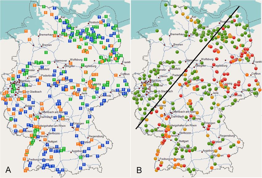
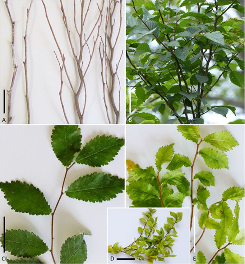
Schneider et al. BMC Microbiology   (2020) 20:74                                                               Page 2 of 12

   Elm yellows was first described in 1938 in North            absence of disease symptoms found in this study,
America [9], but historic reports indicate earlier sight-      prompted a nationwide survey of the pathogen’s distri-
ings [10, 11]. Most North American elm species are             bution and occurrence in the native elm species U.
highly susceptible to an infection by the bacterium and        glabra, U. laevis and U. minor. The results of this survey,
show a dramatic course of disease progression [12–14].         comprising a distribution map with incidence levels at
The trees usually die within two years post-infection,         sampling sites and a newly designed ‘Ca. P. ulmi’-spe-
displaying a number of characteristic symptoms such as         cific qPCR assay, are presented herein.
leaf yellowing, witches’ broom formation and phloem
necrosis. In the US, the disease spread gradually from         Results
the mid-western states to the east and to the south,           Wide absence of yellows disease symptoms in elm in
causing a considerable loss of native elm trees [14]. In       Germany
Europe, the disease was first reported in Italy and then       A total of 6486 elm samples from 339 sites were col-
later on in France, Bulgaria, Serbia and Croatia [15–17].      lected. The plant samples comprised 2630 Scots elm,
The disease symptoms displayed by the European elm             204 8 European white elm and 180 8 field elm samples
species resembled those of the North American elm spe-         (Table 1, Fig. 3a). The individuals’ ages ranged from
cies, but phloem necrosis did not occur. Therefore, the        one-year-old seedlings to trees of more than 400 years.
European elm species were considered less susceptible          The trunk diameter of the trees ranged from 0.5 cm to
than their American relatives [18].                            3.5 m.
   Since 1975, ‘Ca. P. ulmi’ has been regarded in the EU          ‘Ca. P. ulmi’-specific symptoms were rarely observed,
as a harmful organism and regulated by the Council             considering the number of trees, the infection rate at
Directive 2000/29/EC [19]. A comprehensive analysis on         some sites, their different ages and environment. How-
the re-categorisation of ‘Ca. P. ulmi’ was conducted by        ever, in 2017, 2018 and 2019, more than 15 symptomatic
the European Food and Safety Authority in 2014 [20],           Scots elms, approximately 2 to 3 m in height, were ob-
but due to limited information on its distribution, strain     served along roadsides in Müncheberg, Brandenburg
virulence, potential insect vectors and effects on Euro-       (Fig. 1a, b). The trees showed numerous witches’ brooms
pean elm species, the report remained inconclusive.            clearly visible in winter time and during new shoot de-
Ensuing reports of elm yellows findings from the UK,           velopment in July/August. These plants showed an early
the Czech Republic, Poland and Belgium, however,               bud break at the witches’ broom sites in March com-
demonstrated that ‘Ca. P. ulmi’ is more widespread in          pared to non-symptomatic parts of the trees. The
the EU than previously thought [21–24]. In response to         branches and leaves outside the witches’ brooms resem-
the new situation the European Plant Protection                bled those of healthy trees. One tree, about the same
Organization moved ‘Ca. P. ulmi’ in 2017 from Annex I          height and in the same area, showed all branches se-
list A1, as a pathogen absent from the EU, to list A2          verely stunted, and the leaves small and brittle. In 2018,
[25]. In December 2019, the Council Directive 2016/            field elms displaying little leaf, yellowing and stunting
2031 has deprived the quarantine status of ‘Ca. P. ulmi’       symptoms were observed in Ingelheim am Rhein,
for continental Europe, but the status for Great Britain       Rhineland-Palatinate (Fig. 1c, d, e) and near Haßfurt,
remains in place [26].                                         Bavaria. In many natural habitats of Scots elms and field
   In Germany, ‘Ca. P. ulmi’ was first reported in 1992        elms, the assessment on the presence of elm yellows
from a single Scots elm (Ulmus glabra) displaying              symptoms was severely compromised by the Dutch elm
witches’ broom symptoms in south-western Germany               disease which caused wilting and dieback. These symp-
[27]. DNA-DNA hybridisation studies with elm yellows-          toms were aggravated during the 2018 summer drought
specific probes resulted in identical hybridisation profiles   in Germany. Despite the fungal infection, samples were
between German, French, Italian and American acces-            included in the survey.
sions, thereby providing evidence that the European elm
yellows strains were closely related to the American           ‘Ca. P. ulmi’-specific TaqMan assay
strains [16]. In a more recent study, 59 European white        A universal quantitative real-time assay for the detection
elm (Ulmus laevis) trees, four of which showed stunted         of phytoplasma presence and for control of the template
growth and leaf chlorosis, were examined in the states of      quality (18Sr DNA plant) was routinely used [30]. A
Brandenburg and Berlin [28]. Based on 16Sr DNA                 specific-assay for the detection of ‘Ca. P. ulmi’ was de-
nested PCR assays, half of the tree samples were identi-       veloped in this work. The universal and the specific
fied as phytoplasma-positive, and sequence analyses            assay have been applied within this study enabling the
confirmed the presence of ‘Ca. P. ulmi’. However, all          detection of phytoplasma presence and of ‘Ca. P. ulmi’
symptomatic elm trees tested phytoplasma-negative. The         in particular, respectively. The selected forward and re-
unexpected high presence of ‘Ca. P. ulmi’, and the             verse primers and the TaqMan probe of the specific
Schneider et al. BMC Microbiology       (2020) 20:74                                                                      Page 3 of 12

Table 1 Summary of elm samples collected during the survey in Germany
Federal state and relative geographic                  Elm species and no. of samples                                            Total
position in Germany                                                                                                              no.
                                                       U. glabra                   U. laevis             U. minor
                                                                                                                                 per
                                                                                                                                 state
Baden-Württemberg (south-west)                         371                         90                    265                     726
Bavaria (south-east)                                   374                         90                    124                     588
Berlin (east)                                          0                           34                    46                      80
Brandenburg (east)                                     204                         412                   304                     920
Hamburg (north)                                        0                           0                     29                      29
Hesse (central)                                        70                          15                    25                      110
Lower Saxony (north)                                   113                         59                    102                     274
Mecklenburg West-Pomerania                             467                         494                   40                      1001
(north-east)
Rhineland-Palatinate (south-west)                      160                         91                    129                     380
North Rhine-Westphalia (west)                          223                         228                   231                     682
Saarland (west)                                        29                          0                     31                      60
Saxony (south-east)                                    270                         180                   150                     600
Saxony-Anhalt (east)                                   46                          138                   170                     354
Schleswig-Holstein (north)                             215                         142                   78                      435
Thuringia (south-east)                                 88                          75                    84                      247
Number of species and total number                     2630                        2048                  1808                    6486

assay are located in the spacer region at positions 1677                  showed a dynamic range of amplification with Ct values
to 1698, 1700 to 1721 and 1754 to 1772, respectively,                     of 18.2 to 37.6 for the lowest and highest dilution, re-
relative to the first nucleotide of the ‘Ca. P. ulmi’ se-                 spectively, with serial dilution steps differing by Ct
quence deposited in GenBank under the accession num-                      values of 2 to 3 (Table 2).
ber AF122911 and amplified a fragment of 96 bp. The
‘Ca. P. ulmi’ sequences showed the majority of the dif-                   Quantitative real-time PCR results highlight high
ferences to the sequences of alder witches’ broom, “fla-                  incidence of infection in elm stands
vescence dorée”, rubus stunt, ‘Ca. P. balanitae’ and ‘Ca.                 The internal 18Sr DNA amplification control showed
P. ziziphi’ in the region between the forward primer                      strong amplification (Ct values 9 to 22), due to the high
(fEY_spacer-rt) and the TaqMan probe (qEY_spacer-rt)                      content of plant DNA but also confirmed the template
annealing sites. The reverse primer (rEY_spacer-rt) dif-                  quality (data not shown). Samples with a Ct value > 22
fers only by a T or a G at the 3′ end relative to the se-                 were re-assayed, or the DNA extraction was repeated.
quences of alder witches’ broom, “flavescence dorée”,                     With the universal phytoplasma assay, 1803 of the 6486
rubus stunt and ‘Ca. P. ziziphi’, resepectively, or by an                 elm samples were rated phytoplasma-positive, represent-
internal nucleotide difference relative to ‘Ca. P. balanitae’             ing an infection rate of 27.8% based on the total number
(Fig. 2). Temperature gradient assays revealed an                         of samples (Table 3). With the specific qPCR assay 1801
optimum temperature of 56 °C, for both product yield                      samples tested positive (Table 3). To estimate the num-
and assay specificity. At this temperature, only the ‘Ca.                 ber of phytoplasmas present in the positive samples, four
P. ulmi’ strains ULW and EYC maintained in Cathar-                        Ct categories were established. With the universal qPCR
anthus roseus and the ‘Ca. P. ulmi’-infected field samples                assay the majority of the positive samples grouped in the
were amplified (data not shown).                                          Ct range > 22 ≤ 28 followed by the Ct range > 18 ≤ 22
   To assess the number of phytoplasmas in the phloem                     representing 80% of all positive samples. Almost 7% of
tissue of elm samples, DNA standards consisting of                        the positive samples revealed Ct values below 18. The
cloned 16S–23S ribosomal DNA from strain ULW with                         specific qPCR assay had a different performance. Here,
copy numbers from 1008 to 1001 per μl were PCR-                           the majority of positive samples grouped in the Ct rage
amplified with the universal phytoplasma and ‘Ca. P.                      > 22 ≤ 28, followed by the Ct range > 28 ≤ 34. In com-
ulmi’-specific assays. One μl of DNA extract from a                       parison with the universal qPCR assay, 73% of positive
healthy elm tree was added to each reaction, to simulate                  samples grouped between Ct 18 and Ct 28. Only 5 sam-
the assay conditions with unknown samples. Both assays                    ples showed Ct values of 18 or below. However,
Schneider et al. BMC Microbiology (2020) 20:74 Page 4 of 12 Fig. 1 ‘Ca. P. ulmi’-specific disease symptoms. a Branches of a healthy (left) and diseased (right) U. glabra tree in winter time. b Rare witches’ broom formation of U. glabra in summer time. For an overview of trees see Schneider and Kube, 2019 [29]. c Twig of a healthy U. minor with normal leaf size and leaf colouration showing mottle similar to virus infection. d U. minor branch with stunted growth. e U. minor with little leaf symptom and leaf chlorosis. The scale bar represents a length of 8 cm considering the set threshold level of Ct ≤ 34 both assays However, there was no correlation between the phyto- identified the same number of ‘Ca. P. ulmi’-positive plasma titre and ‘Ca. P. ulmi’-symptoms (data not samples. shown). The amplification curves in assays with the Scots elm trees showed in general a higher phyto- DNA of a non-infected elm tree and a no-template con- plasma titre compared to the other elm species. trol always ranged below the threshold line. Fig. 2 Alignment of 16S–23S spacer regions (5′ – 3′) of ‘Ca. P. ulmi’ and closely related phytoplasma strains. Nucleotide differences of the other sequences to the ‘Ca. P. ulmi’ sequence are indicated by lowercase letters. The positions of the forward primer, the TaqMan probe and the reverse primer are underlined and given from left to right, respectively. -, indicates gaps in the alignment. ≈, indicates bases not depicted. Numbers above the alignment correspond to nucleotide positions relative to the ‘Ca. P. ulmi’ sequence deposited under acc. no. AF122911
Schneider et al. BMC Microbiology           (2020) 20:74                                                                                Page 5 of 12

Table 2 Ct values of DNA standards after qPCR with the universal phytoplasma- and ‘Ca. P. ulmi’-specific spacer qPCR assay
Copy number/assay                                          Universal phytoplasma assaya                            ‘Ca. P. ulmi’-specific spacer assaya
     08
10                                                         19.7                                                    21.6
1007                                                       21.4                                                    23.2
     06
10                                                         22.5                                                    24.3
1005                                                       25.7                                                    27.1
     04
10                                                         30.0                                                    30.4
1003                                                       31.3                                                    32.0
     02
10                                                         34.5                                                    35.4
1001                                                       37.2                                                    35.1
a
 , Mean of four technical replicates including DNA from a non-infected elm tree

Comparison of qPCRs revealed rare occurrence of other                             deposited in GenBank under the accession numbers
phytoplasmas                                                                      MN394841 and MN394842.
The ‘Ca. P. ulmi’-specific assay detected two positive
samples less than the universal phytoplasma assay (Table
                                                                                  Distribution map of ‘Ca. P. ulmi’ in Germany highlights
3). The difference in the two cases was due to trees in-
                                                                                  the presence of hot spots
fected by other phytoplasma strains. Partial sequence
                                                                                  Elm samples were collected at 339 sites in Germany.
analysis of two P1/P7 PCR fragments revealed that one
                                                                                  The elm species were not homogeneously distributed
phytoplasma sequence (1631 bp) was identical to the se-
                                                                                  across the territory (Fig. 3a). The approximate species
quence of “flavescence dorée” phytoplasma strain FD70
                                                                                  frequency in the federal states is reflected by the num-
(acc. no. AF176319) from France, whilst the other se-
                                                                                  bers given in Table 1. The detected presence of ‘Ca. P.
quence (1631 bp) was identical to phytoplasmas found in
                                                                                  ulmi’ was likewise not homogeneous (Fig. 3b). Regions
Artemisia vulgaris (acc. no. MK440304) and Alnus gluti-
                                                                                  with sites showing an infection rate of more than 66.7%
nosa (acc. no. MK440303) in Poland. An alignment of
                                                                                  were clustered in Saxony, Saxony-Anhalt and Branden-
both sequences to the ‘Ca. P. ulmi’ sequence AF122911
                                                                                  burg. Other hotspots occurred along the upper Rhine
revealed six nucleotide exchanges within the 16S gene
                                                                                  valley, and some were present in Bavaria and Hesse.
and mispairing at the binding sites of primers and probe
                                                                                  Sites showing a lower infection rate were mostly found
of the specific assay. However, the 16Sr RNA gene of
                                                                                  in the vicinity of these hotspots. The infection rate de-
both phytoplasmas was identical to “flavescence dorée”
                                                                                  creased towards the west and north, and only five sites
phytoplasma strain FD70 which is a member of the elm
                                                                                  were found above a virtual line drawn from Trier to Ros-
yellows group 16SrV-C. The sequences have been
                                                                                  tock (Fig. 3b). At one of these sites in North Rhine-
                                                                                  Westphalia, U. laevis showed an infection rate of 30%
                                                                                  (six out of 20 samples). The four sites in Schleswig-
Table 3 Number of samples listed by Ct categories obtained
                                                                                  Holstein showed infection rates of 2.5% (one out of 40
with the universal phytoplasma- and specific qPCR assays
                                                                                  samples) and 20% (four out of 20 samples) for U. glabra,
qPCR Assays          Ct Category a                                       Sum
and elm species                                                          of
                                                                                  and 5% (one out of 20 samples) and 15% (six out of 40
                     > 28 ≤ 34       > 22 ≤ 28    > 18 ≤ 22       ≤ 18            samples) for U. laevis and U. minor, respectively.
                                                                         rows
Universal phytoplasma assay
     U. glabra       66              314          271             100    751      Infection rate correlated to altitude or tree age
     U. laevis       103             465          90              7      665      The majority of sampling sites were located in the Ger-
     U. minor        74              196          103             14     387      man lowlands at altitudes ≤100 m above the average
Sum of columns       243             975          464             121    1803
                                                                                  mean sea level (AMSL). However, quite a few sites were
                                                                                  also located in the low mountain range, from 300 to
‘Ca. P. ulmi’-specific assay
                                                                                  1100 m AMSL. About the same number of sites ranged
     U. glabra       105             514          127             4      750      in between (Fig. 4). The proportion of sites free of phy-
     U. laevis       240             412          11              1      664      toplasmas, and those with a low (up to 1/3 of individ-
     U. minor        130             235          22              –      387      uals), high (up to 2/3 of individuals) and extreme
Sum of columns       474             1161         160             5      1801     infection rate (up to 100%), were almost identical among
a,
 Columns refer to Ct categories and the associated number of
                                                                                  the zones, thereby indicating a broad habitat for poten-
positive samples                                                                  tial insect vectors.
Schneider et al. BMC Microbiology (2020) 20:74 Page 6 of 12 Fig. 3 a, sampling sites with predominant elm species. Blue square, U. glabra; Green square, U. laevis; Orange square, U. minor. b, presence of ‘Ca. P. ulmi’ at the sampling sites based on qPCR results. Green dot, no phytoplasmas found; Yellow dot, up to one-third of trees infected; Orange dot, more than one-third and up to two-thirds of trees infected; Red dot, more than two-thirds and up to 100% of trees infected Except for monumental trees and seedlings, the ages of youngest age group, and this number did not change much trees were unknown and calculated on the basis of the trunk in the other age categories. The graph also shows the dimin- diameter. For trees up to 5 cm, 10 cm, 20 cm and 50 cm in ishing number of older trees for Scots elms and field elms, diameter, age was calculated at 10, 20, 39 and 98 years, re- due to the mortal effects of Dutch elm disease on their popu- spectively. The oldest tree was a European white elm in lation. Of particular interest was the situation of old and Gülitz (Brandenburg), estimated to be 400 to 700 years old monumental trees of more than 100 cm in trunk diameter. and with a diameter of 3.5 m. The number of infected indi- 111 trees with an estimated age of 195 years and older were viduals was determined in relation to trunk diameter (Fig. 5). included in this survey. Thirty-eight individuals were infected The graph revealed different disease progressions for the by ‘Ca. P. ulmi’, comprising 29 U. laevis-, eight U. glabra- three species. While U. glabra showed a steady increase of and one U. minor tree. infection with age, U. laevis showed a strong increase of in- fected individuals in trees up to 20 years of age, reaching a All-season colonisation of elm plateau thereafter. A different situation was observed for U. To assess the seasonal fluctuation of pathogen numbers, minor. An infection rate of 20% was determined in the a monthly screening of different plant parts from an Fig. 4 Overall infection rate of elm species at different altitude levels. Altitude, number of sites and number of individuals examined indicated. Left (light grey), percentage of sites with non-infected trees. In a clockwise direction, percentage of sites with up to one-third, more than one- third and up to two-thirds and more than two-thirds of individuals infected by ‘Ca. P. ulmi’
Schneider et al. BMC Microbiology       (2020) 20:74                                                                                    Page 7 of 12

 Fig. 5 Infection rate of U. minor, U. laevis and U. glabra in relation to trunk diameter. Grey bars represent non-infected individuals. White bars
 represent phytoplasma-infected individuals. The number of individuals considered is given in the bars. The infection percentage in an age group
 is indicated on the right

infected Scots elm tree was performed with the universal                    seasonal fluctuations, reaching a low in late winter to
qPCR assay. The mean monthly Ct values of all samples                       early summer [31, 32]. To obtain a first insight on the
within the examined period ranged from 24.2 to 28.6.                        colonisation pattern of an elm tree by ‘Ca. P. ulmi’ a nat-
The June to December Ct means were slightly lower                           urally infected U. glabra tree was taken as study object.
than the January to May means. The Ct means between                         The analysis revealed that ‘Ca. P. ulmi’ colonises roots
trunk samples and root samples of the same month                            and shoots all year round in a constantly high titre.
never differed by more than 3. The Ct values of the indi-                   Therefore, branches or trunk material were sampled
vidual monthly trunk samples were close and never                           throughout the survey. Even though petioles or leaf mid-
apart by more than four cycles. The lowest phytoplasma                      ribs would have been easier to collect, the choice fell on
titre was recorded from January to March in buds, with                      branches or trunk material, as the sampling period could
Ct means ranging from 28.4 to 31.5. Considering the                         be extended with this material. However, a more ex-
lowest average Ct (24.2) value, a phytoplasma number of                     tended monitoring of infected elm trees is necessary to
1008 per gram of phloem tissue was calculated. The                          generalise these preliminary colonisation behaviour re-
highest Ct average (Ct 31.5) found in bud material rep-                     sults also for infected U. laevis and U. minor trees.
resents a phytoplasma number two orders of magnitude                           The qPCR assays corroborated the results of the sea-
lower.                                                                      sonal course study, as in most trees a fairly high number
                                                                            of ‘Ca. P. ulmi’ was identified, exemplified by a Ct value
Discussion                                                                  ≤28 representing an organism titre of 1006 per gram of
A recent survey in the states of Brandenburg and Berlin                     phloem tissue and higher. Both qPCR assays worked re-
identified an unexpected high infection rate of U. laevis                   liably, although the specific assay showed a slightly lower
by ‘Ca. P. ulmi’ [28]. To verify the situation on a nation-                 performance in respect to the Ct values. This was most
wide scale a representative survey with a high number of                    likely caused by the lower G + C content of the primers
samples has been conducted covering most of the nat-                        and probe and the reduced binding strength compared
ural habitats of the three elm species. Preliminary results                 to the oligonucleotides of the universal phytoplasma
of this survey have been published [29].                                    assay. The substitution of adenine bases with 2,6-diami-
  At the beginning of the survey, no information was                        nopurin, to increase the melting temperature of the Taq-
available on the colonisation of ‘Ca. P. ulmi’ in infected                  Man spacer probe, did not change the performance
elm trees. In the well-studied phytoplasma diseases of                      significantly compared to the assay with the non-
pome fruit trees, the phytoplasma titre displays strong                     modified probe. Even though, in comparison with the
Schneider et al. BMC Microbiology (2020) 20:74 Page 8 of 12 universal qPCR assay, the spacer assay proved to be a re- European elm species tolerate a ‘Ca. P. ulmi’ infection liable diagnostic tool in terms of positive calls for the quite well, although the number of symptom sightings presence of ‘Ca. P. ulmi’. from southern Europe [33–35] for U. minor and U. It is evident that ‘Ca. P. ulmi’ was present in almost laevis seem to be more frequent compared to reports 28% of the elm samples, thus indicating the high number from central and northern regions of the continent. of infected trees in the German elm populations. A However, this could also be linked to regional differ- higher overall ‘Ca. P. ulmi’ infection rate of 46% was ences of ‘Ca. P. ulmi’ strain virulence, climatic factors, found in Croatian elm populations [33]. Those and genetic background of elm populations or other undeter- higher numbers were also observed in Germany if the mined stress factors. evaluation would had focused on phytoplasma hotspots The different infection rates of the elm species and only. The high overall incidence rate in Croatia is there- within the age categories are difficult to explain. The fore a matter of sample size. Other studies reported of progressive infection in aging U. glabra populations is infection rates as high as 85%, but this is caused by a easily comprehensible, but the plateau phase for U. preferential collection of material from symptomatic laevis, and the constant infection rate of U. minor age trees which does not reflect the real infection rate [34]. classes, is not as easy to deduce. The same is true for the The three elm species showed different disease rates. old and monumental trees. How some of these trees es- While almost one-third of the U. laevis samples tested caped phytoplasma infection remains unclear, as they ‘Ca. P. ulmi’-positive, U. glabra and U. minor were in- were well located in regions of high infection pressure. fected to a lesser degree. This difference is not due to Therefore, it seems that a certain degree of resistance is sample size, as the number of tested trees from each present in the populations. However, other factors might species was about similar. In Croatia, elm species were be involved too, like recovery [36] or the protective role also infected to a different degree, in which almost 75% of the plants’ microbiome [37], albeit insufficient infor- of U. laevis trees were infected, followed by U. minor mation is available to assess their influence so far. with 10.8% and U. glabra with 4% [33]. However, in this Infection hotspots were located in the eastern, central work, the latter two species were also infected by other and south-western parts of Germany, whereas infected phytoplasmas, and therefore the infection rates are not sites became rare towards the north and north-western directly comparable. regions. The most plausible explanation is that an insect In all, except for two elm samples, ‘Ca. P. ulmi’ was vector migrated from the southern-to-eastern side into identified. These phytoplasmas were detected by the the territory moving towards the north and west. Beside different specificity of the two qPCR assays employed. the verified ‘Ca. P. ulmi’ vector Macropsis mendax for Sequence analyses of ribosomal fragments revealed that Italy [38], phytoplasmas of the elm yellows group have both phytoplasmas were members of the 16SrV, or elm been identified by PCR and PCR-RFLP analysis in yellows group. But in contrast to ‘Ca. P. ulmi’ which Hyalesthes luteipes in Serbia, in Iassus scutellaris, taxonomically group in subgroup A, both isolates Allygidius furcatus and Cixus sp. in France, but no trans- belonged to subgroup C. ‘Ca. Phytoplasma solani’ and mission experiments were performed [39, 40]. For ‘Ca. Phytoplasma asteris’, have been described in U. gla- Germany, no data are available. The fact that infected bra and U. minor displaying rather unspecific symptoms trees were found from sea level up to an altitude of 750 of leaf yellowing and drying [33]. These symptoms could m might indicate a vector different to M. mendax, as not be observed in the two phytoplasma-infected trees. this insect is only known to occur up to an altitude of The presence of other phytoplasmas in elms is most 400 m [41]. A spread of the bacterium through the ex- likely due to an occasional feeding of infected insect change or trade of infected elms for planting purposes vectors. However, the fact that only 16SrV-group phyto- can be excluded, as elm timber has little forest use. In plasmas were found in elm trees might indicate a host- addition, root bridges can be discounted, as this would pathogen specificity at the plant host- or insect vector only explain spread in a small area. level, or on both. This survey demonstrated a nationwide distribution of Despite the high number of phytoplasmas in the sieve ‘Ca. P. ulmi’ in all three native elm species. The number tubes, German elms seem to react in a tolerant way of infected individuals is such that the eradication of this upon infection, which stands in striking contrast to the pathogen is impossible. The recent finding of ‘Ca. P. reactions of American and Asian elm species [14, 18]. ulmi’ in Belgium [24] demonstrates its presence also The few symptomatic U. glabra and U. minor trees that west of Germany, although the confirmed number of in- were found, however, displayed typical ‘Ca. P. ulmi’ fected elms is still low. symptoms with witches’ brooms, stunting and leaf chlor- This work has established a sound basis for future osis. Mittempergher (2000) also concluded after ex- research with respect to transmission and phytoplasma- tended observations in Italian elm breeding stations that host interaction. Examples of tolerance are often
Schneider et al. BMC Microbiology   (2020) 20:74                                                             Page 9 of 12

overlooked by phytopathologists and are rarely reported.      Waldsieversdorf (both Brandenburg). Additional elm
It is remarkable that the closely related elm yellows and     trees were randomly sampled during the surveys at road-
alder yellows phytoplasmas share this feature [42], which     sides or in public parks. The diameter of each trunk was
might be the result of a long-term co-evolution of these      measured at a height of 1.3 m, and the approximate age
phytoplasmas with their hosts. A deeper understanding         was estimated by software provided on the website
of such a particular phytoplasma-host interaction may         www.baumportal.de [44]. Elm shoot samples were col-
provide the key to developing new strategies to cope          lected from October 2017 until May 2019. Where pos-
with phytoplasma associated diseases in agriculture and       sible, two shoot samples, about 25 cm in length and 0.5
forestry.                                                     to 4 cm in diameter, were collected from 20 randomly
                                                              selected trees per site. The phytoplasma strains ULW
Conclusions                                                   (elm yellows phytoplasma, subgroup 16SrV-A) and ALY
This work presents the first nationwide survey of ‘Ca. P.     (alder yellows phytoplasma, subgroup 16SrV-C) main-
ulmi’ presence in native elm species in Germany provid-       tained in Catharanthus roseus were used as reference
ing representative figures of infection incidence in the      strains [14, 45]. The coordinates of all collected elm ac-
federal states. Almost 28% of all elm accessions tested       cessions were recorded in WGS84 format, using a port-
‘Ca. P. ulmi’-positive. Elm species were infected to a        able GPS device. The seasonal colonisation of ‘Ca. P.
different degree with regional disparity of infection. Hot-   ulmi’ was monitored for one year in an infected Scots
spots of infection were identified in East-, Southeast and    elm 3 m in height and 6 cm in diameter. Samples were
Central Germany while infection rates in West- and            taken on a monthly basis from the roots, at trunk
North Germany were low. Despite the high infection            ground level and then at distances of 25 cm up to a
rate, disease symptoms were rarely found indicating a         height of 1.75 m. Phloem tissue at these sites was ex-
high degree of tolerance of native elm species to infec-      tracted from a square of 0.5 cm2. Midrib or bud samples
tion. An infection of elm trees by other phytoplasmas         from the top and bottom of the tree were also examined.
was only detected in two cases. A specific qPCR Taq-
Man assay based on 16S–23S spacer sequence motives
has been developed providing sensitive and reliable de-       DNA extraction
tection of the pathogen. The occurrence of infected elm       DNA from all elm shoot samples was extracted from
trees in regions beyond 400 m of altitude suggests insect     125 mg of phloem tissue using 3 ml of CTAB buffer ac-
vectors different to the verified ‘Ca. P. ulmi’-vector M.     cording to a procedure described by Ahrens and See-
mendax. The American vector species Scaphoideus               müller [46]. The tissue was homogenised with a steel
luteolus and Allygus atomarius do not occur in                ball homogeniser in plastic extraction bags (Bioreba,
Germany, except Philaenus spumarius, which is widely          Grenzach). The nucleic acids pellet was resuspended in
distributed but failed to vector ‘Ca. P. ulmi’ in transmis-   200 μl of sterile water and stored at − 20 °C until use.
sion trials. Therefore, it is likely that other unknown       Small-scale phloem DNA extractions (≤ 15 mg) were
vectors are involved in ‘Ca. P. ulmi’ transmission.           performed with a Beadruptor (Biolab, Bebendorf) in
                                                              microfuge tubes containing ceramic beads and 250 μl of
Methods                                                       CTAB buffer. The nucleic acids pellet was resuspended
This study aims to clarify the infection status of elms in    in 50 μl of sterile water and stored as described above.
Germany with respect to ‘Ca. P. ulmi’. Therefore, the         DNA from other phytoplasma strains (‘Candidatus Phy-
survey comprises the sampling of plant material from          toplasma asteris’ strain AAY [subgroup 16SrI-B]; ‘Candi-
elms in Germany, followed by DNA extraction providing         datus Phytoplasma mali’ strain AT [subgroup 16SrX-A];
the templates for screening by quantitative PCR assays.       ‘Candidatus Phytoplasma cynodontis’ strain BGWL
Diagnostic qPCR assays were performed by applying             [subgroup 16SrXII]; ‘Ca. P. ulmi’ strain EYC [subgroup
universal phytoplasma primers [30] and a new specific         16SrV-A]; Western X disease, Green Valley strain GVX
primer set for ‘Ca. P. ulmi’ (this study).                    [subgroup 16SrIII-A]; ‘Candidatus Phytoplasma auranti-
                                                              folia’ strain WBDL [subgroup16SrII]; ‘Candidatus Phyto-
Sampling of plant material from elms                          plasma australiense’ [subgroup 16SrXII-B]; ‘Candidatus
Elm samples were collected at 339 sites, based on a sur-      Phytoplasma rubi’ strain RuS [subgroup 16SrV-E]) main-
vey published in 2007 on the genetic resources of elm         tained in C. roseus and “flavescence dorée” strain FD70
species in Germany [43]. The monumental elm tree sites        (subgroup 16SrV-C), maintained in Vicia faba, was ob-
were taken from the web resource on monumental trees          tained from Kerstin Zikeli and Michael Maixner (Julius
(https://www.monumentaltrees.com). In addition, one-          Kuehn-Institute, Institute for Plant Protection in Fruit
to two-year-old seedlings were obtained from nurseries        Crops and Viticulture, Germany) and were used as posi-
in Riedlingen (Baden-Württemberg), Müncheberg and             tive or negative controls.
Schneider et al. BMC Microbiology (2020) 20:74 Page 10 of 12
Quantitative PCR standard each forward and reverse primer and a probe (5′-FAM/
The 16S-23Sr DNA of the ‘Ca. P. ulmi’ strain ULW was BHQ1–3′) for phytoplasma detection, 3.3 pmol of each
amplified with P1/P7 primers [47, 48] as described previ- forward and reverse primer and a probe (5′-Cy5/BHQ3–
ously [49]. The 1.8 kb PCR fragment was ligated into the 3′) for plant 18Sr DNA amplification and 5 μl of 2 x pri-
cloning vector pGEM-T (Promega, Madison), and the maQUANT mastermix (Steinbrenner, Wiesenbach). The
insert was verified by sequencing with vector primers reactions were cycled as described previously [26] in a
(M13, M13rev) and the universal phytoplasma primers qTower (Analytik Jena AG, Jena), except that the initial
fU5 and rU3 [46]. The recombinant plasmid was bulk- 50 °C and 95 °C steps were replaced by a three-minute
extracted, its quantity determined by Qubit fluorometric denaturation step at 95 °C. The plant 18Sr DNA assay
quantification (Invitrogen, Carlsbad) and serially diluted served as an internal amplification control to exclude
with sterile water to obtain plasmid concentrations ran- the presence of inhibitory co-extracted compounds. The
ging from 1008 to 1001 copies per μl (referred to as ‘Ca. P. ulmi’-specific spacer assay was performed in 10 μl
‘DNA standard’). P1/P7 PCR fragments of phytoplasma reactions containing 10 pmol of each forward and re-
field strains were sequenced as described above. Se- verse primer and probe, albeit without plant-specific
quences differing from ‘Ca. P. ulmi’ database entries primers and probe. The cycling parameters were as fol-
were deposited at GenBank. The standard was only used lows: One cycle for 3 min at 95 °C, 40 cycles at 95 °C for
to roughly estimate the plant phytoplasma titre and was 15 s and 56 °C for 25 s. Data evaluation was done via the
not included in routine screenings. cycler software provided by the manufacturer. Due to
the high number of samples, verification of real-time
Calculation of phytoplasma titres PCR results was performed on a step-by-step basis. Sam-
The phytoplasma titre was determined by the Ct values ples were re-examined if the Ct value between universal
of the above mentioned serially diluted DNA standard and specific assays differed by more than three, or if one
relative to the Ct value of the elm samples. The calcula- of the assays was rated negative. In each run, a
tion considered the amount of plant tissue represented phytoplasma-positive field sample with a Ct value of 29,
in the nucleic acids pellet, the volume of the reconsti- ULW DNA (positive control), DNA of a healthy elm and
tuted nucleic acids pellet and the copy number of the a water control (negative controls) were included, to ver-
target gene. The calculated figures are approximate ify run consistency. The Ct values were assessed strin-
values. gently, and samples with Ct values > 34 were considered
negative.
Design of ‘Ca. P. ulmi’-specific primers
The 16S–23S spacer sequences from ‘Ca. P. ulmi’ data- Abbreviations
EU: European Union; Ct: Cycle threshold; AMSL: Average mean sea level;
base entries with acc. Nos. AF122911, AF189214 and IPWG: International Phytoplasmologist Working Group; EPPO: European Plant
EU184021 and spacer regions of related phytoplasmas Protection Organisation; CTAB: Cetyltrimethylammoniumbromide
(“flavescence dorée” phytoplasma strain FD70, acc. no.
AF176319; rubus stunt phytoplasma, acc. no. Y16395; ‘Ca. Acknowledgements
We thank Marlies Karaus for her excellent technical assistance in this study.
P. balanitae’, acc. no. AB689678; ‘Ca. P. ziziphi’, acc. no.
KC478660 and alder witches’ broom phytoplasma, acc. no. Authors’ contributions
MK440303) were aligned using Clustal X [50]. The BS, RK and MK designed the sampling. BS carried out the surveys, performed
primers and probe were selected based on regions of all experiments and drafted the manuscript. MK designed the study and
contributed to manuscript preparation. All authors have read and approved
complete homology to ‘Ca. P. ulmi’ sequences and the the manuscript.
maximal number of base differences or gaps to the related
phytoplasmas. The derived primers and probe were as fol- Funding
lows (5′ – 3′): Forward primer fEY_spacer-rt, ATAT- This project (no. 22026316) was funded by the “Fachagentur
Nachwachsende Rohstoffe e.V.” (FNR), a promotor of the German Federal
CAGGAAAATATTTACTAC; reverse primer rEY_ Ministry for Food and Agriculture.
spacer-rt, CGCCCTTACTTTCTTCAAT; TaqMan probe
pEY_spacer-rt, FAM-TTGAAGAAAGTTCTTTGAAAA Availability of data and materials
G-BHQ1. Double underlined nucleotides were replaced by All data generated or analysed during this study are included in this
published article. Specific datasets on coordinates of elm stands analysed
2,6-diaminopurin to increase the melting temperature. during the current study are available from the corresponding author on
reasonable request.
Diagnostic qPCR assays
A TaqMan real-time PCR assay for universal phyto- Ethics approval and consent to participate
Not applicable.
plasma detection was performed [30] with the following
modifications. The assay was performed in 10 μl reac- Consent for publication
tions containing 1 μl of nucleic acids extract, 10 pmol of Not applicable.Schneider et al. BMC Microbiology (2020) 20:74 Page 11 of 12
Competing interests 21. EPPO: First report of ‘Candidatus Phytoplasma ulmi’ in the United Kingdom.
The authors declare that they have no competing interests. In. https://gd.eppo.int: EPPO Global Database; 2014.
22. EPPO: First confirmed report of ‘Candidatus Phytoplasma ulmi’ in the Czech
Author details Republic. In. https://gd.eppo.int: EPPO Global Database; 2015.
1
Thuenen-Institute of Forest Genetics, Eberswalder Chaussee 3A, 15377 23. EPPO: First report of ‘Candidatus Phytoplasma ulmi’ in Belgium. In. https://
Waldsieversdorf, Germany. 2Landeskompetenzzentrum Forst Eberswalde, gd.eppo.int: EPPO Global Database; 2018.
Alfred-Möller-Straße 1, 16225 Eberswalde, Germany. 3Department of 24. De Jonghe K, Deeren AM, Goedefroit T, Ronse A. First report of ‘Candidatus
Integrative Infection Biology Crops-Livestock, University of Hohenheim, Phytoplasma ulmi’ on elm in Belgium. Plant Dis. 2019;103(7):1763.
Garbenstr. 30, 70599 Stuttgart, Germany. 25. EPPO: Changes made to the EU list of regulated pests. In. https://gd.eppo.
int: EPPO Global Database; 2017.
Received: 19 December 2019 Accepted: 10 March 2020 26. EU DRdEU: Council directive 2016/2031/EC of 26 October 2016 on
protective measures against pests of plants, amending regulations (EU) no
228/2013, (EU) no 652/2014 and (EU) no 1143/2014 of the European
Parliament and of the council and repealing council directives 69/464/EEC,
References
74/647/EEC, 93/85/EEC, 98/57/EC, 2000/29/EC, 2006/91/EC and 2007/33/EC.
1. IRPCM. ‘Candidatus Phytoplasma’, a taxon for the wall-less, non-helical
EUR-Lex Access to European Union law 2016.
prokaryotes that colonize plant phloem and insects. Int J Syst Evol
27. Seemüller E. Laubgehölzmycoplasmosen in Europa. Nachrichtenblatt des
Microbiol. 2004;54(4):1243–55.
deutschen Pflanzenschutzdiensts. 1992;44:145–8.
2. Sugio AM, AM, Kingdom HN, Grieve VM, Manimekalai R, Hogenhout SA:
28. Eisold A-M, Kube M, Holz S, Büttner C. First report of ‘Candidatus
Diverse targets of phytoplasma effectors: from plant development to
Phytoplasma ulmi’ in Ulmus laevis in Germany. Commun Agric Appl Biol Sci.
defense against insects. Annu Rev Phytopathol 2011, 49:175–195.
2015;80(3):575–8.
3. Seemüller E, Garnier M, Schneider B. Mycoplasmas of plants and insects. In:
29. Schneider B, Kube M. Occurrence of ‘Candidatus Phytoplasma ulmi’ in native
Razin S, Hermmann R, editors. Molecular Biology and Pathology of
elm trees in Germany. Phytopathogenic Mollicutes. 2019;9(1):51–2.
Macoplasmas. London: Kluwer Academic/Plenum Publishers; 2002. p. 91–
116. 30. Christensen NM, Nicolaisen M, Hansen M, Schulz A. Distribution of
4. Lee I-M, Martini M, Marcone C, Zhu SF. Classification of phytoplasma strains phytoplasmas in infected plants as revealed by real-time PCR and
in the elm yellows group (16SrV) and proposal of 'Candidatus Phytoplasma bioimaging. Mol Plant-Microbe Interact. 2004;17(11):1175–84.
ulmi' for the phytoplasma associated with elm yellows. Int J Syst Evol 31. Seemüller E, Schaper U, Zimbelmann F. Seasonal variation in the
Microbiol. 2004;54(2):337–47. colonization patterns of mycoplasma-like organisms associated with apple
5. Martini M, Murari E, Mori N, Bertaccini A. Identification and epidemic proliferation and pear decline. J Plant Dis Protect. 1984;91:371–82.
distributions of two “favescence dorée”-related phytoplasmas in Veneto 32. Marzachi C. Molecular diagnosis of phytoplasmas. Phytopathol Mediterr.
(Italy). Plant Dis. 1999;83(10):925–30. 2004;43(2):228–31.
6. Malembic-Maher S, Salar P, Filippin L, Carle P, Angelini E, Foissac X. Genetic 33. Katanić Z, Krstin L, Ježić M, Zebec M, Ćurković-Perica M. Molecular
diversity of European phytoplasmas of the 16SrV taxonomic group and characterization of elm yellows phytoplasmas in Croatia and their impact
proposal of 'Candidatus Phytoplasma rubi'. Int J Syst Evol Microbiol. 2011; on Ulmus spp. Plant Pathol. 2016;65(9):1365–3059.
61(9):2129–34. 34. Jović J, Cvrković T, Mitrović M, Petrović A, Krstić O, Krnjajić S, Toševski I.
7. Jung HY, Sawayanagi T, Kakizawa S, Nishigawa H, Wei W, Oshima K, Miyata Multigene sequence data and genetic diversity among ‘Candidatus
S, Ugaki M, Hibi T, Namba S. ‘Candidatus Phytoplasma ziziphi’, a novel Phytoplasma ulmi’ strains infecting Ulmus spp. in Serbia. Plant Pathol. 2011;
phytoplasma taxon associated with jujube witches'-broom disease. Int J Syst 60(2):356–68.
Evol Microbiol. 2003;53(4):1037–41. 35. Marcone C, Ragozzino A, Seemüller E. Identification and characterization of
8. Lederer W, Seemüller E. Occurrence of mycoplasma-like organisms in the phytoplasma associated with elm yellows in southern Italy and its
diseased and non-symptomatic Alder trees (Alnus spp.). Eur J For Pathol. relatedness to other phytoplasmas of the elm yellows group. Eur J Forest
1991;21(2):90–6. Pathol. 1997;27(1):45–54.
9. Swingle RU. A phloem necrosis of elm. Phytopathology. 1938;28(10):757–9. 36. Carraro L, Ermacora P, Loi N, Osler R. The recovery phenomenon in apple
10. Garman H. The elms and their diseases. Kentucky Agricultural Exp Station proliferation-infected apple trees. J Plant Pathol. 2004;86(2):141–6.
Bull. 1899;80:51–75. 37. Gonella E, Musetti R, Crotti E, Martini M, Casati P, Zchori-Fein E.
11. Forbes SA. What is the matter with the elms in Illinois? Urbana, University of Microbe relationships with phytoplasmas in plants and insects. In:
Illinois Agricultural Experiment Station. 1912;154:1–22. Phytoplasmas: plant pathogenic bacteria-II. Singapore: Springer; 2019. p.
12. Carter JC, Carter LR. An urban epiphytotic of phloem necrosis and Dutch 207–35.
elm disease 1944-1972. Illinois Natural History Service Bulletin. 1974;31(1– 38. Carraro L, Ferrini F, Ermacora P, Loi N, Martini M, Osler R. Macropsis mendax
10):113–43. as a vector of elm yellows phytoplasma of Ulmus species. Plant Pathol.
13. Braun EJ, Sinclair WA. Phloem necrosis of elms: symptoms and 2004;53(1):90–5.
histopathological observationsin tolerant hosts. Phytopathology. 1979;69(4): 39. Jović J, Cvrković T, Mitrović M, Petrović A, Krstić O, Krnjajić S, Toševski I.
354–8. Genetic variability among ‘Candidatus Phytoplasma ulmi’strains infecting
14. Sinclair WA, Townsend AM, Griffiths HM. Responses of six Eurasian Ulmus elms in Serbia and survey of potential vectors. COST action FA0807. Spain:
cultivars to a north American elm yellows Phytoplasma. Plant Dis. 2000; Sitges; 2010. p. 18.
84(12):1266–70. 40. Boudon-Padieu E, Larrue J, Clair D, Hourdel A, Jeanneau A, Sforza R, Collin E.
15. Goidànich G. Gli scopazzi dell’olmo. Informatore Fitopatologico. 1951;14:8. Detection and prophylaxis of elm yellows phytoplasma in France. Invest
16. Mäurer R, Seemüller E, Sinclair WA. Genetic relatedness of mycoplasma-like Agrar, Sist Recu For. 2004;13(1):71–80.
organisms affecting elm, alder, and ash in Europe and North America. 41. Kunz G, Nickel H, Niedringhaus R. Fotoatlas der Zikaden Deutschlands:
Phytopathology. 1993;83:971–6. photographic atlas of the planthoppers and leafhoppers of Germany.
17. Pleše N, Juretić N. Virus disease of field elm (Ulmus minor mill.) in Croatia. Wissenschaftlich Akademischer Buchvertrieb: Fründ, Germany; 2011.
Šumarski List. 1999;123(3/4):95–100. 42. Holz S, Duduk B, Büttner C, Kube M. Genetic variability of Alder yellows
18. Mittempergher L. Elm yellows in Europe. In: Boston DCP, editor. The Elms: phytoplasma in Alnus glutinosa in its natural Spreewald habitat. For Pathol.
Breeding, Conservation, and Disease Management. Massachusetts, USA: 2016;46(1):11–21.
Kluwer Academic Publisher; 2000. p. 103–19. 43. Kätzel R: Erfassung der genetischen Ressourcen der Ulmen-Arten in
19. EU DRdEU: Council Directive 2000/29/EC of 8 May 2000 on protective Deutschland. Schlussbericht des Auftrages: "Erfassung und Dokumentation
measures against the introduction into the Community of organisms genetischer Resourcen der Schwarzpappel und Ulmenarten in
harmful to plants or plant products and against their spread within the Deutschland", Teillos 2: Erfassung der genetischen Ressourcen der Ulmen-
Community. In.: EUR-Lex Access to European Union law; 2000. Arten (Ulmus spp.) in der Bundesrepublik Deutschland", Aktenzeichen: 541–
20. EFSA-Panel-of-Plant-Health. Scientific Opinion on the pest categorisation of 73.01/05BE001. 2007.
Elm phloem necrosis mycoplasm. EFSA J. 2014;12(7):3773. 44. baumportal.de.Schneider et al. BMC Microbiology (2020) 20:74 Page 12 of 12
45. CRA M, Seemuller E. Dodder transmission of alder yellows phytoplasma to
the experimental host Catharanthus roseus (periwinkle). Eur J For Pathol.
1997;27(6):347–50.
46. Ahrens U, Seemüller E. Detection of DNA of plant pathogenic mycoplasma-
like organisms by a polymerase chain reaction that amplifies a sequence of
the 16S rRNA gene. Phytopathology. 1993;82(8):828–32.
47. Deng S, Hiruki C. Amplification of 16S rRNA genes from culturable and
nonculturable mollicutes. J Microb Methods. 1991;14(1):53–61.
48. Schneider B, Seemüller E, Smart CD, Kirkpatrick BC. Molecular and diagnostic
procedures in mycoplasmology. New York; 1995.
49. Lorenz KH, Schneider B, Ahrens U, Seemüller E. Detection of the apple
proliferation and pear decline phytoplasmas by PCR amplification of
ribosomal and nonribosomal DNA. Phytopathology. 1995;85(7):771–6.
50. Larkin MA, Blackshields G, Brown NP, Chenna R, McGettigan PA, McWilliam
H, Valentin F, Wallace IM, Wilm A, Lopez R. Clustal W and Clustal X version
2.0. Bioinformatics. 2007;23(21):2947–8.
Publisher’s Note
Springer Nature remains neutral with regard to jurisdictional claims in
published maps and institutional affiliations.You can also read