The changing epidemiology worldwide of Mycobacterium ulcerans - Cambridge University Press
←
→
Page content transcription
If your browser does not render page correctly, please read the page content below
Epidemiology and Infection                            The changing epidemiology worldwide of
        cambridge.org/hyg
                                                              Mycobacterium ulcerans
                                                              D. P. O’Brien1,2,3, I. Jeanne4, K. Blasdell5, M. Avumegah1 and E. Athan1
                                                              1
                                                                Geelong Centre for Emerging Infectious Diseases, Geelong, Australia; 2Department of Medicine and Infectious
        Review                                                Diseases, Royal Melbourne Hospital, University of Melbourne, Melbourne, Australia; 3Medecins Sans Frontieres,
        Cite this article: O’Brien DP, Jeanne I, Blasdell     London, UK; 4School of Medicine, Deakin University, Geelong, Australia and 5CSIRO Health and Biosecurity,
        K, Avumegah M, Athan E (2019). The changing           Australian Animal Health Laboratory, Geelong, Australia
        epidemiology worldwide of Mycobacterium
        ulcerans. Epidemiology and Infection 147, e19,          Abstract
        1–8. https://doi.org/10.1017/
        S0950268818002662                                       Mycobacterium ulcerans is recognised as the third most common mycobacterial infection
                                                                worldwide. It causes necrotising infections of skin and soft tissue and is classified as a
        Received: 9 April 2018
                                                                neglected tropical disease by the World Health Organization (WHO). However, despite
        Revised: 15 July 2018
        Accepted: 29 August 2018                                extensive research, the environmental reservoir of the organism and mode of transmission
                                                                of the infection to humans remain unknown. This limits the ability to design and implement
        Key words:                                              public health interventions to effectively and consistently prevent the spread and reduce the
        Buruli ulcer; epidemiology; Mycobacterium               incidence of this disease. In recent years, the epidemiology of the disease has changed. In
        ulcerans; spread; transmission
                                                                most endemic regions of the world, the number of cases reported to the WHO are reducing,
        Author for correspondence:                              with a 64% reduction in cases reported worldwide in the last 9 years. Conversely, in a smal-
        D. P. O’Brien, E-mail: daniel.obrien@                   ler number of countries including Australia and Nigeria, reported cases are increasing at a
        amsterdam.msf.org                                       rapid rate, new endemic areas continue to appear, and in Australia cases are becoming more
                                                                severe. The reasons for this changing epidemiology are unknown. We review the epidemi-
                                                                ology of M. ulcerans disease worldwide, and document recent changes. We also outline and
                                                                discuss the current state of knowledge on the ecology of M. ulcerans, possible transmission
                                                                mechanisms to humans and what may be enabling the spread of M. ulcerans into new
                                                                endemic areas.

                                                              Background
                                                              Mycobacterium ulcerans is a slow-growing organism that causes necrotising infections of skin
                                                              and soft tissue and is classified as a neglected tropical disease by the World Health
                                                              Organization (WHO) [1]. Ulcers are the most common form of disease, but it can also mani-
                                                              fest as a subcutaneous nodule, plaque or as a diffuse and aggressive oedematous form, and can
                                                              be complicated by osteomyelitis [2–4] (Fig. 1). The disease is known internationally as Buruli
                                                              ulcer (BU) after the county in Uganda where cases were described in the 1960s. Previously,
                                                              wide surgical excision was the treatment of choice [5], but dual antibiotic combinations
                                                              have recently been shown to be highly effective at curing lesions [6, 7] and are now the recom-
                                                              mended first-line treatment [2, 8]. Surgery is used to aid wound healing and prevent deform-
                                                              ity, or if antibiotics are not tolerated, contraindicated or declined [8]. If diagnosed and treated
                                                              early, outcomes are excellent, but if left untreated, the disease can progress resulting in high
                                                              levels of morbidity and permanent disability [4, 9].
                                                                  In Africa, the disease affects mainly children [4] with more than 50% of cases occurring in
                                                              those 5 to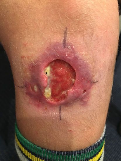
2                                                                                                                                                        D.P. O’Brien et al.

                                                                                                  In Australia, it has also been reported from tropical areas in
                                                                                                  Queensland, where it is known as the Daintree ulcer [49–51],
                                                                                                  and in the Northern Territory [52]. Importantly, case numbers
                                                                                                  reported by countries may be influenced by political stability,
                                                                                                  access to health care, funding for case detection activities, quality
                                                                                                  of reporting systems and availability of diagnostics. For example,
                                                                                                  an exhaustive field survey conducted in DRC involving more
                                                                                                  than 39 000 households showed that only 7% of active BU
                                                                                                  cases were captured in the hospital-based reporting system [53].
                                                                                                     In recent years, the number of disease cases reported to the
                                                                                                  WHO worldwide has been steadily decreasing; from 5156 cases
                                                                                                  in 2008 to 1864 cases in 2016 – a reduction of 64% (Fig. 3). This
                                                                                                  mainly reflects reductions in Africa where there has been a decline
                                                                                                  in most of the highest prevalence countries (Table 1). For example,
                                                                                                  Cote D’Ivoire reported 2679 cases in 2009 and only 376 cases in
                                                                                                  2016 (86% reduction), Ghana’s reported cases reduced from 1048
                                                                                                  in 2010 to 371 in 2016 (65% reduction) and Benin’s from 1203
                                                                                                  cases in 2007 to 312 in 2016 (74% reduction). An African country
                                                                                                  going against the trend is Nigeria, where cases were first reported in
                                                                                                  2009 and have increased from 24 in that year to 235 in 2016 (879%
                                                                                                  increase). Case incidence reported from French Guyana has also
                                                                                                  decreased from 6.07 cases per 100 000 person-years in 1969–1983
                                                                                                  to 4.77 cases per 100 000 person-years in 1984–1998 and to 3.49
                                                                                                  cases per 100 000 person-years in 1999–2013 [38].
                                                                                                     Conversely, in Australia, the number of reported cases has
       Fig. 1. A severe Mycobacterium ulcerans lesion on the knee of an 11-year-old boy.          been increasing with 186 reported in 2016 compared with 42 in
                                                                                                  2010 (343% increase). This mainly reflects a rapidly increasing
                                                                                                  number of cases reported from the coastal regions of the south-
       and Fig. 2). Confirmed cases of Buruli were then reported from                             eastern state of Victoria where there has been a 248% increase
       three countries in the 1950s; Democratic Republic of Congo                                 in cases in the last 4 years (79 cases in 2014 to 275 in 2017). In
       (DRC) [19], Mexico [20] and Uganda [21]. Nine countries across                             this region, the disease has emerged in new geographical areas
       three continents reported their first cases in the 1960s; in Central                       including the Mornington Peninsula outside of Melbourne, and
       Africa (Angola [20], Congo [22] and Gabon [23]), in West Africa                            the proportion of cases presenting with severe disease has doubled
       (Nigeria) [20], in Asia and the Pacific (Papua New Guinea [24],                            since 2010 [10]. Paradoxically, in two adjacent peninsulas sepa-
       Malaysia [25] and Indonesia [20]) and in South America (Peru                               rated by only a few kilometres of ocean with similar climate
       [20] and French Guiana [20]). During the 1970s, cases were                                 and resident populations, there are diverging epidemics – increas-
       reported from West Africa for the first time (Benin [20], Ghana                            ing case numbers on the Mornington Peninsula and reducing case
       [26], Sierra Leone [20] and Cameroon [27]). In the 1980s, Ivory                            numbers on the Bellarine Peninsula [54].
       Coast [28] and Liberia [29] were added to the list of West
       African countries recording their first cases, along with Japan
       [30], Kiribati [31] and Suriname [32]. In the 1990s, four countries                        Ecology of BU
       were added from West Africa (Burkina Faso [33], Equatorial                                 Evidence indicates that M. ulcerans likely evolved from M. mari-
       Guinea [20], Togo [34] and Guinea [20]) as well as Sri Lanka                               num by acquiring a virulence plasmid that produces its patho-
       [20] and China [35] from Asia. In the 2000s, Brazil reported                               genic mycolactone toxin [55] and allowed it to adapt to a
       cases for the first time [36] in addition to the East and Central                          specific environmental niche [56]. Laboratory conditions that
       African countries Kenya [37], Malawi [20], South Sudan [20]                                favour the growth of M. ulcerans are low oxygen [57], relatively
       and Central African Republic [18].                                                         low temperatures (28–33 °C) [58, 59], moderately acidic environ-
                                                                                                  ments (pH 5.4–7.4) [60] and low levels of ultra violet rays [58].
                                                                                                  This may explain why M. ulcerans is often found at the bottom
       Current epidemiological situation
                                                                                                  of aquatic habitats or protected by biofilms [61]. However, despite
       The greatest burden of disease is found in West and Central                                extensive research, the environmental reservoir of the organism
       Africa where the highest number of cases are reported from                                 and mode of transmission of the infection remain unknown. A
       Cote D’Ivoire [39], Benin [40], Ghana [41], Cameroon [42] and                              major factor limiting this understanding is that the organism
       the DRC [43]. Estimated incidence rates include 21.5 per 100                               can rarely be cultured from the environment [62], although
       000/year in parts of Benin [40] and 20.7 per 100 000/year in                               PCR testing of water, aquatic plants, soil and detritus from
       Ghana overall with up to 158.8 per 100 000/year in some affected                           swamps can show evidence of M. ulcerans [47, 63–67].
       districts [41]. Cases continue to be reported from South America                               In endemic areas, the disease is highly focal with endemic and
       (mainly in French Guiana) [38, 44, 45], Asia and the Pacific                               non-endemic areas separated by only a few kilometres [12, 13]. It
       (mainly in Papua New Guinea) [18,20]. BU is predominantly                                  is usually associated with wetlands, especially those with slow-
       found in tropical and subtropical climates, apart from south-                              flowing or stagnant waters such as floodplains or swampy areas
       eastern Australia [12, 46] (with estimated incidence rates of up                           [13, 65]. Studies have suggested that farming activities close to riv-
       to 404 per 100 000/year) [47], China and Japan [48] (Table 1).                             ers [39] and swimming in rivers in endemic areas [68] are risk

Downloaded from https://www.cambridge.org/core. IP address: 46.4.80.155, on 12 Apr 2021 at 14:41:44, subject to the Cambridge Core terms of use, available at https://www.cambridge.org/core/terms.
https://doi.org/10.1017/S0950268818002662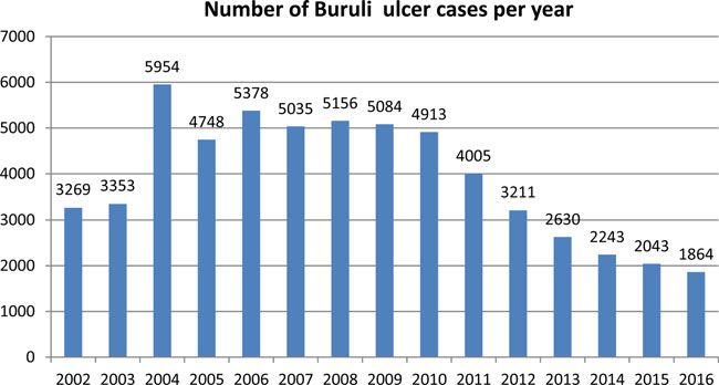
Epidemiology and Infection                                                                                                                                                     3

        Table 1. Countries with published reports of Buruli ulcer cases including year of initial report and changes in numbers of cases reported over time

                                           Year cases first            Year of peak disease      Peak number of cases               2016 cases          Percentage change in 2016
                                            reported and              cases reported to WHO      reported to WHO in a               reported to         from peak reported cases in
             Country                          reference                     2002–2016                    year                          WHO                        a year

             Angola                      1960 [20]                   NA                          NA                             NA                      –
             Australia                   1940 [17]                   2016                        186                            186                     Peak
             Benin                       1977 [20]                   2007                        1203                           312                     −74%
             Brazil                      2007 [36]                   NA                          NA                             NA                      –
             Burkina Faso                1998 [33]                   NA                          NA                             NA                      –
             Cameroon                    1973 [27]                   2004                        914                            85                      −91%
             Central African             2008 [18]                   2008                        3                              NA                      –
             Republic
             China                       1997 [35]                   NA                          NA                             NA                      –
             Congo                       1966 [22]                   2006                        370                            NA                      –
             Democratic                  1950 [19]                   2004                        487                            175                     −64%
             Republic of
             Congo
             Equatorial Guinea           1998 [20]                   2005                        3                              NA                      –
             French Guiana               1969 [20]                   2002                        27 [38]                        NA                      –
             Gabon                       1968 [23]                   2005                        91                             39                      −57%
             Ghana                       1971 [26]                   2006                        1048                           371                     −65%
             Guinea                      1993 [20]                   2006                        279                            72                      –74
             Indonesia (not              1960 [20]                   NA                          NA                             NA                      –
             confirmed)a
             Ivory Coast                 1980 [28]                   2009                        2679                           376                     −86%
             Japan                       1989 [30]                   2011,2013                   10                             2                       −80%
             Kenya                       2008 [37]                   NA                          NA                             NA                      –
             Kiribati (not               1987 [31]                   NA                          NA                             NA                      –
             confirmed)
             Liberia                     1981 [29]                   2015                        105                            NA                      –
             Malawi (not                 2001 [20]                   NA                          NA                             NA                      –
             confirmed)a
             Malaysia                    1964 [25]                   NA                          NA                             NA                      –
             Mexico                      1953 [20]                   NA                          NA                             NA                      –
             Nigeria                     1967 [20]                   2016                        235                            235                     Peak
             Papua New                   1962 [24]                   2004                        31                             16                      –48
             Guinea
             Peru                        1969 [20]                   NA                          NA                             NA                      –
             Sierra Leone                1975 [20]                   2011                        28                             NA                      –
             South Sudan                 2001 [20]                   2002                        568                            NA                      –
             Sri Lanka (not              1992 [20]                   NA                          NA                             NA                      –
             confirmed)a
             Suriname                    1984 [32]                   NA                          NA                             NA
             Togo                        1996 [34]                   2004                        800                            83                      −90%
             Uganda                      1958 [21]                   2002                        117                            NA
        a
            The presence of M. ulcerans was not microbiologically confirmed in this report.

        factors for acquisition of BU. The construction of dams and irri-                            districts [70]. A major process of land-use change, deforestation,
        gation systems have also been associated with increased cases [69],                          is known to result in increased erosion, which has been speculated
        although in French Guyana, a reduction in cases has been                                     to result in run-off contamination of water bodies with M. ulcer-
        observed, likely related to reduced flooding of downstream                                   ans [71]. Deforestation has also been found to alter the

Downloaded from https://www.cambridge.org/core. IP address: 46.4.80.155, on 12 Apr 2021 at 14:41:44, subject to the Cambridge Core terms of use, available at https://www.cambridge.org/core/terms.
https://doi.org/10.1017/S0950268818002662
4 D.P. O’Brien et al.
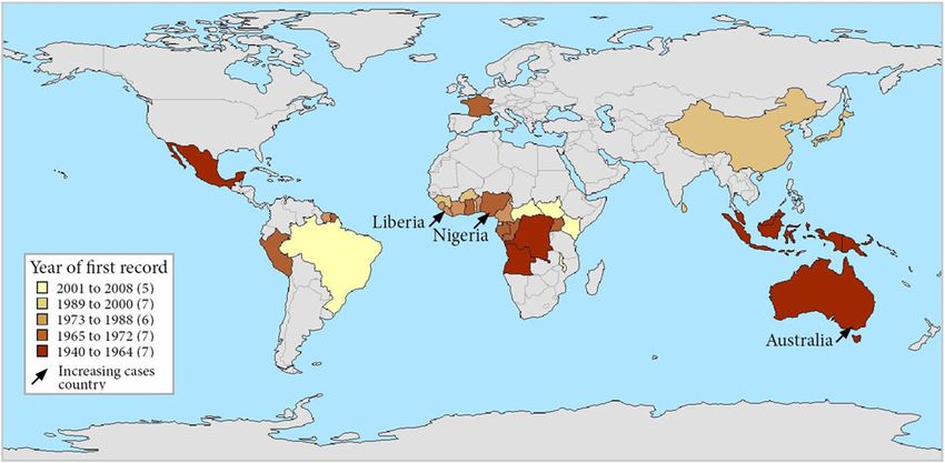
Fig. 2. Map of countries reporting Buruli ulcer cases, stratified by year of first report. Note that each country is represented by its administrative area and that Buruli
ulcer did not occur throughout each country. France is represented for its overseas department French Guiana – there has been no case in metropolitan France.
composition of freshwater communities in French Guiana, the introduction and expansion of M. ulcerans into new environ-
impacting the abundance of M. ulcerans [72]. ments rather than an awakening of quiescent pathogens [79]. Also
In Victoria, Australia, native and domestic mammals including by analysing the population genomics of isolates from 11 different
possums, dogs, cats, koalas, horses and alpacas have developed countries in Africa, Vandelannoote et al. concluded that the
disease [47], but whether they are intimately involved in transmis- spread of M. ulcerans across Africa was a relatively modern phe-
sion, or accidental hosts, remains unclear. Outside of Australia, nomenon and one that had escalated since the late 19th and the
M. ulcerans has rarely been detected in vertebrates, although early 20th centuries [80]. Their work suggested human-induced
lesions on a wild mouse (Mastomys sp.) from Ghana [73] and changes and activities were behind the expansion of M. ulcerans
on a goat and dog from Benin have been found PCR-positive in Africa with humans with active BU lesions inadvertently con-
for M. ulcerans [74], as have domestic duck faeces in Cameroon taminating aquatic environments during water contact activities.
and wild agouti faeces in Ivory Coast [75, 76]. In contrast, a In Australia, it is possible that the dispersal of possums or their
range of aquatic invertebrates from numerous taxa representing active transfer by humans from one area to another may promote
several orders have been found positive for M. ulcerans DNA the introduction of M. ulcerans into new areas. Urban develop-
from many locations in Africa [65, 77]. ment may also increase the disease risk because possums can
Recent evidence from Australia suggests that whatever the reach high population densities in remaining refuge habitats
source in the environment, it may only persist for a short time (e.g. parks, ‘bush-style’ gardens) due to their generalist nature
[78]. In 21 patients with M. ulcerans who were part of a family and ability to utilise human environments and food sources
cluster, the median time to diagnosis between family members [81, 82]. Regardless of whether they are directly involved in intro-
was 2.8 months, and none were diagnosed more than 23 months ducing disease, as possums themselves develop BU, they could act
apart in a cohort spanning 18 years and nearly 2000 combined as sentinel animals for detecting the emergence of M. ulcerans
years of elapsed time since diagnosis. This suggests that in this disease in new areas in Australia [83].
setting the exposure risk is short term, and thereafter diminishes.
Environmental studies in Cameroon found that samples from a
water hole used by local people remained PCR-positive for Transmission of BU to humans
more than 2 years, and at least 12 months after all local human
M. ulcerans disease cases were treated and cured [75]. This sug- Insects such as mosquitoes [84, 85] and aquatic biting arthropods
gests that although the risk of disease transmission has dimin- [59, 86] have been proposed as vectors for transmission, but this
ished, the organism may continue to persist for longer periods remains an open question [66]. Mosquitoes in Australia have
in some environments. tested positive for M. ulcerans by PCR [85] and there are epi-
demiological links such as the use of insect repellent on exposed
body surfaces and the use of mosquito nets being associated with
a reduction in M. ulcerans incidence [84, 87]. Additionally, in
Spread of BU into new areas
Australian towns in an endemic area, a strong dose–response rela-
The mechanism by which M. ulcerans is introduced into new tionship was found between the detection of M. ulcerans in mos-
areas is unknown. However in Australia, research using popula- quitoes and the risk of human disease [88]. Possible mechanisms
tion genomics suggests that the organism has moved from east for infection may involve direct inoculation of the organism
to west in the southern state of Victoria, and that this relates to under the skin via a bite, as suggested by a recent study showing
Downloaded from https://www.cambridge.org/core. IP address: 46.4.80.155, on 12 Apr 2021 at 14:41:44, subject to the Cambridge Core terms of use, available at https://www.cambridge.org/core/terms.
https://doi.org/10.1017/S0950268818002662Epidemiology and Infection 5
Fig. 3. Number of Buruli ulcer cases worldwide reported to the
WHO from 2002 to 2016.
that if the skin already surface contaminated with M. ulcerans is determine if possums play a pivotal role in the transmission of
subjected to a puncturing injury in the form of a needle or a disease to humans or whether they are simply accidental hosts.
bite from a live mosquito, then M. ulcerans lesions can develop In Africa, M. ulcerans has been detected by PCR in aquatic
at the puncture site [89]. Alternatively, infection may result insect species in the order Hemiptera (families: Naucoridae and
from a bite leading to a wound which is secondarily infected by Belostomatidae), which are known to bite humans, suggesting
M. ulcerans from environmental sources such as soil. Although that transmission may occur through these bites [59]. This is sup-
arguing against this is evidence from a guinea pig model where ported by the detection of M. ulcerans in the salivary glands of
applying M. ulcerans organisms directly to abraided skin did these insects after eating snails containing the organism [94],
not establish infection – instead infection was only established and the finding that it can also be transmitted to laboratory
when organisms were inoculated under the skin [90]. It has mice via their bite [86]. In an outbreak in Philip Island,
been proposed that mosquitoes carry the organism on their pro- Australia, it was postulated that aerosols generated by spray irriga-
boscis following contact or feeding with contaminated environ- tion using contaminated water may have disseminated M. ulcer-
mental sources and then directly transmit it through their bite ans and infected humans via the respiratory tract, or through
[85], although the widespread nature and potential travel of mos- contamination of skin lesions and minor abrasions [95], but
quitoes both inside and outside the restricted geographic regions this has not been proven.
affected by BU argues against this. Nevertheless, it is possible that Another recent study on an Australian cohort [78] confirms
mosquito movements between the affected and unaffected areas previous suggestions that human-to-human transmission does
may be limited as some implicated mosquito species, such as not occur [96]. Although cases were often clustered amongst fam-
Aedes notocsriptus, have short flight distances and low dispersal ilies (6.5% of cases had another family member affected), the
ability [91], whilst specific larval habitat requirements can also short time period between the diagnosis of family clustered
restrict distribution [92]. If affected mosquito populations in cases (median 2.8 months) was shorter than the estimated incu-
BU endemic areas were relatively isolated from other populations, bation period of the disease (4.5 months) [78]. Additionally,
this could result in the geographic restriction observed. whole genome SNP analysis of isolates from three paired family
The strongest evidence for a zoonosis comes from Australia clusters revealed isolates derived from two of the three family
involving native mammal species; the common ringtail clusters were not genetically identical and family cluster isolates
(Pseudocheirus peregrinus) and common brushtail (Trichosurus were not any more genetically related than those of six random
vulpecula) possums. Research has found that 19% of these ani- isolates from the same geographic region [78].
mals in an endemic area (Point Lonsdale on the Bellarine The location of clinical M. ulcerans lesions provides some
Peninsula) had M. ulcerans clinical disease, whilst a further 14% information about possible transmission mechanisms. A study
were asymptomatic but had high levels of M. ulcerans DNA of 649 lesions in 579 Australian patients revealed that most lesions
detected on PCR examination of their faeces [47]. In addition, were on exposed body areas, notably upper and lower limbs, and
the location, proportion and concentration of M. ulcerans DNA were commonly over a joint. Few lesions were found on the head
in possum faeces strongly correlated with that of human M. ulcer- and neck, palms of hands, soles of feet and trunk [97].
ans disease cases in at least two outbreaks where it has been mea- Furthermore, the distribution was non-random, with a strong pre-
sured: Sorrento on the Mornington Peninsula [83] and Point dilection for ankles, elbows and calves. Differences in the pattern
Lonsdale on the Bellarine Peninsula [47]. Additionally, in nearby of lesion distribution were also found between genders (men had
areas with no cases of human disease possum faeces were not more lesions on upper limbs and less on lower limbs than
found to contain M. ulcerans DNA [47]. It is theoretically possible women), age groups (those aged ⩾65 years were less likely to
that infected possums amplify the organism in the environment have proximal upper limb lesions compared with those6 D.P. O’Brien et al.
activities undertaken. Similar findings have also been reported a reduced burden of disease in humans through improved case
from a smaller study in Cameroon [98]. Case–control studies in detection and increasing antibiotic use may be responsible for
Africa have also identified wearing short lower body clothing the reduction in cases in Africa [80]. However, the situation of
whilst farming as risk factors for M. ulcerans [99] and covering increasing cases in Australia, where there is good access to med-
limbs during farming as protective for M. ulcerans [100]. These ical care and antibiotic treatment is widely used argues against
findings suggest that M. ulcerans transmission and pathogenesis this explanation. An alternative explanation may be that improve-
may be similar across the world despite very different geograph- ments in the accuracy of diagnosis since PCR confirmation was
ical and climatic conditions. introduced have reduced over-reporting of cases [105].
Transmission also appears to be seasonal. In Australia, the Importantly, cases of disease have been decreasing in most
large majority of cases (>70%) are likely acquired in the warmest countries in recent years indicating a hopeful, positive trend.
6 months of the year [97], and in Cameroon, the likely time of However, despite recent insights, it still has not been conclusively
infection was seasonal and highest between the months of determined how this pathogen circulates in the environment, or
August and October [101]. Studies have also reported an associ- how transmission to humans occurs. This requires the availability
ation with rainfall; studies from Ghana and Cameroon report of robust scientific knowledge acquired by a thorough and
that the proportion of M. ulcerans-positive samples from the exhaustive examination of the environment, local fauna, human
environment was higher during the months with higher rainfall behaviour and characteristics, and the interactions between
levels than during the dry season months [101, 102] and reports them [106]. Only with this knowledge can control strategies or
from French Guyana [70] and Australia suggest an increase in early warning systems be designed and implemented that effect-
cases associated with periods of high rainfall followed by dry per- ively and consistently prevent the spread and reduce the incidence
iods [103]. of this disease.
Conflicts of interest. None.
Future prospects
The epidemiology of M. ulcerans has clearly changed over time
and is expected to continue evolving into the future. Although References
the explanations for this are not fully understood, the processes
1. World Health Orgainisation (2010) Working to Overcome the Global
associated with increasing anthropogenic land-use change such
Impact of Neglected Tropical Diseases: First WHO Report on Neglected
as deforestation, road construction, flooding and population Tropical Diseases. Geneva, Switzerland: World Health Orgainisation.
settlement may have significant impacts on this environmental 2. World Health Organisation (2012) Treatment of Mycobacterium ulcer-
pathogen, affecting both its future distribution and human expos- ans Disease (Buruli Ulcer): Guidance for Health Workers. Geneva,
ure risk [93]. For example, the increase in cases in recent years Switzerland: World Health Orgainisation.
observed in south-eastern Victoria, Australia, may be reflective 3. O’Brien DP et al. (2014) Clinical features and risk factors of oedematous
of the impact of new residential developments which may have Mycobacterium ulcerans lesions in an Australian population: beware cel-
altered the environment and impacted both aquatic and terrestrial lulitis in an endemic area. PLoS Neglected Tropical Diseases 8, e2612.
communities, including a proposed reservoir of M. ulcerans, the 4. Vincent QB et al. (2014) Clinical epidemiology of laboratory-confirmed
possum. Buruli ulcer in Benin: a cohort study. The Lancet Global Health 2, e422–
e430.
Flooding, through environmental disturbance and contamin-
5. Sizaire V et al. (2006) Mycobacterium ulcerans infection: control, diag-
ation of aquatic habitats, has regularly been linked to outbreaks nosis, and treatment. The Lancet Infectious Diseases 6, 288–296.
of BU [93]. As dams alter the degree and frequency of down- 6. Nienhuis WA et al. (2010) Antimicrobial treatment for early, limited
stream flooding, the increase in dam building occurring in Mycobacterium ulcerans infection: a randomised controlled trial. The
many regions where BU is endemic, may also alter the distribu- Lancet 375, 664–672.
tion of this pathogen. Climate change will likely also be influen- 7. Friedman ND et al. (2016) Increasing experience with primary oral
tial, through altered temperatures, increased frequency of medical therapy for Mycobacterium ulcerans disease in an Australian
extreme weather events and intense flooding events, and inunda- cohort. Antimicrobial Agents and Chemotherapy 60, 2692–2695.
tion of coastal foci, such as the Victorian hot-spot, through 8. O’Brien DP et al. (2014) Treatment and prevention of Mycobacterium
changes in sea level. The increased mobility of today’s societies ulcerans infection (Buruli ulcer) in Australia: guideline update. The
Medical Journal of Australia 200, 267–270.
may additionally help to modify the distribution of M. ulcerans,
9. Ellen DE et al. (2003) Assessment of functional limitations caused by
by altering the genetic variants of this pathogen present in an Mycobacterium ulcerans infection: towards a Buruli ulcer functional limi-
area (as found in the Offin river valley in Ghana) [104], and by tation score. Tropical Medicine & International Health: TM & IH 8, 90–
providing opportunities for the establishment of new foci where 96.
suitable environmental conditions exist. 10. Tai AYC et al. (2018) Increased severity and spread of Mycobacterium
ulcerans, Southeastern Australia. Emerging Infectious Diseases 24, 58–64.
11. Sopoh GE et al. (2007) Buruli ulcer surveillance, Benin, 2003–2005.
Conclusions Emerging Infectious Diseases 13, 1374–1376.
The reasons for the changing epidemiology of BU worldwide are 12. Boyd SC et al. (2012) Epidemiology, clinical features and diagnosis of
unknown. Possibilities include changing environmental condi- Mycobacterium ulcerans in an Australian population. The Medical
Journal of Australia 196, 341–344.
tions such as rainfall and temperature in the era of climate
13. Anon (2008) Buruli ulcer: progress report, 2004–2008. Releve epidemio-
change, changes in population dynamics and land use, improved logique hebdomadaire/Section d’hygiene du Secretariat de la Societe des
sanitation and access to healthcare or reduction in exposure Nations = Weekly Epidemiological Record/Health Section of the
through such things as increasing mosquito net use, or spill-over Secretariat of the League of Nations 83, 145–154.
into humans from epidemics in animal reservoirs. Additionally, if 14. O’Brien DP et al. (2014) Management of BU-HIV co-infection. Tropical
humans represent the disease reservoir, it has been speculated that Medicine & International Health: TM & IH 19, 1040–1047.
Downloaded from https://www.cambridge.org/core. IP address: 46.4.80.155, on 12 Apr 2021 at 14:41:44, subject to the Cambridge Core terms of use, available at https://www.cambridge.org/core/terms.
https://doi.org/10.1017/S0950268818002662Epidemiology and Infection 7
15. Christinet V et al. (2014) Impact of HIV on the severity of Buruli ulcer 40. Debacker M et al. (2004) Mycobacterium ulcerans disease (Buruli ulcer)
disease: results from a retrospective study in Cameroon. Open Forum in rural hospital, Southern Benin, 1997–2001. Emerging Infectious
Infectious Diseases 1, ofu021. Diseases 10, 1391–1398.
16. Yeboah-Manu D et al. (2012) Sero-epidemiology as a tool to screen 41. Amofah G et al. (2002) Buruli ulcer in Ghana: results of a national case
populations for exposure to Mycobacterium ulcerans. PLoS Neglected search. Emerging Infectious Diseases 8, 167–170.
Tropical Diseases 6, e1460. 42. Noeske J et al. (2004) Buruli ulcer disease in Cameroon rediscovered.
17. MacCallum P, Tolhurst JC et al. (1948) A new mycobacterial infection The American Journal of Tropical Medicine and Hygiene 70, 520–526.
in man. The Journal of Pathology and Bacteriology 60, 93–122. 43. Kibadi K et al. (2009) [Relapse after surgical treatment of mycobacterium
18. World Health Organization. Global Health Observatory data repository - ulcerans infection (Buruli ulcer): study of risk factors in 84 patients in the
Buruli ulcer. Available at http://apps.who.int/neglected_diseases/ntddata/ Democratic Republic of the Congo]. Medecine Tropicale: Revue Du Corps
buruli/buruli.html (accessed 30 June 2018). De Sante Colonial 69, 471–474.
19. van Oye E and Ballion M (1950) [Is it necessary to take into account a 44. Guerra H et al. (2008) Mycobacterium ulcerans disease, Peru. Emerging
new infection from acid-resistant bacilli in Africa? Preliminary note]. Infectious Diseases 14, 373–377.
Annales De La Societe Belge De Medecine Tropicale (1920) 30, 619–627. 45. Sambourg E et al. (2014) [Paradoxical reactions and responses during
20. Janssens B et al. (2005) Buruli ulcer: an historical overview with updat- antibiotic treatment for Mycobacterium ulcerans infection (Buruli
ing to 2005. Bulletin Des Séances Académie Royale Des Sciences ulcer). Four cases from French Guiana]. Annales De Dermatologie Et
D’outre-Mer 51, 265–299. De Venereologie 141, 413–418.
21. Clancey JK et al. (1961) Mycobacterial skin ulcers in Uganda. The 46. Johnson PD et al. (1996) The emergence of Mycobacterium ulcerans
Lancet 2, 951–954. infection near Melbourne. The Medical Journal of Australia 164, 76–78.
22. Perquis P and Muret G (1965) [Tropical ulcers due to mycobacteria. 47. Fyfe JA et al. (2010) A major role for mammals in the ecology of
Apropos of 2 cases]. Bulletin De La Societe De Pathologie Exotique Et Mycobacterium ulcerans. PLoS Neglected Tropical Diseases 4, e791.
De Ses Filiales 58, 789–795. 48. Kondo M et al. (2009) Leg ulcer caused by Mycobacterium ulcerans ssp.
23. Carayon A et al. (1968) [Ulcers due to mycobacteria in Africa]. Bulletin shinshuense infection. International Journal of Dermatology 48, 1330–1333.
De La Societe Medicale D’afrique Noire De Langue Francaise 13, 670–679. 49. Jenkin GA et al. (2002) Acute, oedematous Mycobacterium ulcerans
24. Reid IS (1967) Mycobacterium ulcerans infection: a report of 13 cases at infection in a farmer from far North Queensland. The Medical Journal
the Port Moresby General Hospital, Papua. The Medical Journal of of Australia 176, 180–181.
Australia 1, 427–431. 50. Steffen CM, Smith M and McBride WJ (2010) Mycobacterium ulcerans
25. Pettit JH, Marchette NJ and Rees RJ (1966) Mycobacterium ulcerans infection in North Queensland: the ‘Daintree ulcer’. ANZ Journal of
infection. Clinical and bacteriological study of the first cases recognized Surgery 80, 732–736.
in South East Asia. The British Journal of Dermatology 78, 187–197. 51. Francis G, Whitby M and Woods M (2006) Mycobacterium ulcerans
26. Bayley AC (1971) Buruli ulcer in Ghana. British Medical Journal 2, 401– infection: a rediscovered focus in the Capricorn Coast region of central
402. Queensland. The Medical Journal of Australia 185, 179–180.
27. Ravisse P (1977) [Skin ulcer caused by Mycobacterium ulcerans in 52. Quinn JV and Crotty JM (1963) Mycobacterium ulcerans infections in
Cameroon. I. Clinical, epidemiological and histological study]. Bulletin the Northern Territory. The Medical Journal of Australia 2, 317–319.
De La Societe De Pathologie Exotique Et De Ses Filiales 70, 109–124. 53. Mavinga Phanzu D et al. (2013) Burden of Mycobacterium ulcerans disease
28. Perraudin ML, Herrault A and Desbois JC (1980) [Mycobacterium (Buruli ulcer) and the underreporting ratio in the territory of Songololo,
ulcerans skin ulcers (or Buruli ulcers) (author’s transl)]. Annales De Democratic Republic of Congo. PLoS Neglected Tropical Diseases 7, e2563.
Pediatrie 27, 687–692. 54. Department of Human Services Victoria. Surveillance of notifiable con-
29. Ziefer A, Connor DH and Gybson DW (1981) Mycobacterium ulcerans: ditions in Victoria – regional comparisons. Available at http://www.
infection of two patients in Liberia. International Journal of Dermatology health.vic.gov.au/ideas/downloads/daily_reports/rptVS_SNIDSRegional
20, 362–367. Comparisons_GR.pdf (accessed 1 March 2018).
30. Tsukamura M et al. (1989) [A taxonomic study on a mycobacterium 55. Doig KD et al. (2012) On the origin of Mycobacterium ulcerans, the
which caused a skin ulcer in a Japanese girl and resembled causative agent of Buruli ulcer. BMC Genomics 13, 258.
Mycobacterium ulcerans]. Kekkaku: [Tuberculosis] 64, 691–697. 56. Stinear TP et al. (2007) Reductive evolution and niche adaptation
31. Christie M (1987) Suspected Mycobacterium ulcerans disease in Kiribati. inferred from the genome of Mycobacterium ulcerans, the causative
The Medical Journal of Australia 146, 600–604. agent of Buruli ulcer. Genome Research 17, 192–200.
32. Portaels F et al. (1996) Variability in 3′ end of 16S rRNA sequence of 57. Palomino J et al. (1998) Effect of oxygen on growth of Mycobacterium
Mycobacterium ulcerans is related to geographic origin of isolates. ulcerans in the BACTEC system. Journal of Clinical Microbiology 36,
Journal of Clinical Microbiology 34, 962–965. 3420–3422.
33. Ouoba K et al. (1998) [Buruli ulcers in Burkina Faso: apropos of 6 58. Portaels F et al. (2001) Mycobacterium ulcerans in wild animals.
cases]. La Tunisie Medicale 76, 46–50. Revue Scientifique Et Technique (International Office of Epizootics) 20,
34. Meyers WM et al. (1996) Mycobacterium ulcerans infection (Buruli 252–264.
ulcer): first reported patients in Togo. The British Journal of 59. Portaels F et al. (1999) Insects in the transmission of Mycobacterium
Dermatology 134, 1116–1121. ulcerans infection. The Lancet 353, 986.
35. Faber WR et al. (2000) First reported case of Mycobacterium ulcerans 60. Portaels F and Pattyn SR (1982) Growth of mycobacteria in relation to
infection in a patient from China. Transactions of the Royal Society of the pH of the medium. Annales De Microbiologie 133, 213–221.
Tropical Medicine and Hygiene 94, 277–279. 61. Marsollier L et al. (2007) Impact of Mycobacterium ulcerans biofilm on
36. dos Santos VM et al. (2007) Mycobacterium ulcerans infection in Brazil. transmissibility to ecological niches and Buruli ulcer pathogenesis. PLoS
The Medical Journal of Australia 187, 63–64. Pathogens 3, e62.
37. Walsh DS et al. (2009) Short report: clinical and molecular evidence for 62. Portaels F et al. (2008) First cultivation and characterization of
a case of Buruli ulcer (Mycobacterium ulcerans infection) in Kenya. The Mycobacterium ulcerans from the environment. PLoS Neglected
American Journal of Tropical Medicine and Hygiene 81, 1110–1113. Tropical Diseases 2, e178.
38. Douine M et al. (2017) Mycobacterium ulcerans infection (Buruli ulcer) 63. Ross BC et al. (1997) Detection of Mycobacterium ulcerans in environ-
in French Guiana, South America, 1969–2013: an epidemiological study. mental samples during an outbreak of ulcerative disease. Applied and
The Lancet Planetary Health 1, e65–e73. Environmental Microbiology 63, 4135–4138.
39. Marston BJ et al. (1995) Emergence of Buruli ulcer disease in the Daloa 64. Stinear T et al. (1999) Identification and characterization of IS2404 and
region of Cote d’Ivoire. The American Journal of Tropical Medicine and IS2606: two distinct repeated sequences for detection of Mycobacterium
Hygiene 52, 219–224. ulcerans by PCR. Journal of Clinical Microbiology 37, 1018–1023.
Downloaded from https://www.cambridge.org/core. IP address: 46.4.80.155, on 12 Apr 2021 at 14:41:44, subject to the Cambridge Core terms of use, available at https://www.cambridge.org/core/terms.
https://doi.org/10.1017/S09502688180026628 D.P. O’Brien et al.
65. Williamson HR et al. (2008) Distribution of Mycobacterium ulcerans in 85. Johnson PD et al. (2007) Mycobacterium ulcerans in mosquitoes cap-
Buruli ulcer endemic and non-endemic aquatic sites in Ghana. PLoS tured during outbreak of Buruli ulcer, southeastern Australia. Emerging
Neglected Tropical Diseases 2, e205. Infectious Diseases 13, 1653–1660.
66. Benbow ME et al. (2008) Aquatic invertebrates as unlikely vectors of 86. Marsollier L et al. (2002) Aquatic insects as a vector for Mycobacterium
Buruli ulcer disease. Emerging Infectious Diseases 14, 1247–1254. ulcerans. Applied and Environmental Microbiology 68, 4623–4628.
67. Vandelannoote K et al. (2010) Application of real-time PCR in Ghana, a 87. Landier J et al. (2011) Adequate wound care and use of bed nets as pro-
Buruli ulcer-endemic country, confirms the presence of Mycobacterium tective factors against Buruli ulcer: results from a case control study in
ulcerans in the environment. FEMS Microbiology Letters 304, 191–194. Cameroon. PLoS Neglected Tropical Diseases 5, e1392.
68. Aiga H et al. (2004) Assessing water-related risk factors for Buruli ulcer: 88. Lavender CJ et al. (2011) Risk of Buruli ulcer and detection of
a case-control study in Ghana. The American Journal of Tropical Mycobacterium ulcerans in mosquitoes in southeastern Australia. PLoS
Medicine and Hygiene 71, 387–392. Neglected Tropical Diseases 5, e1305.
69. Marion E et al. (2011) Geographic expansion of Buruli ulcer disease, 89. Wallace JR et al. (2017) Mycobacterium ulcerans low infectious dose and
Cameroon. Emerging Infectious Diseases 17, 551–553. mechanical transmission support insect bites and puncturing injuries in
70. Morris A et al. (2014) Complex temporal climate signals drive the emer- the spread of Buruli ulcer. PLoS Neglected Tropical Diseases 11, e0005553.
gence of human water-borne disease. Emerging Microbes & Infections 3, 90. Williamson HR et al. (2014) Mycobacterium ulcerans fails to infect
e56–e56. through skin abrasions in a guinea pig infection model: implications
71. Hayman J (1991) Postulated epidemiology of Mycobacterium ulcerans for transmission. PLoS Neglected Tropical Diseases 8, e2770.
infection. International Journal of Epidemiology 20, 1093–1098. 91. Foley DH, Russell RC and Bryan JH (2004) Population structure of the
72. Morris AL et al. (2016) Deforestation-driven food-web collapse linked to peridomestic mosquito Ochlerotatus notoscriptus in Australia. Medical
emerging tropical infectious disease, Mycobacterium ulcerans. Science and Veterinary Entomology 18, 180–190.
Advances 2, e1600387. 92. Johnston E et al. (2014) Mosquito communities with trap height and
73. Narh CA et al. (2015) Source tracking Mycobacterium ulcerans infec- urban-rural gradient in Adelaide, South Australia: implications for dis-
tions in the Ashanti region, Ghana. PLoS Neglected Tropical Diseases 9, ease vector surveillance. Journal of Vector Ecology: Journal of the
e0003437. Society for Vector Ecology 39, 48–55.
74. Zeukeng F (2017) Mycobacterium ulcerans infected wounds found in 93. Combe M et al. (2017) Global and local environmental changes as dri-
domestic animals in the Buruli ulcer endemic area of Sedje-Denou, vers of Buruli ulcer emergence. Emerging Microbes & Infections 6, e22.
Southern Benin. In World Health Organization annual scientific meeting 94. Marsollier L et al. (2004) Aquatic snails, passive hosts of Mycobacterium
on Buruli ulcer. Geneva, Switzerland. ulcerans. Applied and Environmental Microbiology 70, 6296–6298.
75. Bratschi MW et al. (2014) Mycobacterium ulcerans persistence at a vil- 95. Veitch MG et al. (1997) A large localized outbreak of Mycobacterium
lage water source of Buruli ulcer patients. PLoS Neglected Tropical ulcerans infection on a temperate southern Australian island.
Diseases 8, e2756. Epidemiology and Infection 119, 313–318.
76. Tian RB et al. (2016) Detection of Mycobacterium ulcerans DNA in the 96. Merritt RW et al. (2010) Ecology and transmission of Buruli ulcer dis-
environment, Ivory Coast. PLoS ONE 11, e0151567. ease: a systematic review. PLoS Neglected Tropical Diseases 4, e911.
77. Garchitorena A et al. (2014) Mycobacterium ulcerans ecological dynam- 97. Yerramilli A et al. (2017) The location of Australian Buruli ulcer lesions
ics and its association with freshwater ecosystems and aquatic communi- – implications for unravelling disease transmission. PLoS Neglected
ties: results from a 12-month environmental survey in Cameroon. PLoS Tropical Diseases 11, e0005800.
Neglected Tropical Diseases 8, e2879. 98. Bratschi MW et al. (2013) Geographic distribution, age pattern and sites
78. O’Brien DP et al. (2017) Exposure risk for infection and lack of of lesions in a cohort of Buruli ulcer patients from the Mape Basin of
human-to-human transmission of Mycobacterium ulcerans disease, Cameroon. PLoS Neglected Tropical Diseases 7, e2252.
Australia. Emerging Infectious Diseases 23, 837–840. 99. Pouillot R et al. (2007) Risk factors for Buruli ulcer: a case control study
79. Buultjens AH et al. (2018) Comparative genomics shows Mycobacterium in Cameroon. PLoS Neglected Tropical Diseases 1, e101.
ulcerans migration and expansion has preceded the rise of Buruli ulcer in 100. Kenu E et al. (2014) Risk factors for Buruli ulcer in Ghana – a case con-
south-eastern Australia. Applied and Environmental Microbiology 84, trol study in the Suhum-Kraboa-Coaltar and Akuapem South Districts of
e02612-17. the eastern region. PLoS Neglected Tropical Diseases 8, e3279.
80. Vandelannoote K et al. (2017) Multiple introductions and recent spread 101. Landier J et al. (2015) Seasonal patterns of Buruli ulcer incidence,
of the emerging human pathogen Mycobacterium ulcerans across Africa. Central Africa, 2002–2012. Emerging Infectious Diseases 21, 1414–1417.
Genome Biology and Evolution 9, 414–426. 102. Aboagye SY et al. (2017) Seasonal pattern of Mycobacterium ulcerans,
81. Stow A et al. (2006) Genetic structure infers generally high philopatry the Causative Agent of Buruli ulcer, in the environment in Ghana.
and male-biased dispersal of brushtail possums (‘Trichosurus vulpecula’) Microbial Ecology 74, 350–361.
in urban Australia. Wildlife Research 33, 409–415. 103. van Ravensway J et al. (2012) Climate and landscape factors associated
82. Giffney R, Russell T and Kohen J (2009) Age of road-killed common with Buruli ulcer incidence in Victoria, Australia. PLoS ONE 7, e51074.
brushtail possums (Trichosurus vulpecula) and common ringtail possums 104. Lamelas A et al. (2016) Spatiotemporal co-existence of two
(Pseudocheirus peregrinus) in an urban environment. Australian Mycobacterium ulcerans clonal complexes in the Offin River Valley of
Mammalogy 31, 137–142. Ghana. PLoS Neglected Tropical Diseases 10, e0004856.
83. Carson C et al. (2014) Potential wildlife sentinels for monitoring the 105. O’Brien DP (2017) Unlocking of the secrets of Mycobacterium ulcerans
endemic spread of human Buruli ulcer in South-East Australia. PLoS disease transmission. The Lancet Planetary Health 1, e52–e53.
Neglected Tropical Diseases 8, e2668. 106. O’Brien DP et al. (2018) Tackling the worsening epidemic of Buruli
84. Quek TY et al. (2007) Risk factors for Mycobacterium ulcerans infection, ulcer in Australia in an information void: time for an urgent scientific
southeastern Australia. Emerging Infectious Diseases 13, 1661–1666. response. The Medical Journal of Australia 208, 287–289.
Downloaded from https://www.cambridge.org/core. IP address: 46.4.80.155, on 12 Apr 2021 at 14:41:44, subject to the Cambridge Core terms of use, available at https://www.cambridge.org/core/terms.
https://doi.org/10.1017/S0950268818002662You can also read