Glycophorins and the MNS blood group system: a narrative review
←
→
Page content transcription
If your browser does not render page correctly, please read the page content below
Review Article
                                                                                                                                      Page 1 of 16

Glycophorins and the MNS blood group system: a narrative review
Genghis H. Lopez1,2, Catherine A. Hyland1,3, Robert L. Flower1,3
1
Clinical Services and Research Division, Australian Red Cross Lifeblood, Kelvin Grove, Queensland, Australia; 2School of Medical Science, Griffith
Health, Griffith University, Gold Coast, Queensland, Australia; 3School of Biomedical Sciences, Faculty of Health, Queensland University of
Technology, Brisbane, Queensland, Australia
Contributions: (I) Conception and design: All authors; (II) Administrative support: None; (III) Provision of study materials or patients: None; (IV)
Collection and assembly of data: All authors; (V) Data analysis and interpretation: All authors; (VI) Manuscript writing: All authors; (VII) Final
approval of manuscript: All authors.
Correspondence to: Genghis H. Lopez, PhD. Clinical Services and Research Division, Australian Red Cross Lifeblood, 44 Musk Avenue, Kelvin Grove,
Queensland 4059, Australia. Email: glopez@redcrossblood.org.au.

                Abstract: The MNS blood group system, International Society of Blood Transfusion (ISBT) 002, is second
                after the ABO system. GYPA and GYPB genes encode MNS blood group antigens carried on glycophorin A
                (GPA), glycophorin B (GPB), or on variant glycophorins. A third gene, GYPE, produce glycophorin E (GPE)
                but is not expressed. MNS antigens arise from several genetic mechanisms. Single nucleotide variants (SNVs)
                contribute to the diversity of the MNS system. A new antigen SUMI (MNS50), p.Thr31Pro on GPA has
                been described in the Japanese population. Unequal crossing-over and gene conversion are the mechanisms
                forming hybrid glycophorins, usually from parent genes GYPA and GYPB. GYPE also contributes to gene
                recombination previously only described with GYPA. Recently, however, GYPE was shown to recombine
                with GYPB to form a GYP(B-E-B) hybrid. A GYP(B-E-B) hybrid allele encodes a mature GP(E-B) molecule
                expressing a trypsin-resistant M antigen but no S/s. Another novel glycophorin GP.Mot has been described
                carrying Mia, Mur, MUT, and KIPP antigens. GP.Mot is encoded by a GYP(A-B-A) hybrid allele. Newly
                reported cases of haemolytic transfusion reaction (HTR) or haemolytic disease of the fetus and newborn
                (HDFN) due to antibodies to MNS antigens is a constant reminder of the clinical significance of the MNS
                system. In one HDFN case, anti-U and anti-D were detected in an Indian D–, S–s–U– mother. The S–s–
                U– phenotype is rare in Asians and Caucasians but it is more commonly found in the African populations.
                Several types of novel GYPB deletion alleles that drive the S–s–U– phenotype have been recently described.
                Two large GYPB deletion alleles, over 100 kb, were identified as the predominant alleles in the African
                population. The use of advanced DNA sequencing techniques and bioinformatic analysis has helped uncover
                these large gene-deletion variants. Molecular typing platforms used for MNS genotyping are also discussed
                in this review. In conclusion, this review considers currently recognised MNS antigens and variants, new
                hybrid alleles and GYPB gene deletion alleles as well as clinical case studies. These new discoveries contribute
                to our understanding of the complexity of the MNS system to guide decision-making in genetic analysis and
                transfusion medicine.

                Keywords: MNS blood group system; blood group antigens; hybrid glycophorins; variant glycophorins; Mia
                antigen (MNS7); anti-Mia antibodies; blood group genetics; blood type

                Received: 22 January 2021; Accepted: 28 April 2021.
                doi: 10.21037/aob-21-9
                View this article at: http://dx.doi.org/10.21037/aob-21-9

© Annals of Blood. All rights reserved.                                                      Ann Blood 2021 | http://dx.doi.org/10.21037/aob-21-9
Page 2 of 16                                                                                                   Annals of Blood, 2021

Introduction                                                    produced by GYPE was shown to be very unstable affecting
                                                                GPE expression (18). GPE has not been detected on the
After the discovery of the ABO blood group system in 1900,
                                                                surface of RBC. However if expressed, GPE would be a
Landsteiner and Levine searched for more human blood
                                                                59-amino-acid molecule carrying an M antigen as the Exon
groups (1). In 1927, rabbits injected with human red blood
                                                                2 sequence for GYPE is identical to GYPA (14,19).
cells (RBCs) produced antibodies against M (MNS1) and
                                                                   G PA a n d G P B a r e s i n g l e - p a s s t r a n s m e m b r a n e
N (MNS2) blood group antigens (1,2). The names of these
                                                                sialoglycoproteins heavily glycosylated with abundant
antigens came from the word “immune” and this discovery
                                                                O-glycans (10). Only GPA carries N-glycan (10). These
created the second blood system now known as the MNS
                                                                carbohydrate molecules contribute a strong net negative
blood group system (2). Examples of human anti-M and
                                                                charge to the surface of RBCs preventing RBC aggregation
anti-N were reported in later years (3).
                                                                thus maintaining blood flow in the circulation (10). It is
   In 1947, Walsh and Montgomery reported a female
                                                                estimated that there are 1×10⁶ copies of GPA and 2.5×10⁵
patient with puerperal fever who developed an antibody
                                                                copies of GPB per RBC (15,20).
recognising the S antigen (MNS3) (4). The S antigen was
named after Sydney, the capital city of New South Wales,
Australia. The s antigen (MNS4), antithetical to S, was         Genes of the MNS blood group system
described in 1951 (4,5). Two years later, the fifth antigen U   GYPA, GYPB and GYPE genes form a 350-kb gene cluster on
(stands for universal, MNS5) in this system was discovered      chromosome 4q31.21, Figure 2A (21,22). Analysis of this region
(6,7). Currently, 50 antigens in the MNS system are             suggests that GYPA was the ancestral gene and that a series of
recognised by the International Society of Blood Transfusion    molecular events formed GYPB and GYPE genes (23). Firstly,
(ISBT) Working Party (WP) on Red Cell Immunogenetics            ancestral GYPA is duplicated. Two chromatid strands,
and Blood Group Terminology (RCIBGT), Table 1 (8).              each carrying a duplicated GYPA, misaligned. This was
These antigens are carried on glycophorin A (GPA),              followed by unequal crossing-over occurring within the Alu
glycophorin B (GPB) or variant glycophorins. Amongst            sequences present in each strand producing a progenitor
antibodies to MNS antigens, many are regarded as                GYPB/GYPE genomic segment (23). Subsequent duplication
clinically significant with reported cases of haemolytic        of this segment gave rise to independent GYPB and GYPE
disease of the fetus and newborn (HDFN) and haemolytic          genes. The 3' sequences for GYPB and GYPE were acquired
transfusion reactions (HTRs). The history of the MNS            from an unrelated genomic segment (23).
system has been detailed in reference textbooks (1,2,9-11)         GYPA, GYPB and GYPE genes show a high degree of
and review articles (12-17). We present the following article   homology, over 95%, from the 5' flanking sequences to the
in accordance with the Narrative Review reporting checklist     Alu sequence located in Intron 5 which is approximately 1
(available at http://dx.doi.org/10.21037/aob-21-9).             kb downstream from Exon 5 (21,23). Sequence homology
                                                                and the intron-exon gene structure organisation (Figure 2B)
GPA, GPB, and glycophorin E (GPE)                               is thought to facilitate the numerous gene recombination
                                                                events occurring in these three glycophorin genes (14).
GYPA and GYPB genes encode GPA, 150 amino acids, and            Furthermore, an approximately 1 kb region was identified
GPB, 91 amino acids, respectively. GYPA and GYPB Exon           as a major recombination hotspot spanning between
1 up to the 5' end of Exon 2 encode a 19-amino acid leader      Intron 2–Exon 3 junction and Intron 3–Exon 4 junction
sequence, from the 3' end of Exon 2 up to Exon 4 encode         (10). The presence of multiple direct repeat sequences and
amino acids residing in the extracellular region, Exon 5        palindromic sequences, particularly the 35-bp complex
in the transmembrane and Exon 6–7 in the intracellular          palindrome within Exon 3 of GYPA and pseudoexon 3 of
domain. The leader sequence is cleaved after the protein        GYPB, are distinct features of these genes (10). Reference
is inserted into the cell membrane (1). Therefore, the          sequences associated with these genes are listed in Table 2 (8).
mature GPA and GPB molecule has 131 and 72 amino
acid residues, respectively, Figure 1 (10). GYPE encodes
                                                                Variant glycophorins: genetic mechanisms
GPE which is a 78-amino acid molecule that includes a
19-residue leader sequence (18). The mRNA transcript            Several genetic mechanisms contribute to the diversity

© Annals of Blood. All rights reserved.                                          Ann Blood 2021 | http://dx.doi.org/10.21037/aob-21-9
Annals of Blood, 2021 Page 3 of 16
Table 1 Classification of MNS antigens in the general population
n Antigens
Polymorphic antigens n=4 M, N, S, s
High-frequency antigens n=10 U, Ena, ENKT, ‘N’, ENEP, ENEH, ENAV, ENDA, ENEV, JENU
Low-frequency antigens n=36 He, Mia, Mc, Vw, Mur, Mg, Vr, Me, Mta, Sta, Ria, Cla, Nya, Hut, Hil, Mv, Far, sD, Mit, Dantu, Hop, Nob,
Or, DANE, TSEN, MINY, MUT, SAT, ERIK, Osa, HAG, MARS, MNTD, SARA, KIPP, SUMI
Exon 2 Exon 3 Exon 4 Exon 5 Exon 6 Ex 7
GPA M RBC membrane
GPA N
RBC membrane
GPB S
GPB s
Exon 2 Exon 4 Exon 5 Ex 6
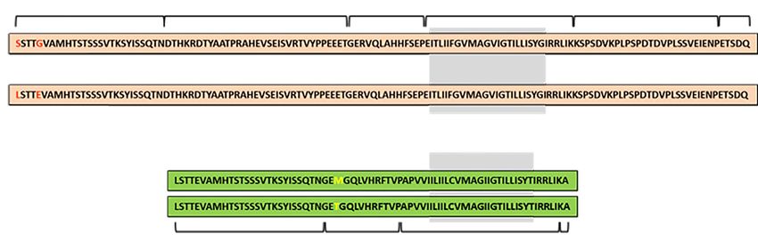
Figure 1 Amino acid sequences for GPA and GPB. The red texts on GPA show the amino acid differences for M (p.Ser20; p.Gly24) and
N (p.Leu20; p.Glu24). In GPB, yellow texts indicate S (p.Met48) and s (p.Thr48). The regions of GPA and GPB predicted to be in the
transmembrane domain are indicated with a gray background (1). GPA, glycophorin A; GPB, glycophorin B.
A Chromosome 4 q31.21 of variant glycophorins (10,12). Unequal crossing-
over and gene conversion are the two main mechanisms
forming hybrid glycophorin variants (10,12). Currently,
143.9Mb 144.0Mb 144.1Mb
there are over 30 hybrid genes in the MNS system (8).
GYPE GYPB GYPA Examples of these hybrid alleles are shown in
Table 3. The third mechanism is single nucleotide
polymorphism (10,12).
B
GYPA
A1 A2 A3 A4 A5 A6 A7 Unequal crossing-over
tt
GYPB In this mechanism, GYPA and GYPB genes misaligns during
meiosis (13). The two sister chromatid strands exchange
B1 B2 B3 B4 B5 B6
at at genetic material of unequal length generating two hybrid
GYPE genes in reciprocal arrangements. As a result, one strand
E1 E2 E3 E4 E5 E6 received less (Lepore type) and the other received more
(anti-Lepore type) genetic material than what each initially
Figure 2 Location of MNS glycophorin genes on chromosome 4.
gave (14). Unequal crossing-over forms GYP(A-B) and
(A) Schematic diagram of the glycophorin gene cluster. (B) GYPA,
GYP(B-A) hybrid alleles, Table 3.
has 7 exons, GYPB has 5 exons and 1 pseudoexon (B3), and GYPE
has 4 exons and 2 pseudoexons (E3 and E4). Instead of a functional
splice site “gt”, GYPB and GYPE pseudoexons carry defective Gene conversion
donor splice sites “tt” and “at”, respectively (1,2).
This mechanism also occurs during meiosis when nucleotide
© Annals of Blood. All rights reserved. Ann Blood 2021 | http://dx.doi.org/10.21037/aob-21-9Page 4 of 16 Annals of Blood, 2021
Table 2 Reference sequences for MNS glycophorin genes
Source GYPA GYPB GYPE
Genomic NG_007470.3 NG_007483.3 NG_009173.1
Transcript NM_002099.8 NM_002100.6 NM_002102.4
Protein NP_002090.4 NP_002091.4 NP_941391.2
Table 3 Mechanisms forming variant glycophorins
Mechanisms Variant alleles
Unequal crossing-over
GYP(A-B) e.g., GYP*Hil, GYP*JL
GYP(B-A) e.g., GYP*Dantu, GYP*Sch
Gene conversion
GYP(A-B-A) e.g., GYP*Vw, GYP*Hut, GYP*Zan, GYP*Mot
GYP(B-A-B) e.g., GYP*Mur, GYP*Bun, GYP*Hop, GYP*HF, GYP*Kip
GYP(A-E-A) e.g., GYP*Mar
GYP(B-E-B) e.g., GYP*Man, GYP*Ros, GYP*Dia, GYPB-E(2-4)-B
Single nucleotide polymorphism causing missense mutations or splicing variants
GYPA c.91A>C e.g., GYPA*SUMI
GYPB c.161G>A e.g., GYPB*Mit
GYPA c.232G>A e.g., GYPA*Erik or GYP*EBH
The four examples of GYP(B-E-B) hybrid alleles listed above do not encode S or s (24,25).
sequences from a donor chromatid strand replace a encoded by a novel variant glycophorin GYPB-E(2-4)-B (24).
homologous sequence in the acceptor chromatid strand (10). Exons 3 and 4 of this allele are pseudoexons. The final protein
This transfer of genetic material is non-reciprocal. Gene is a 59-amino-acid GP(E-B) molecule expressing M antigen.
conversion between GYPA and GYPB forms GYP(A- To our knowledge, this was the first report of a glycophorin
B-A) and GYP(B-A-B) hybrid genes (13,14). Examples of molecule expressing a GYPE product. GYPB-E(2-4)-B
GYP(B-A-B) hybrid genes are formed when the defective has a similar structure to GYPB-E-B.Ros allele reported by
donor splice site “tt” in GYPB pseudoexon 3 is replaced Willemetz et al. in a Caucasian individual from Portugal (25).
by the functional splice site “gt” from GYPA Exon 3 (10), However, the 5' gene breakpoint for GYPB-E-B.Ros has not
Table 3. Conversely, an active splice site on GYPA Exon 3 been fully defined (24,25). Two other examples of GYPB-E-B
replaced by the inactive splice site from GYPB pseudoexon alleles were identified in African individuals—GYPB-E-B.Man
3 produces a variant glycophorin called GP.Zan (26). (Gambia) and GYPB-E-B.Dia (Mali) (25).
GYPE also recombines with GYPA forming GYP(A-E-A)—
encoding GP.Mar—and with GYPB to form GYP(B-E-B)
Single nucleotide variants (SNVs)
(24,25). Few examples of GYP(B-E-B) alleles have been
identified and they differ from each other based on the SNVs in the exon or intron regions of GYPA and GYPB
position and length of the GYPE gene insert, Table 3 (24,25). genes produce variant glycophorins either by an amino acid
Recently, a trypsin-resistant M antigen was identified in change, or disrupting the normal splicing mechanisms if
0.05% of the Japanese population (24). This antigen was the nucleotide is adjacent to or near the splice site, Table 3.
© Annals of Blood. All rights reserved. Ann Blood 2021 | http://dx.doi.org/10.21037/aob-21-9Annals of Blood, 2021 Page 5 of 16
Table 4 GYPB deletion types
GYP deletion size described by Gassner
ISBT allele name Gassner et al. (30) [2020] Lane et al. (31) [2020] Leffler et al. (32) [2017]
et al. (30) [2020]
GYPB*05N.01 110.24 kb deletion includes the entire GYPB 110-kb deletion DEL_B_LEFT DEL1
GYPB*05N.02 103.26 kb deletion includes the entire GYPB 103-kb deletion DEL_B_RIGHT DEL2
GYPB*05N.03 18.61 kb deletion includes GYPB exon 2–6 19-kb deletion DEL_PART_B DEL8
Based on the similarity in size and location of the deletion within the GYP locus, the authors of this manuscript assigned DEL_PART_B allele
as GYPB*05N.03. ISBT, International Society of Blood Transfusion.
Splice sites are important markers during splicing, when to S–s–U– phenotype (Table 4). The two most common
introns are removed and exons are fused together, by alleles identified in these studies were GYPB*05N.01
spliceosomes. Single base substitutions in the donor splice and GYPB*05N.02 alleles (30-32). Both alleles have a
site (gt) will cause skipping of the preceding exon (27). deletion span of over 100 kb in the GYP locus which
Examples of glycophorin variants formed by this mechanism includes the entire GYPB gene. These were observed in
include GP.EBH and GP(P2). individuals from West Africa (The Gambia, Sierra Leone,
SNV in the exon—the GP.EBH phenotype arose Nigeria, Burkina Faso and Cameroon) and East Africa
from a c.232G>A (p.Gly59Arg) in GYPA Exon 3. (Tanzania and Kenya) (30,32). In addition, GYPB*05N.01
This SNV is located adjacent to the “gt” splice site was identified in a sample from North Africa (Algeria),
in Intron 3 and produces several transcripts. One Central Africa (DR Congo) and Southern Africa (South
transcript forms GPA carrying the ERIK antigen Africa) (30). A 19-kb deletion within the GYPB gene,
(p.Gly59Arg). Another forms a GPA molecule, GYPB*05N.03, was identified in an African Barbadian
lacking the Exon 3 product, expressing Sta (MNS15) individual (31).
antigen (28).
SNV in the intron—the GYPB*P2 allele has a
S–s–U– phenotype in Asians
c.270+5G>T polymorphism located in Intron 5. This
base change results in skipping of Exon 5 forming Rare examples of S–s–U– have been reported in Asians (33).
GP(P2) expressing a S–s–U+var phenotype (29). In 1972, a pregnant woman of Indian heritage was typed
as U– (33). She had post-partum transfusion after her first
pregnancy. In her third pregnancy, she delivered a U+
Loss of GYPB gene (S–s–U– phenotype)
baby showing signs of mild HDFN (33). Anti-U and anti-c
Homozygous deletion of glycophorin genes generate antibodies were detected in the mother and were eluted
null phenotypes such as M kM k (deletion of GYPA and from the baby’s RBCs. The mother has two siblings who
GYPB), En(a–) (a deletion of GYPA) and S–s–U– (deletion were also S–s–U– (33). Another case of HDFN due to
of GYPB) (8). The S–s–U– phenotype is present in anti-D and anti-U antibodies in a pregnant D–, S–s–U–
approximately 1% of individuals of African heritage woman in India was described (34).
and the predominant GYPB deletion alleles have been Three GYPB deletion types were identified in Asian
identified (30-32). However, the major alleles responsible individuals: (I) GYPB*05N.03 in a Bengali individual
for the S–s–U– in Asians, Caucasians and other population (31,32), (II) a 112-kb deletion which includes GYPA Exons
groups are yet to be determined. 4–7 to GYPB Exon 1 identified in a Gujarati Indian (31),
and (III) a 224-kb deletion, DEL_EB-1c, in the GYP locus
that includes the whole GYPB and GYPE genes in a Sri
S–s–U– phenotype in Africans
Lankan Tamil individual (31,32). Three DEL_EB types
Studies by Leffler et al. (32), Gassner et al. (30), and Lane described by Lane et al. (31) resembles the DEL6 variant
et al. (31) identified several examples of GYPB deletion identified by Leffler et al. (32). The predominant GYPB
alleles (whole and partial gene deletion) that give rise deletion allele in Asians is not known.
© Annals of Blood. All rights reserved. Ann Blood 2021 | http://dx.doi.org/10.21037/aob-21-9Page 6 of 16 Annals of Blood, 2021
S–s–U– phenotype in Caucasians personal communication, 27 October 1988) stated that
‘Kip’ is the short form for the name Kippenhahn, the
The S–s–U– phenotype was reported in a Caucasian blood
German propositus. GP.Kip was also described in an
donor during routine phenotyping (35). The propositus was
Australian blood donor (40). DNA sequencing for the
identified as “Fav.”. Three family members of “Fav.” were
Australian example revealed GYP*Kip as a hybrid GYP(B-
also S–s–U– (35). DNA analysis for “Fav.” showed that
A-B) gene (41). GP.Kip is Mi a + and carries p.Ser51
GYPB Exon 2–5 and GYPE Exon 1 were deleted (18). This
which is distinct from other Mi a+ GYP(B-A-B) hybrid
allele is designated as GYPB*01N (18,31,35). GYPB*01N
glycophorins (41). Several Japanese individuals have
allele was not described in the cohort of African and Asian
been identified as GP.Kip (42-44). The KIPP antigen on
population groups in recently published studies (30-32).
GP.Kip is recognised by two anti-Hop(+Nob) antisera,
Other examples of S–s–U– in Caucasians (Finland) have
Anek and Raddon (40). Another Mia+ hybrid glycophorin
been reported (1).
called GP.Mot also express KIPP antigen (45).
S–s–U– phenotype in the Americas JENU (MNS49)
Lane et al. identified two other 224-kb GYPB deletion types: A Thai individual with thalassemia was transfused with
DEL_EB-1a in an African from Barbados and DEL_EB-1b RBCs (46). Following transfusion, anti-E, anti-c, anti-
in a Peruvian individual (31). S–s– phenotype was found in Jkb, anti-S and an antibody to a high-frequency antigen on
two Central American Indians (Honduras) (1). GPB were identified in the patient’s serum (46). Epitope
mapping analysis using 12-mer peptides, representing the
MNS antigens recently recognised by ISBT extracellular domain of GPB, showed that an antibody
in the patient’s plasma recognised an epitope with
Review articles on the MNS system published before 2014 the sequence SYISSQTNGETG (46). This sequence
included 46 blood group antigens. Since then, four new is encoded by GYPB Exon 2 and Exon 4 producing
blood group antigens have been added (1,2,13,14). 38
SYISSQTN45 and 46GETG49, respectively. This epitope
is called JENU (46). The name JENU is a combination of
SARA (MNS47) ‘JE’—the first two letters from surname of the antibody
producer, and ‘NU’ from the high-frequency antigens
A regular blood donor whose cells were used as a reagent ‘N’ (MNS30) and U (MNS5) on GPB. Phenotyping and
RBC for antibody identification reacted to a serum from genotyping showed the patient was GP.Mur homozygote
a patient (36). Serological studies showed the antigen (GP.Mur/GP.Mur) (46). GP.Mur/GP.Mur individuals
is novel and inherited (36). This antigen was originally do not express normal GPB and are, therefore, JENU-
named “SARAH” but is now called SARA (8,36). A whole- negative (46). These individuals can produce antibodies to
exome sequencing study on SARA+ individuals, from two GPB including anti-JENU (46,47).
independent families, showed that GYPA c.240C>T was the
genetic basis for the SARA antigen (37). In 2015, the ISBT
SUMI (MNS50)
WP on RCIBGT assigned SARA as MNS47 (37). At least
two cases of HDFN due to antibodies against SARA antigen A patient’s serum was found reactive to RBCs from a blood
have been reported (38,39). donor during compatibility testing but non-reactive to the
antibody identification panel of RBCs (48). Subsequent
serological investigations were performed and called this
KIPP (MNS48)
antigen—SUMI. An anti-SUMI monoclonal-antibody
RBCs from a blood donor of German origin showed a producing cell line was created. Anti-SUMI was used to
unique reactivity profile using antibodies with known screen 541,522 blood donors and identified 23 were SUMI-
specificity to low-frequency MNS antigens (40). This positive (48). Molecular analysis showed all 23 individuals
phenotype was called GP.Kip (40). A laboratory report (J. carried a single nucleotide change GYPA c.91A>C
Poole, International Blood Group Reference Laboratory, (p.Thr31Pro). SUMI is a low-frequency antigen on GPA
© Annals of Blood. All rights reserved. Ann Blood 2021 | http://dx.doi.org/10.21037/aob-21-9Annals of Blood, 2021 Page 7 of 16
Table 5 Serological profile of Mia+ hybrid glycophorins
GP Mia Vw Hut Mur MUT Hop Hil TSEN MINY KIPP
GP.Vw + + – – – – – – – –
GP.Hut + – + – + – – – – –
GP.Mur + – – + + – + – + –
GP.Hop + – – + + + – + + +
GP.Bun + – – + + + + – + +
GP.HF + – – – + – + – + –
GP.Kip + – – + + – + – + +
GP.Mot + – NT + + – – NT – +
The serological profile for GP.Kip was assembled from several published sources (40,42,49-51). Anek antiserum was used to detect the
presence or absence of KIPP antigen on Mia+ hybrid glycophorins (51,52). NT, not tested.
Table 6 Frequency of Mia in blood donor
Country moAb used Total tested Mia+ donors Frequency
Japan [2019] (44) CBC-172 826,379 831 0.1%
Australia [2020] (56) CBC-172 5,098 11 0.22%
#
USA [2019] (57) GAMA210 4,600 103 ND
India [2016] (58) GAMA210 1,000 1 0.1%
#
, number of blood donations from Asian American blood donors. ND, not determined.
with a prevalence of 0.0042% in blood donors in Japan (48). The Mia epitope is recognised by two murine
SUMI antigen was designated as MNS50 by the ISBT WP monoclonal antibodies—CBC-172, binding to epitope
on RCIBGT. 48-HKRDTYAA-55, and GAMA210, binding to epitopes
44-TNDKHKRD-51, and 43-QTNDMHKR-50
(53,54). These moAb equally gave strong agglutination
Mia and its associated hybrid glycophorins
reactions (3+ to 4+ in a “0–4” scale) against a panel Mia
Mia (MNS7) is immunogenic and its clinical significance is positive RBCs (55). CBC-172 and GAMA210 monoclonal
widely reported (16). Currently, eight hybrid glycophorins antibodies have been used to screen for Mia in large blood
express Mi a , Table 5. A new Mi a + hybrid glycophorin donor populations, Table 6 (44,56-58).
called GP.Mot was recently described in a Japanese blood Generally, Mi a is rare in Caucasian (9) and African
donor (45). GP.Mot encoded by GYP*Mot—a GYP(A- (59,60) population groups and is more commonly found
B-A) gene. GYP*Mot is formed when a section of GYPA in Asian populations (61-64). In the 1960s, studies on the
is replaced by homologous sequences from GYPB indigenous population in the American continents reported
involving Exon 2 and part of pseudoexon 3 (45). The Mia in Seneca Indians in North America, Quecha Indians in
resulting structure for GYP*Mot is A1-B2-(BA)3-A(4–7). Ecuador (65) and in a Mataco individual in Argentina (66).
An email (K. Ogasawara and M. Uchikawa, personal Of all the Mi a+ hybrid glycophorins, GP.Mur is most
communication, 12 April 2021) confirmed that the Exon commonly encountered while others are geographically
3 sequence, predicting amino acid sequence DKHKRD or ethnically-specific. The availability of anti-Mia typing
TYPAHTANEVSEISVTTVSPPEEET, for GYP*Mot is reagents and in combination with molecular typing
identical to GYP*Kip (41). Both GP.Mot and GP.Kip are allowed identification of Mia+ hybrid glycophorins in other
KIPP+, Table 5. population groups, Table 7.
© Annals of Blood. All rights reserved. Ann Blood 2021 | http://dx.doi.org/10.21037/aob-21-9Page 8 of 16 Annals of Blood, 2021
Table 7 Population groups where Mia+ hybrid glycophorins have been identified
GP Initial reports Current reports
GP.Vw Reported in Caucasians (9) and Thais (62). One Kekchi One Vw+ blood donor was reported in India (68) and 17 in Japan (44)
individual from Guatemala was Vw+ (67)
GP.Hut Previously reported in Caucasians (9) and Thais (62) GP.Hut was described in two African American blood donors (60) and
in 182 Japanese blood donors (44)
GP.Bun GP.Bun is usually found in the Thai population (52,69) GP.Bun blood donors were identified in China (64,70), the US (57),
Japan (44), and Australia (56)
GP.Hop Two GP.Hop blood donors were reported. One in the UK, Nine blood donors in Thailand were phenotyped as GP.Hop (73)
GP.Hop (MH) (71), and the other is from Australia, GP.Hop
(ES) (71,72)
GP.HF Identified in the Japanese population (44,51,74) First report of GYP*HF in a Chinese blood donor (64)
GP.Kip GP.Kip was reported in a German individual and in an Several GP.Kip blood donors were identified in Japan (42,44)
Australian blood donor (40)
GP.Mot No prior reports Japanese blood donor (46)
Table 8 Reactivity profile of hybrid glycophorins to anti-s reagents (46,75,76)
Monoclonal IgG 771002
GP Polyclonal Z186 Quotient Monoclonal IgM P3BER Polyclonal IgG bioCSL Monoclonal IgG P3YAN3
Lorne
GP.Mur + + – + +
GP.Bun + + – Not tested Not tested
GP.Hil + + – Not tested Not tested
Variant glycophorins with altered antigen to a panel of anti-s typing reagents (46,75,76,78). Anti-s
expression monoclonal antibody P3BER does not recognise the s antigen
on GP.Mur, GP.Bun, and GP.Hil RBCs suggesting these
Qualitative expression of s (MNS4) in GP.Mur, GP.Bun
hybrid glycophorins express a variant s antigen (46,75,76).
and GP.Hil
Anti-s has been reported in a s+ GP.Mur individual (1).
The products of GYPB Exon 2 (B2) and Exon 4 (B4) form In one study, plasma from an alloimmunised GP.Mur/
the extracellular region of GPB. The s antigen, found on GP.Mur individual was tested against synthetic 12-
the B4 segment of GPB, resides near the B2-B4 junction mer peptides representing the extracellular domain of
site (10). In s+ hybrid glycophorins, the B2-B4 junction GPB (46). Peptide mapping analysis showed three distinct
site does not exist. This is either due to the insertion of reactivity domains (46). The first domain (peptides 4–7)
an Exon 3 product between B2 and B4 (e.g., GP.Mur or represents the JENU epitope. The second (peptide 9)
GP.Bun) or that the B2 segment was replaced by products and third (peptides 11–13) domains represent epitopes
of GYPA (e.g., GP.Hil). Studies have shown that structural for s and U, respectively (46). Data suggests that three
changes adjacent to B4 alter the s presentation and may antibodies were present: anti-JENU, anti-s, and anti-U.
not be recognised by some anti-s typing reagents, Table 8 However, at the time of publication, only anti-JENU was
(10,46,75,76). reported (46).
Cleghorn reported that in one GP.Mur+ Chinese family,
one in five potent anti-s antisera failed to react with the
Qualitative U (MNS5) expression in GP.Mur
s/(Hil) antigen (77). This is consistent with recent studies
showing GP.Mur homozygote RBCs reacted variably GP.Mur homozygote individuals do not possess normal
© Annals of Blood. All rights reserved. Ann Blood 2021 | http://dx.doi.org/10.21037/aob-21-9Annals of Blood, 2021 Page 9 of 16
GPB and are at risk of alloimmunisation when exposed to donors, respectively (44).
RBCs carrying normal GPB (46). A case was reported in a
pregnant GP.Mur/GP.Mur individual of Thai ethnicity (47).
Cases demonstrating the clinical significance of
Plasma from the patient reacted positive with all routine
hybrid glycophorins
screening panel cells. Antibody identification investigations
showed that the patient’s plasma failed to reactive with S– Antibodies to MNS antigens are frequently naturally
s–U– and MkMk RBCs but were weakly positive with S–s– occurring and can be ignored unless these antibodies are
U+ cells identifying an anti-U antibody (47). This suggests reactive at 37 °C (14). There are rare examples of anti-M
that GP.Mur/GP.Mur individuals express a variant form of and anti-N antibodies reactive at 37 °C causing immediate
U antigen and can form anti-U antibody when exposed to and delayed HTR (84). Antibodies to low-frequency MNS
normal GPB. antigens Mia, Hut, and Mur are also clinically significant
and have been implicated in immediate and delayed HTR
and HDFN (15,16,84). The case studies presented below
Altered S (MNS3) expression in GPB.Mit
are consistent with previous reports (16).
The Mit (MNS24) antigen, GYPB c.161G>A, is carried on
GPB.Mit (1). Mit+ RBCs are usually associated with reduced
HDFN due to anti-Mur [2016]
S expression (1). In one case report, an apparent alloanti-S
was detected in a S+s+ male, Caucasian patient (79). A baby exhibited jaundice 24 hours post-delivery and was
The patient’s RBCs were tested using multiple anti-S treated with phototherapy. One week later, jaundice was still
reagents and consistently gave positive results. Molecular evident and another round of phototherapy was given (85).
typing by SNP-microarray predicted S–s+ while DNA The mother, father and baby were all Group A, D+ (85).
sequencing predicted S+s+ (79). In addition, a c.161G>A Mother’s plasma failed to react with standard antibody screening
(p.Arg54His) was detected predicting Mit antigen. cells, although reactive positive with RBCs from the baby and
This is the first report demonstrating GPB.Mit RBCs father. Serological investigations showed that antibody from the
express an altered S and Mit+ individuals are at risk of mother reacted with Mur+ RBCs. DNA typing showed the father
alloimmunisation producing alloanti-S antibody (79). was homozygous for GYP*Mur (85). Based on serological and
molecular data, the authors concluded that the antibody
most likely caused HDFN was anti-Mur. The ancestry of
Antibodies to hybrid glycophorins in patients
the mother is Chinese and the father is Vietnamese (85).
and blood donors
The authors signalled that in the United States, it is prudent
Mia+ screening cells are used to detect antibodies against to consider antibodies to variant glycophorins in patients of
antigens carried on hybrid glycophorins. Antibodies to Asian ancestry when investigating for HDFN (85).
these antigens are commonly reported in several patient
groups in Asia. The incidence of anti-Mi a antibodies
HTR due to anti-Mur antibodies in a patient with
was reported at 0.08% in 20,283 patients of Guangxi,
leukemia [2017]
China (80) and 2.07% in 143 thalassemia patients in
Thailand (81). In Malaysia, anti-MUT, anti-Mur, and anti- A 41-year-old male Hispanic individual was diagnosed
Mur + MUT was detected in 0.60% (n=70,543) patients in with leukemia (86). The patient is transfusion-dependent
a tertiary care hospital (82). In a study in Brazil, Nakasone requiring 1–2 units of RBCs every 1–2 weeks. After one such
et al. reported the prevalence of anti-Mia in 7,119 patients blood transfusion event, the patient’s haemoglobin dropped
was 0.41% (83). Nakasone et al. recommended the use of to 6.6 g/dL. Two months post-transfusion, anti-Jka and anti-
Mia+ screening cells in Brazil in areas with a significant Mur antibodies were detected in the patient’s plasma (86). A
Asian population (83). lookback study was undertaken to determine the ethnicity of
In Japan, the frequency of GP.Hil in blood donors is blood donors linked to the RBCs received by the patient. Of
0.03% (4/13,546) (44). GP.Hil and GP.JL RBCs were used the 30 blood donors, four had Asian ancestry (86). In regions
to screen sera from 137,340 blood donors (44). Anti-Hil where a significant population of blood donors are of Asian
and anti-MINY antibodies were detected in 10 and 3 blood ancestry, screening for anti-Mur in chronically-transfused
© Annals of Blood. All rights reserved. Ann Blood 2021 | http://dx.doi.org/10.21037/aob-21-9Page 10 of 16 Annals of Blood, 2021
patients could help prevent HTR. Genotyping for hybrid glycophorins
Hemagglutination technique is the conventional
Suspected HTR due to anti-Mia and anti-Vw antibodies in method to identify blood group antigens (90). However,
a sickle cell disease patient [2019] serological typing has limitations. It is difficult to
accurately phenotype recently transfused patients or when
A regularly-transfused African American patient with sickle
reliable typing reagents are not available (90). Commercial
cell anemia received a unit of packed RBCs. Following-
monoclonal antibody GAMA210, a typing reagent for Mia,
transfusion, the patient experienced severe back pain (87).
has lately become available (57). However, characterising
Post-transfusion heart rate and blood pressure readings
hybrid glycophorins requires more than one typing reagent
were higher compared to pre-transfusion (87). Pre- and
and is performed only in specialised laboratories who have
post-transfusion samples from the patient did not react
access to rare antisera. Molecular typing can overcome
with an antibody screening RBC panel. Antibody screening
these challenges. A brief description of genotyping
using RBCs expressing low-prevalence antigens showed the
techniques used to type for hybrid glycophorins is
patient had multiple antibodies: anti-Vw, anti-Mia and anti-
described below. This list is non-exhaustive.
Goa (87).
Polymerase chain reaction-sequence specific primer (PCR-
HTR due to anti-Hut in a geriatric patient [2020]
SSP)
A 74-year-old female individual presented with rigors,
Palacajornsuk et al. designed two sets of primers to detect six
tachycardia, and fever during transfusion. Post-transfusion
hybrid glycophorin genes (91). The first primer set produces
sample from the patient indicated hyperbilirubinemia (88).
two types of amplicons depending on the gene present—
The patient’s pre- and post-transfusion samples were
148-bp band for GYP*Mur, GYP*Hop, and GYP*Bun and
negative with the reagent RBC panel and but reacted with
a 151-bp for GYP*Hut and GYP*HF. The second set of
one of two donor units of RBCs. RBC from the donor was
primers targets GYP*Vw producing a 296-bp band (91). A
M+ N‒ Mia+. Patient’s plasma was positive with GP.Hut
434-bp human-growth hormone band was used an internal
(Mia+ Mur‒ Hut+ MUT+) cells and negative with GP.Mur
DNA control. While one primer set was specific for only
cells (Mia+ Mur+ Hut‒ MUT+). This is the first report on
one hybrid glycophorin, GYP*Vw, the other targets five
anti-Hut causing HTR (88).
hybrid glycophorins and would require DNA sequencing to
define the specific hybrid glycophorin gene present (91).
HDFN due to anti-Mia [2020]
T he M u r a n t ig e n (MN S1 0 ), ex p res s ed b y hybr id High-resolution melting (HRM) analysis
glycophorins such as GP.Mur (Table 6), is considered
HRM analysis is a powerful screening tool to detect
low-frequency in Caucasian and African populations
polymorphisms based on the melting property of double-
but is more commonly found in Southeast Asian and
stranded DNA (dsDNA) (92). HRM requires a real-time
East Asian populations (89). A clinical case involving a
PCR equipment to perform and uses an intercalating
pregnant individual of Chinese and Filipino ethnicity
dye. This dye emits fluorescence only when bound to
delivered a baby showing signs of HDFN. The infant’s
dsDNA (92). Fluorescence is monitored throughout the
RBC were DAT positive. Serological studies showed anti-
testing process. HRM is a two-step process. The first
Mia was present in the maternal plasma (89). The father’s
step is a standard PCR procedure. As more amplicons are
RBC were Mi a + Hil+ and MINY+. DNA sequencing
generated, fluorescence is increased. The second is the
analysis showed the father was GYPB/GYP*Mur. Anti-Mia
HRM analysis step. At this stage, heat is gradually increased
antibodies are not routinely detected in North America
to promote denaturation of dsDNA. As amplicons dissociate,
because screening cells used in the laboratory are not
fluorescence is decreased. Fragment length, GC content,
Mia+. The authors advocate the use of Mia+ screening cells
sequence and heterozygosity influence the unique DNA
to improve detection of anti-Mia especially in individuals
melting profile of amplicons (92). This signature melt profile
of Asian background suspected with antibodies to low-
is the basis for genotyping assignments in comparison to
frequency antigens (89).
© Annals of Blood. All rights reserved. Ann Blood 2021 | http://dx.doi.org/10.21037/aob-21-9Annals of Blood, 2021 Page 11 of 16
known DNA controls. HRM have been successfully applied nucleotides labelled with biotin. Biotin-labelled amplicons
to genotype for GYP*Mur, GYP*Bun, and GYP*HF and to hybridises to oligonucleotide probes. Following this step,
determine zygosity for hybrid glycophorin genes (46,64). a fluorescent molecule called streptavidin-phycoerythrin is
added that will bind to biotin. Fluorescence signal is then
detected by Luminex flow analyser (97). Data is analysed by
Matrix-assisted laser desorption/ionisation, time-of-flight
ID Core XT analysis software (97). ID Core XT platform
mass spectrometry (MALDI-TOF MS)
targets nucleotide at GYP c.140 to predict Mia (MNS7) (98).
The MassARRAY (Agena Bioscience) system combines
PCR and MALDI-TOF MS technologies to detect single
Next-generation sequencing (NGS)
nucleotide polymorphisms (93). After PCR amplification,
custom-designed primers hybridise to target regions (93,94). NGS also called Massively Parallel Sequencing (MPS) is
Annealed primers are extended by a single base, mass- a high throughput DNA sequencing platform (90). MPS-
modified dideoxynucleotide terminators, specific to the based whole-genome sequencing (WGS) and whole-exome
complementary nucleotide on the template (94). Products sequencing using Illumina HiSeq and MiSeq, respectively,
are spotted onto a chip and then shot with a laser beam to have been used to predict multiple RBC antigens (99-101).
desorb and ionise. The time it takes for the ionised molecules Briefly in these MPS platforms, DNA is fragmented
to travel towards the detector—TOF—is calculated. TOF is and adapters are attached to the end of the fragments in
proportional to mass of the extended product (94). MALDI- preparation for DNA amplification. After amplification,
TOF MS is powerful in detecting SNV alleles and hybrid dsDNA fragments are denatured forming single-stranded
glycophorin alleles (72,93). MALDI-TOF MS phenotype templates. Sequencing by synthesis begins when primers are
predictions for M/N and S/s antigens have been shown to extended with fluorescent-labelled dNTPs. Laser is applied
be highly concordant with serology (93). and the fluorescent signal is detected. MiSeq sequencing
platform, in particular, can generate sequence reads up to
300 bp. Sequence reads are then aligned to the reference
Multiplex ligation-dependent probe amplification (MLPA)
sequence and a variant call file is generated. Data is interpreted
The MLPA genotyping platform was developed by MRC to predict blood group antigens. Short-read sequences
Holland, The Netherlands (95,96). This technique uses produced by MPS is powerful in detecting SNVs (99-101).
two oligonucleotide probes to interrogate a particular However, using short-read sequences to characterise large
target sequence. One of two probes carry a unique length of structural variants, especially those formed by RHD/RHCE
nucleotide sequences making it distinct with other probes genes, and GYPA/GYPB genes can be challenging (100).
in a multiplex PCR set-up (95). Once the two adjacent Hybrid alleles, exon/intron deletions, and exon/intron
probes annealed to their target sequences, ligation occurs duplications are examples of these structural variants. Long-
creating a single fragment (95). The ligated probes are read sequences, over 5 kb, can overcome this limitation (102).
then amplified and PCR products are size-separated by
capillary electrophoresis. MLPA analysis software converts
Discussion
these fragments as peaks and are analysed to predict
phenotype and zygosity (95). MLPA has been evaluated The cumulative reports of new gene GYPB deletion alleles,
for RBC genotyping and can detect several types of hybrid hybrid alleles, and blood group antigens demonstrate
glycophorins including GYP*Mur (95,96). the diversity and complexity of the MNS blood group
system. Case reports of HTR and HDFN highlight the
immunogenic potential and clinical significance of MNS
Fluidic microarray
blood group antigens. Gene conversion and unequal
The ID Core XT genotyping platform (Progenika/Grifols) is crossing-over are genetic mechanisms frequently associated
a multiplex PCR, hybridisation-based assay to detect multiple with the MNS system. These molecular mechanisms that
alleles encoding blood group antigens belonging to 10 blood produce novel blood group antigens are the same genetic
group systems (97,98). This platform uses fluorochrome- mechanisms that disrupt them. For example, in GP.Mur,
labelled microspheres coupled with an allele-specific gene conversion facilitated expression of Mia, Mur, MUT,
oligonucleotide probe (97,98). DNA is amplified using Hil, MINY antigens but also disrupted the expression of
© Annals of Blood. All rights reserved. Ann Blood 2021 | http://dx.doi.org/10.21037/aob-21-9Page 12 of 16 Annals of Blood, 2021
JENU antigen. Newly reported case studies associated with Attribution-NonCommercial-NoDerivs 4.0 International
antibodies to MNS antigens were an enduring reminder of License (CC BY-NC-ND 4.0), which permits the non-
the clinical significance of this system. The identification commercial replication and distribution of the article with
of Mia+ hybrid glycophorins in other population groups the strict proviso that no changes or edits are made and the
suggests these variant glycophorins may not be as original work is properly cited (including links to both the
geographically or ethnically exclusive as first thought. The formal publication through the relevant DOI and the license).
introduction of DNA sequencing technology has allowed See: https://creativecommons.org/licenses/by-nc-nd/4.0/.
us to identify variant glycophorins when antisera is not
available. Recent history suggests that the MNS story is
References
not finished. With the utilisation of current and emerging
typing platforms, discovery of new alleles and gene variants 1. Daniels G. Human blood groups. 3rd ed. Oxford: Wiley-
is almost inevitable. Blackwell Publishing, 2013.
2. Reid ME, Lomas-Francis C, Olsson ML. The blood group
antigen facts book. 3rd ed. San Diego: Elsevier Academic
Acknowledgments
Press, 2012.
We thank Candice Davison, Eunike McGowan, and Brett 3. Farr AD. Blood group serology--the first four decades
Wilson for reviewing this manuscript. (1900--1939). Med Hist 1979;23:215-26.
Funding: The Australian governments fund Australian Red 4. Walsh RJ, Montgomery CM. A new human iso-agglutinin
Cross Lifeblood for the provision of blood, blood products subdividing the MN blood groups. Nature 1947;160:504-5.
and services to the Australian community. 5. Sanger R, Race RR. Subdivisions of the MN blood groups
in man. Nature 1947160:505.
6. Wiener AS, Unger LJ, Gordon EB. Fatal hemolytic
Footnote
transfusion reaction caused by sensitization to a new
Provenance and Peer Review: This article was commissioned blood factor U: report of a case. J Am Med Assoc
by the Guest Editor (Yann Fichou) for the series “Molecular 1953;153:1444-6.
Genetics and Genomics of Blood Group Systems” published 7. Wiener AS, Unger LJ, Cohen L. Distribution and heredity
in Annals of Blood. The article has undergone external peer of blood factor U. Science 1954;119:734-5.
review. 8. International Society of Blood Transfusion (ISBT). Red
cell immunogenetics and blood group terminology. 2020.
Reporting Checklist: The authors have completed the Available online: http://www.isbtweb.org/working-parties/
Narrative Review reporting checklist. Available at http:// red-cell-immunogenetics-and-blood-group-terminology/
dx.doi.org/10.21037/aob-21-9 9. Race RR, Sanger R. Blood groups in man. 6th ed. Oxford:
Blackwell Scientific Publications, 1975.
Conflicts of Interest: The authors have completed the 10. Huang CH, Blumenfeld OO. MNS blood groups and
ICMJE uniform disclosure form (available at http://dx.doi. major glycophorins: molecular basis for allelic variation.
org/10.21037/aob-21-9). The series “Molecular Genetics In: Cartron JP, Rouger P. editors. Molecular basis of
and Genomics of Blood Group Systems” was commissioned human blood group antigens 6. New York: Plenum Press,
by the editorial office without any funding or sponsorship. 1995:153-88.
RLF serves as an editorial board member of Annals of Blood. 11. Issitt PD, Anstee DJ. Applied blood group serology. 4th
The authors have no other conflicts of interest to declare. ed. Durham: Montgomery Scientific Publications, 1998.
12. Blumenfeld OO, Huang CH. Molecular genetics
Ethical Statement: The authors are accountable for all of glycophorin MNS variants. Transfus Clin Biol
aspects of the work in ensuring that questions related 1997;4:357-65.
to the accuracy or integrity of any part of the work are 13. Palacajornsuk P. Review: molecular basis of MNS blood
appropriately investigated and resolved. group variants. Immunohematology 2006;22:171-82.
14. Reid ME. MNS blood group system: a review.
Open Access Statement: This is an Open Access article Immunohematology 2009;25:95-101.
distributed in accordance with the Creative Commons 15. Lomas-Francis C. Miltenberger phenotypes are
© Annals of Blood. All rights reserved. Ann Blood 2021 | http://dx.doi.org/10.21037/aob-21-9Annals of Blood, 2021 Page 13 of 16
glycophorin variants: a review. ISBT Science Series Prevalent ∼100-kb GYPB deletions causative of the GPB-
2011;6:296-301. deficient blood group MNS phenotype S-s-U- in Black
16. Heathcote DJ, Carroll TE, Flower RL. Sixty years of Africans. Transfus Med Hemother 2020;47:326-36.
antibodies to MNS system hybrid glycophorins: what have 31. Lane WJ, Gleadall NS, Aeschlimann J, et al. Multiple
we learned? Transfus Med Rev 2011;25:111-24. GYPB gene deletions associated with the U– phenotype in
17. Castilho L. An update on the MNS blood group system. those of African ancestry. Transfusion 2020;60:1294-307.
Immunohematology 2019;35:61-2. 32. Leffler EM, Band G, Busby GBJ, et al. Resistance to
18. Vignal A, Rahuel C, London J, et al. A novel gene member malaria through structural variation of red blood cell
of the human glycophorin A and B gene family. Molecular invasion receptors. Science 2017;356:eaam6393.
cloning and expression. Eur J Biochem1990;191:619-25. 33. Moores P. Four examples of the S–s–U–phenotype in an
19. Rahuel C, Elouet JF, Cartron JP. Post-transcriptional Indian family. Vox Sanguinis 1972;23:452-4.
regulation of the cell surface expression of glycophorins A, 34. Kulkarni S, Jadhav S, Vasantha K, et al. Identification of
B, and E. J Biol Chem 1994;269:32752-8. rare S–s–U– blood group phenotype in RHD negative
20. Gardner B, Parsons SF, Merry AH, et al. Epitopes on woman: first report from India. Vox Sang 2020;115:3-387
sialoglycoprotein alpha: evidence for heterogeneity in the [Abstract P-497].
molecule. Immunology 1989;68:283-9. 35. Sondag-Thull D, Girard M, Blanchard D, et al. S-s-U-
21. Kudo S, Fukuda M. Structural organization of glycophorin phenotype in a Caucasian family. Exp Clin Immunogenet
A and B genes: glycophorin B gene evolved by homologous 1986;3:181-6.
recombination at Alu repeat sequences. Proc Natl Acad Sci 36. Stern DA, Hawksworth DN, Watt JM, et al. A new
U S A 1989;86:4619-23. low-frequency red cell antigen, 'SARAH'. Vox Sang
22. Fukuda M. Molecular genetics of the glycophorin A gene 1994;67:64-7.
cluster. Semin Hematol 1993;30:138-51. 37. McBean RS, Hyland CA, Hendry JL, et al. SARA: a "new"
23. Onda M, Kudo S, Rearden A, et al. Identification of a low-frequency MNS antigen (MNS47) provides further
precursor genomic segment that provided a sequence evidence of the extreme diversity of the MNS blood group
unique to glycophorin B and E genes. Proc Natl Acad Sci system. Transfusion 2015;55:1451-6.
U S A 1993;90:7220-4. 38. Towns D, Hannon J, Hendry J, et al. Hemolytic disease
24. Tsuneyama H, Isa K, Watanabe-Okochi N, et al. An of the fetus and newborn caused by an antibody to
unusual variant glycophorin expressing protease-resistant a low-prevalence antigen, anti-SARA. Transfusion
M antigen encoded by the GYPB-E(2-4)-B hybrid gene. 2011;51:1977-9.
Vox Sang 2020;115:579-85. 39. Venkataraman R, Yusuf K. Intravenous immunoglobulin
25. Willemetz A, Nataf J, Thonier V, et al. Gene conversion in the management of a rare cause of hemolytic disease of
events between GYPB and GYPE abolish expression the newborn: Anti-SARA antibodies. J Neonatal Perinatal
of the S and s blood group antigens. Vox Sang Med 2017;10:329-32.
2015;108:410-6. 40. Green C, Poole J, Ford D, et al. A postulated glycophorin
26. Huang CH, Reid ME, Blumenfeld OO. Exon skipping B-A-B hybrid demonstrating heterogeneity of anti-
caused by DNA recombination that introduces a defective Hop and anti-Nob sera. [Abstract P64]. Transfus Med
donor splice site into the human glycophorin A gene. J 1992;2:67.
Biol Chem 1993;268:4945-52. 41. Lopez GH, Wei L, Ji Y, et al. GYP*Kip, a novel GYP(B-
27. Storry JR, Olsson ML. Genetic basis of blood group A-B) hybrid allele, encoding the MNS48 (KIPP) antigen.
diversity. Br J Haematol 2004;126:759-71. Transfusion 2016;56:539-41.
28. Huang CH, Reid M, Daniels G, et al. Alteration of 42. Uchikawa M, Ogasawara K, Suzuki Y, et al. A new
splice site selection by an exon mutation in the human GP(B-A-B) hybrid molecule (GP.Yak) with Miltenberger
glycophorin A gene. J Biol Chem 1993;268:25902-8. Phenotype. Vox Sanguinis 2012;103:214 [Abstract P-464].
29. Storry JR, Reid ME, Fetics S, et al. Mutations in GYPB 43. Flower RL, Wei L, Ji YL, et al. GP.Kipp and GP.Yak
exon 5 drive the S–s–U+(var) phenotype in persons of BAB hybrid glycophorins: No difference in sequence or
African descent: implications for transfusion. Transfusion serology. Vox Sang 2013;105:10 [Abstract 2A-S02-02].
2003;43:1738-47. 44. Kaito S, Wang Z, Tanaka C, et al. Miltenberger related
30. Gassner C, Denomme GA, Portmann C, et al. Two glycophorin phenotypes including GP.Hil (Mi.V) and
© Annals of Blood. All rights reserved. Ann Blood 2021 | http://dx.doi.org/10.21037/aob-21-9Page 14 of 16 Annals of Blood, 2021
antibodies to Hil and MINY antigens among Japanese type O blood donors with Mia phenotype. Transfusion
donors. Vox Sang 2019;114:5-135 [Abstract P-88]. 2019;59:3767-75.
45. Oda A, Suzuki Y, Isa K, et al. GP.Mot, a novel 58. Makroo RN, Bhatia A, Chowdhry M, et al. Frequency of
glycophorin hybrid molecule with miltenberger Mi(a) antigen: a pilot study among blood donors. Indian J
phenotype in Japanese blood donor. Vox Sang 2018;113 Med Res 2016;143:633-5.
Suppl 1:5-347 [Abstract P-548]. 59. Mohn JF, Lambert RM, Rosamilia HG. Incidence of
46. Lopez GH, Wilson B, Liew YW, et al. An alloantibody the blood group antigen Mia in the Caucasian and
in a homozygous GYP*Mur individual defines JENU Negro populations of Western New York. Transfusion
(MNS49), a new high-frequency antigen on glycophorin B. 1961;1:392-3.
Transfusion 2017;57:716-7. 60. Lopez M, Kalvelage M, Billingsley K. Molecular detection
47. Poole J, Warke N, King MJ, et al. An antibody to a novel of glycophorin A and B hybrid phenotypes for prediction of
glycophorin B epitope made in a GP.Mur/GP.Mur (MiIII/ Mia antigen. Transfusion 2019;59:125A [Abstract P-IM-26].
MiIII) individual. Transfus Med 2005;15 Suppl 1:30-31 61. Prathiba R, Lopez CG, Usin FM. The prevalence of GP
[Abstract SI46]. Mur and anti-"Mia" in a tertiary hospital in Peninsula
48. Ito S, Kaito S, Miyazaki T, et al. A new antigen SUMI Malaysia. Malays J Pathol 2002;24:95-8.
carried on glycophorin A encoded by the GYPA*M with 62. Chandanayingyong D, Pejrachandra S. Studies on the
c.91A>C (p.Thr31Pro) belongs to the MNS blood group miltenberger complex frequency in Thailand and family
system. Transfusion 2020;60:1287-93. studies. Vox Sanguinis 1975;28:152-5.
49. Reid ME, Poole J, Green C, et al. MINY: A novel MNS- 63. Lin M, Broadberry RE. Immunohematology in Taiwan.
related blood group antigen. Vox Sanguinis 1992;63:129-32. Transfus Med Rev 1998;12:56-72.
50. Reid ME, Moore BPL, Poole J, et al. TSEN: A novel 64. Wei L, Lopez GH, Zhang Y, et al. Genotyping analysis of
MNS-related blood group antigen. Vox Sanguinis MNS blood group GP(B-A-B) hybrid glycophorins in the
1992;63:122-8. Chinese Southern Han population using a high-resolution
51. Tippett P, Reid ME, Poole J, et al. The Miltenberger melting assay. Transfusion 2018;58:1763-71.
subsystem: is it obsolescent? Transfus Med Rev 65. Matson GA, Sutton HE, Swanson J, et al. Distribution of
1992;6:170-82. hereditary blood groups among Indians in South America.
52. Chandanayingyong D, Pejrachandra S, Poole J. Three I. In Ecuador. Am J Phys Anthropol 1966;24:51-69.
antibodies of the MNSs system and their association with 66. Matson GA, Sutton HE, Swanson J, et al. Distribution of
the Miltenberger Complex of antigens. I. Anek serum. Vox hereditary blood groups among Indians in South America.
Sang 1977;32:272-3. VII. In Argentina. Am J Phys Anthropol 1969;30:61-83.
53. Reid ME, Lisowska E, Blanchard D. Section 3: Epitope 67. Matson GA, Swanson J. Distribution of hereditary
determination of monoclonal antibodies to glycophorin blood antigens among Indians in Middle America: III. In
A and glycophorin B. Coordinator's report. Antibodies Guatemala. Am J Phys Anthropol 1963;21:301-17.
to antigens located on glycophorins and band 3. Transfus 68. Shah A, Jariwala K, Gupte S, et al. Pattern of distribution
Clin Biol 2002;9:63-72. of 35 red cell antigens in regular voluntary blood donors of
54. Chen V, Halverson G, Wasniowska K, et al. Direct South Gujarat, India. Transfus Apher Sci 2018;57:672-5.
evidence for the existence of Miltenberger antigen. Vox 69. Giles CM, Chandanayingong D, Webb AJ. Three
Sang 2001;80:230-3. antibodies of the MNSs system and their association
55. Fraser NS, Roots NM, Lopez GH, et al. Comparison with the Miltenberger complex of antigens. III. Anek,
of two monoclonal antibodies for the detection of Mia Raddon and Lane antisera in relation to each other and the
(MNS7) in MNS hybrid glycophorins. Pathology 2018;50 Miltenberger complex. Vox Sang 1977;32:277-9.
Suppl 1:abstr S104. 70. Wei L, Shan ZG, Flower RL, et al. The distribution of
56. Lopez GH, Wilson B, Turner RM, et al. Frequency of MNS hybrid glycophorins with Mur antigen expression in
Mia (MNS7) and classification of Mia-positive hybrid Chinese donors including identification of a novel GYP.
glycophorins in an Australian blood donor population. Bun allele. Vox Sang 2016;111:308-14.
Transfus Med Hemother 2020;47:279-86. 71. Storry JR, Poole J, Condon J, et al. Identification of
57. Lin X, Rubio G, Patel J, et al. Hybrid glycophorin and a novel hybrid glycophorin gene encoding GP.Hop.
red blood cell antigen genotyping in Asian American Transfusion 2000;40:560-5.
© Annals of Blood. All rights reserved. Ann Blood 2021 | http://dx.doi.org/10.21037/aob-21-9You can also read