Fluorescence in situ hybridization analysis with a tissue
←
→
Page content transcription
If your browser does not render page correctly, please read the page content below
Pathology International 2010; 60: 543–550                                                        doi:10.1111/j.1440-1827.2010.02561.x

Review Article
Fluorescence in situ hybridization analysis with a tissue
microarray: ‘FISH and chips’ analysis of pathology archives                                                                   pin_2561   543..550

Haruhiko Sugimura,1 Hiroki Mori,1 Kiyoko Nagura,1 Shin-ichiro Kiyose,1 Tao Hong,1 Masaru Isozaki,2
Hisaki Igarashi,1 Kazuya Shinmura,1 Akio Hasegawa,2 Yasuhiko Kitayama3 and Fumihiko Tanioka4
1
 Department of Pathology, Hamamamatsu University School of Medicine, Higashi-ward, Hamamatsu, 2Department of
Diagnostic Pathology and Laboratory Medicine, Odawara Municipal Hospital, Odawara, 3Department of Pathology,
International University of Health and Welfare Mita Hospital, Tokyo 4Division of Pathology and Laboratory Medicine,
Iwata City Hospital, Iwata, Japan

Practicing pathologists expect major somatic genetic                      labourious4 that only limited information on chromosomal
changes in cancers, because the morphological deviations                  abnormalities in human solid tumors in situ was available
in the cancers they diagnose are so great that the somatic                until recently. The latest methodologies that involve the use
genetic changes to direct these phenotypes of tumors are
                                                                          of human genome information, however, have provided us
supposed to be correspondingly tremendous. Several lines
                                                                          techniques that make it possible to identify any locus-specific
of evidence, especially lines generated by high-throughput
genomic sequencing and genome-wide analyses of cancer                     chromosomal changes in a tumor. Several examples of appli-
DNAs are verifying their preoccupations. This article                     cations of these state-of-the-art methodologies are essential
reviews a comprehensive morphological approach to                         diagnostic tools in diagnostic laboratories to, for example,
pathology archives that consists of fluorescence in situ                  identify translocation in certain solid tumors.5–7
hybridization with bacterial artificial chromosome (BAC)                     New information is being obtained every day in genetic
probes and screening with tissue microarrays to detect
                                                                          research on human solid tumors (especially carcinomas).
structural changes in chromosomes (copy number alter-
                                                                          The high-throughput, ‘genome-wide’ approach to genetic
ations and rearrangements) in specimens of human solid
tumors. The potential of this approach in the attempt to                  changes in human tumors has been widely adopted in
provide individually tailored medical practice, especially in             every branch of medicine, and it is now known that there
terms of cancer therapy, is discussed.                                    are extensive somatic changes, including multiple point
                                                                          mutations,8,9 copy number alterations,10,11 and further
Key words: copy number alteration, fluorescence in situ hybrid-           complex rearrangements12 in every kind of tumor. Since
ization (FISH), formalin-fixed paraffin- embedded (FFPE) tissue,          most of these somatic changes have been identified in the
pathology archives, tissue microarray (TMA)                               analysis of the DNAs of advanced primary tumors and
                                                                          tumor cell lines, questions about when and where these
                                                                          genetic changes occur during cancer development in the
                         INTRODUCTION                                     human body remain to be answered by pathologists.
                                                                          Human pathology archives contain specimens of human
Extreme copy number alterations (aneuploidy) are the norm                 tumors in various stages of development, from the incipient
in human solid tumors.1–3 Karyotyping solid tumors is so                  stage to the metastatic stage, and they are a treasure trove
                                                                          in the post-human-genome-sequencing era. The know-
                                                                          hows of two methods are important, especially for diagnos-
Correspondence: Haruhiko Sugimura, MD, PhD, Department of
Pathology, Hamamamatsu University School of Medicine, 1-20-1,             tic pathologists: intensive application of bacterial artificial
Handayama, Higashi-ward, Hamamatsu 431-3192, Japan. Email:                chromosome (BAC) clones as probes that have exact
hsugimur@hama-med.ac.jp                                                   ‘addresses’ in the whole genome and construction of tissue
  Declaration of conflicts of interest to declare: Shinichiro Kiyose is   microarrays (TMAs) which consist of hundreds of tissue
an employee of Jokoh Inc.
                                                                          specimens on a single slide. Using a combination of these
  Received 23 March 2010. Accepted for publication 7 April 2010.
© 2010 The Authors                                                        two know-hows is a strategy that facilitates identification of
Journal compilation © 2010 Japanese Society of Pathology and              changes at any genomic locus in several hundreds of tissue
Blackwell Publishing Asia Pty Ltd                                         samples at once.
544    H. Sugimura et al.

   Use of some of the specific BAC probes has already                steps must be carefully performed including labeling and
acquired a niche in routine examinations in diagnostic labo-         hybridizing them to DNA. The BAC clone must be confirmed to
ratories as a means of verifying a diagnosis, selecting sub-         be the correct one, because assignments of BAC clones often
jects for particular molecularly targeted therapies, and for         change to reflect the daily process of refining the human
predicting recurrence.13–20 Use of BAC probes by diagnostic          genome database. The information on exact location of each
pathologists, however, is still not widespread because of the        BAC probe according to the most recent Build (Build 37 in
difficulty of accessing and making the BAC probes for inter-         March, 2010) of the human genome is necessary. Although
ests of their own. In this article we review the various facets      the reason is usually unclear, some BAC clones hybridize with
of the latest advances in the application of BAC probes to           multiple sites (more than 4) in normal interphase cells, and
diagnostic pathology and describe some of our own experi-            logically they cannot be used to evaluate human tumors. Thus,
ences with using many BAC probes to investigate pathology            commercial BAC probes must be tested to determine whether
archives. We think that using numerous BAC probes will               they are hybridized to the two corresponding sites (or two pairs
soon become a popular diagnostic practice, the same as the           of the signals on the sister chromatids) in the metaphase
current use of monoclonal antibodies.                                chromosome spread before they are applied to human tissues
   Actually, several ambitious pathology laboratories around         containing cancer cells (Fig. 1). Sequencing of part of the BAC
the world that possess these methods in their arsenals, have         probes is of some help in further confirming the correctness of
started to propose an agenda of TMA-FISH (‘Fish and chips’)          the BAC probes.
approaches to tumor DNA analysis.21–30 The recent observa-              In addition to the above-mentioned hurdles to obtaining the
tion of repositioning of chromosomal loci during carcinogen-         right BAC probes, there is another stumbling block to comple-
esis has further encouraged the analysis of human tumor              tion of a FISH procedure: the labeling step. Several labeling
specimen in various clinicopathological settings.31,32               methods are available, and some are commercially available
                                                                     and packaged in the form of a kit. Sufficiently efficient label-
                                                                     ing is sometimes achieved only in an heuristic manner.
  APPLICATIONS OF FISH TO DETECTION OF COPY                             The following limitations in interpretation must be consid-
   NUMBER ALTERATIONS IN HUMAN TUMORS IN                             ered when using a FISH procedure to enumerate chromo-
             PATHOLOGY ARCHIVES                                      somes in paraffin-embedded tissue sections. The signals can
                                                                     be weak for many reasons. Clinical practice has been stan-
The development and modifications of the FISH procedure,             dardized only for the system for detection of HER2 amplifi-
especially for use in formalin-fixed-paraffin-embedded (FFPE)        cation in breast cancer cases.36 The merits of protease
tissues have been extensively reviewed.30 Equivalent hybrid-         treatment, microwave treatment, heating, and other treat-
ization efficiency of probes for the arrayed pieces of tissue        ments such as using various detergents have been debated.
after different fixation times and storage methods is necessary      Some ‘pre-treatment’ kits are commercially available, but
to correctly evaluate copy number amplification. In many             retrieval efficiency usually depends on the condition of the
studies, the FISH procedure has been performed as a means            specimen, and individual adjustments must be made each
of validation, that is, to verify amplification data generated by    time in each laboratory. For example, the recommended pre-
other methodologies, such as by quantitative PCR, array-             treatment to augment signal strength in the two kits available,
based comparative genomic hybridization (aCGH), and single           the Hercep test (Abbott, Tokyo, Japan) and the HISTRA
nucleotide polymorphism (SNP) arrays,33 and comparisons              (Jokoh, Tokyo, Japan) are different.37 Based on our own
between methods and the interpretations of the results               experience, one technical tip for generating stable, sensitive
obtained by each method have sometimes been a matter of              signals in pathology archives that have been fixed by various
controversy.34,35 FISH analysis, especially of FFPE tissues, is      methods and stored for a long period is appropriate, careful
often technically demanding, and standardized quality control,       pretreatment with protease.
which is very important in practical settings, has just begun.          Since overlapping cells and cells whose nuclei are partially
There are large inconsistencies between the prevalence of            cut cause miscounting of the numbers of signals, cut-off
amplification of well-known and familiar genes that we con-          values must be set based on preliminary evaluation of the
sider clinically useful and that are routinely used in practice      signals in several non-tumorigenic tissues.38,39 Several
without rigorous quality control guidelines.34,35 Thousands of       quality controls are necessary before applying the new
BAC clones are commercially available, and, in theory, any of        probes to clinical uses the same as for the HER2 probe.
them can be used as FISH probes. The BAC clones or labeled
probes can be ordered from at least two Japanese companies                 MERITS OF TMAs FOR SCREENING BY FISH
(Advanced GenoTechs Co., Tsukuba, Japan; GSP laboratory,
Kawasaki, Japan). When we use these BAC clones for FISH              The preparation of FISH probes is a tedious task that
procedures in paraffin-embedded tissue sections, several             includes several hurdles described in the previous section
                                                                                                                 © 2010 The Authors
                                        Journal compilation © 2010 Japanese Society of Pathology and Blackwell Publishing Asia Pty Ltd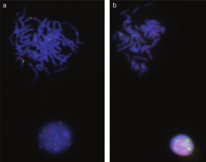
FISH analysis with TMA         545

Figure 1 A metaphase spread (top) for testing a bacterial artificial
chromosome (BAC) probe. (a) Two signals (green) with the corre-
sponding centromere probe (red signals) are seen in the same
chromosome. (b) Red and green signals are seen in different chro-
mosomes, although they were supposed to be in the same chromo-
some according to the information in the database. Interphase cells
exhibit two (pairs of) signals each (bottom).

                                                                       Figure 2 Tissue microarray gauges, prefabricated recipient blocks
                                                                       with holes, commercially available, and embedded blocks from top to
                                                                       bottom. The core diameters are 3 mm, 2 mm, and 1 mm in diameter
                                                                       (left to right).

Figure 3 Amplification of kinase loci
detected in FFPE tissues from the undif-
ferentiated carcinoma of the stomach.
Symbol genes are FMS related tyrosine
kinase 3 (FLT3) (a), Activated p21CDC42
kinase(ACK1) (b), V-SRC avian sarcoma
(Schimidt-Ruppin A-2) viral oncogene
(SRC) (c), and Cyclin dependent kinase 8
(CDK8) (d). The probes were labeled with
Spectrum Orange (Abbott, Abbott Park,
IL, USA), and the nuclei were stained with
4, 6-diamino-2-phenyl indole dihydrochlo-
ride (DAPI, Abbot). The method is
described in detail in the previous
literature.

© 2010 The Authors
Journal compilation © 2010 Japanese Society of Pathology and Blackwell Publishing Asia Pty Ltd
546 H. Sugimura et al.
that must be overcome. Many investigators have constructed specific and stage-specific gains or losses of particular
tissue microarrays for efficient use of probes they had labo- regions of chromosomes.46–49 Much of the information gen-
riously prepared, especially in retrospective studies. The idea erated by aCGH itself is used as a diagnostic or prognostic
of embedding many pieces in a single block existed in the tool in pathology laboratories.50–52 Information on genome-
early days of anatomical pathology, but several embedding wide genetic changes in cancer DNA are now viewed as
instruments for this purpose recently became popular, and academic knowledge that is only useful to the graduate stu-
technical refinements are under way. One well-circulating dents and researchers, but sooner or later it will be an essen-
brand of microarray instruments is Beecher Instruments tial tool of the diagnostic pathologist facing daily challenges
(Beecher Instruments, Inc. Sun Prairie, WI, USA). Their in diagnosis and management. There are many issues in
models have 0.6 mm, 1 mm, 2 mm cylinders, and the conventional pathology research and practice to which
Azumaya model KIN-1 and model FIN-3 (Azumaya Coopera- human genome data can be applied.53 Sano et al. conducted
tion, Tokyo) have wider cores that are 2 mm, 3 mm, 5 mm, a chromosome-wide survey to the archives of adenomatous
and 7 mm in diameter. There are pros and cons in regard to hyperplasia of the lung38 and proposed ‘adenocarcinoma in
using the smaller cores, and several problems encountered adenomatous hyperplasia’ as an early stage of carcinogen-
in using the instruments with various sized-cores are esis of lung adenocarcinoma. Although the tools were
addressed in the instructions included with each of the instru- genetic, the story they told was morphological. Very recently,
ments. A validation study in regard to possible sampling error a more powerful system, an SNP array platform containing
when small core specimens are collected was performed and more than 500 000 SNP sites has come into widespread use,
the results were published.40 Very recently, donor blocks con- and copy number estimation by several algorithms has facili-
taining multiple slots and an apparatus for making them have tated identification of copy number changes, such as loss of
become commercially available (Fig. 2) (Patent Application heterozygosity, uniparental disomy, and amplification, in
2009-028167), and many other variations will become avail- many clinical tumors. Midorikawa et al.54 integrated the data
able commercially. In addition to genomic and immunohis- based on pathological examination of ‘nodule in nodule’ in
tochemical studies, a proteomics approach by imaging mass resected liver tissue with the results of a comprehensive copy
spectrometry on a TMA platform is also feasible.41 number survey with the Affymetrix SNP array that were con-
firmed by FISH, and succeeded in clarifying genetic process
in human hepatocarcinogenesis in detail.
APPLICATION OF TMAS TO BIO-BANKS Research on structural changes and balanced transloca-
AND ETHICS CONSIDERATION tion of chromosomes in solid epithelial tumors is also a
cutting edge area of research today.7,51,55–57 The numbers of
Preparations of TMAs and requests to prepare them will candidate probes that should be investigated for clinical sig-
become more frequent in both investigative and diagnostic nificance seem huge. Several points need to be addressed
pathology laboratories, and as members of institutional when interpreting the results generated when an aCGH
review boards (IRBs) pathologists are sometimes respon- array and SNP array are used to analyze a human tumor
sible for appropriate control of these TMA bioresources. The genome. The first point is that many platforms are available
categories of pathology specimens are described in several to analyze copy number alterations, and a few papers on the
documents and on several websites,42–44 and IRBs are characteristics of each platform have been published.58–60
required to facilitate research proposals of making or using Furthermore, since many algorithms are available to enu-
TMAs to implement the research smoothly and ethically. merate copy numbers on the same platform, the character-
TMAs are a major component of tissue banks,45 which are istics of the platforms themselves and the benchmarks of the
tissue resources for future personalized medicine and algorithms need to be known. Most algorithms for estimating
national and international bio-bank systems are now being the copy number of loci set the reference dosage of tumor
established (websites: http://www.stn.org.sg, http:// autosomes as 2 (diplotype), but this reference number is not
www.ukbiobank.ac.uk, http://www.bbmri.eu, and http:// valid for most common epithelial malignant tumors. Ng
www.src.riken.go.jp/english/project/person/index.html). et al.61 recently refined the protocol for ploidy-specific copy
number estimation, and obtained a better threshold for
detecting CNA in cell lines, and Suzuki et al. performed a
COPY NUMBER ALTERATIONS DETECTED BY aCGH benchmark test of two widely used algorithms and exten-
AND SNP ARRAY: USEFUL DATA FOR FISH ANALYSIS sively characterized the features of the algorithms in terms of
different formulas for setting the gain or loss thresholds of
Data on copy number alterations in solid tumors deposited in genetic loci.62 Because of the intrinsic limitations of each
databases and publications have rapidly accumulated since method, two or three methods need to be used simulta-
the introduction of aCGH led to the discovery of many tumor- neously for the same tumor.
© 2010 The Authors
Journal compilation © 2010 Japanese Society of Pathology and Blackwell Publishing Asia Pty LtdFISH analysis with TMA 547
Figure 4 Distribution of the numbers of
the loci amplified in any of the 60 cases
(20 gastric cancer cases, 20 lung cancer
cases, and 20 colon cancer cases) in a
discovery set. More than half (51) of the
100 loci tested were not amplified in any of
the 60 cases tested. Five or more loci
were amplified in 5 (8%) of the 60 cases
tested.
Figure 5 Distribution of cases according
to numbers of loci amplified (vertical axis).
From 0 to 11 of the 70 or more (as many
as 100) loci successfully tested were
amplified. None of the 100 loci were ampli-
fied in 29 of the cases. Seven or more loci
were amplified in 3 cases.
WILL FISH BECOME A POPULAR AND ACCEPTED survival after surgical resection of non-small cell lung cancer.68
DIAGNOSTIC TOOL IN PATHOLOGY PRACTICE, Amplifications of PIK3CA is found in a considerable percent-
ESPECIALLY IN GUIDING INDIVIDUAL age of non-small cell lung cancers, and it and PIK3CA muta-
CANCER THERAPY? tion are mutually exclusive.69 The list of the amplified
segments continues to increase, although validation of their
Only a few FISH kits have been authorized for clinical use, but clinical significance awaits further study. The list of tumors in
many are available for use in research. Translocation detec- which amplification of certain gene product(s)can be indenti-
tion kits are often used to confirm diagnoses.63,64 Mori et al. fied has been growing, meaning that the list of the promising
recently used tens of BAC probes to make the differential targets of therapy is also growing. Comprehensive copy
diagnosis between adrenal tumors.65 However, the clinical number analysis by large-scale sequence technology has
significance of copy number alterations warrants further accu- revealed that a copy number gain of an unexpectedly high
mulations of retrospective and prospective data. The rationale proportion of genes that encode kinases in cancers.11 We
for the efficacy of molecularly targeted drugs varies with the tested 100 BAC probes containing different kinase loci in a
mutation, overexpression, and genomic amplification of the gastric, colorectal, and lung cancer detection sets (20 cases
target molecules, such as HER2 and EGFR. Fu et al. investi- for each organ) by TMA-FISH technology, and found amplifi-
gated copy number changes and expression of GATA-6 in cation of at least one kinase gene in a considerable number of
pancreatic cancer and reported finding consistency between cases, or, expressed another way, found that unexpected
the results for overexpression and amplification of the kinase loci were amplified in a significant proportion of human
genomic area of the GATA-6 locus,66 and they also validated common solid tumors (Figs 3–5). The discovery blocks we
their findings observation by FISH. Amplification itself, used consisted of tumor tissues in both early and advanced
however, does not always imply activation of the molecules or stages, and various histological types. The observation above
pathways of the genes on that genomic locus. Actually the has also provided us with the following perspectives. Combi-
EGFR immunohistological findings in lung cancer cells are not natory chemistry has already generated many drugs targeted
always consistent with the FISH data,67 and borderline grades to kinase genes or their products, thus amplifications of spe-
of immunostaining of HER2(2+) require FISH analysis to cific sites on certain kinase genes are amenable to pharma-
determine whether the HER2 gene has been amplified. cological intervention which that will lead to the establishment
Another receptor kinase gene, MET, has been evaluated as a of the target specific therapy. When observations like ours are
potential target of tailor made therapy in the same manner as validated and refined for clinical evaluation, the FISH diagnos-
the EGFR gene and HER2 gene have, and in some studies tic system with particular kinase probes may serve as another
MET amplification has been found to predict shorter patient basis of tailor-made cancer therapy.
© 2010 The Authors
Journal compilation © 2010 Japanese Society of Pathology and Blackwell Publishing Asia Pty Ltd548 H. Sugimura et al.
Major issues, however, remain to be resolved for before 2 Bialy H. Oncogenes, Aneuploidy, and AIDS: A Scientific Life and
authorization of FISH-based diagnostic tools even if scientifi- Times of Peter H. Duesberg. Barkeley, CA: North Atlantic
Books, 2004.
cally validated. Cost-benefit analysis of so-called targeted 3 Beroukhim R, Merml CH, Porter D et al. The landscape of
therapy is just starting in the tight-fisted health insurance somatic copy-number alteration across human cancers. Nature
environment, and there are gloom and doom forecasts that a 2010; 463: 899–905.
bonanza of new authorized diagnostic kits is unlikely to arrive 4 de Ravel TJ, Devriendt K, Fryns JP, Vermeesch JR. What’s new
in karyotyping? The move towards array comparative genomic
anytime soon. The time-line of the last few decades, hybridisation (CGH). Eur J Pediatr 2007; 166: 637–43.
however, in which many antibodies eventually became 5 Yoshimoto M, Cutz JC, Nuin PA et al. Interphase FISH analysis
essential in pathology labs, evokes us a very different picture. of PTEN in histologic sections shows genomic deletions in 68%
of primary prostate cancer and 23% of high-grade prostatic
intra-epithelial neoplasias. Cancer Genet Cytogenet 2006; 169:
CONCLUSIONS 128–37.
6 Frohling S, Dohner H. Chromosomal abnormalities in cancer. N
Engl J Med 2008; 359: 722–34.
The basic knowledge required to perform the combination of
7 Soda M, Choi YL, Enomoto M et al. Identification of the trans-
TMA and FISH with many BAC probes is familiar to diagnos- forming EML4-ALK fusion gene in non-small-cell lung cancer.
tic pathologists, but that is different from actually running it Nature 2007; 448: 561–6.
(TMA-FISH with BACs) in real pathology practice. Obtaining 8 Velculescu VE. Defining the blueprint of the cancer genome.
Carcinogenesis 2008; 29: 1087–91.
an ample numbers of BAC probes, labeling, and expensive
9 Wood LD, Parsons DW, Jones S et al. The genomic landscapes
fluorescence microscopes may be hurdles for modestly of human breast and colorectal cancers. Science 2007; 318:
equipped community hospitals. In the previous issue of 1108–13.
Pathology International, Kato et al.70 have reported their 10 Weir BA, Woo MS, Getz G et al. Characterizing the cancer
genome in lung adenocarcinoma. Nature 2007; 450: 893–
experience with using of a commercialized product that
898.
applies chromogenic in situ hybridization, a friendlier method 11 Kubo T, Kuroda Y, Shimizu H et al. Resequencing and Copy
that allows the use of ordinary microscope. Number Analysis of the Human Tyrosine Kinase Gene Family in
Many DNA probes labeled ‘research use only’ are actually Poorly Differentiated Gastric Cancer. Carcinogenesis 2009; 30:
1857–64.
used in sarcoma diagnosis,20 and standardization and quality
12 Campbell PJ, Stephens PJ, Pleasance ED et al. Identification of
control of only a few FISH diagnostic systems have been somatically acquired rearrangements in cancer using genome-
achieved. Most of DNA probes are expensive, and there are wide massively parallel paired-end sequencing. Nat Genet
few ‘generic’ diagnostic kits. 2008; 40: 722–9.
13 Watters AD, Ballantyne SA, Going JJ, Grigor KM, Bartlett JM.
Over the coming decades, DNA probes will become a
Aneusomy of chromosomes 7 and 17 predicts the recurrence of
familiar diagnostic tool to the pathologists in community hos- transitional cell carcinoma of the urinary bladder. BJU Int 2000;
pitals, and the information obtained by using them will 85: 42–7.
suggest therapeutic guidance as well as the diagnosis. At the 14 Skacel M, Ormsby AH, Pettay JD et al. Aneusomy of chromo-
somes 7, 8, and 17 and amplification of HER-2/neu and epider-
same time, the accumulation of the data generated by TMA-
mal growth factor receptor in Gleason score 7 prostate
FISH approach will complement numerous OMICS data that carcinoma: A differential fluorescent in situ hybridization study of
have been accumulating in other disciplines of medicine. In Gleason pattern 3 and 4 using tissue microarray. Hum Pathol
other words, the TMA-FISH approach may be one of the 2001; 32: 1392–7.
15 Bayani J, Squire JA. Application and interpretation of FISH in
smartest harvest (exit) strategies among OMICS projects
biomarker studies. Cancer Lett 2007; 249: 97–109.
related to human cancer, and many investments have been 16 Tibiletti MG, Bernasconi B, Dionigi A, Riva C. The applications
made in it over the last two decades. of FISH in tumor pathology. Adv Clin Path 1999; 3: 111–8.
17 Giltnane JM, Murren JR, Rimm DL, King BL. AQUA and FISH
analysis of HER-2/neu expression and amplification in a small
ACKNOWLEDGMENTS cell lung carcinoma tissue microarray. Histopathology 2006; 49:
161–9.
18 Lee CH, Huntsman DG, Cheang MC et al. Assessment of
This work was supported by a Grant-in-Aid for priority
Her-1, Her-2, And Her-3 expression and Her-2 amplification in
areas from the Japanese Ministry of Education, Culture, advanced stage ovarian carcinoma. Int J Gynecol Pathol 2005;
Sports, Science and Technology (20014007), from the Japa- 24: 147–52.
nese Ministry of Health, and from the Smoking Research 19 Hicks DG, Tubbs RR. Assessment of the HER2 status in breast
cancer by fluorescence in situ hybridization: A technical review
Foundation.
with interpretive guidelines. Hum Pathol 2005; 36: 250–61.
20 Iwasaki H, Nabeshima K, Nishio J et al. Pathology of soft-tissue
tumors: Daily diagnosis, molecular cytogenetics and experimen-
REFERENCES
tal approach. Pathol Int 2009; 59: 501–21.
21 Schraml P, Kononen J, Bubendorf L et al. Tissue microarrays
1 Weinberg RA. Biology of Cancer. New York, NY: Garland Pub, for gene amplification surveys in many different tumor types.
2006. Clin Cancer Res 1999; 5: 1966–75.
© 2010 The Authors
Journal compilation © 2010 Japanese Society of Pathology and Blackwell Publishing Asia Pty LtdFISH analysis with TMA 549 22 Al Kuraya K, Simon R, Sauter G. Tissue microarrays for high- 42 Shapiro HT. Research involving human biologicalmaterials: throughput molecular pathology. Ann Saudi Med 2004; 24: 169– Ethical issues and policy guidance. Commission NBA ed. Vol. 1. 74. Rockville 1999; 1–138. 23 Drev P, Grazio SF, Bracko M. Tissue microarrays for routine 43 WHO. World Health Organization, Proposed International diagnostic assessment of HER2 status in breast carcinoma. Guidelines on Ethical Issues in Medical Genetics and Genetic Appl Immunohistochem Mol Morphol 2008; 16: 179–84. Services. WHO/HGN/GL/ETH/98.1. ed, 1998. 24 Dhir R. Tissue microarrays: An overview. Methods Mol Biol 44 Wolf S. The American point of view regarding personal informa- 2008; 441: 91–103. tion protection [homepage on the Internet]: Japanese Ministry of 25 Toncheva D, Petrova D, Tzenova V et al. Tissue microarray Health, Welfare, and Labor I, 47–51. 2000. (cited 7 June 2010) analysis of cyclin D1 gene amplification and gain in colorectal Series Title. Available from: http://www.mc.pref.osaka.jp/ocr/ carcinomas. Tumour Biol 2004; 25: 157–60. ocr_hcr/registry/security/report/wolff.pdf. 26 Koynova DK, Tsenova VS, Jankova RS, Gurov PB, Toncheva 45 Ornes S. What happens to a donated tumor? CR 2009; 36– DI. Tissue microarray analysis of EGFR and HER2 oncogene 45. copy number alterations in squamous cell carcinoma of the 46 Zhou X, Rao NP, Cole SW, Mok SC, Chen Z, Wong DT. larynx. J Cancer Res Clin Oncol 2005; 131: 199–203. Progress in concurrent analysis of loss of heterozygosity and 27 Okon K, Sinczak-Kuta A, Klimkowska A et al. Tissue microarray comparative genomic hybridization utilizing high density single FISH applied to colorectal carcinomas with various microsatel- nucleotide polymorphism arrays. Cancer Genet Cytogenet lite status. Pol J Pathol 2006; 57: 99–103. 2005; 159: 53–7. 28 Brown LA, Huntsman D. Fluorescent in situ hybridization on 47 Kallioniemi A. CGH microarrays and cancer. Curr Opin Biotech- tissue microarrays: Challenges and solutions. J Mol Histol 2007; nol 2008; 19: 36–40. 38: 151–7. 48 Bejjani BA, Shaffer LG. Clinical utility of contemporary molecu- 29 von Schalburg KR, Rise ML, Cooper GA et al. Fish and chips: lar cytogenetics. Annu Rev Genomics Hum Genet 2008; 9: Various methodologies demonstrate utility of a 16,006-gene 71–86. salmonid microarray. BMC Genomics 2005; 6: 126. 49 Inazawa J, Inoue J, Imoto I. Comparative genomic hybridization 30 Sugimura H. Detection of chromosome changes in pathology (CGH)-arrays pave the way for identification of novel cancer- archives: An application of microwave-assisted fluorescence in related genes. Cancer Sci 2004; 95: 559–63. situ hybridization to human carcinogenesis studies. Carcinogen- 50 Gouas L, Goumy C, Veronese L, Tchirkov A, Vago P. Gene esis 2008; 29: 681–7. dosage methods as diagnostic tools for the identification of 31 Meaburn KJ, Gudla PR, Khan S, Lockett SJ, Misteli T. Disease- chromosome abnormalities. Pathol Biol (Paris) 2008; 56: 345– specific gene repositioning in breast cancer. J Cell Biol 2009; 353. 187: 801–12. 51 Gunn SR, Robetorye RS, Mohammed MS. Comparative 32 Gunter C. Reliable repositioning in cancer. Nat Rev Cancer genomic hybridization arrays in clinical pathology: Progress and 2010; 10: 84–85. challenges. Mol Diagn Ther 2007; 11: 73–7. 33 Holst F, Stahl PR, Ruiz C et al. Estrogen receptor alpha (ESR1) 52 Callagy G, Jackson L, Caldas C. Comparative genomic hybrid- gene amplification is frequent in breast cancer. Nat Genet 2007; ization using DNA from laser capture microdissected tissue. 39: 655–60. Methods Mol Biol 2005; 293: 39–55. 34 Brown LA, Hoog J, Chin SF et al. ESR1 gene amplification in 53 Ooi A, Zen Y, Ninomiya I et al. Gene Amplification of ERBB2 breast cancer: A common phenomenon? Nat Genet 2008; 40: and EGFR in Adenocarcinoma in situ and Intramucosal Adeno- 806–7, author reply 10–2. carcinoma of Barrett’s Esophagus. Pathol Int 2010; 60: 466– 35 Horlings HM, Bergamaschi A, Nordgard SH et al. ESR1 gene 471. amplification in breast cancer: A common phenomenon? Nat 54 Midorikawa Y, Yamamoto S, Ishikawa S et al. Molecular karyo- Genet 2008; 40: 807–8, reply 10–2. typing of human hepatocellular carcinoma using single- 36 Wolff AC, Hammond ME, Schwartz JN et al. American Society nucleotide polymorphism arrays. Oncogene 2006; 25: 5581–90. of Clinical Oncology/College of American Pathologists guideline 55 Shinmura K, Kageyama S, Tao H et al. EML4-ALK fusion tran- recommendations for human epidermal growth factor receptor 2 scripts, but no NPM-, TPM3-, CLTC-, ATIC-, or TFG-ALK fusion testing in breast cancer. J Clin Oncol 2007; 25: 118–45. transcripts, in non-small cell lung carcinomas. Lung Cancer 37 Kasami M, Uematsu T, Honda M et al. Comparison of estrogen 2008; 61: 163–9. receptor, progesterone receptor and Her-2 status in breast 56 Pleasance ED, Stephens PJ, O’Meara S et al. A small-cell lung cancer pre- and post-neoadjuvant chemotherapy. Breast 2008; cancer genome with complex signatures of tobacco exposure. 17: 523–7. Nature 463: 184–90. 38 Sano T, Kitayama Y, Igarashi H et al. Chromosomal numerical 57 Shinmura K, Kageyama S, Igarashi H et al. EML4-ALK fusion abnormalities in early stage lung adenocarcinoma. Pathol Int transcripts in immunohistochemically ALK-positive non-small 2006; 56: 117–25. cell lung canrcinomas. Exp Ther Med 2010; 1: 271–75. 39 Kitayama Y, Igarashi H, Watanabe F, Maruyama Y, Kanamori 58 Lo KC, Bailey D, Burkhardt T, Gardina P, Turpaz Y, Cowell JK. M, Sugimura H. Nonrandom chromosomal numerical abnormal- Comprehensive analysis of loss of heterozygosity events in ity predicting prognosis of gastric cancer: A retrospective study glioblastoma using the 100K SNP mapping arrays and compari- of 51 cases using pathology archives. Lab Invest 2003; 83: son with copy number abnormalities defined by BAC array com- 1311–20. parative genomic hybridization. Genes Chromosomes Cancer 40 O’Grady A, Flahavan CM, Kay EW, Barrett HL, Leader MB. 2008; 47: 221–37. HER-2 analysis in tissue microarrays of archival human breast 59 Coe BP, Macaulay C, Lam WL, Ylstra B, Carvalho B, Meijer GA. cancer: Comparison of immunohistochemistry and fluorescence Comment re: A comparison of DNA copy number profiling plat- in situ hybridization. Appl Immunohistochem Mol Morphol 2003; forms. Cancer Res 2008; 68: 4010, author reply 10. 11: 177–82. 60 Greshock J, Feng B, Nogueira C et al. A comparison of DNA copy 41 Morita Y, Ikegami K, Goto-Inoue N et al. Imaging mass spectrom- number profiling platforms. Cancer Res 2007; 67: 10173–80. etry of gastric carcinoma in formalin-fixed paraffin-embedded 61 Ng G, Huang J, Roberts I, Coleman N. Defining ploidy-specific tissue microarray. Cancer Science 2010; 101: 267–273. thresholds in array comparative genomic hybridization to © 2010 The Authors Journal compilation © 2010 Japanese Society of Pathology and Blackwell Publishing Asia Pty Ltd
550      H. Sugimura et al.

      improve the sensitivity of detection of single copy alterations in   66 Fu B, Luo M, Lakkur S, Lucito R, Iacobuzio-Donahue CA. Fre-
      cell lines. J Mol Diagn 2006; 8: 449–58.                                quent genomic copy number gain and overexpression of
62    Suzuki M, Nagura K, Igarashi H et al. Copy number estimation            GATA-6 in pancreatic carcinoma. Cancer Biol Ther 2008; 7:
      algorithms and fluorescence in situ hybridization to describe           1593–601.
      copy number alterations in human tumors. Pathol Int 2009; 59:        67 Suzuki M, Kageyama S, Shinmura K et al. Inverse relationship
      218–228.                                                                between the length of the EGFR CA repeat polymorphism in
63    Yoshida H, Nagao K, Ito H, Yamamoto K, Ushigome S. Chro-                lung carcinoma and protein expression of EGFR in the carci-
      mosomal translocations in human soft tissue sarcomas by inter-          noma. J Surg Oncol 2008; 98: 457–61.
      phase fluorescence in situ hybridization. Pathol Int 1997; 47:       68 Cappuzzo F, Marchetti A, Skokan M et al. Increased MET gene
      222–9.                                                                  copy number negatively affects survival of surgically resected
64    Ten Heuvel SE, Hoekstra HJ, Suurmeijer AJ. Diagnostic accu-             non-small-cell lung cancer patients. J Clin Oncol 2009; 27:
      racy of FISH and RT-PCR in 50 routinely processed synovial              1667–74.
      sarcomas. Appl Immunohistochem Mol Morphol 2008; 16: 246–            69 Okudela K, Suzuki M, Kageyama S et al. PIK3CA mutation and
      50.                                                                     amplification in human lung cancer. Pathol Int 2007; 57: 664–
65    Mori H, Nagata M, Nishijima N et al. Malignant pheochromocy-            71.
      toma in a young adult forming the structure simulating Homer         70 Kato N, Itoh H, Serizawa A, Hatanaka Y, Umemura S, Osamura
      Wright rosette: Differentiation from neuroblastoma on repeating         Y. Evaluation of HER2 Gene Amplification in Invasive Breast
      fluorescence in situ hybridization. Pathol Int 2008; 58: 518–           Cancer Using a Dual-color Chromogenic In Situ Hybridization
      23.                                                                     (Dual CISH). Pathol Int 2010; 60: 510–5.

                                                                                                                      © 2010 The Authors
                                             Journal compilation © 2010 Japanese Society of Pathology and Blackwell Publishing Asia Pty Ltd
You can also read