Review Article The Domestic Cat as a Large Animal Model for Characterization of Disease and Therapeutic Intervention in Hereditary Retinal Blindness
←
→
Page content transcription
If your browser does not render page correctly, please read the page content below
Hindawi Publishing Corporation
Journal of Ophthalmology
Volume 2011, Article ID 906943, 8 pages
doi:10.1155/2011/906943

Review Article
The Domestic Cat as a Large Animal Model for
Characterization of Disease and Therapeutic Intervention
in Hereditary Retinal Blindness

          Kristina Narfström,1, 2 Koren Holland Deckman,3 and Marilyn Menotti-Raymond4
          1 Department   of Veterinary Medicine and Surgery, College of Veterinary Medicine, Mason Eye Institute,
            University of Missouri-Columbia, MO 65211, USA
          2 Department of Ophthalmology, Mason Eye Institute, University of Missouri-Columbia, MO 65212-0001, USA
          3 Department of Chemistry, Gettysburg College, Gettysburg, PA 17325, USA
          4 Laboratory of Genomic Diversity, National Cancer Institute-Frederick, Frederick, MD 21702-1201, USA

          Correspondence should be addressed to Kristina Narfström, narfstromk@missouri.edu

          Received 19 July 2010; Revised 4 October 2010; Accepted 24 January 2011

          Academic Editor: Radha Ayyagari

          Copyright © 2011 Kristina Narfström et al. This is an open access article distributed under the Creative Commons Attribution
          License, which permits unrestricted use, distribution, and reproduction in any medium, provided the original work is properly
          cited.

          Large mammals, including canids and felids, are affected by spontaneously occurring hereditary retinal diseases with similarities to
          those of humans. The large mammal models may be used for thorough clinical characterization of disease processes, understanding
          the effects of specific mutations, elucidation of disease mechanisms, and for development of therapeutic intervention. Two well-
          characterized feline models are addressed in this paper. The first model is the autosomal recessive, slowly progressive, late-onset,
          rod-cone degenerative disease caused by a mutation in the CEP290 gene. The second model addressed in this paper is the autosomal
          dominant early onset rod cone dysplasia, putatively caused by the mutation found in the CRX gene. Therapeutic trials have been
          performed mainly in the former type including stem cell therapy, retinal transplantation, and development of ocular prosthetics.
          Domestic cats, having large human-like eyes with comparable spontaneous retinal diseases, are also considered useful for gene
          replacement therapy, thus functioning as effective model systems for further research.

1. Introduction                                                          retinal pathology. Though 157 genes have been identified as
                                                                         causative of nonsyndromic human retinitis pigmentosa (RP;
The value of appropriate animal models to advance our                    http://www.sph.uth.tmc.edu/retnet/home.htm), over 50% of
understanding and treatment of human retinal disease pro-                the genetic causality of RP still remains uncharacterized [6].
cesses that cause severe visual impairment or blindness                      Though the mouse has been the classic animal model of
cannot be overemphasized. Animal models have led to the                  retinal disease, the advent of comprehensive genetic maps of
identification of disease genes, and elucidation of the molec-           many mammals has led to the identification of a number
ular genetic and cellular mechanisms underlying retinal                  of non-rodent animal models of human hereditary retinal
pathology. Moreover, they provide the basis for testing the              disease. Many large animal models offer a complement
efficacy of therapeutic approaches, including the use of                   to existing rodent models. The size of the rodent eye is
drugs and gene replacement [1], novel genetic approaches                 restrictive for visualization using regular clinical ophthalmic
(siRNA) [2], stem cell therapy [3], surgical intervention,               instrumentation and also in conjunction with therapeutic
such as retinal transplantation [4], and the use of ocular               intervention. Even for detailed morphological studies the
or retinal prosthetics [5]. Additionally, animal models can              small size of the mouse eye may be a problem. As a case in
lead to the identification of novel genes underlying human               point Pazour et al. previously reported that in their research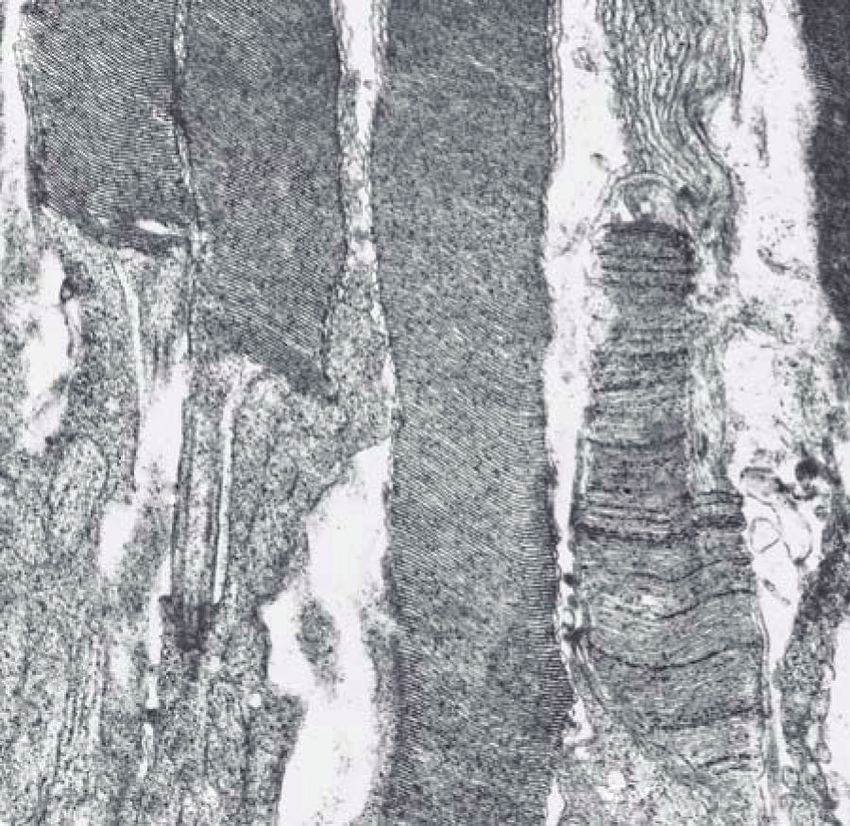
2                                                                                                    Journal of Ophthalmology

examining the trafficking of ciliary protein in photoreceptor        (SOD1) in the Pembroke Welsh corgi with 38 affected and 17
cells, the physical limitations of the mouse retina led them to    control individuals. Extended linkage disequilibrium (LD) in
resort to the use of a bovid eye [7].                              dog breeds [19, 34, 35] contributes to the success of GWA
                                                                   mapping in dogs and is an important factor underlying
                                                                   successful mapping with small sample sizes in the dog.
2. Dogs and Cats as Large Animal Models of                         Preliminary studies suggest that extended blocks of LD are
   Spontaneous Retinal Disease                                     also observed in cat breeds, though the length of LD appears
                                                                   to be abbreviated to that which is observed in dog breeds
Dog and cat populations offer a wealth of potential as large        [29].
animal models of human retinal disease. Small effective
population sizes, the use of popular sires and inbreeding have
contributed to the “load” of inherited diseases, especially
                                                                   4. The Abyssinian Retinal Degeneration
in dog breeds [8]. Hereditary and primary photoreceptor               Cat Model (rdAc )
diseases, or progressive retinal atrophies (PRA) have been         The female Abyssinian cat (Cinnamon), subject of the feline
described in more than 100 dog breeds [9], many of which           whole genome sequencing efforts, is a member of a pedigree
are likely to be caused by the same mutation which is              developed for genetic mapping of the gene defect for the
observed across related breeds. This phenomenon has been           rdAc (retinal degeneration in Abyssinian cats) model, first
observed in a number of gene-defining phenotypes in the            described in 1982 [36]. The autosomal recessive (AR) trait,
dog [10–12]. Thirteen genes have been mapped and charac-           rdAc, has become an important model of human RP [37, 38].
terized as causative of canine PRA, including ADAM9 [13],          At birth, affected cats have normal vision, but, by 1.5–2 years
CCDC66 [14], CNGB3 [15], PDE6α [16], PDE6β [17], PRCD              of age they develop early changes that can be observed by
[18], RD3 [19], RHO [20], RPE65 [21], RPHP4 [22], RPGR             ophthalmoscopy [39] (Figures 1(a)–1(c)). By 7 months of
[23], RPGRIP1 [24], and VMD2 [25].                                 age, affected cats demonstrate significantly reduced retinal
    Cats have been considered to be affected less frequently        function by electroretinography (ERG; Figure 1(d)). ERG
by hereditary disease. However, the informative website            a-wave amplitudes are then reduced more than 50% as
Online Mendelian Inheritance in Animals (http://omia.              compared to normal individuals, with a parallel reduction
angis.org.au/) catalogues 288 distinctive pathologies with an      in retinal oxygen tension [40]. Rod photoreceptor outer
inherited component in the cat, with cited references. Only        segments exhibit the first morphological changes in indi-
in recent years have specific mutations been elucidated for        viduals 5–8 months of age, observed as a disorganization
hereditary retinal diseases in cats [26, 27], clinically similar   and disruption of rod outer segment lamellar discs and the
to the PRA complex in dogs [28]. Domestic dogs and cats            appearance of vacuoles near the connecting cilium [41].
of today experience a level of medical surveillance second         Progression of the disease results in further degeneration
only to human kind, thus increasing the likelihood, that           of the rods (Figures 2(a) and 2(b)), followed also by
individuals with rare or unique mutations are identified.          disruption of the cone photoreceptors. By 3–5 years of age,
                                                                   the clinical end stage of the disease has been reached with
3. Sequencing of the Cat Genome                                    generalized photoreceptor degeneration, and subsequently
                                                                   retinal atrophy leads to blindness [42].
Report of two partial sequences (1.9X, 3X) of the cat genome           The molecular genetic basis of rdAc was recently estab-
[29, 30] has been invaluable in the initial mapping and            lished in the CEP290 gene. A single-nucleotide polymor-
characterization of feline hereditary diseases [26, 27, 31]. A     phism in an intron of the felid CEP290 gene generates
full genome sequence (14X) of the cat has currently been           a novel strong canonical splice-donor site resulting in a
completed (Wes Warren, Washington University, personal             4-bp insertion, a frame shift, and the introduction of a
communication). The identification of single-nucleotide            premature stop codon (Figures 3(a) and 3(b)). The putative
polymorphisms (SNP) in cat breeds, an integral part of the         truncated CEP290 peptide would lack the more 3 KIDV
14X full genome sequence project and the 3X sequencing of          and VI domains. The protein is an important component of
the cat genome [30] is currently being utilized in develop-        the intraflagellar transport (IFT) system whereby specialized
ment of a domestic cat SNP chip. With the availability of          proteins critical for phototransduction are transferred from
these genomic resources, the mapping and characterization          their site of synthesis in the inner segment of photoreceptors
of feline monogenic disorders will largely be dependent on         through the connecting cilium to the outer segment [7].
obtaining an appropriate sample set. Genome-wide associ-           As the rod photoreceptor discs are in a constant state
ation (GWA) studies in dog breeds are proving extremely            of regeneration, a fully functional IFT system is critical
successful in identifying genes associated with breed-defining     for the maintenance of the photoreceptors [7]. In the
phenotypes and monogenic disorders [13, 32, 33]. Often             rd16 mouse model, the phototransduction proteins opsin
this is accomplished with surprisingly small sample sizes.         and rhodopsin are found concentrated in the inner segment,
The mapping of the canine cone-rod dystrophy 3 gene                which led Chang et al. [44] to propose a ciliary trafficking
(ADAMS9) in the Glen of Imaal Terrier breed was recently           role for the CEP290 protein. Mutations in CEP290 have been
accomplished with as few as 22 unaffected and 19 affected            reported in RP, Leber’s congenital amaurosis (LCA), as well
individuals [13] while Awano et al. reported identification        as the syndromic retinopathies, Joubert, Meckel-Gruber, and
of the gene causative of canine degenerative myelopathy            Bardet-Biedl [45–48].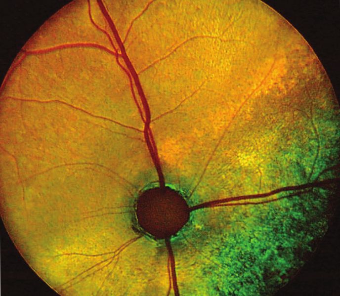
Journal of Ophthalmology 3
(a) (b)
250 μV
50 ms
WT
S2
S4
(c) (d)
Figure 1: Fundus appearance and electroretinograms (ERGs) of rdAc individuals with the CEP290 mutation. Fundus photographs demon-
strate (a) a 1-year-old unaffected Abyssinian cat (wildtype, WT), (b) a 2-year-old affected Abyssinian cat with an early disease stage (S2)
[39], and (c) a 6-year-old Abyssinian with an advanced disease stage (S4) [39]. Arrows in (b) and (c) indicate retinal vasculature that is
attenuated, more so in the advanced stage (c) than in early stage of disease (b). For the same three cats, the waveforms of the dark-adapted
full-field flash ERG recordings are shown, using 4 cd.s/m2 of white light stimulation for each of the recordings. Amplitude and implicit time
calibrations are indicated in the figure, vertically and horizontally, respectively. Reproduced with permission from [43].
am
ros ros
ros ros
cc
cos
cc
ris
ris
ris cis
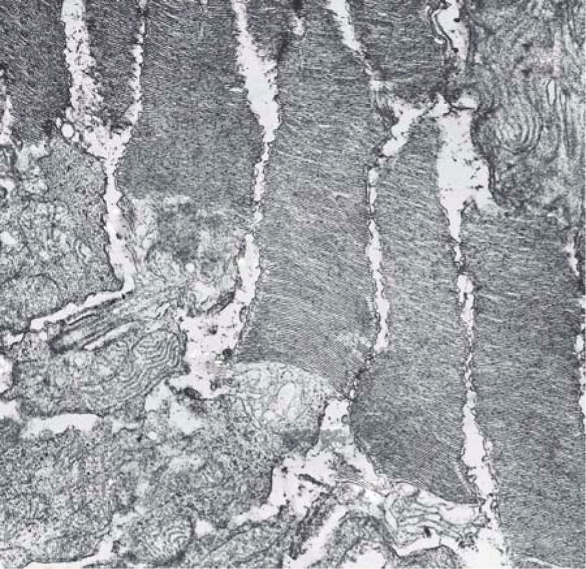
(a) (b)
Figure 2: Electron micrographs of outer retina showing photoreceptor outer and inner segments of normal (a) Abyssinian cat and young
affected (b) rdAc cat. Note abnormalities at the base of the rod outer segments near the connecting cilium in (b); membranes are not formed
as in the normal (a), instead there is vacuolization and degeneration (arrows) of membranes in the affected retina. Am: apical microvilli
of the retinal pigment epithelium, ros: rod outer segments, ris: rod inner segments, cos: cone outer segments, cis: cone inner segments, cc:
connecting cilium of the photoreceptor. Original magnification: ×19152. Reproduced with permission from [27].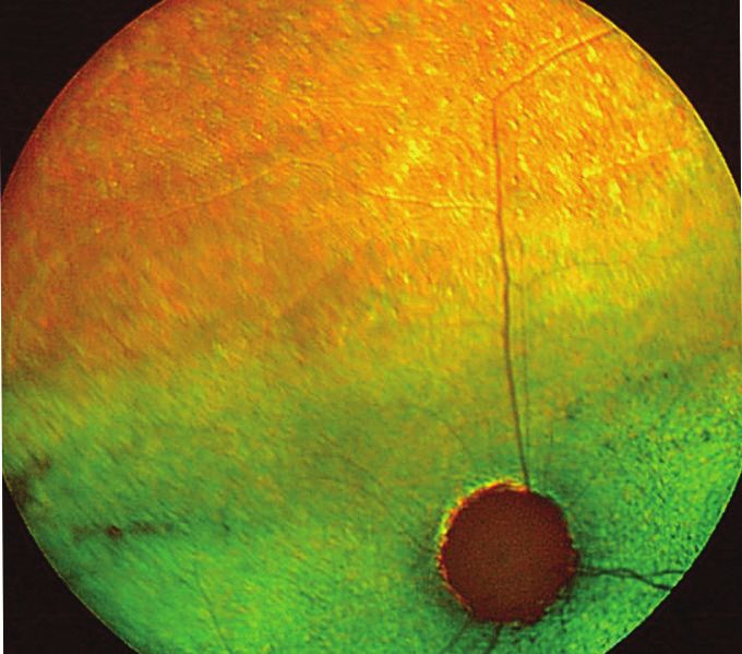
4                                                                                                       Journal of Ophthalmology

                                                                     abnormal photoreceptor development at 22 days of age. The
                                                                     disease leads rapidly to blindness usually within the first few
                                                                     months of life. Further characterization of the dystrophy
                                                   Unaffected        has demonstrated that the photoreceptors never develop
     C A A g c a a g t a a T t t t t             genomic DNA         normally, and the disease has therefore been designated as
     Exon 50               Intron 50                                 a rod-cone dysplasia with early onset degeneration of both
                                                                     cones and rods.
                                                                         The molecular genetic basis for Rdy was recently eluci-
                                                    Affected         dated [26]. A single-base deletion in the CRX gene intro-
     C A A G C A A g t a a G t t t t             genomic DNA         duces a frameshift and a stop codon immediately down-
         Exon 50               Intron 50                             stream, truncating a region previously demonstrated as
                                                                     critical for gene function [26, 54] (Figure 4). The CRX
                               (a)                                   gene product is critical in transcriptional activation of a
                                                                     number of genes involved in photoreceptor development
                                               rdAc unaffected
                                                neutral retinal      and maintenance [55, 56]. In humans, mutations in CRX
     A C A A       A T T G A G         A T T       cDNA              are associated with human AD cone-rod dystrophy (CoRD),
        GLN         ILE   GLU           ILE                          and both AD and AR Leber’s congenital amaurosis (LCA)
                                                                     [54, 57–59]. The Rdy cat is the first large animal model for
                                                rdAc affected        CRX-linked spontaneous retinal disease. A large screening
                                                neutral retinal
                                                                     of cat breeds has failed to detect any other domestic feline
     A C A A       G C A A A T         T G A       cDNA
                                                                     breeds with the disease allele [26].
        GLN         ALA   ASN           Stop
                                                                         The Rdy model provides one of the very few large animal
                               (b)                                   models of an autosomal dominant retinal disease. These
Figure 3: (a) Electropherograms of genomic DNA of CEP290             disorders are challenging from a therapeutic standpoint.
sequenced in unaffected and rdAc affected individuals of exon          Causality of the disease can arise from haploinsufficiency of
50/intron 50 junction. Arrow indicates position of SNP in intron     product, or in some circumstances from gain of function or
50, which uncovers a canonical GT splice donor site, resulting in    competition from a truncated or aberrant protein product
alternative splicing in affected individuals. Exon 50 (red letters)   [60]. The presence of both mutant and wildtype RNA in Rdy
and intron 50 (blue letters) nucleotides were identified by cDNA     individuals, initially suggestive that a truncated CRX product
sequence analysis. GenBank Accession No. for feline CEP290:          might be generated [26] has been supported by recent find-
EF028068. (b) Electropherograms of cDNA for CEP290 3 region         ings (K. Holland Deckman, unpublished). The truncated
of exon 50 generated from neural retinal tissue in affected and       peptide would retain the CRX motifs involved in nuclear
unaffected individuals. Alternative splicing in affected individuals
                                                                     localization and DNA-binding, but lack the region critical
results in a frame shift and introduction of a premature STOP
codon. Reproduced with permission from [27].
                                                                     for transcriptional activation of photoreceptor specific genes
                                                                     [61]. This truncated product could thus compete with the
                                                                     wildtype CRX product and other transcription factors for
                                                                     promoter binding regions of target genes, which is currently
    Recently, it has been shown that rdAc cats exhibit some          under investigation.
degree of phenotypic variation, with end-stage blindness
reached in individuals anywhere from three to seven years
[49]. Interestingly, it appears that the slow progression of         6. Other Cat Models under Investigation
disease may be one of the factors leading to the cat’s excep-
                                                                     In the late 1960s, a new feline breed, the Bengal, which has
tional ability to adapt to decreasing retinal function [49].
                                                                     gained huge popularity, was developed through hybridiza-
The condition thus evaded detection by both owners and
                                                                     tion of domestic cats and the Asian leopard cat [62]. Recently,
veterinarians in a highly popular cat breed, the Siamese,
                                                                     a novel, early onset autosomal recessive disorder was
which demonstrates a high frequency (∼33%) for the
                                                                     described in this breed [63]. The disease is under investiga-
rdAc disease allele [49]. Breeding practices have caused the
                                                                     tion but appears to be an early onset primary photoreceptor
CEP290 mutation to spread to multiple cat breeds [49], and
                                                                     disorder, leading to blindness within the first year of age.
to exhibit a worldwide distribution [49].
                                                                     Genetic mapping and further characterization of the disorder
                                                                     are in progress. A second feline retinal disease model has
5. The Rod Cone Dysplasia Cat Model (Rdy )                           been described in the Persian cat breed [64]. The rod cone
                                                                     dysplasia demonstrates an autosomal recessive mode of
A second feline model of human retinal dystrophy, the Rdy            inheritance [64]. Affected individuals showed clinical signs
cat, was first described in a single Abyssinian cat, from which      of disease 2-3 weeks after birth and clinical blindness at
a pedigree was developed and extensively studied on a phe-           16 weeks of age. Photoreceptors in affected individuals
notypic level [50–53]. The disease is an early onset primary         never reach full maturity. The molecular genetic defect for
photoreceptor disorder with an autosomal dominant (AD)               both of these disorders has not as yet been elucidated. It
mode of inheritance in which affected individuals exhibit             appears that the Bengal cat retinal disease should become
Journal of Ophthalmology                                                                                                                5

              ∗   Exon:   2 3                  3 4                                                                       X

                           Homeobox                                WSP                        TTD1           TTD2    OTX
                                                                   (a)
                                   ∗                                                                 X
                                       Exon:    2 3            3 4

                                                 Homeobox                               WSP
                                                                   (b)

Figure 4: CRX protein structure in Felis catus. Wildtype feline CRX protein (a) compared to the putative truncated CRX protein (b). The
exon splice junctions are noted as “Y”. The start codon and stop codons are labeled as (∗ ) and (X), respectively. The protein domains are
highlighted as shaded boxes and defined as the homeobox, the WSP domain, the transcriptional transactivation domains 1 and 2 (TTD1
and TTD2), and the OTX tail. Domains are drawn to scale. Reproduced with permission from [26].

an important animal model for the research community in                      Novel therapeutic interventions have recently been devel-
regards to the study of various treatment modalities. The                oped to target aberrant RNA species that survive nonsense
disease starts out from a mainly normal appearing retina but             mediated decay. Short interfering RNA (shRNA), short
due to the fast progression of the disorder, retinal atrophy             double stranded RNA molecules, can be designed to degrade
ensues comparatively early thus functioning as an effective               specific target mRNAs [67], while ribozymes, which are small
model system for retinal research.                                       catalytic RNAs, are designed to cleave complementary RNA
                                                                         sequences [2]. RNA interference-mediated suppression and
                                                                         replacement aims to remove both wildtype and aberrant
7. Therapeutic Intervention                                              RNA copies of a targeted gene while replacing wildtype
The retinas of large animal models more closely approximate              expression with a copy of the gene.
that of humans, and are thus more easily amenable for                        Other methods of treatment include retinal transplan-
visualization and imaging [65] of the disease process, for sur-          tation of viable cells or tissue. Experimentation in this
gical intervention, and for clinical evaluation of therapeutic           regard includes the replacement of dying visual cells with
effects. Dogs and cats also offer the potential of long-term               healthy neuroblastic progenitor cells and retinal pigment
followup studies in conjunction with treatment trials.                   epithelial (RPE) cells as sheets of normal tissue [68]. It
    The rod and cone photoreceptors (the latter; short and               has been demonstrated that retinal transplants in rats can
middle wavelength sensitive cones) of both species are dis-              morphologically reconstruct a damaged retina and restore
tributed in the retina in a mosaic pattern comparable to                 retinal sensitivity [69]. Affected cats with the CEP290 defect
that of the human retina. Neither cats nor dogs have a                   (rdAc) have been used in trials with transplantation of sheets
macula. However, in cats, in the same region as the human                of allogeneic fetal retinal tissue [70]. Surgeries have been
macula, there sre a cone-rich region called the area centralis           successful as to graft survival in the retina, although cellular
where the concentration of cones in comparison to that                   connectivity has not been shown and ERG testing has not
of rods is higher than at any other location. Along with                 demonstrated improvement in retinal function. So far the cat
the holangiotic configuration of the retinal vasculature,                model in regards to transplantation of large sheets of normal
the cat retina becomes structurally similar to the human                 tissue has shown a comparatively high risk for complications.
counterpart. Further, cats in particular, have historically been         The tight structures of the cat eye presents difficulties to
important models in neuroanatomy and neurophysiology,                    manipulate the globe in the orbit in comparison to other
especially with respect to visual function.                              large animal models (such as dog, pig, and rabbits) and the
    Successful therapeutic intervention is the ultimate goal             high frequency of hemorrhage from the deep venous plexus
of research using animal models for human retinal disease                region of the domestic cat renders this surgery difficult even
processes. In recent years, groundbreaking research has been             for experienced surgeons [71].
performed by independent groups in regards to gene therapy                   Transplantation of stem and neural progenitor cells
using dogs with spontaneous hereditary retinal disease. Proof            appears to offer considerable promise. Subretinal transplan-
of principle that the technology works was achieved by                   tation of neural progenitor cells in rats has shown evidence
an in vivo study by Acland et al. [66], using AAV2/2 as                  of cellular repopulation of damaged retinas and retardation
a safe and effective vector. The well-characterized rdAc                  of ongoing retinal degeneration [72, 73]. Neural progenitor
and Rdy feline models of spontaneous hereditary retinal                  cells can also be engineered to secrete specific growth factors
disease, now with known mutations, are excellent candidates              such as glial cell line-derived neurotrophic factor (GDNF).
for gene therapy-based approaches, especially for the late               When used for transplantation studies such cells contributed
onset type of retinal degeneration (Jean Bennett, personal               to enhanced cellular survival, neuronal differentiation, and
communication, 2007). Gene therapy approaches targeting                  improved host cognitive function following brain injury, in
the Rdy model, which has been recently elucidated on the                 comparison to transplantation of nontransduced neuronal
molecular genetic level, are currently under investigation.              progenitor cells [74]. Recent studies, using rdAc animals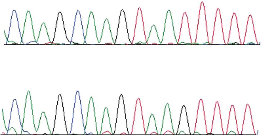
6                                                                                                               Journal of Ophthalmology

have shown promising results when retinal progenitor cells              [2] M. Gorbatyuk, V. Justilien, J. Liu, W. W. Hauswirth, and A.
from transgenic fluorescent red cats were transplanted to                   S. Lewin, “Preservation of photoreceptor morphology and
cats affected by the CEP290 mutation (rdAc) by subretinal                    function in P23H rats using an allele independent ribozyme,”
injections of progenitor cell suspensions. No adverse reac-                 Experimental Eye Research, vol. 84, no. 1, pp. 44–52, 2007.
tions were observed in the transplanted cat eyes. There was             [3] O. Comyn, E. Lee, and R. E. MacLaren, “Induced pluripotent
                                                                            stem cell therapies for retinal disease,” Current Opinion in
further development and migration of transplanted cells
                                                                            Neurology, vol. 23, no. 1, pp. 4–9, 2010.
in the outer and inner retina, and development of donor
                                                                        [4] R. B. Aramant and M. J. Seiler, “Progress in retinal sheet
progenitor cells specifically into Müller-like cells observed by           transplantation,” Progress in Retinal and Eye Research, vol. 23,
immunohistochemistry [63]. Further studies are in progress.                 no. 5, pp. 475–494, 2004.
    Another modality under development using the feline                 [5] J. Dowling, “Current and future prospects for optoelectronic
species is intraocular implantation of retinal prosthesis [75].             retinal prostheses,” Eye, vol. 23, no. 10, pp. 1999–2005, 2009.
Either epiretinal or subretinal implantation can be utilized            [6] E. Pomares, G. Marfany, M. J. Brión, A. Carracedo, and R.
in the degenerate retina. The electrodes in the prosthesis                  Gonzàlez-Duarte, “Novel high-throughput SNP genotyping
may emit electrical currents and stimulate residual retinal                 cosegregation analysis for genetic diagnosis of autosomal
cells, such as second- and third- order neurons, for example,               recessive retinitis pigmentosa and Leber congenital amauro-
bipolar and ganglion cells. Signals to the visual cortex are                sis,” Human Mutation, vol. 28, no. 5, pp. 511–516, 2007.
transmitted to produce a visual sensation. It appears that              [7] G. J. Pazour, S. A. Baker, J. A. Deane et al., “The intraflagellar
                                                                            transport protein, IFT88, is essential for vertebrate photore-
the cat eye, with the visual processes already thoroughly
                                                                            ceptor assembly and maintenance,” Journal of Cell Biology, vol.
investigated, would be an optimal animal model for further                  157, no. 1, pp. 103–113, 2002.
development of research in regards to retinal prosthesis.               [8] L. Asher, G. Diesel, J. F. Summers, P. D. McGreevy, and L. M.
                                                                            Collins, “Inherited defects in pedigree dogs. Part 1: disorders
                                                                            related to breed standards,” Veterinary Journal, vol. 182, no. 3,
8. Future Directions                                                        pp. 402–411, 2009.
                                                                        [9] S. M. Petersen-Jones, “A review of research to elucidate the
Through discoveries of causative mutations and their detri-
                                                                            causes of the generalized progressive retinal atrophies,” Veteri-
mental effects on retinal cell function, new insights into                   nary Journal, vol. 155, no. 1, pp. 5–18, 1998.
retinal degenerative disease mechanisms have been gained.              [10] E. K. Karlsson, I. Baranowska, C. M. Wade et al., “Efficient
It is now possible to aim therapies at correcting disease                   mapping of mendelian traits in dogs through genome-wide
mutations in the eye directly or indirectly. The cat species,               association,” Nature Genetics, vol. 39, no. 11, pp. 1321–1328,
with disease entities that are comparable to those of humans,               2007.
and with large human-like eyes, is amenable to treatment               [11] H. G. Parker, B. M. VonHoldt, P. Quignon et al., “An expressed
using similar surgical techniques and instrumentation as                    fgf4 retrogene is associated with breed-defining chondrodys-
those used for humans. We now have an effective model                        plasia in domestic dogs,” Science, vol. 325, no. 5943, pp. 995–
system that can be used for cell replacement therapy, retinal               998, 2009.
transplantation using tissue from healthy retinas or retinal           [12] E. Cadieu, M. W. Neff, P. Quignon et al., “Coat variation in the
progenitor cells, artificial retinal prosthesis, or combinations            domestic dog is governed by variants in three genes,” Science,
                                                                            vol. 326, no. 5949, pp. 150–153, 2009.
of one or more of the above. There definitely is some hope of
                                                                       [13] O. Goldstein, J. G. Mezey, A. R. Boyko et al., “An ADAM9
further advancement in the field of spontaneously occurring                 mutation in canine cone-rod dystrophy 3 establishes homol-
hereditary retinal blinding disease using the cat as a valuable             ogy with human cone-rod dystrophy 9,” Molecular Vision, vol.
large animal model.                                                         16, pp. 1549–1569, 2010.
                                                                       [14] G. Dekomien, C. Vollrath, E. Petrasch-Parwez et al., “Pro-
                                                                            gressive retinal atrophy in Schapendoes dogs: mutation of the
Acknowledgments                                                             newly identified CCDC66 gene,” Neurogenetics, vol. 11, pp.
                                                                            163–174, 2009.
This paper has been funded in part with federal funds from             [15] D. J. Sidjanin, J. K. Lowe, J. L. McElwee et al., “Canine CNGB3
the National Cancer Institute, National Institutes of Health,               mutations establish cone degeneration as orthologous to the
under contract N01-CO-12400. The authors would like to                      human achromatopsia locus ACHM3,” Human Molecular
thank the Lincy Foundation, the Discovery Eye Foundation                    Genetics, vol. 11, no. 16, pp. 1823–1833, 2002.
and the Grousbeck Family Foundation for their financial                [16] S. M. Petersen-Jones, D. D. Entz, and D. R. Sargan, “cGMP
support. The content of this publication does not necessarily               phosphodiesterase-α mutation causes progressive retinal atro-
reflect the views or policies of the Department of Health                   phy in the Cardigan Welsh corgi dog,” Investigative Ophthal-
and Human Services, nor does mention of trade names,                        mology and Visual Science, vol. 40, no. 8, pp. 1637–1644, 1999.
commercial products, or organizations imply endorsement                [17] M. L. Suber, S. J. Pittler, N. Qin et al., “Irish setter dogs affected
by the US Government.                                                       with rod/cone dysplasia contain a nonsense mutation in the
                                                                            rod cGMP phosphodiesterase β-subunit gene,” Proceedings
                                                                            of the National Academy of Sciences of the United States of
References                                                                  America, vol. 90, no. 9, pp. 3968–3972, 1993.
                                                                       [18] B. Zangerl, O. Goldstein, A. R. Philp et al., “Identical
 [1] A. J. Smith, J. W. Bainbridge, and R. R. Ali, “Prospects for           mutation in a novel retinal gene causes progressive rod-cone
     retinal gene replacement therapy,” Trends in Genetics, vol. 25,        degeneration in dogs and retinitis pigmentosa in humans,”
     no. 4, pp. 156–165, 2009.                                              Genomics, vol. 88, no. 5, pp. 551–563, 2006.
Journal of Ophthalmology                                                                                                                      7

[19] A. V. Kukekova, O. Goldstein, J. L. Johnson et al., “Canine           [34] M. M. Gray, J. M. Granka, C. D. Bustamante et al., “Linkage
     RD3 mutation establishes rod-cone dysplasia type 2 (rcd2) as               disequilibrium and demographic history of wild and domestic
     ortholog of human and murine rd3,” Mammalian Genome,                       canids,” Genetics, vol. 181, no. 4, pp. 1493–1505, 2009.
     vol. 20, no. 2, pp. 109–123, 2009.                                    [35] N. B. Sutter, M. A. Eberle, H. G. Parker et al., “Extensive
[20] J. W. Kijas, A. V. Cideciyan, T. S. Aleman et al., “Naturally              and breed-specific linkage disequilibrium in Canis familiaris,”
     occurring rhodopsin mutation in the dog causes retinal                     Genome Research, vol. 14, no. 12, pp. 2388–2396, 2004.
     dysfunction and degeneration mimicking human dominant                 [36] K. Narfström, “Hereditary progressive retinal atrophy in the
     retinitis pigmentosa,” Proceedings of the National Academy of              Abyssinian cat,” Journal of Heredity, vol. 74, no. 4, pp. 273–
     Sciences of the United States of America, vol. 99, no. 9, pp. 6328–        276, 1983.
     6333, 2002.                                                           [37] K. Narfström, G. B. Arden, and S. E. G. Nilsson, “Retinal
[21] G. D. Aguirre, V. Baldwin, S. Pearce-Kelling, K. Narfström, K.            sensitivity in hereditary retinal degeneration in Abyssinian
     Ray, and G. M. Acland, “Congenital stationary night blindness              cats: electrophysiological similarities between man and cat,”
     in the dog: common mutation in the RPE65 gene indicates                    British Journal of Ophthalmology, vol. 73, no. 7, pp. 516–521,
     founder effect,” Molecular Vision, vol. 4, pp. 23–29, 1998.                 1989.
[22] A. C. Wiik, C. Wade, T. Biagi et al., “A deletion in nephronoph-      [38] S. G. Jacobson, C. M. Kemp, K. Narfström, and S. E.
     thisis 4 (NPHP4) is associated with recessive cone-rod dys-                G. Nilsson, “Rhodopsin levels and rod-mediated function
     trophy in standard wire-haired dachshund,” Genome Research,                in Abyssinian cats with hereditary retinal degeneration,”
     vol. 18, no. 9, pp. 1415–1421, 2008.                                       Experimental Eye Research, vol. 49, no. 5, pp. 843–852, 1989.
[23] Q. Zhang, G. M. Acland, W. X. Wu et al., “Different RPGR               [39] K. Narfström, “Progressive retinal atrophy in the Abyssinian
     exon ORF15 mutations in Canids provide insights into                       cat. Clinical characteristics,” Investigative Ophthalmology and
     photoreceptor cell degeneration,” Human Molecular Genetics,                Visual Science, vol. 26, no. 2, pp. 193–200, 1985.
     vol. 11, no. 9, pp. 993–1003, 2002.                                   [40] J. J. Kang Derwent, L. Padnick-Silver, M. McRipley, E.
[24] C. S. Mellersh, M. E. G. Boursnell, L. Pettitt et al., “Canine             Giuliano, R. A. Linsenmeier, and K. Narfström, “The elec-
     RPGRIP1 mutation establishes cone-rod dystrophy in minia-                  troretinogram components in Abyssinian cats with hereditary
     ture longhaired dachshunds as a homologue of human Leber                   retinal degeneration,” Investigative Ophthalmology and Visual
     congenital amaurosis,” Genomics, vol. 88, no. 3, pp. 293–301,              Science, vol. 47, no. 8, pp. 3673–3682, 2006.
     2006.                                                                 [41] K. Narfström and S. E. Nilsson, “Morphological findings
                                                                                during retinal development and maturation in hereditary
[25] K. E. Guziewicz, B. Zangerl, S. J. Lindauer et al., “Bestrophin
                                                                                rod-cone degeneration in Abyssinian cats,” Experimental Eye
     gene mutations cause canine multifocal retinopathy: a novel
                                                                                Research, vol. 49, no. 4, pp. 611–628, 1989.
     animal model for best disease,” Investigative Ophthalmology
                                                                           [42] K. Narfström and S. E. Nilsson, “Progressive retinal atrophy
     and Visual Science, vol. 48, no. 5, pp. 1959–1967, 2007.
                                                                                in the Abyssinian cat. Electron microscopy,” Investigative
[26] M. Menotti-Raymond, K. H. Deckman, V. David, J. Myrkalo,
                                                                                Ophthalmology and Visual Science, vol. 27, no. 11, pp. 1569–
     S. J. O’Brien, and K. Narfström, “Mutation discovered in a
                                                                                1576, 1986.
     feline model of human congenital retinal blinding disease,”
                                                                           [43] S. Thompson, R. E. H. Whiting, R. H. Kardon, E. M. Stone,
     Investigative Ophthalmology & Visual Science, vol. 51, no. 6,
                                                                                and K. Narfström, “Effects of hereditary retinal degeneration
     pp. 2852–2859, 2010.
                                                                                due to a CEP290 mutation on the feline pupillary light reflex,”
[27] M. Menotti-Raymond, V. A. David, A. A. Schäffer et al.,                    Veterinary Ophthalmology, vol. 13, no. 3, pp. 151–157, 2010.
     “Mutation in CEP290 discovered for cat model of human                 [44] B. Chang, H. Khanna, N. Hawes et al., “In-frame deletion in
     retinal degeneration,” Journal of Heredity, vol. 98, no. 3, pp.            a novel centrosomal/ciliary protein CEP290/NPHP6 perturbs
     211–220, 2007.                                                             its interaction with RPGR and results in early-onset retinal
[28] K. Narfström and S. Petersen-Jones, “Diseases of the canine               degeneration in the rd16 mouse,” Human Molecular Genetics,
     ocular fundus,” in Veterinary Ophthalmology, K. N. Gelatt, Ed.,            vol. 15, no. 11, pp. 1847–1857, 2006.
     vol. 2, pp. 944–1025, Blackwell, Ames, Iowa, USA, 2007.               [45] C. C. Leitch, N. A. Zaghloul, E. E. Davis et al., “Hypomorphic
[29] J. U. Pontius, J. C. Mullikin, D. R. Smith et al., “Initial                mutations in syndromic encephalocele genes are associated
     sequence and comparative analysis of the cat genome,”                      with Bardet-Biedl syndrome,” Nature Genetics, vol. 40, no. 4,
     Genome Research, vol. 17, no. 11, pp. 1675–1689, 2007.                     pp. 443–448, 2008.
[30] J. C. Mullikin, N. F. Hansen, L. Shen et al., “Light whole            [46] V. Frank, A. I. den Hollander, N. O. Brüchle et al., “Mutations
     genome sequence for SNP discovery across domestic cat                      of the CEP290 gene encoding a centrosomal protein cause
     breeds,” BMC Genomics, vol. 11, no. 1, article 406, 2010.                  meckel-gruber syndrome,” Human Mutation, vol. 29, no. 1,
[31] J. C. Fyfe, M. Menotti-Raymond, V. A. David et al., “An ∼140-              pp. 45–52, 2008.
     kb deletion associated with feline spinal muscular atrophy            [47] J. A. Sayer, E. A. Otto, J. F. O’Toole et al., “The centrosomal
     implies an essential LIX1 function for motor neuron survival,”             protein nephrocystin-6 is mutated in Joubert syndrome and
     Genome Research, vol. 16, no. 9, pp. 1084–1090, 2006.                      activates transcription factor ATF4,” Nature Genetics, vol. 38,
[32] K. M. Meurs, E. Mauceli, S. Lahmers, G. M. Acland, S.                      no. 6, pp. 674–681, 2006.
     N. White, and K. Lindblad-Toh, “Genome-wide association               [48] E. M. Valente, J. L. Silhavy, F. Brancati et al., “Mutations
     identifies a deletion in the 3 untranslated region of Striatin in         in CEP290, which encodes a centrosomal protein, cause
     a canine model of arrhythmogenic right ventricular cardiomy-               pleiotropic forms of Joubert syndrome,” Nature Genetics, vol.
     opathy,” Human Genetics, vol. 128, pp. 315–324, 2010.                      38, no. 6, pp. 623–625, 2006.
[33] T. Awano, G. S. Johnson, C. M. Wade et al., “Genome-                  [49] K. Narfström, V. David, O. Jarret et al., “Retinal degeneration
     wide association analysis reveals a SOD1 mutation in canine                in the Abyssinian and Somali cat (rdAc): correlation between
     degenerative myelopathy that resemblesnamyotrophic lateral                 genotype and phenotype and rdAc allele frequency in two
     sclerosis,” Proceedings of the National Academy of Sciences of the         continents,” Veterinary Ophthalmology, vol. 12, no. 5, pp. 285–
     United States of America, vol. 106, no. 8, pp. 2794–2799, 2009.            291, 2009.
8                                                                                                                Journal of Ophthalmology

[50] K. C. Barnett and R. Curtis, “Autosomal dominant progressive         [66] G. M. Acland, G. D. Aguirre, J. Ray et al., “Gene therapy
     retinal atrophy in Abyssinian cats,” Journal of Heredity, vol. 76,        restores vision in a canine model of childhood blindness,”
     no. 3, pp. 168–170, 1985.                                                 Nature Genetics, vol. 28, no. 1, pp. 92–95, 2001.
[51] R. Curtis, K. C. Barnett, and A. Leon, “An early-onset retinal       [67] M. Gorbatyuk, V. Justilien, J. Liu, W. W. Hauswirth, and A. S.
     dystrophy with dominant inheritance in the Abyssinian cat.                Lewin, “Suppression of mouse rhodopsin expression in vivo
     Clinical and pathological findings,” Investigative Ophthalmol-            by AAV mediated siRNA delivery,” Vision Research, vol. 47, no.
     ogy and Visual Science, vol. 28, no. 1, pp. 131–139, 1987.                9, pp. 1202–1208, 2007.
[52] A. Leon and R. Curtis, “Autosomal dominant rod-cone                  [68] M. J. Seiler, B. Rao, R. B. Aramant et al., “Three-dimensional
     dyksplasia in the Rdy cat 1. Light and electron microscopic               optical coherence tomography imaging of retinal sheet
     findings,” Experimental Eye Research, vol. 51, no. 4, pp. 361–            implants in live rats,” Journal of Neuroscience Methods, vol. 188,
     381, 1990.                                                                no. 2, pp. 250–257, 2010.
[53] A. Leon, A. A. Hussain, and R. Curtis, “Autosomal dominant           [69] M. J. Seiler, R. B. Aramant, B. B. Thomas, Q. Peng, S. R. Sadda,
     rod-cone dysplasia in the Rdy cat,” Experimental Eye Research,            and H. S. Keirstead, “Visual restoration and transplant con-
     vol. 53, no. 4, pp. 489–502, 1991.                                        nectivity in degenerate rats implanted with retinal progenitor
[54] S. Chen, Q. L. Wang, Z. Nie et al., “Crx, a novel Otx-                    sheets,” European Journal of Neuroscience, vol. 31, no. 3, pp.
     like paired-homeodomain protein, binds to and transactivates              508–520, 2010.
     photoreceptor cell-specific genes,” Neuron, vol. 19, no. 5, pp.      [70] M. J. Seiler, R. B. Aramant, M. W. Seeliger, R. Bragadottir,
     1017–1030, 1997.                                                          M. Mahoney, and K. Narfström, “Functional and structural
[55] T. H. C. Hsiau, C. Diaconu, C. A. Myers, J. Lee, C. L. Cepko,             assessment of retinal sheet allograft transplantation in feline
     and J. C. Corbo, “The Cis-regulatory logic of the mammalian               hereditary retinal degeneration,” Veterinary Ophthalmology,
     photoreceptor transcriptional network,” PLoS One, vol. 2, no.             vol. 12, no. 3, pp. 158–169, 2009.
     7, article e643, 2007.                                               [71] R. Bragadóttir and K. Narfström, “Lens sparing pars plana vit-
[56] A. K. Hennig, G. H. Peng, and S. Chen, “Regulation of pho-                rectomy and retinal transplantation in cats,” Veterinary Oph-
     toreceptor gene expression by Crx-associated transcription                thalmology, vol. 6, no. 2, pp. 135–139, 2003.
     factor network,” Brain Research, vol. 1192, no. C, pp. 114–133,      [72] M. J. Young, J. Ray, S. J. O. Whiteley, H. Klassen, and F.
     2008.                                                                     H. Gage, “Neuronal differentiation and morphological inte-
[57] C. L. Freund, C. Y. Gregory-Evans, T. Furukawa et al., “Cone-             gration of hippocampal progenitor cells transplanted to the
     rod dystrophy due to mutations in a novel photoreceptor-                  retina of immature and mature dystrophic rats,” Molecular and
     specific homeobox gene (CRX) essential for maintenance of                 Cellular Neurosciences, vol. 16, no. 3, pp. 197–205, 2000.
     the photoreceptor,” Cell, vol. 91, no. 4, pp. 543–553, 1997.         [73] D. M. Gamm, S. Wang, B. Lu et al., “Protection of visual
[58] T. Furukawa, E. M. Morrow, and C. L. Cepko, “Crx, a                       functions by human neural progenitors in a rat model of
     novel otx-like homeobox gene, shows photoreceptor-specific                retinal disease,” PLoS One, vol. 2, no. 3, article e338, 2007.
     expression and regulates photoreceptor differentiation,” Cell,        [74] A. Bakshi, S. Shimizu, C. A. Keck et al., “Neural progenitor
     vol. 91, no. 4, pp. 531–541, 1997.                                        cells engineered to secrete GDNF show enhanced survival,
[59] A. Swaroop, Q. L. Wang, W. Wu et al., “Leber congenital                   neuronal differentiation and improve cognitive function fol-
     amaurosis caused by a homozygous mutation (R90W) in the                   lowing traumatic brain injury,” European Journal of Neuro-
     homeodomain of the retinal transcription factor CRX: direct               science, vol. 23, no. 8, pp. 2119–2134, 2006.
     evidence for the involvement of CRX in the development of            [75] K. Narfström, “Subretinal implantation: a step forward to
     photoreceptor function,” Human Molecular Genetics, vol. 8,                restoring dying photoreceptors,” Expert Reviews in Ophthal-
     no. 2, pp. 299–305, 1999.                                                 mology, vol. 2, pp. 521–524, 2007.
[60] T. Rio Frio, N. M. Wade, A. Ransijn, E. L. Berson, J. S.
     Beckmann, and C. Rivolta, “Premature termination codons
     in PRPF31 cause retinitis pigmentosa via haploinsufficiency
     due to nonsense-mediated mRNA decay,” Journal of Clinical
     Investigation, vol. 118, no. 4, pp. 1519–1531, 2008.
[61] S. Chen, Q. L. Wang, S. Xu et al., “Functional analysis of
     cone-rod homeobox (CRX) mutations associated with retinal
     dystrophy,” Human Molecular Genetics, vol. 11, no. 8, pp. 873–
     884, 2002.
[62] B. Fogle, The New Encyclopedia of the Cat, DK Publishing, New
     York, NY, USA, 2001.
[63] K. Narfström, “Development of Müller-like cells after subreti-
     nal transplantation of feline red fluorescent retinal progenitors
     in Abyssinian cat hereditary retinal degeneration,” in The
     Association for Research in Vision and Ophthalmology Annual
     Meeting Abstracts, Fort Lauderdale, Fla, USA, May 2010.
[64] H. Rah, D. J. Maggs, T. N. Blankenship, K. Narfström, and
     L. A. Lyons, “Early-onset, autosomal recessive, progressive
     retinal atrophy in Persian cats,” Investigative Ophthalmology
     and Visual Science, vol. 46, no. 5, pp. 1742–1747, 2005.
[65] M. W. Seeliger, S. C. Beck, N. Pereyra-Muñoz et al., “In
     vivo confocal imaging of the retina in animal models using
     scanning laser ophthalmoscopy,” Vision Research, vol. 45, no.
     28, pp. 3512–3519, 2005.
You can also read


