ROOT-SHOOT ANATOMY AND POST-HARVEST VEGETATIVE CLONAL DEVELOPMENT IN LOPHOPHORA WILLIAMSII (CACTACEAE: CACTEAE): IMPLICATIONS FOR CONSERVATION
←
→
Page content transcription
If your browser does not render page correctly, please read the page content below
ROOT-SHOOT ANATOMY AND POST-HARVEST VEGETATIVE
  CLONAL DEVELOPMENT IN LOPHOPHORA WILLIAMSII
(CACTACEAE: CACTEAE): IMPLICATIONS FOR CONSERVATION
                    Martin Terry                                 James D. Mauseth
                  Department of Biology                          Section of Integrative Biology
                 Sul Ross State University                        College of Natural Sciences
                Alpine, Texas 79832, U.S.A.                         The University of Texas
                                                                   Austin, Texas 78712, U.S.A.

                                              ABSTRACT
Over the last four decades, the size and density of populations of Lophophora williamsii (peyote)
have diminished markedly in large areas of South Texas where licensed peyote distributors harvest
the cactus for ceremonial use by the Native American Church. Part of the problem lies in the fact
that some harvesters are cutting plants too low on the subterranean stem or taproot. That practice
precludes the regeneration of new stems and ultimately results in the death of the decapitated plants.
To address this problem, we describe the anatomical distinctions between subterranean stem and
root in L. williamsii as follows: The stem cortex can be distinguished by the cortical bundles running
through the parenchyma, in contrast to the root cortex, which consists of pure parenchyma without
cortical bundles. The pith at the center of the stem is pure parenchyma (without xylem) and is readily
distinguished from the dilatated metaxylem (with masses of dark-staining metaxylem tracheary
elements) occupying the center of the root. With these new anatomical tools, it is now possible to set
up titration experiments, first in the greenhouse and then in the field, to generate practical biomet-
ric data to determine the maximum depth at which the peyote harvesters can cut the plants without
significantly reducing the survival rate of the rootstocks left in the ground after harvest.

                                              RESUMEN
Durante las últimas cuatro décadas, el tamaño y densidad de las poblaciones de Lophophora
wiilliamsii (peyote) han disminuido drásticamente en grandes extensiones del sur de Texas donde se
dan permisos a distribuidores de peyote para recolectar especimenes de uso en las ceremonias
religiosas de la Native American Church. Parte del problema estriba en el hecho de que algunos
colectores están cortando plantas muy abajo, o en el tallo subterráneo o en la raíz pivotante. Esta
práctica impide la regeneración de nuevos tallos y finalmente ocasiona la muerte de las plantas
decapitadas. Para abordar este problema, nosotros describimos las características anatómicas que
distinguen el tallo subterráneo y la raíz de L. williamsii como sigue: El tejido cortical del tallo se
distingue por la presencia de haces corticales que se distribuyen por el parénquima, atributo que
contrasta con el tejido cortical de la raíz, el cual consiste en puro parénquima sin haces corticales. En
el centro del tallo la médula es puro parénquima (sin xilema), y se reconoce claramente del metaxilema
dilatado (parches de elementos traqueales del metaxilema teñidos de oscuro) que ocupa el centro de
la raíz. En base a estas características anatómicas será posible hacer experimentos de corte, primero
en invernadero y después en el campo, con la finalidad de generar datos que permitan determinar la
profundidad máxima a la cual los recolectores de peyote pueden cortar las plantas sin reducir
significativamente la supervivencia de los individuos que quedan en el suelo después de la colecta.

SIDA 22(1): 565 – 592. 2006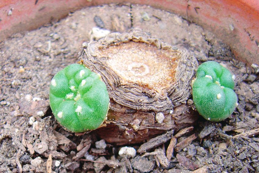
566                                                                   BRIT.ORG/SIDA 22(1)

                                 INTRODUCTION

Peyote harvest as a conservation problem
Lophophora williamsii (Lem. ex Salm-Dyck) J.M. Coult. 1894, commonly called
peyote, is a small, napiform, spineless cactus native to the Chihuahuan Desert
and the Tamaulipecan Thornscrub of northeastern Mexico and adjacent Texas.
While the mescaline-containing stems are ingested in various forms for their
psychotropic effects, this behavior was generally proscribed by federal drug
legislation in 1970. However, federal law provides protection for the use of peyote
for bona fide religious ceremonial purposes by members of the Native Ameri-
can Church. The supply of peyote for such purposes is regulated by the Drug
Enforcement Administration and the Texas Department of Public Safety. The
regulated commerce in peyote begins with the harvest of peyote from wild popu-
lations by licensed peyote distributors or their agents. Commercial quantities
of peyote occur in the U.S. only in Starr, Zapata, Webb and Jim Hogg Counties
in South Texas, within 70 km of the Rio Grande, and all four currently licensed
peyote distributors are based where peyote grows in those counties. Historically
the peyote distributors have gained access to harvestable populations of peyote
through peyote-specific lease agreements with private landowners. Most of the
actual harvesting of peyote is done by contract laborers who are paid in accor-
dance with the number and size of freshly cut “buttons” (tops of stems) of peyote
that they deliver to the licensed distributors.
     There has been a decrease in the number, size, extent and density of peyote
populations in South Texas (Anderson 1995; Moreno 2005) over the past four
decades. Much of this reduction in peyote numbers can be attributed to habitat
destruction associated with urban development and agricultural practices such
as rootplowing of native brush, and some adverse effects on population num-
bers may have been due to illicit harvesting. Such phenomena are difficult to
quantify and virtually impossible to prevent. One factor that is quantifiable in
the decline of peyote in South Texas is the regulated commercial harvest of
peyote by the licensed distributors for ceremonial use by the Native American
Church. Approximately two million peyote buttons per year have been har-
vested by these distributors over the last two decades (Texas Department of
Public Safety, unpublished data). And the technique used in the harvest of those
plants could logically have a substantial impact on the observed decline—or
the potential recovery—of peyote populations in the Tamaulipecan Thornscrub
of South Texas.
     The proper technique for harvesting peyote is that the crown (i.e., the aerial,
photosynthetic portion of the stem) of the peyote cactus is cut off at or imme-
diately below its base, and the subterranean portion of the plant, including all
or most of the subterranean portion of the stem, is left in the ground to regen-
erate one or more new crowns. Such harvesting of the commercially valuable
crown of the cactus may be accomplished by cutting through the plant trans-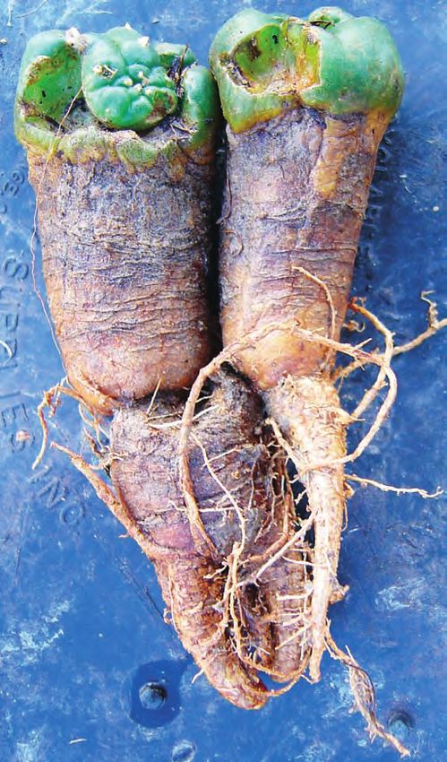
TERRY AND MAUSETH, ROOT AND SHOOT ANATOMY OF LOPHOPHORA WILLIAMSII               567

versely at the level of the surface of the ground, at or near the interface of the
green crown and the brown subterranean portion of the stem, using a machete,
a cutting tool with a broad flat blade and a handle about 60 cm long (such as a
hand edger), or virtually any kind of knife (M. Terry, pers. obs., Fig. 1a–c).
     When proper harvesting technique is adhered to, the decapitated subter-
ranean portion of the stem and the more distal taproot of the cactus (Fig. 2)
remain intact and in situ, where the viable subterranean stem tissue will nor-
mally begin to regenerate one or more new crowns by lateral branching from
axillary (areolar) buds within a few months after loss of the apical meristem
(Fig. 3). Such regenerated crowns may in turn be sustainably harvested after
they reach maturity, some years later (Fig. 4).

Peyote harvest—degeneration and death of the decapitated plant
Not every peyote plant responds in the same manner to removal of its apical
meristem along with the crown at harvest. The simplest response is that de-
scribed above: regeneration of one or more new crowns by lateral branching
from (usually subterranean) stem tissue (Fig. 3). But frequently we observe a
more complex, gradual response, which begins with simple lateral branching
from the remaining stem of the decapitated plant. The difference is that the new
stem branches, instead of remaining dependent on the taproot of the original
plant, put down their own adventitious taproots and eventually become inde-
pendent plants that detach themselves from the original plant, which—if its crown
was completely removed at harvest—degenerates and dies in this process.
     The unusual aspect of this second type of response of peyote to removal of
its apical meristem in the harvesting process, is that the regeneration of new
stems and the generation of new adventitious taproots from the new stem
branches proceed in a seamless developmental process until at some point it
must be recognized that the new, increasingly autonomous shoot-root units have
become independent vegetative clones of the original plant. That is to say, where
we started with a single decapitated plant undergoing development of new stem
branches, we end up with what must be recognized as a moribund parent plant
with viable clonal progeny.
     Another remarkable feature of this process is that as the clonal progeny
become larger and more nutritionally independent, the stem tissue connection
between the parent plant and each of the vegetative clones degenerates to a slen-
der tube consisting mostly of vascular tissue, while the parent plant (which
has no photosynthetic capabilities of its own) steadily decreases in size and
density as the nutrients stored in its parenchymal tissues are depleted and uti-
lized by the growing clonal offspring. In the final phase of the process, the con-
nection between the parent plant and the now nutritionally independent prog-
eny disintegrates, and what is left of the decapitated parent plant dies. This
series of events is depicted in Figures 5–12. All the photos in these figures are of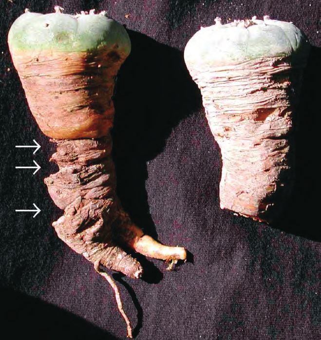
568                                                                                                    BRIT.ORG/SIDA 22(1)

FIG. 1a. Proper peyote harvesting technique. Plant is cut transversely at base of crown, i.e., at ground level.

specimens that the first author collected from populations near Rio Grande City,
Starr County, Texas.
     This natural process of vegetative clone production in response to the ex-
cision of the apical meristem that occurs when peyote crowns are harvested, is
remarkably congruent with the folk belief that harvesting peyote results in an
increase in the number of plants in a population, and that where one plant grew
before harvesting, several plants may be found after allowing an adequate pe-
riod of time for the peyote to “grow back.” A necessary condition for this pro-
cess to occur, however, is that the harvesting be done in a manner that does not
preclude the production of new stems from the subterranean stem of a plant
from which the crown has been harvested. What does this mean in terms that
could serve as a practical guideline for sustainable harvesting?
     The answer must be based on an understanding of the anatomy of the root
and stem of Lophophora williamsii. We begin with the observation that of all
the people working with this plant—and here we include peyote distributors,
members of the Native American Church, cactus hobbyists in countries where
peyote cultivation is legal, and even botanists—very few appreciate the distinc-
tion between true root and subterranean stem. Yet this distinction is crucial to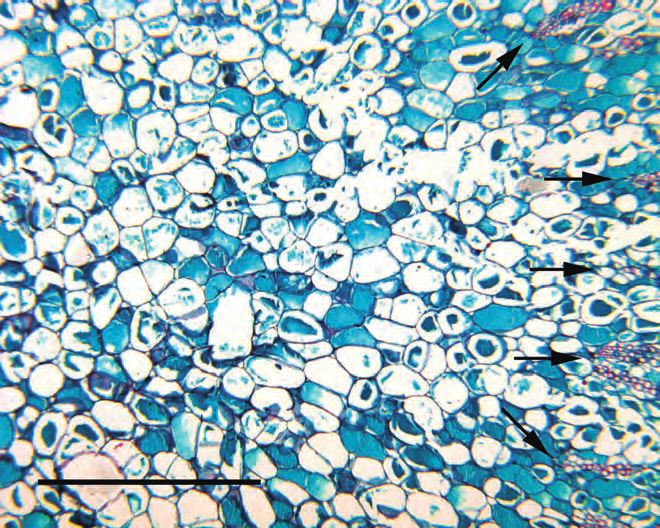
TERRY AND MAUSETH, ROOT AND SHOOT ANATOMY OF LOPHOPHORA WILLIAMSII                                                    569

FIG. 1b Proper peyote harvesting technique. Cut has been made parallel to ground surface, and harvested crown (below,
about 5 cm in diameter, with 8 ribs) has been removed from subterranean portion of stem (above, remaining in the
ground). Cut surface shows cross section of vascular cylinder (ring of yellow tissue near center of stem), pith within the
vascular cylinder, and yellowish green parenchymal tissue of the cortex of the stem (between vascular ring and
epidermis).

an understanding of how to harvest peyote so that it will “grow back,” because,
as far as we have been able to determine from observations to date, new stem
branches will develop only from stem tissue, not from root tissue. Therefore, if
in harvesting the crown one cuts so deeply below the crown that all or most of
the subterranean stem tissue is removed along with the crown, then there will
be no possibility of new stem development, and the stemless root left in the
ground will simply perish. With that as the operating premise, let us now con-
sider the anatomy of the root and the shoot of L. williamsii, and how to distin-
guish between them.
     Ideally we should like to be able to identify the shoot-root transition zone
in order that at least some portion of the shoot should remain with the root of
the plant left in the ground after harvest. Unfortunately, the plants normally
protrude only 1–3 cm above the surface of the ground and have a large subter-
ranean shoot that tapers gradually until it ends in a taproot. The shoot-root
transition zone does not occur near the soil level where the seed germinated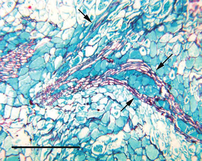
570                                                                                               BRIT.ORG/SIDA 22(1)

FIG. 1c. Proper peyote harvesting technique. Underside of harvested crown (left) shows very narrow (2–3 mm wide) ring
of bark at perimeter of cut surface, indicating that the cut was barely below base of crown, in uppermost portion of
subterranean stem. Cut surfaces match, except that crown parenchyma (left) is slightly greener due to higher chloro-
phyll content, whereas subterranean stem parenchyma (right) is more yellowish.

but instead occurs at various depths (higher in smaller plants, lower in larger
plants). It is not possible to identify the shoot/root transition zone merely by
examining an intact plant with the naked eye.
     In most seed plants, young shoots and roots can be distinguished from each
other because the shoot has a pith and cortex whereas the root lacks both these
structures (Mauseth 1988). However, several other cacti grow like peyote—having
a large below-ground shoot that tapers into a large taproot—and the shoot-root
nature of those structures has not been clarified (Stone-Palmquist & Mauseth
2002). Early workers on the anatomy of Lophophora (Rouhier 1927; Bravo 1931;
Janot & Bernier 1933) gave fair to very good anatomical descriptions of root and
shoot, but even Bravo, whose description of the subterranean stem and root of
Lophophora was the best available in its day, admitted that “[it is most difficult to
know in which region the stem ends and in which the root begins]…” (Bravo 1931,
translated from the Spanish). Because better harvest techniques may aid the sur-
vival of this species in areas subject to intensive harvesting for human consump-
tion, as in South Texas, we undertook a histologic study of the anatomy of roots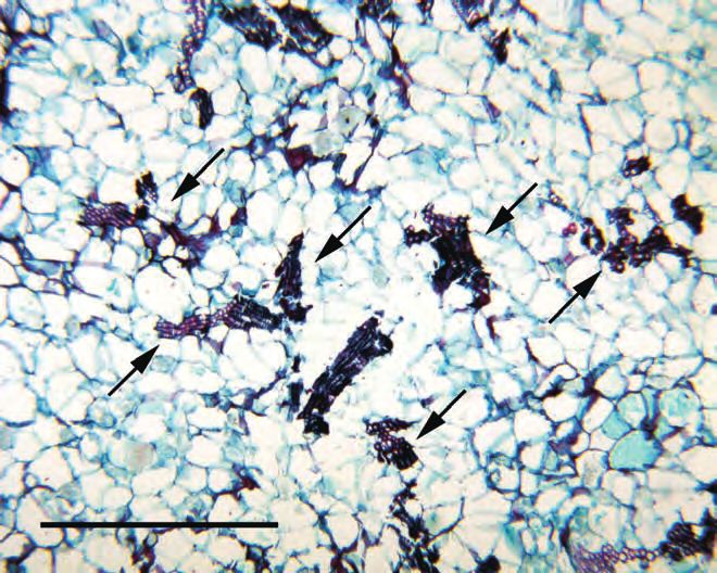
TERRY AND MAUSETH, ROOT AND SHOOT ANATOMY OF LOPHOPHORA WILLIAMSII                                            571

FIG. 2. Peyote plant immediately after most of crown has been harvested, showing cut just above base of harvested
crown (green tissue at top), subterranean stem (brown bark-covered tissue immediately below cut base of crown) ca-
pable of regenerating new crown(s), and tapering taproot (bottom part of plant with a few visible lateral roots).

and shoots in L. williamsii to determine if there are reliable criteria for distin-
guishing the root from the shoot (and particularly the subterranean portion of
the shoot). We especially looked for criteria that could be used in the field.

                                     MATERIALS AND METHODS

Plants were collected by M. Terry (DEA Researchers Registration No. RT0269591)
from a wild population of L. williamsii near Rio Grande City in Starr County,
Texas, or, in the case of one specimen (the very large plant), donated for research
purposes by law enforcement personnel. Specimens examined included two very
small plants, two plants of intermediate size and one very large plant (Table 1).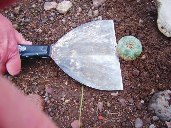
572                                                                                    BRIT.ORG/SIDA 22(1)

TABLE 1. Dimensions of the three adult plants studied, listed from shortest to longest. All values are
in millimeters. Shoots of plants #1212 and #1213 were sampled at ground level and at two below-
ground levels; shoot of plant #1214 was sampled at only one below-ground level. The root of each
plant was sampled only at the top of the root, where it most resembled a portion of shoot.

Sample                    Plant    Aerial   Plant diam. Shoot cortex Shoot pith Root cortex- Root pith-
                          length   height   at soil level thickness  diam.      like zone    like region
                                                                                thickness    diam.

Plant 1212, at ground     135      24       55          20            4
level
   1212, at -20 mm                                      13            6
   1212, at -25 mm                                      9             6
   1212, root, -35 mm                                                            1.5          10
Plant 1214, at ground     140      44       78          32            10
level
   1214, at -35 mm                                      6             27
   1214, root, -41mm                                                             3.0          27
Plant 1213, at ground     160      40       59          14            6
level
   1213, at -20 mm                                      14            6
   1213, at -35 mm                                      9             8
   1213, root, -45 mm                                                            1.0          3.4

     Plants were dissected with care being taken to obtain samples of material
that was definitely root (provisionally defined as the region below the upper-
most point at which a lateral root had emerged), definitely shoot (namely the
aerial portion of the shoot—specifically known as the crown, sensu Schultes
(1938)—which has a blue-gray to blue-green epidermis, photosynthetic tissue
and axillary buds), and definitely hypocotyl (the transition zone between shoot
and root) samples being obtained by taking numerous samples between obvious
root and obvious shoot). In all but the smallest, youngest plants, the plant ma-
terial that was easily recognizable as root was located at least 35 mm below soil
level (45 mm below in plant #1213; Table 1). Since the nature of the higher sub-
terranean portions of L. williamsii was in question, samples were taken from
all plants examined, with the position of each sample being carefully measured
with the soil level (taken as the level of the base of the crown) as reference.
     As tissue samples were obtained during dissection, they were immediately
immersed in Navashin’s solution, then aspirated in a vacuum chamber to remove
air and permit rapid penetration of fixative. Tissues were fixed for 24 hours, de-
hydrated through mixtures of ethanol and tertiary butanol, then embedded in
Paraplast Plus. After microtoming, sections were stained with Safranin and Fast
Green by a procedure designed especially for cacti (Mauseth et al. 1985).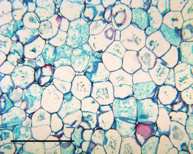
TERRY AND MAUSETH, ROOT AND SHOOT ANATOMY OF LOPHOPHORA WILLIAMSII                                                  573

FIG. 3. Two young peyote crowns (“pups”) regenerating by lateral branching from the upper edge of the subterranean
stem of a plant that was decapitated 7.5 months previously. This plant is the same individual shown in Figs. 1a–1c. Each
of the new crowns is ca. 1.5 cm in diameter

                                                     RESULTS

Morphology
All plants tapered gradually from an unbranched aerial shoot to a region of
subterranean shoot, then to hypocotyl, and finally to taproot. The taper was
uniform in most plants, without any abrupt change in diameter that might in-
dicate the boundary between shoot and hypocotyl or between hypocotyl and
root. The two smallest plants that we examined (whose above-ground, photo-
synthetic crowns were 15 mm in diameter) were only 31mm long (9 mm above
ground, 22 mm below ground) and 50 mm long (10 mm above ground), so seeds
must have germinated at or slightly below ground level. However, in the three
adults we examined, the root/shoot junction was located at least 35 mm below
the soil level (45 mm below in plant #1213), so plants of Lophophora williamsii
must have contractile roots pulling the root/shoot junction deeper as the plant
ages. All above-ground portions of shoot were covered with a blue epidermis;
all subterranean portions were covered with thin, flaking brown bark. Ribs and
axillary buds (often called areoles in cacti) were obvious on all above-ground
portions, and withered areoles were occasionally detected (as much as 17 mm
below soil level on plant #1213).
     Slender lateral roots (1–3 mm diameter) emerged from taproots, but were
extremely sparse, with only two or three present on any plant. This could be in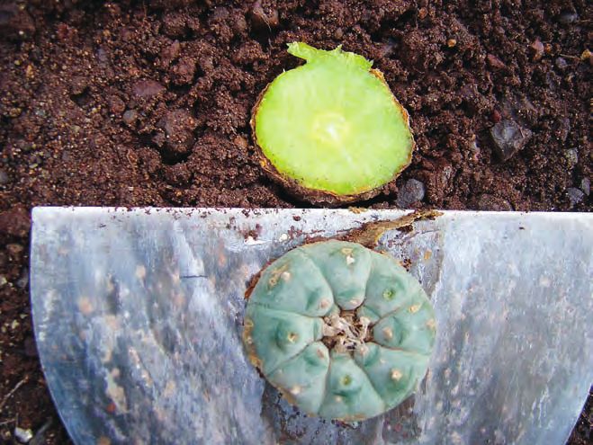
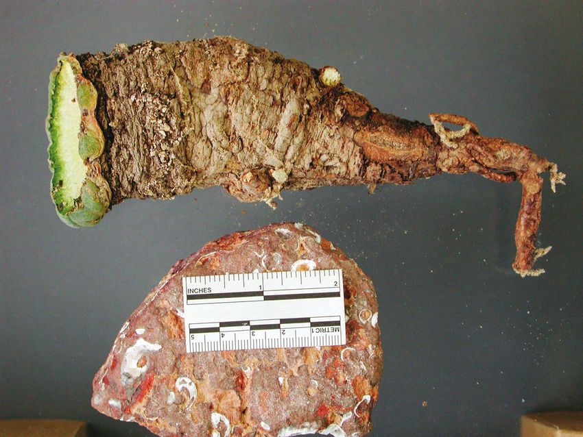
574                                                                                                BRIT.ORG/SIDA 22(1)

FIG. 4. Repeated harvesting of successive crowns from the same peyote plant. Individual on left bears notches (see ar-
rows) indicating stem regeneration after having its crown harvested three times in the past. Individual on right has
never had its crown harvested (the distal end of the root broke off when the plant was dug up for examination).

part because most lateral roots in this species are ephemeral (emerging in re-
sponse to moisture, then being shed in conditions of drought), small, and frag-
ile (M. Terry, pers. obs.). The result is that all but the few largest lateral roots are
broken off and left in the ground when one uproots the plant from its natural
growth site in habitat—no matter how carefully one goes about extracting the
plant. When one grows the plant in loose, friable soil under artificial condi-
tions, more lateral roots and their finer branches remain intact when the plant
is depotted. The plants used in this study, however, were recently uprooted from
TERRY AND MAUSETH, ROOT AND SHOOT ANATOMY OF LOPHOPHORA WILLIAMSII                                                   575

FIG. 5. This is a normal peyote plant with a single long lateral root and no obvious evidence of previous harvesting. The
symmetry of the annular constrictions on the long subterranean stem suggests reduced growth rates in periods of
winter and/or drought. The crown of this mature plant measures ca. 6 cm in diameter and has eight ribs. The shallow,
elongated indentation in the side of the crown facing the reader is a scar from tissue sampling for DNA analysis 18
months previously.

the gravelly soil of their habitat and this would account for some reduction in
the number of intact (and therefore observed) lateral roots.
     Lateral roots emerged only from areas that were later shown to be taproot
or hypocotyl, not from shoot tissue. But one plant examined in the field but not
dissected and studied here had a root emerging from its side at about the same
level as a lateral stem branch; that root might have emerged from the hypo-
cotyl rather than the shoot but that is not known. This isolated field observa-
576 BRIT.ORG/SIDA 22(1) FIG. 6. This is a good example of a plant that was de- FIG. 7. This plant shows evidence of having been previously har- capitated exactly once, several years ago, judging vested at least twice. The small sizes of the two crowns on lat- from the size of the new crown (ca. 5 cm diameter). eral branches from the original subterranean stem suggest that Note that the crown-bearing lateral branch is mark- the most recent harvesting of this plant was perhaps 2–4 years edly offset from the center of the original subterra- ago. (The larger of the two crowns is about 2.5 cm in diameter. nean stem seen at the base of the lateral branch. Both are 5-ribbed.) That is a consequence of the fact that the lateral branch originated from an areole on the side of the subterranean stem. tion should be interpreted in light of the possibility that some of what appear to be uppermost lateral roots of Lophophora may prove to be adventitious roots emerging from subterranean stem tissue. Whether ordinary subterranean stem tissue can produce adventitious roots, or whether adventitious roots can be pro- duced only by regenerative lateral branches from a plant whose apical mer- istem has been removed, is an anatomical issue still to be resolved.
TERRY AND MAUSETH, ROOT AND SHOOT ANATOMY OF LOPHOPHORA WILLIAMSII                                                    577

FIG. 8. This plant has been harvested several times. Only one of the two new crowns is visible from this view, but the two
adventitious taproots, each originating in a stem branch bearing a crown, are both visible (protruding to lower right).
Each of the two crown-bearing stems has recently developed by lateral budding from a previous harvest-associated
lateral branch that was itself decapitated near ground level. The two new crowns, each with its own adventitious tap-
root, are well on their way to becoming independent of the parent rootstock. The original/parental plant, represented
by its bark-covered taproot protruding to lower left from the original subterranean stem (also bark-covered), is degen-
erating, but its subterranean stem is still alive and still attached to the subterranean stems of the two clonal progeny.
578                                                                                                   BRIT.ORG/SIDA 22(1)

FIG. 9. Two vegetative clones that originated as lateral branches of the stem of a decapitated parent plant, which is
degenerating but still clearly recognizable as the bark-covered subterranean stem with taproot extending down be-
tween and beyond the taproots of the two new plants. There is still a connection (not shown directly, but inferable from
the inter-adherence of the plants in the photo) between the new plants and the parental plant, but each of the young
plants has its own functional taproot and is virtually independent of the parental rootstock by now. Visible damage to
the crowns is attributed to the activity of natural herbivores, probably terrestrial snails, which are known to consume L.
williamsii crown tissue.
TERRY AND MAUSETH, ROOT AND SHOOT ANATOMY OF LOPHOPHORA WILLIAMSII                                                         579

FIG. 10. This pair of “sister” vegetative clones is different from that shown in Fig. 9, in that the lateral branch on the left
has effectively co-opted the taproot of the parent rootstock as its own taproot, whereas the lateral branch on the right
has developed an adventitious taproot of its own, which will lead to its eventual separation from the parent rootstock.

     Several plants had shrunken, withered tubercles on their subterranean
portions. They were wrinkled and covered with bark just like all other subter-
ranean portions, but their centers contained living parenchyma cells and an
apical meristem, apparently the shoot apical meristem of the tubercle. The low-
est one found on each adult plant was 15 mm below soil level on plant #1212, 30
mm on plant #1213, and 22 mm on plant #1214.
     Withered tubercles were identifiable on subterranean, bark-covered por-
580 BRIT.ORG/SIDA 22(1) FIG. 11. Three clonal sister peyote plants—each anatomically and functionally on its way to independence with its individual crown and taproot—still attached to the parental subterranean stem (visible as the dark brown structure between the taproots of the middle and right clonal sister plants). tions of two of the adult plants. Tubercles were reliably identifiable only in the uppermost 4.0 mm of subterranean shoot; below that, they had withered so much that the only visible remnants were peg-like structures 2.0–3.0 mm in diameter and 1.0–2.0 mm tall, which appeared to be deteriorated areolar tufts of trichomes. Bark was sufficiently wrinkled and rough that some of its irregu- larities resembled withered tubercles, making identification of tubercles diffi- cult. Subterranean withered tubercles should have been aligned with the rows of tubercles on the aerial portions (Fig. 13), but there were only two cases in
TERRY AND MAUSETH, ROOT AND SHOOT ANATOMY OF LOPHOPHORA WILLIAMSII                                              581

FIG. 12.The amorphous, decaying mass (right) that is tenuously attached to the subterranean stem of the live cactus
(left), is the dead remnant of the subterranean stem of a parent plant that was decapitated by peyoteros sometime in
the past. The young specimen that developed from the then-living parental subterranean stem, is now fully formed,
with its own taproot, and is fully independent. The very low density of the attached mass suggests that it is in an
advanced stage of decomposition.
582                                                                                                BRIT.ORG/SIDA 22(1)

FIG. 13. L. williamsii young tubercle with areole bearing woolly trichomes. The apical meristem from which the areoles
emerge radially is immediately below the field of the photograph.

which a row of withered tubercles could be identified by their areolar tufts (Fig.
14). We followed rows of tubercles from the aerial portions of shoots down into
subterranean portions but usually could not find any identifiable withered
tubercles, apparently the areolar tufts abscise from the plant.
Structure of the root
Very young regions of root (less than 0.5 mm in diameter) had an organization
typical of most dicots. There was an epidermis, a thin cortex only a few cells
thick, endodermis and vascular tissue consisting of small bundles of primary
phloem alternating with arms of protoxylem, and metaxylem occupied the very
center. An important point is that metaxylem contained significant amounts
of living xylem parenchyma cells, it did not consist entirely of dead tracheary
elements. This organization was found in only the two smallest, youngest plants,
in samples taken from closest to root tips. Older portions of roots (2.5 to 30 mm
in diameter) had altered their organization. They had lost their epidermis and
cortex, and parenchyma cells in the metaxylem had begun to proliferate. Sec-
TERRY AND MAUSETH, ROOT AND SHOOT ANATOMY OF LOPHOPHORA WILLIAMSII                                                    583

FIG. 14. L. williamsii tubercles with tufted areoles (red arrows) on lower portion of crown (green tissue in upper half of
photo). On the corrugated brown surface of the subterranean stem (lower half of photo) in the transition between light
and shadow are two lighter-colored protuberances, situated on a diagonal curve containing the two tufted areoles
above in the crown. These protuberances (white arrows) are the visible areolar remnants of stem tubercles, which are
the source of lateral branches of stem that develop in response to removal of the apical meristem.

ondary xylem (wood) and phloem were present, but epidermis, cortex and en-
dodermis had been replaced by a bark consisting of thin flakes of cork. Just
interior to the bark was a band of parenchyma that appeared to be cortex but
which was really secondary phloem parenchyma. This cortex-like region was
about 0.3 to 3.0 mm thick and extended inward from the bark almost to the
vascular cambium. Thickness was correlated with root size: roots less than 4.0
mm in diameter had a cortex-like region only about 0.3 to 0.4 mm thick; roots
about 10–12 mm in diameter had a cortex-like region 1.0 mm thick; and very
large roots 30 mm in diameter had a cortex-like region 3.0 mm thick. The cor-
tex-like region was recognizable as secondary phloem only because it had traces
of collapsed sieve tube members in it. There were no vascular bundles in the
cortex-like region other than very rare connections with lateral roots, and these
were oriented vertically rather than radially or tangentially.
584                                                                  BRIT.ORG/SIDA 22(1)

      Metaxylem parenchyma proliferated in roots, producing such abundant
amounts of parenchyma that the center of the root appeared to have a pith.
Metaxylem vessel elements were pushed apart (such proliferation in an other-
wise mature tissue is called dilatation), and parenchyma cells in the innermost,
first-formed wood also underwent dilatation growth. This pith-like region could
be identified as dilatated xylem (rather than true pith) by the presence of iso-
lated vessel elements within it; these were easily visible with a handlens and
dissecting microscope. In roots about 2.5 to 5.0 mm in diameter, the pith-like
region was about 0.5 to 1.2 mm in diameter, but it was 3.4 mm in diameter in
roots 12 mm wide, 10.0 mm in diameter in roots 22 mm wide, and 27 mm wide
in roots 35 mm wide. Dilatation occurred in both the innermost, first-formed
secondary xylem as well as the middle regions, but the outer regions of second-
ary xylem (the outermost 1.0–2.0 mm) had ordinary wood organization.
      Root wood consisted of a ray system and an axial system (containing axi-
ally oriented cells such as vessel elements). Rays were extremely narrow, only
7.3 sd 2.3 µm wide and consisted of large, rounded parenchyma cells. The axial
system consisted of vessel elements, paratracheal parenchyma in immediate
contact with the vessels, and wide-band tracheids (WBTs). Wide-band trache-
ids are an unusual type of cell found in almost all cacti; they are short (range in
Lophophora: 315 to 525 µm), broad spindle-shaped tracheids with secondary
walls that are annular or helical (Mauseth et al. 1995; Mauseth 2004). There
were no fibers in the wood. Just as rays were narrow, so were axial masses (98.4
sd 54 µm wide), and cross sections of root wood appeared to be rather solid
when viewed with the naked eye or by dissecting microscope.
Structure of the shoot
Young regions of shoot differed from older regions by still having epidermis
but lacking secondary xylem, phloem and bark. Epidermis was present on all
aerial portions of shoots and had a blue-gray color. Hypodermis consisted of
one layer of parenchyma cells. Both epidermis and hypodermis cells definitely
did not have thickened walls so the shoot surface was very soft. Shoot cortex
was always much thicker than the root’s cortex-like region of secondary ph-
loem. The thinnest cortex in an adult plant was 6 mm (in an old, below-ground
portion of plant #1214) and the thickest was 32 mm at soil level in the same
plant. Cortex was only 1.5 mm thick in the seedlings. The outermost cortex cells
were columnar and aligned in palisades, the palisade cortex was about 3.5 mm
thick. Cells of the inner cortex (located between the base of the palisade cortex
and the phloem) consisted of large, rounded parenchyma cells. Cortical bundles
were abundant throughout the inner cortex, extending to the base of the pali-
sade cortex, and each bundle contained both xylem and phloem. Cortical
bundles were easily visible by handlens and dissecting microscope.
     A slender pith was present in the center of all stems. It was only 1.5 mm in
TERRY AND MAUSETH, ROOT AND SHOOT ANATOMY OF LOPHOPHORA WILLIAMSII 585
diameter in seedlings, from 4 to 6 mm in plants of medium size, and 10 to 27
mm in diameter in the largest plant (i.e., plant #1214, which had the greatest
girth). It consisted of just parenchyma cells with very rare spherical crystals
and no mucilage. There were no medullary bundles at all and no dilated met-
axylem. The lack of xylem in the pith was easily visible by handlens and dis-
secting microscope: with both of these, shoot pith looked very clean and homo-
geneous whereas the root’s pith-like region was coarse and granular due to
xylem in the dilatated region.
Young shoots had a ring of collateral vascular bundles located between
pith and cortex, older shoots had secondary xylem and phloem as well. Sec-
ondary xylem in shoots was similar to root wood. Rays were narrow (149 sd 134
µm, just one or two cells wide) and consisted of parenchyma cells with no
sclerification at all. The axial system consisted of small numbers of vessels and
paratracheal parenchyma but large amounts of WBTs. As in roots, axial masses
were narrow, only about 318 sd 179 µm wide. No xylem fibers were present in
any sample. Due to the narrow rays and axial masses and the lack of fibers,
shoot wood resembled root and the two could not be distinguished if a micro-
scope view contained only wood and no other tissues. Secondary phloem in shoots
did not produce a cortex-like region as it did in roots; instead, as the sieve tube
members stopped conducting, phloem collapsed into a thin, tangential band.
All subterranean portions of Lophophora shoots were covered by bark simi-
lar to that on older portions of roots. An unusual feature was that shoot bark
occasionally contained crystals and vascular bundles, indicating that the cork
cambium had arisen deeply enough in the shoot cortex to cut across cortical
bundles; however, both crystals and vascular bundles were too small to be vis-
ible with a handlens examination of bark.
Structure of the hypocotyl
The hypocotyl is the short (less than 10 mm long) region located between the
seedling shoot (epicotyl) and the seedling root. The structure of the hypocotyl
in L. williamsii had characters of both the root and shoot. The center of all hy-
pocotyls was root-like because it consisted of dilatated metaxylem and inner-
most secondary xylem, so it too was pith-like. It could be identified as not be-
ing a true pith by the presence of vessel elements and WBTs interspersed with
the parenchyma cells. The outermost regions were true cortex, and even though
a hypocotyl is not a part of the shoot, the hypocotyls of Lophophora had corti-
cal bundles. Hypocotyl cortex width was wider than that of the cortex-like
region in roots, narrower than the true cortex of shoots in each plant. All hypo-
cotyl samples had abundant secondary xylem and phloem, which was similar
to that in both roots and shoots. Hypocotyl bark was similar to that of shoots,
having occasional bits of cortical bundle that had been cut off by a cork cam-
bium located deep within the cortex.586 BRIT.ORG/SIDA 22(1)
TABLE 2. Distinguishing characters of shoots and roots of L. williamsii.
Shoots Roots
Outer tissues True cortex; appears granular due to Cortex-like region; appears smooth
presence of cortical bundles. At least due to lack of cortical bundles. At
5 mm or more thick. most only 3 mm thick.
Center True pith; appears smooth due to Pith-like region; appears granular due
lack of medullary bundles and lack of to dilatated metaxylem and innermost
dilatated metaxylem. Width is not a secondary xylem.
reliable criterion.
Withered Sometimes present, not always easy Never present.
tubercles to identify if bark is rough.
Lateral roots Never present on shoots? May be Common on taproots, but could be
confused with post-harvest absent.
adventitious roots.
DISCUSSION
This study shows that roots and shoots of L. williamsii differ significantly in
several features. At least in fresh plants, the two organs can be distinguished
easily and reliably using just a handlens or dissecting microscope (Table 2). Both
root and shoot have an outer region that resembles cortex, but the true cortex
of shoots has a granular appearance because it contains numerous cortical
bundles (Fig. 15), as is true of many cacti (Sajeva & Mauseth 1991; Mauseth &
Sajeva 1992). In contrast, the outer region of roots resembles cortex but is in fact
an accumulation of secondary phloem, which has a very smooth appearance
as seen with a handlens. The vascular bundles of lateral roots pass through
this cortex-like region of secondary phloem, but because lateral roots are so
sparse and because their vascular bundles are oriented vertically, there is little
chance of confusing the shoot and root outer tissues. Roots of other cacti also
have this outermost cortex-like region (Stone-Palmquist & Mauseth 2002). The
true cortex of the root of L. williamsii is pure parenchyma (Fig. 16) and does
not contain secondary phloem.
The center of shoots is occupied by true pith, which is homogeneous in
appearance due to the lack of medullary bundles in Lophophora williamsii (Fig.
17). Medullary bundles are common in many species of cacti but lacking in
others (Mauseth 1993). In the roots of most species, metaxylem either has no
parenchyma or if it does, the parenchyma does not undergo proliferation, so
that roots of most plants have no pith-like region at all and can easily be distin-
guished from shoots (Mauseth 1988). The pith-like region of roots in L.
williamsii (Fig. 18) makes the roots look like shoots at first glance or with just
the naked eye, but because the pith-like region in cacti originates by cell divi-
sion in root metaxylem (Gibson 1978; Loza-Cornejo & Terrazas 1996), it has a
granular appearance when examined with a handlens.TERRY AND MAUSETH, ROOT AND SHOOT ANATOMY OF LOPHOPHORA WILLIAMSII 587
FIG. 15. Cortex of shoot of L. williamsii. Arrows indicate cortical bundles running through the parenchyma. All visible
parenchyma cells are cortex parenchyma. Scale bar (lower left) = 1 mm.
Two characters might be useful for distinguishing shoots from roots with-
out cutting plants open to examine the cortex-like regions and pith-like regions.
Lateral roots emerged from the sides of other roots and from the sides of hypo-
cotyls, but none was seen on any part of the three adult shoots we examined. It
is possible that shoots had produced adventitious roots which had either bro-
ken off when the plants were collected or which had abscised before collection.
No remnants of such roots were seen when we examined the sides of subterra-
nean portions of shoots with a dissecting microscope, but these plant parts were
so wrinkled and bark-covered that we could have missed any that were present.
However, as we examined the microscope slides of subterranean portions of
shoots, we did not encounter any vascular bundles that would have indicated
adventitious roots had been present. One plant, examined in the field, had a
root emerging from its side at about the same level as a lateral branch; if that
root was emerging from the shoot rather than the hypocotyl then shoots as well
as roots might bear roots. Now that anatomical characters can be used to dis-
tinguish roots from shoots, it will be possible to examine more plants in the588 BRIT.ORG/SIDA 22(1)
FIG. 16. Cortex of root of L. williamsii. All cells are cortical parenchyma cells. Absence of cortical bundles is conspicuous.
Scale bar = 1 mm.
field to see how frequently shoots bear roots from their sides, and the extent to
which such adventitious root development is associated with branching of sub-
terranean stem tissue in response to peyote harvesting or removal of the apical
meristem by natural processes.
If the below-ground portion of the plant has withered tubercles, it must be
part of the shoot rather than root or hypocotyl. However, we did not find any
withered tubercles on one of our adult specimens despite a search with a dis-
secting microscope. Apparently they either wither so much that they become
unrecognizable or they abscise, so their absence cannot be used as proof that
the structure is either hypocotyl or root. When trying to find withered tubercles
in the field, one should search in a line continuous with the line formed by the
rows of tubercles in the aerial shoot, because all tubercles are formed in rows
(just as on the ribs of columnar cacti: Mauseth 2000).
If a plant remnant is to sprout and continue growing after its top has been
harvested, the presence and health of these withered tubercles is important. If
a plant is harvested by being cut too low, only root or hypocotyl will remain inTERRY AND MAUSETH, ROOT AND SHOOT ANATOMY OF LOPHOPHORA WILLIAMSII 589
FIG. 17. Pith of center of shoot of L. williamsii, showing pith as pure parenchyma without xylem (center and left). Arrows
(right) indicate the innermost primary xylem of the vascular bundles in the ring of bundles at the perimeter of the pith.
Only part of the ring of vascular bundles is shown (right), and all the cells on the left are true pith. Scale bar = 1 mm.
the ground and neither of these have axillary buds, so neither can produce a
bud to replace the harvested shoot. If the plant is harvested by being cut through
the subterranean shoot, and if the remaining portion of shoot has healthy tu-
bercles—withered but with an axillary bud—then the remaining portion should
be able to sprout and grow and be ready for harvesting again in a few years. But
if the remaining shoot has abscised all its tubercles, or if they have withered so
much that they are no longer healthy, then the remaining piece of plant will
not be able to sprout and will instead eventually die for lack of photosynthetic
tissues. Tubercles located higher on the subterranean portion of the shoot are
younger and presumably healthier than those lower down, deeper in the soil
and closer to the root. If plants are harvested by cutting the subterranean shoot
rather high—closer to soil level—the greater the chances are that the residual
piece of plant will have healthy tubercles and will be able to re-sprout.
With the information discovered in the current study, we now have the tools
to examine in detail the potential for regrowth after different types of harvest-590 BRIT.ORG/SIDA 22(1)
FIG. 18. Dilatated metaxylem of center of root of L. williamsii. All arrows indicate masses of dark-staining metaxylem
tracheary elements. All parenchyma cells in the image are metaxylem parenchyma cells, not pith. Scale bar = 1 mm.
ing. A set of plants could be harvested at various depths below soil level with
the certainty that all had been cut high enough that some shoot tissue had been
left on the remaining plant portion. The harvested top of each could be exam-
ined for withered tubercles. Presumably if plants are cut so high that their har-
vested tops have several recognizable withered tubercles near the cut, then the
remaining portion also has at least a few tubercles and will sprout. But if plants
are cut so low that the harvested tops have few or no identifiable withered tu-
bercles near the cut—and if the cut does pass through shoot tissue and not root
tissue—then probably the remaining shoot also has few or no tubercles capable
of sprouting. It is even possible to try cutting the plants through the hypocotyl
to see if it is capable of forming adventitious shoot buds; that capacity is rare in
hypocotyls but is known to occur in a few species.
Preliminary data on plants under greenhouse conditions were collected on
11 plants over a period of three years (not synchronically). Six plants were cut
low (approximately 1.5 crown diameters below the base of the crown, and in
every case at least 50 mm below the base of the crown). The distal subterra-TERRY AND MAUSETH, ROOT AND SHOOT ANATOMY OF LOPHOPHORA WILLIAMSII 591
nean portions of those plants were then observed for at least eight months. No
new stem branches were observed on any of the six low-cut plants. The other
five plants were cut high (at about the base of the crown, and in no case more
than 5 mm below the base of the crown) and similarly observed. One or more
crown-bearing lateral branches from the decapitated subterranean stem were
observed within five months on three of the five plants, and within eight months
on all five high-cut plants.
Future studies include a similar greenhouse experiment on regrowth, with
substantially larger numbers of plants and with varying measured depths of
cut expressed as a fraction (or multiple) of crown diameter, so that crown di-
ameter could be used as a practical guide for harvesting peyote in the field. It
will be noted whether the cut goes through root, stem, or hypocotyl. That titra-
tion of the effect of depth of cut on regrowth in the greenhouse will be followed
by a similar experiment conducted in the field, with individually identified
and permanently marked plants. It is anticipated that results in the field may
differ from greenhouse results, due to harsher environmental conditions in the
field and the possibility that some of the smaller plants in the field may have
been harvested previously, perhaps leaving less than the critical mass of sub-
terranean stem tissue needed to regenerate viable photosynthetic stem tissue.
These factors may affect regeneration and survivorship in all experimental
groups.
ACKNOWLEDGEMENTS
We thank Phil Dering for the photo in Figure 2, and Shirley Powell for the pho-
tos in Figures 13 and 14. Kendall Craig and Tim Parsons did minor miracles in
adding needed layers to some photographs and retrieving high-resolution ver-
sions from low-resolution derivative versions. Special thanks are due to Mike
Powell for his ongoing support extended from the SRSC herbarium.
REFERENCES
ANDERSON, E.F. 1995. The “peyote gardens” of South Texas: a conservation crisis? Cact. Succ.
J. (Los Angeles) 67:67–73.
BRAVO, H. 1931. Nota acerca de la histología del peyote, Lophophora willliamsii Lemaire.
Anales Inst. Biol. Univ. Mexico 2:3–14.
GIBSON, A.C. 1978. Structure of Pterocactus tuberosus, a cactus geophyte. Cact. Succ. J. (Los
Angeles) 50:41–43
JANOT, M. and M. BERNIER. 1933. Essai de localisation des alkaloides dans le peyotl. Bull. Sci.
Pharm. 40:145–153.
LOZA-CORNEJO and TERRAZAS. 1996. Bol. Soc. Bot. México 59:13–23.
MAUSETH, J.D., G. MONTENEGRO, and A.M. WALCKOWIAK. 1985. Host infection and flower forma-
tion by the parasite Tristerix aphyllus (Loranthaceae). Canad. J. Bot. 63:567–581.
MAUSETH, J.D. 1988. Plant anatomy. Benjamin/Cummings. Menlo Park, California.592 BRIT.ORG/SIDA 22(1) MAUSETH, J.D. and M. SAJEVA. 1992. Cortical bundles in the persistent, photosynthetic stems of cacti. Ann. Bot. 70:317–324. MAUSETH, J.D. 1993. Medullary bundles and the evolution of cacti. Amer. J. Bot. 80:928–932. MAUSETH, J.D., Y. UOZUMI, B.J. PLEMONS, and J.V. LANDRUM. 1995. Structural and systematic study of an unusual tracheid type in cacti. J. Pl. Res. 108:517–526. MAUSETH, J.D. 2000. Theoretical aspects of surface-to-volume ratios and water-storage ca- pacities of succulent shoots. Amer. J. Bot. 88:1107–1115. MAUSETH, J.D. 2004. Wide-band tracheids are present in almost all species of Cactaceae. J. Pl. Res. 117:69–76. MORENO, S. 2005. For Native Americans, peyote is dwindling. Austin American-Statesman, Monday, September 19, 2005, p. B3. ROUHIER, A. 1927. Le peyotl—la plante qui fait les yeux emerveilles. Doin, Paris. SAJEVA, M. and J.D. MAUSETH. 1991. Leaflike structure in the photosynthetic, succulent stems of cacti. Ann. Bot. 68:405–411. SCHULTES, R.E. 1938. The appeal of peyote (Lophophora williamsii) as a medicine. Amer. Anthropol., n.s. 40:698–715. STONE-PALMQUIST, M. and J.D. MAUSETH. 2002. The structure of enlarged storage roots in cacti. Int. J. Pl. Res. 163:89–98. TEXAS DEPARTMENT OF PUBLIC SAFETY. 2005. Peyote sales totals and distributors of Texas. Unpub- lished data. Austin, Texas.
You can also read