ALPHA PROTONS AS NMR PROBES IN DEUTERATED PROTEINS - MPG.PURE
←
→
Page content transcription
If your browser does not render page correctly, please read the page content below
Journal of Biomolecular NMR (2019) 73:81–91
https://doi.org/10.1007/s10858-019-00230-y
ARTICLE
Alpha protons as NMR probes in deuterated proteins
Kumar Tekwani Movellan1 · Eszter E. Najbauer1 · Supriya Pratihar1 · Michele Salvi1 · Karin Giller1 · Stefan Becker1 ·
Loren B. Andreas1
Received: 10 December 2018 / Accepted: 28 January 2019 / Published online: 14 February 2019
© The Author(s) 2019
Abstract
We describe a new labeling method that allows for full protonation at the backbone Hα position, maintaining protein side
chains with a high level of deuteration. We refer to the method as alpha proton exchange by transamination (α-PET) since
it relies on transaminase activity demonstrated here using Escherichia coli expression. We show that α-PET labeling is par-
ticularly useful in improving structural characterization of solid proteins by introduction of an additional proton reporter,
while eliminating many strong dipolar couplings. The approach benefits from the high sensitivity associated with 1.3 mm
samples, more abundant information including Hα resonances, and the narrow proton linewidths encountered for highly
deuterated proteins. The labeling strategy solves amide proton exchange problems commonly encountered for membrane
proteins when using perdeuteration and backexchange protocols, allowing access to alpha and all amide protons including
those in exchange-protected regions. The incorporation of Hα protons provides new insights, as the close Hα–Hα and Hα–HN
contacts present in β-sheets become accessible, improving the chance to determine the protein structure as compared with
HN–HN contacts alone. Protonation of the Hα position higher than 90% is achieved for Ile, Leu, Phe, Tyr, Met, Val, Ala, Gln,
Asn, Thr, Ser, Glu, Asp even though LAAO is only active at this degree for Ile, Leu, Phe, Tyr, Trp, Met. Additionally, the
glycine methylene carbon is labeled preferentially with a single deuteron, allowing stereospecific assignment of glycine alpha
protons. In solution, we show that the high deuteration level dramatically reduces R2 relaxation rates, which is beneficial for
the study of large proteins and protein dynamics. We demonstrate the method using two model systems, as well as a 32 kDa
membrane protein, hVDAC1, showing the applicability of the method to study membrane proteins.
Keywords Isotopic labeling · NMR · Membrane proteins · Structural restraints · Transamination · l-Amino acid oxidase
Introduction relaxation, and therefore narrow lines, by elimination of
strong proton–proton dipolar couplings.
The study of proteins by nuclear magnetic resonance (NMR) Proton detected magic-angle spinning (MAS) NMR stud-
has been continuously evolving to improve sensitivity in ies have employed different combinations of spinning fre-
order to resolve signals in multidimensional spectra, which quency and deuteration to optimize sensitivity and resolution
serve as the basis for studies of structure and dynamics. For (Andreas et al. 2015; Zhang et al. 2015; Wang and Ladizhan-
large proteins that tumble slowly in solution, as well as for sky 2014; Brown 2012; Chevelkov et al. 2006; Zhou et al.
proteins in the solid state, a high level of deuteration with 2007; Lewandowski et al. 2011; Akbey et al. 2010). Cur-
introduction of selective protons is used to improve proton rently, many applications of proton detected MAS NMR
are applied at about 60 kHz with 1.3 mm rotors, a spinning
frequency that for fully protonated samples is not enough
Electronic supplementary material The online version of this to average the strong network of 1H–1H dipolar couplings.
article (https://doi.org/10.1007/s10858-019-00230-y) contains
supplementary material, which is available to authorized users. This results in proton line broadening and about 200–300 Hz
proton linewidths (Andreas et al. 2015). The advantage of
* Loren B. Andreas this spinning frequency is that narrow lines are observed
land@nmr.mpibpc.mpg.de at high sensitivity when selected sites are labeled to 100%
1
Department of NMR Based Structural Biology, Max Planck
incorporation of protons, while others are deuterated.
Institute for Biophysical Chemistry, Am Fassberg 11,
Göttingen, Germany
13
Vol.:(0123456789)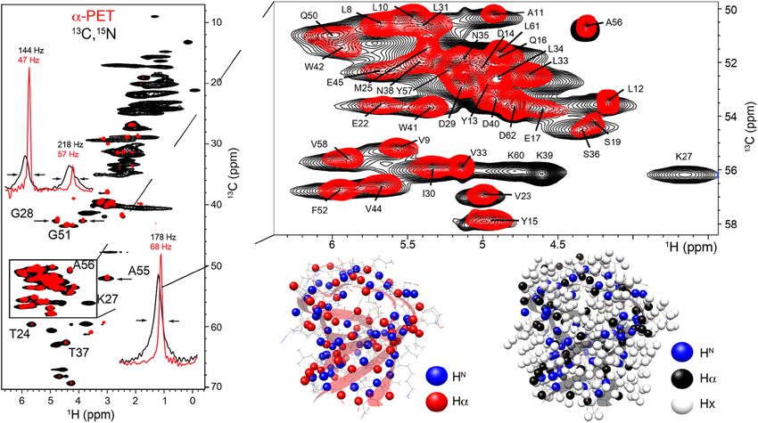
82 Journal of Biomolecular NMR (2019) 73:81–91 In the most straightforward approach, Escherichia coli media (O’Brien et al. 2018; Fiaux et al. 2004), exchange of expression in D 2O is followed by exchange with H 2O/D2O to amide moieties occurs, but results in only 10–50% incorpo- produce a protein with a specific protonation level at amides, ration of alpha protons for hydrophobic residues (Löhr et al. and perdeuteration at non-exchangeable sites (Chevelkov 2003). et al. 2006; Akbey et al. 2010; Lemaster 1990). A pro- We show an alternative approach that results in up to 100% tonation level of 100% at amide positions results in high Hα incorporation by supplying keto acids. The keto acids are resolution when using 40–60 kHz MAS in microcrystalline converted by E. coli transaminases to the respective amino samples, (Lewandowski et al. 2011) enabling structure deter- acids, while adding a proton at the alpha position from the mination based on backbone resonances (Zhou et al. 2007). water pool. This avoids any problems due to racemic amino Accessing aliphatic protons is still an area of active devel- acid mixtures, since the correct l-amino acids are generated opment. Sidechain protons can be selectively introduced enzymatically. The major pathways of amino acid synthesis using metabolic precursors, which has the advantage that a from glucose and glycerol carbon sources are depicted in single isotopomer is typically present and deuterium isotope Fig. 1a for E. coli. Keto acids are often the direct precursor to shifts do not result in broadening, even at 100% protonation an amino acid, indicating that provided a source of keto acids, of the selected sites. In various ways, methyl groups of I, L, protons can be introduced via transaminase activity (Fig. 1b), V, T, A can be incorporated (Tugarinov et al. 2003; Isaacson a method hereafter referred to as ‘alpha proton exchange by et al. 2007; Velyvis et al. 2012). Exquisite control of labe- transamination’ (α-PET). This method, as with any where the ling can be afforded using a synthetic approach known as growth medium is based on H2O, results in protonation of the SAIL labeling, with the only downside being the high cost, amide position during protein expression, such that both the which has typically restricted applications to labeling only alpha and amide positions of the protein are protonated. selected amino acid types in cell free expression systems Some transaminases are amino acid-specific, like the (Kainosho et al. 2006). For fully protonated samples, high glutamate–pyruvate aminotransferase that transfers the magnetic fields and very fast MAS ~ 100 kHz are required N H 3 from glutamate to pyruvate forming alanine and to sufficiently narrow proton resonances. At lower spinning α-ketoglutarate (Kim et al. 2010). Others are less specific frequencies, deuteration is still required, and approaches to for their substrate, such as branched-chain-amino-acid resolve aliphatic protons involve using mixtures of H 2O and transaminase (BCAT) involved in leucine, isoleucine and D2O during protein expression, along with combinations of valine anabolism (Rudman and Meister 1953). E. coli has protonated or deuterated carbon sources such as glycerol or a high diversity of such transaminase enzymes, resulting in glucose (Lemaster 1990; Asami et al. 2010). While utiliz- effective labeling for the majority of residue types. ing the residual protons in perdeuterated samples results in We generated keto acids by l-amino acid oxidase (LAAO) exquisite spectra (Agarwal and Reif 2008), the low labeling treatment of a commercial growth medium that is primarily level severely limits the ability to measure proton–proton comprised of 2H, 13C, 15N-amino acids. LAAO enzymes are distances. At higher proton concentrations, the carbon reso- found in many organisms (Hossain et al. 2014), with differ- nances are broadened due to deuterium isotope shifts (Asami ent specificity for the substrate amino acids (Nuutinen et al. et al. 2012). Although this is a small effect for Hα, since only 2012; Sun et al. 2010). We chose as the enzyme source a one proton is directly attached, it would still be desirable to crude snake venom containing LAAO, which can be applied limit the protons introduced in the sidechains, since such directly to the commercial growth medium (Fig. 1c). Addi- protons broaden the alpha resonance, and are a magnetiza- tional metabolic pathways might also be important, for tion sink during proton–proton transfer. example, we observed stereospecific labeling of glycine, We therefore sought a strategy that would allow labeling of which can occur through transaminase, but also by conver- alpha protons in E. coli at a cost that allows widespread adop- sion of serine and threonine (Fig. 1d). tion of the approach for structure determination and dynamics Here we show successful introduction of alpha protons for investigations. Previously, an approach for Hα labeling was 13 amino acids, with a high deuteration level that improves introduced, where the proton was chemically exchanged in a transverse relaxation rates in both solid and liquid samples. deuterated amino acid mixture, producing a D/L mixture of amino acids, the L portion of which can be utilized directly by bacteria (Yamazaki et al. 1997). Although this previous Methods method was successful, the alpha proton incorporation level was a problem for several amino acids, and serine and threo- L‑amino acid oxidase stock nine were lost during the acetylation and deacetylation reac- tion. d-amino acids may also inhibit bacterial growth at high 10 mg of LAAO powder (crude extract from the snake concentrations (Hishinuma et al. 1969; Bardaweel 2014). It venom of Cortalus admanteus, Sigma Aldrich) were was also noted that during growth on deuterated amino acid 13

Journal of Biomolecular NMR (2019) 73:81–91 83
Fig. 1 Amino acid metabolic
pathways and the different
enzymatic stages of the α-PET
labeling method. The metabolic
pathways of the TCA cycle are
shown in a. In b, the transami-
nation reaction is shown, which
is the main route for Hα incor-
poration. In c, the generation of
α-keto acids from amino acids
by the enzyme LAAO is shown.
d Shows the main biosynthesis
pathways of glycine with the
observed stereospecific labeling
dissolved in 1 ml of 100 mM sodium phosphate, 100 mM were re-suspended in 1L of M9 salts with 4 g of Silantes
KCl at pH 7.4. The solution can be kept at 4 °C for several media either as received, or treated with LAAO. The cells
weeks. were adapted to the new media for 30 min before induc-
tion at O D 600nm = 0.8 with 1 mM of isopropyl β-D-1-
Preparation of keto acid mix thiogalactopyranoside (IPTG). Ubiquitin samples, includ-
ing a 13C, 15N-ubiquitin reference sample were purified as
The amino acid mix (SILEX rich growth media as powder) previously described (Lazar et al. 1997).
from Silantes was used as starting material. To obtain keto Using this media exchange protocol, four different sam-
acids, 1 g of powder was dissolved in 150 ml of H2O. To this ples of ubiquitin were produced, two using 2H Silantes
mixture, 10 µl of bovine liver catalase solution was added at powder treated with LAAO or as received and two others
0 and 12 h (Sigma, fivefold water dilution from crystalline using 2H, 13C, 15N Silantes powder again LAAO treated or
suspension, 10,000–40,000 units/mg). In total, 3–4 mg of as received.
l-amino acid oxidase (LAAO, Sigma) was used per gram Two samples of α-PET SH3 were produced, a media-
powder media, added in equal amounts at 0, 3, 6, 9 and exchanged sample (as for ubiquitin), and a second α-PET
12 h. The solution was kept shaking at 37 °C for 1 day, then SH3 grown in the presence of glucose. Specifically, the
lyophilized. growth was started with a low concentration of 1.25 g/L
12
C-glucose in 800 ml of M9 media. Cells were grown
Protein expression until O D 600nm reached 0.6–0.8. Then 4 g/L of treated
Silantes media solubilized in 200 ml of H 2O were added.
All proteins were expressed in E. coli BL21(DE3). Two The culture was switched to 30 °C for about 30 min until
NMR model proteins were used, ubiquitin in solution, and OD600nm = 0.7–0.8, and protein expression was induced
microcrystalline chicken alpha-spectrin SH3 (SH3). In addi- using 1 mM IPTG. A reference sample (13C, 15N-SH3) was
tion, the 32 kDa voltage dependent anion channel (VDAC), a expressed and all samples purified as previously described
beta barrel membrane protein was prepared in lipid bilayers. (Pauli et al. 2000). In brief, the protein was purified by
For α-PET ubiquitin, a change of medium was used anion exchange chromatography (Q-TRAP, GE Healthcare)
prior to expression. E. coli cells were grown in 1 L of M9 followed by gel filtration on a Superdex-75 column (GE
using 1 g/L of 15N ammonium chloride and 4 g/L of 13C Healthcare). The purified protein sample was extensively
glucose until the O D 600nm reached 0.6–0.8. Then cells dialyzed against H 2O–HCl pH 3.5 for 2 days (exchang-
were spun down at 7000 g at 4 °C for 20 min. The cells ing the dialysis solution every 12 h). The protein was then
13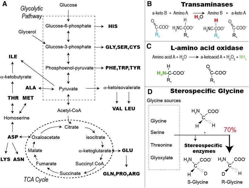
84 Journal of Biomolecular NMR (2019) 73:81–91
concentrated (Amicon, 3.5 kDa cut-off) to 20 mg/ml before
lyophilization. The samples were resuspended in H2O–HCl
pH 3.5 or D2O-HCl pH 3.5 at 15–20 mg/ml. Microcrys-
tals were obtained using a pH shift protocol as previously
described (Chevelkov et al. 2007).
α-PET VDAC was expressed at 37 °C in dilute glucose
media (as for SH3) and purified and reconstituted in 2D
crystalline arrays as previously described (Eddy et al. 2012;
Dolder et al. 1999). The E73V, C127A, C232S variant of
human VDAC was used.
NMR measurements
Solution NMR data were recorded in a 400 MHz Bruker
spectrometer at 298 K. We recorded a set of spectra to
characterize the labeling pattern: 15N-HSQC, 13C-HSQC
in D2O, 1H–15N TOCSY-HSQC, and 1D proton spectra.
Quantification of Hα was done from a 13C-HSQC spectrum Fig. 2 Effective incorporation of Hα protons in a ubiquitin sample,
at 950 MHz at 310 K. Transverse relaxation rates (R2) were while suppressing many side-chain signals. The solution 13C-HSQC
measured at 277 K using a 600 MHz Bruker spectrometer of uniformly labelled ubiquitin (blue) is compared with α-PET ubiq-
equipped with a 5 mm cryoprobe. uitin (red). Selected slices show the intensity at backbone and side-
chain sites. Intensities are not corrected for differences in T2
The black spectrum of Fig. 6a was recorded at 105 kHz
MAS on a 950 MHz Bruker spectrometer using a 0.7 mm
HCND probe. All other solid state NMR data were recorded shows selected strips for each of the amino acid types of
on an 800 MHz Bruker spectrometer using a 1.3 mm narrow ubiquitin; the 1H–15N TOCSY-HSQC of α-PET Ubiquitin
bore HCN probe and spinning at 55 kHz MAS. We recorded (red) is compared to the 15N, 13C-labeled reference sample
cross-polarization based (H)NH, (H)CH, and (H)CANH, (H) (black). The TOCSY was implemented with MLEV-17 mix-
NCAHA for resonance assignment of VDAC and SH3. We ing (Bax and Davis 1985). The Hα proton was detectable
measured contacts in H(H)CH, H(H)NH spectra (SH3) and for 13 of the 16 (non-proline) amino acid types present in
(H)C(HH)CH (VDAC) using RFDR for the proton–proton the ubiquitin sequence. Only lysine, arginine and histidine
mixing. The spectra were apodized with a squared cosine remained deuterated at Hα. This can be explained for lysine
function (details in Table S6). The data analysis was per- because Cortalus admanteus LAAO is not able to use it as
formed using CcpNMR and Sparky. substrate (Fig S1), and the deuterated amino acids are taken
up in E. coli, while endogenous synthesis is suppressed
(Zhou et al. 1998). Although LAAO showed some activity
Results and discussion for arginine and histidine, these two amino acids are clearly
relatively poor substrates of LAAO as reported in previous
Characterization of the labeling pattern studies (Arbor 1967) and also herein (Figs. S1 and S4), and
therefore it appears that the resulting keto acid could not
To measure labeling patterns on an amino acid specific be utilized by E. coli, while the remaining amino acid was
basis, we recorded a 13C HSQC spectrum and integrated effectively incorporated in the protein.
isolated peaks in the alpha region (Fig. 2). The level of Hα Of the 13 successful amino acid types, tyrosine, pheny-
incorporation was determined assuming ideal incorporation lalanine, isoleucine, valine, alanine, threonine and aspartic
of hydrophobic residues, based on complete reaction with acid residues show only Hα signals in the 1H–15N TOCSY-
LAAO. The uncorrected and T 2 corrected determinations HSQC spectrum. The anabolic pathway of these residues
are shown in Tables S2 and S3, respectively. A 15N-TOCSY ends with an aminotransferase reaction, with the exception
(Fig. 3) was recorded using a medium-range mixing time of threonine, which explains the labeling. Effective aspar-
(75 ms) to assess suppression of sidechain protons. This tic acid labeling was unexpected since it enters and exits
spectrum cannot be used in a quantitative manner due to the TCA cycle, but is explained by the very high starting
the potential for several isotopomers, differential relaxa- concentration.
tion, and relayed transfer. However, since the beta protons The amino acid mix from Silantes (Table S1) is obtained
are relatively isolated from these effects, we could show from bacterial proteins by an HCl proteolysis and conse-
effective suppression for most amino acid types. Figure 2 quently glutamine, asparagine, tryptophan, and cystein are
13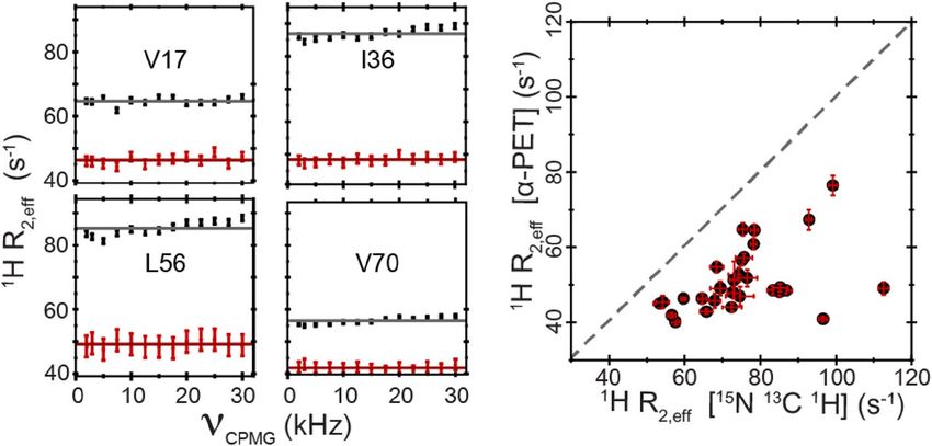
Journal of Biomolecular NMR (2019) 73:81–91                                                                                                  85

Fig. 3  Residue-specific characterization of labeling from 1H–15N TOCSY-HSQC spectra of 1 mM ubiquitin using 75 ms MLEV-17 mixing.
α-PET ubiquitin (red) is compared with 15N, 13C-ubiquitin (black)

not present in the media. Therefore, glutamine and aspara-                acids directly in the Silantes medium. To distinguish the
gine require conversion from the respective acids, which                  signal from the individual amino acid without significantly
explains protonation of beta and gamma protons for these                  changing the composition, we used deuterated Silantes
residues (Fig. 1). Glutamic acid efficiently enters and exits             media, and added only 100 µM of each protonated amino
the TCA cycle, which may explain the incomplete suppres-                  acid. In this way, we rule out potential issues such as com-
sion of beta and gamma protons.                                           petitive binding to the enzyme and determine the approxi-
   Leucine side-chain protons were not expected, but appear               mate starting concentration of all amino- and keto- acids in
to some extent due to LAAO treatment (Fig. S2). If the                    the medium (Table 1).
LAAO treatment is not performed, this sidechain labeling                      Quantification of the labeling for each residue type is
is not observed (Fig. S7), thus it is the crude snake venom               tabulated in Tables S2 and S3 based on intensities extracted
extract that introduces leucine Hγ protons. Details of this               from 13C-HSQC spectra. The intensities were corrected for
side reaction were not investigated further, however we did               the measured proton transverse relaxation rates (Fig. S11)
follow the reaction of LAAO to test efficiency in different               and normalized based on the assumption of complete incor-
buffer conditions for a variety of amino acids (Figs. S1–S5).             poration of isoleucine, phenylalanine, and leucine residues,
   For most amino acids, the reaction proceeded as expected,              which were cleaved completely and are known to effectively
and the snake venom LAAO was particularly efficient for                   incorporate in E. coli (Tugarinov et al. 2003).
hydrophobic amino acids such as phenylalanine and isoleu-                     We also found that efficient transamination occurs when
cine (Crotalus and Allen 2013; Arbor 1967). The degree                    E. coli is grown primarily on amino acids. Some exchange
of conversion to keto acids was also tested for all 20 amino              still occurs at amide positions even without LAAO treatment

Table 1  LAAO activity in deuterated Silantes media, as determined        in the methods section for expression. The remaining alpha signal
by solution NMR. Each amino acid was added in protonated form at a        intensity was used to determine the degree of conversion to keto acid
concentration of 100 µM and LAAO was added exactly as described

Keto acid conversion (%)       Residue                                        Measured Hα incorporation (%)     Residue*

90–100                         Ile, Leu, Phe, Tyr, Trp, Met                   90–100                            Ile, Leu, Phe, Tyr, Met, Val,
                                                                                                                  Ala, Gln, Asn, Thr, Ser, Glu,
                                                                                                                  Asp
10–50                          Val, Arg, His                                  10–50
0–10                           Gly, Pro, Cys, Asn, Gln, Asp, Glu, Ser, Thr,   0–10                              Lys, Arg, His
                                Ala, Lys

*Of 16 amino acids that could be quantified (see SI)

                                                                                                                                   13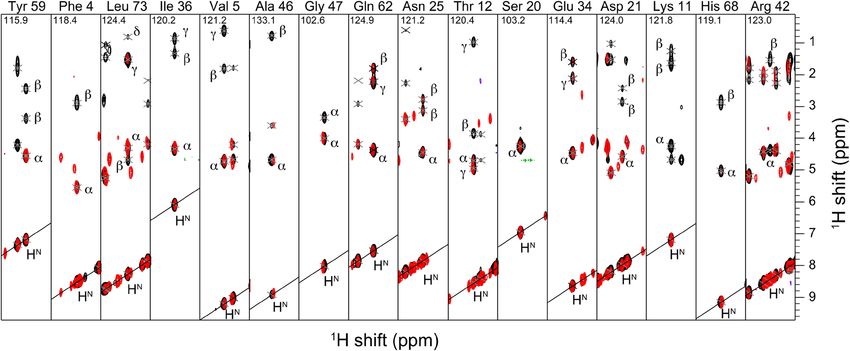
86 Journal of Biomolecular NMR (2019) 73:81–91
(Figs. S6, S7), consistent with a previous report showing liver transaminase acts then R will be the predominant con-
significant Hα labeling for TCA cycle amino acids, but only figuration. Note that in our case, each enzyme will produce
10–50% Hα labeling for hydrophobic residues (Löhr et al. the reverse stereoisomer because the starting amino acid
2003). is deuterated, and the enzymatic reaction occurs in proto-
nated water. By examination of NOE spectra of ubiquitin
Glycine is labeled stereospecifically we observed a cross peak between the glycine 47 Hα and
isoleucine 45 HN, which according to the known structure,
The Hα labeling of glycine attracts particular attention, since indicates that glycine was predominantly the R configu-
one of the two Hα protons is labeled predominantly, result- ration. This is consistent with the stereospecific labelling
ing in stereospecific glycine labeling (Figs. 2, 4). For glycine approach reported previously using cell free extracts (Loscha
28, the intensity ratio between the two alpha protons for and Otting 2013), but results in the opposite labeling, since
microcrystalline 13C, 15N SH3 (Fig. 4, black) is 1–0.93 while we expressed in H 2O rather than D
2O. We can therefore rule
the ratio is 1–0.30 for α-PET SH3 (Fig. 4, red). This effect out deuterated glycine from the medium as the main source
was observed for glycine in all the samples tested, based on of stereospecific glyine found in the expressed protein.
signal intensity in HSQC and CP-HSQC spectra. We also
observed a considerable reduction in line width, by more Resolution and structural data under MAS
than a factor of three. conditions
Glycine can be produced from serine by hydroxymethyl-
transferase, from threonine by l-allo-threonine aldolase, or To demonstrate that the α-PET labeling scheme results in
through serine–glyoxylate or alanine–glyoxylate transami- improved resolution, we prepared a microcrystalline sample
nases. Information in E. coli is limited, but analysis of other of α-spectrin SH3 according to established crystallization
organisms using tritiated water indicates that the stereo protocols (Pauli et al. 2000). The Hα line width is signifi-
specificity depends on the pathways involved (Yoshimura cantly reduced for α-PET SH3 and the effect is particularly
et al. 1996; Dunathan et al. 1968; Wellner 1970). If serine improved for certain residues, by a factor of two and above
transhydroxymethylase acts in tritiated water, the resulting (Fig. 4). The proton resolution is also superior to labeling
glycine will predominantly be the S configuration, but if with deuterated glucose in otherwise protonated media
Fig. 4 Cross-polarization based carbon-proton correlation spectra, peak intensities show stereospecific labeling with preference for R
hCH, of microcrystalline SH3 either uniformly α-PET labeled (red) (α3 protonated) over S (α2) configuration. At the bottom right, the
and 13C, 15N-labled (black) crystalized from a protonated buffer. backbone and side-chain protons are indicated on the solution NMR
Spectra were recorded at a magnetic field of 800 MHz and 30 °C, structure (pdb: 1aey) for α-PET SH3 (red ribbon) and 13C, 15N SH3
55 kHz MAS. 1D slices from the spectrum indicate the improvement (black ribbon)
in linewidths for G28 (top left) and A55 (bottom right). The glycine
13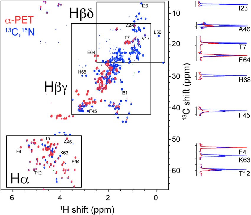
Journal of Biomolecular NMR (2019) 73:81–91 87
(Medeiros-Silva et al. 2016) (Fig. S8). To characterize the observed using cross-polarization based transfer experi-
narrowing of the homogeneous part of the lines, the bulk T2´ ments. Thus 50 Hα peaks are expected for 13C,15N SH3. For
relaxation times at 55 kHz was measured for Hα, Cα, and α-PET SH3 lysine, arginine and histidine are not expected.
CO from 1D (HCAN)H, (HCON)H and (HCA)HA spectra Thus only 41 Hα peaks are expected and indeed 41 peaks
by integrating the full signal. The Hα T 2´ of 3–4 ms for were readily identified in (H)NCAHA spectra.
α-PET SH3 is a dramatic improvement compared to 1 ms Figure 5 shows a comparison between α-PET SH3 in fully
for the fully protonated sample (Fig S9). protonated buffer (red) and α-PET SH3 in fully deuterated
The Hα T2´ of α-PET SH3 crystallized in 100% D2O buffer (blue) in which long-range structural restraints were
buffer ranged from 7 to 15 ms, an improvement over the measured. To characterize the benefit of the restraints pre-
amide protonated sample large enough that we can directly sent with α-PET labeling, we manually selected peaks in
observe an increase in resolution in the 1D spectrum (Fig the H(H)CH and H(H)NH spectra, and used automated shift
S9). The improvement is further characterized for select resi- matching (0.05, 0.5 and 0.5 ppm tolerance, in 1H, 13C and
dues in Figure S12. The H N signals were almost completely 15
N, respectively) to identify contacts. Of 114 automatically
removed in the D2O buffer. assigned peaks from the 3D H(H)NH of α-PET SH3 in pro-
Sequential resonance assignment in Fig. 4 were made tonated buffer, two unambiguous contacts were identified,
using a (H)NCAHA spectrum, and are consistent with those of which one is a long-range HN–HN contact. For the 3D
previously reported (Xue et al. 2017). SH3 has 62 residues, H(H)CH, 132 peaks were selected, and seven unambigu-
of which, two are prolines and the N-terminal seven resi- ous contacts were identified, five of which are long-range
dues and residues 46–48 are flexible and are therefore not restraints. However, for the H(H)CH spectrum in deuterated
Fig. 5 Long-range distance
information is highlighted in a
3D H(H)CH spectrum of α-PET
SH3 (pdb: 1aey) in D2O (blue)
and in H 2O (red). a Shows a
contact between L33 Hα and
V44 Hα. In b, the contact
between T32 Hα and L8 Hα
is readily observed in D 2O (in
blue) while it is much weaker in
the presence of additional pro-
tons in H2O (in red). Recorded
in a 800 MHz Bruker spectrom-
eter at 30 °C and 55 kHz MAS
1388 Journal of Biomolecular NMR (2019) 73:81–91
buffer we found 150 contacts, of which eight are unambigu- The method was also successful for a more challeng-
ous restraints, seven of which were long range corresponding ing system, the human voltage-dependant anion channel
N–Hα or Hα–Hα. One of the additional contacts
to either H (VDAC). The lipid bilayer structure of this protein has
identified in fully deuterated buffer is highlighted in Fig. 5b. been investigated through MAS NMR spectra of VDAC
This method clearly improves the number of structural in liposomes (Schneider et al. 2010) and in 2D crystalline
restraints available at 55 kHz MAS, and in particular, the arrays (Eddy et al. 2015), and narrow proton resonances
unambiguous restraints, a metric that is crucial for the con- were reported for a perdeuterated sample.(Eddy et al.
vergence of commonly used structure calculation methods. 2015a, b) With α-PET labeling, we also observed narrow
A concern with Hα detection is the presence of water and amide proton linewidths of 150 Hz in H 2O, while ~ 100 Hz
other solvent signals in this spectral region. Therefore good lines were observed using D2O buffer, which is slightly
water suppression is needed, but as demonstrated here for better than the ~ 120 Hz linewidths observed for perdeuter-
samples in both H2O and D2O, control of the water is pos- ated and H N back-exchanged protein. This indicates that
sible even without gradient methods. the non-exchangeable protons are slightly narrower, and
For resonance assignment, the α-PET labeling approach that Hα labeling does not significantly impact the spectral
benefits from the implementation of new proton detected quality. In this D2O-exchanged buffer, less improvement in
NMR pulse sequences focused on Hα detection that were Hα T2´ (Fig. S9) was observed as compared with the SH3
recently developed for > 100 kHz MAS (Stanek et al. 2016). domain, which is not unexpected, since approximately
So far, proton detected MAS NMR structures were mostly half the amide protons were protected from exchange (Fig.
based on H N detected experiments or more recently on S9B).
fully protonated samples that are best investigated using To further characterize the potential spectral resolution,
>100 kHz MAS. (Cala-De Paepe et al. 2017) New possibili- α-PET VDAC was measured at 105 kHz MAS at a 950 MHz
ties are opened with the α-PET approach, allowing effective spectrometer (black in Fig. 6a). Surprisingly, the same line
structural measurements with the inherently more sensitive width was obtained at 110 kHz MAS at 950 MHz (~ 110 Hz)
equipment for ~ 60 kHz spinning. and at 55 kHz MAS at 800 MHz (~ 95 Hz), showing that the
inhomogeneous contributions are dominating the linewidth
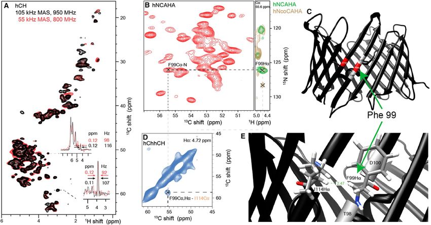
Fig. 6 Identification of a cross beta strand contact (F99–I114 Hα) in Hα is assigned from the strip comparing (H)NCAHA (green) and (H)
the beta barrel membrane protein VDAC in lipid bilayers. a Shows, N(CO)CAHA (brown). In c and e, the contact is shown on the X-ray
the comparison of the (H)CH spectrum at 105 kHz on a 950 MHz structure of mouse VDAC (pdb: 2jk4). d Shows the F99–I114 cross-
spectrometer (black) and at 55 kHz on an 800 MHz spectrometer peak in the carbon–carbon 2D plane of the (H)C(HH)CH spectrum at
(red). b Shows a 13C–15N projection of a (H)NCAHA spectrum. F99 the proton frequency of F99, 4.72 ppm
13Journal of Biomolecular NMR (2019) 73:81–91 89
Fig. 7 Selected residues show-
ing the reduction in proton (Hα)
R2 relaxation rates with α-PET
labeling (red) as compared with
full protonation (black). The
correlation plot (right) shows a
reduction for all residues. The
data is from ubiquitin sam-
ples exchanged in 100% D2O
at 277 K and measured at a
600 MHz spectrometer
at 55 kHz. This shows that even for a highly homogene- transmembrane beta sheet interface, of which VDAC has
ous preparation of a membrane protein, α-PET labeling 19. Further analysis of the expected contacts in VDAC is
efficiently reduces the proton dipolar broadening at 55 kHz. show in in Fig. S10 This demonstrates a successful imple-
The protection from solvent exchange observed for mentation of α-PET labelling for structure determination in
VDAC highlights an issue with perdeuteration for proteins a challenging 32 kDa membrane protein embedded in lipid
that lack refolding protocols. Perdeuteration of membrane bilayers, where structural restraints are particularly difficult
proteins (Medeiros-Silva et al. 2016; Ward et al. 2011) and to identify (Eddy et al. 2015).
large complexes (Andreas et al. 2016) in E. coli often results
in deuterated amides that cannot be exchanged with protons α‑PET labeling for solution NMR
from water. Such exchange protected regions of the pro-
tein become inaccessible in the perdeuteration and back- The α-PET labeling approach is also beneficial for the study
exchange approach, limiting the analysis to solvent acces- of proteins in solution, when deuteration is needed to reduce
sible regions (Andreas et al. 2015; Chevelkov et al. 2006; transverse relaxation rates (LeMaster and Richards 1988;
Fricke et al. 2017; Zhou et al. 2007a, b; Ward et al. 2015), Torchia et al. 1988). Figure 7 and Tables S4–S5 show the
although such limited exchange phenomena can also be used reduction in R2 relaxation rates due to the high level of deu-
to obtain functional information (Ward et al. 2011; Böck- teration in α-PET labeled Ubiquitin. Such improvement in
mann and Guittet 1997; Agarwal et al. 2010). Using α-PET relaxation rates is important for the study of protein dynam-
labeling, we are now able to detect both exchangeable as ics. For example in detection of Hα relaxation dispersion,
well as non-exchangeable amide protons in highly deuterated fractional deuteration was used to improve R 2 (Lundström
samples as shown previously for amino acid based media et al. 2009; Vallurupalli et al. 2009). The current labeling
(Löhr et al. 2003). incorporates the alpha positions at 100% for most residues,
Due to the size of the protein, unambiguous assignment with a high overall deuteration level, which improves sensi-
of important cross-strand contacts was not possible in a 3D tivity as compared with random fractional deuteration.
H(H)CH spectrum of VDAC. We therefore applied the bet-
ter resolved 3D (H)C(HH)CH spectrum to measure cross-
strand contacts (Fig. 6). VDAC assembles as a beta barrel, Conclusions
a topology that places cross-strand Hα pairs in close prox-
imity (~ 2.3 Å), and much closer than sequential Hα spins, Here, we introduced a new method to label Hα protons in a
which are separated by about 4.5 Å. 28 Hα–Hα contacts protein without significant isotopic scrambling, and demon-
were detected from this spectrum, of which we show the strated how this new sensitive magnetic probe in the back-
cross strand contact between residue phenylalanine 99 and bone of the protein adds new structural information even
isoleucine 114, which was assigned based on the existing at below 60 kHz MAS. The α-PET labeling approach has
13
C and 15N assignments of this protein (Eddy et al. 2015a, several advantages, (i) adaptation of the cells to D 2O is not
b) and (H)NCAHA and (H)N(CO)CAHA spectra. The cur- required, (ii) it gives similar yields as deuterated expression
rent published assignments (32% of 283 residues) of VDAC in M9 media, and (iii) costs are similar to production of
do not allow a characterization of all 28 peaks. However, deuterated proteins. It is expected to be particularly useful
resolving 28 peaks is significant, considering that only ~ 4 for deuteration of proteins that lack refolding protocols, such
amide–amide or alpha–alpha contacts are available in each as membrane proteins.
1390 Journal of Biomolecular NMR (2019) 73:81–91
In this demonstration, we used a commercially avail- Arbor A (1967) On the reaction mechanism acid oxidase * of Crotalus
able crude snake venom extract to generate keto acids. This adamanteus. J Biol Chem 242:1259–1264
Asami S, Schmieder P, Reif B (2010) High resolution 1 H-detected
approach results in the designed incorporation of alpha pro- solid-state NMR spectroscopy of protein aliphatic resonances:
tons for Tyr, Phe, Leu, Ile, Gly, Gln, Asn, Asp, Glu and Met. access to tertiary structure information. J Am Chem Soc
In the future, further optimization of the method might entail 132:15133–15135
other LAAOs with different substrate specificity, perhaps Asami S, Szekely K, Schanda P, Meier BH, Reif B (2012) Optimal
degree of protonation for1H detection of aliphatic sites in ran-
in combination with auxotrophic strains to limit unwanted domly deuterated proteins as a function of the MAS frequency. J
reaction pathways. In addition, other amino acid mixtures or Biomol NMR 54:155–168
expression systems could be investigated. This might allow Bardaweel SK (2014) D-amino acids: prospects for new therapeutic
labeling of lysine, arginine, and histidine, which were cur- agents. J Med Bioeng 3:195–198
Bax A, Davis DG (1985) MLEV-17-based two-dimensional homo-
rently left deuterated. nuclear magnetization transfer spectroscopy. J Magn Reson
65:355–360
Böckmann A, Guittet E (1997) Determination of fast proton exchange
rates of biomolecules by NMR using water selective diffusion
Supporting Information experiments. FEBS Lett 418:127–130
Brown SP (2012) Applications of high-resolution 1H solid-state
NMR. Solid State Nucl Magn Reson 41:1–27
Quantification of labeling patterns, LAAO activity, meas- Cala-De Paepe D, Stanek J, Jaudzems K, Tars K, Andreas LB, Pin-
urement of relaxation times under 55 kHz MAS, Hα R2 in tacuda G (2017) Is protein deuteration beneficial for proton
solution and spectral acquisition parameters. detected solid-state NMR at and above 100 kHz magic-angle
spinning? Solid State Nucl Magn Reson 87:126–136
Acknowledgements Open access funding provided by Max Planck Chevelkov V, Rehbein K, Diehl A, Reif B (2006) Ultrahigh resolu-
Society. We thank Pablo Trigo Mourino for helping to set up solution tion in proton solid-state NMR spectroscopy at high levels of
NMR measurements and Christian Griesinger, Tobias Schubeis and deuteration. Angew Chem Int Ed 45:3878–3881
Guido Pintacuda for useful discussions. We also thank Ashok Kumar Chevelkov V, Faelber K, Schrey A, Rehbein K, Diehl A, Reif B
Rout for NOE data in ubiquitin. We acknowledge financial support (2007) Differential line broadening in MAS solid-state NMR
from the MPI for Biophysical Chemistry, and from the Deutsche due to dynamic interference. J Am Chem Soc 129:10195–10200
Forschungsgemeinschaft (Emmy Noether program Grant AN1316/1- Crotalus I, Allen R (2013) Crystalline of Crotalus udamanteus. Pub-
1, SFB803 Grant INST 186/794-3 Project A04). lic Health 235:2013–2018
Dolder M, Zeth K, Tittmann P, Gross H, Welte W, Wallimann T
(1999) Crystallization of the human, mitochondrial voltage-
OpenAccess This article is distributed under the terms of the Crea-
dependent anion-selective channel in the presence of phospho-
tive Commons Attribution 4.0 International License (http://creativeco
lipids. J Struct Biol 127:64–71
mmons.org/licenses/by/4.0/), which permits unrestricted use, distribu-
Dunathan HC, Davis L, Kury PG, Kaplan M (1968) The stereochem-
tion, and reproduction in any medium, provided you give appropriate
istry of enzymatic transamination. Biochemistry 7:4532–4537
credit to the original author(s) and the source, provide a link to the
Eddy MT, Ong TC, Clark L, Teijido O, Van Der Wel PCA, Garces R,
Creative Commons license, and indicate if changes were made.
Wagner G, Rostovtseva TK, Griffin RG (2012) Lipid dynamics
and protein-lipid interactions in 2D crystals formed with the
β-barrel integral membrane protein VDAC1. J Am Chem Soc
134:6375–6387
References Eddy MT, Su Y, Silvers R, Andreas L, Clark L, Wagner G, Pintacuda
G, Emsley L, Griffin RG (2015a) Lipid bilayer-bound conforma-
Agarwal V, Reif B (2008) Residual methyl protonation in perdeuterated tion of an integral membrane beta barrel protein by multidimen-
proteins for multi-dimensional correlation experiments in MAS sional MAS NMR. J Biomol NMR 61:299–310
solid-state NMR spectroscopy. J Magn Reson 194:16–24 Eddy MT, Andreas L, Teijido O, Su Y, Clark L, Noskov SY, Wagner
Agarwal V, Linser R, Fink U, Faelber K, Reif B (2010) Identification of G, Rostovtseva TK, Griffin RG (2015b) Magic angle spinning
hydroxyl protons, determination of their exchange dynamics, and nuclear magnetic resonance characterization of voltage-depend-
characterization of hydrogen bonding in a microcrystallin protein. ent anion channel gating in two-dimensional lipid crystalline
J Am Chem Soc 132:3187–3195 bilayers. Biochemistry 54:994–1005
Akbey Ü, Lange S, Franks WT, Linser R, Rehbein K, Diehl A, Van Fiaux J, Bertelsen EB, Horwich AL, Wüthrich K Uniform and res-
Rossum BJ, Reif B, Oschkinat H (2010) Optimum levels of idue-speci c 15 N-labeling of proteins on a highly deuterated
exchangeable protons in perdeuterated proteins for proton detec- background. J. Biomol. NMR 289–297 (2004)
tion in MAS solid-state NMR spectroscopy. J Biomol NMR Fricke P, Chevelkov V, Zinke M, Giller K, Becker S, Lange A (2017)
46:67–73 Backbone assignment of perdeuterated proteins by solid-state
Andreas LB, Le T, Jaudzems K, Pintacuda G (2015) High-resolution NMR using proton detection and ultrafast magic-Angle spin-
proton-detected NMR of proteins at very fast MAS. J Magn Reson ning. Nat Protoc 12:764–782
253:36–49 Hishinuma F, Izaki K, Takahashi H (1969) Effects of glycine and
Andreas LB, Jaudzems K, Stanek J, Lalli D, Bertarello A, Le Marchand d-amino acids on growth of various microorganisms. Agric Biol
T, Cala-De Paepe D, Kotelovica S, Akopjana I, Knott B, Wegner Chem 33:1577–1586
S, Engelke F, Lesage A, Emsley L, Tars K, Herrmann T, Pinta- Hossain GS, Li J, Shin HD, Du G, Liu L, Chen J (2014) L-amino acid
cuda G (2016) Structure of fully protonated proteins by proton- oxidases from microbial sources: Types, properties, functions,
detected magic-angle spinning NMR. Proc. Natl. Acad. Sci. 113, and applications. Appl Microbiol Biotechnol 98:1507–1515
9187–9192
13Journal of Biomolecular NMR (2019) 73:81–91 91
Isaacson RL, Simpson PJ, Liu M, Cota E, Zhang X, Freemont P, Sun MZ, Guo C, Tian Y, Chen D, Greenaway FT, Liu S (2010) Bio-
Matthews S (2007) A new labeling method for methyl transverse chemical, functional and structural characterization of Akbu-
relaxation-optimized spectroscopy NMR spectra of alanine resi- LAAO: A novel snake venom l-amino acid oxidase from Agkis-
dues. J Am Chem Soc 129:15428–15429 trodon blomhoffii ussurensis. Biochimie 92:343–349
Jain MG, Lalli D, Stanek J, Gowda C, Prakash S, Schwarzer TS, Torchia DA, Sparks SW, Bax A (1988) Delineation of α-helical
Schubeis T, Castiglione K, Andreas LB, Madhu PK, Pintacuda domains in deuteriated staphylococcal nuclease by 2D NOE NMR
G, Agarwal V (2017) Selective 1H– 1H distance restraints in spectroscopy. J Am Chem Soc 110:2320–2321
fully protonated proteins by very fast magic-angle spinning Tugarinov V, Kay LE, Ile (2003) Leu, and Val methyl assignments of
solid-state NMR. J Phys Chem Lett 8:2399–2405 the 723-residue malate synthase G using a new labeling strategy
Kainosho M, Torizawa T, Iwashita Y, Terauchi T, Mei Ono A, Gün- and novel NMR methods. J Am Chem Soc 125:13868–13878
tert P (2006) Optimal isotope labelling for NMR protein struc- Vallurupalli P, Hansen DF, Lundström P, Kay LE (2009) CPMG relaxa-
ture determinations. Nature 440:52–57 tion dispersion NMR experiments measuring glycine 1Hα and
Kim SH, Schneider BL, Reitzer L (2010) Genetics and regulation of 13Cα chemical shifts in the ‘invisible’ excited states of proteins.
the major enzymes of alanine synthesis in Escherichia coli. J Bac- J Biomol NMR 45:45–55
teriol 192:5304–5311 Velyvis A, Ruschak AM, Kay LE (2012) An economical method for
Lazar G, Desjarlais JR, Handel TM (1997) De novo design of the production of 2H,13CH3-threonine for solution NMR studies of
hydrophobic core of ubiquitin. Protein Sci 6:1167–1178 large protein complexes: application to the 670 kDa proteasome.
Lemaster DM (1990) Deuterium labeling in NMR structural-analysis PLoS ONE 7:1–8
of larger proteins. Q Rev Biophys 23:133–174 Wang S, Ladizhansky V (2014) Recent advances in magic angle spin-
LeMaster DM, Richards FM (1988) NMR sequential assignment of ning solid state NMR of membrane proteins. Prog Nucl Magn
Escherichia coli thioredoxin utilizing random fractional deuteri- Reson Spectrosc 82:1–26
ationt. Biochemistry 27:142–150 Ward ME, Shi L, Lake E, Krishnamurthy S, Hutchins H, Brown LS,
Lewandowski JR, Dumez JN, Akbey Ü, Lange S, Emsley L, Oschki- Ladizhansky V (2011) Proton-detected solid-state NMR reveals
nat H (2011) Enhanced resolution and coherence lifetimes in the intramembrane polar networks in a seven-helical transmembrane
solid-state NMR spectroscopy of perdeuterated proteins under protein proteorhodopsin. J Am Chem Soc 133:17434–17443
ultrafast magic-angle spinning. J Phys Chem Lett 2:2205–2211 Ward ME, Ritz E, Ahmed MAM, Bamm VV, Harauz G, Brown LS,
Löhr F, Katsemi V, Hartleib J, Günther U, Rüterjans H (2003) A strat- Ladizhansky V (2015) Proton detection for signal enhancement in
egy to obtain backbone resonance assignments of deuterated pro- solid-state NMR experiments on mobile species in membrane pro-
teins in the presence of incomplete amide2H/1H back-exhange. J teins. J Biomol NMR. https: //doi.org/10.1007/s10858 -015-9997-5
Biomol NMR 25:291–311 Wellner D (1970) Stereospecificity of enzymatic formation and oxida-
Loscha KV, Otting G (2013) Biosynthetically directed2H labelling tion of glycine. Biochemistry 9:2307–2310
for stereospecific resonance assignments of glycine methylene Xue K, Sarkar R, Motz C, Asami S, Camargo DCR, Decker V, Wegner
groups. J Biomol NMR 55:97–104 S, Tosner Z, Reif B Limits of resolution and sensitivity of proton
Lundström P, Hansen DF, Vallurupalli P, Kay LE (2009) Accurate Detected MAS solid-state NMR experiments at 111 kHz in deu-
measurement of alpha proton chemical shifts of excited protein terated and protonated proteins. Sci Rep 7, (2017)
states by relaxation dispersion NMR spectroscopy. J Am Chem Yamazaki T, Tochio H, Furui J, Aimoto S, Kyogoku Y (1997) Assign-
Soc 131:1915–1926 ment of backbone resonances for larger proteins using the 13C-1H
Medeiros-Silva J, Mance D, Daniëls M, Jekhmane S, Houben K, Bal- coherence of a 1H(α)-, 2H-, 13C-, and 15N-labeled sample. J Am
dus M, Weingarth M (2016) 1H-detected solid-state NMR studies Chem Soc 119:872–880
of water-inaccessible proteins in vitro and in situ. Angew Chem Yoshimura T, Jhee K-H, Soda K (1996) Stereospecificity for the Hydro-
Int Ed 55:13606–13610 gen transfer and molecular evolution of pyridoxal enzymes. Biosci
Rudman D, Meister A (1953) Transamination in Escherichia coli. J Biotechnol Biochem 60:181–187
Biol Chem 200:591–604 Zhang R, Nishiyama Y, Ramamoorthy A (2015) Proton-detected 3D
Nuutinen JT, Marttinen E, Soliymani R, Hilden K, Timonen AS (2012) 1H/13C/1H correlation experiment for structural analysis in
L-amino acid oxidase of the fungus Hebeloma cylindrosporum rigid solids under ultrafast-MAS above 60 kHz. J Chem Phys
displays substrate preference towards glutamate. Microbiology 143:164201
158:272–283 Zhou P, Sun LJ, Dötsch V, Wagner G, Verdine GL (1998) Solution
O’Brien ES, Lin DW, Fuglestad B, Stetz MA, Gosse T, Tommos C, structure of the core NFATC1/DNA complex. Cell 92:687–696
Wand AJ (2018) Improving yields of deuterated, methyl labeled Zhou DH, Shah G, Cormos M, Mullen C, Sandoz D, Rienstra CM
protein by growing in H2O. J Biomol NMR 0:0 (2007a) Proton-detected solid-state NMR spectroscopy of fully
Pauli J, Van Rossum B, Förster H, De Groot HJM, Oschkinat H (2000) protonated proteins at 40 kHz magic-angle spinning. J Am Chem
Sample optimization and identification of signal patterns of amino Soc 129:11791–11801
acid side chains in 2D RFDR spectra of the α-Spectrin SH3 Zhou DH, Shea JJ, Nieuwkoop AJ, Franks WT, Wylie BJ, Mullen
domain. J Magn Reson 143:411–416 C, Sandoz D, Rienstra CM (2007b) Solid-state protein-struc-
Schneider R, Etzkorn M, Giller K, Daebel V, Eisfeld J, Zweckstetter ture determination with proton-detected triple-resonance 3D
M, Griesinger C, Becker S, Lange A (2010) The native confor- magic-angle-spinning NMR spectroscopy. Angew Chem Int Ed
mation of the human VDAC1 N terminus. Angew Chem Int Ed 46:8380–8383
49:1882–1885
Stanek J, Andreas LB, Jaudzems K, Cala D, Lalli D, Bertarello A, Publisher’s Note Springer Nature remains neutral with regard to
Schubeis T, Akopjana I, Kotelovica S, Tars K, Pica A (2016) jurisdictional claims in published maps and institutional affiliations.
NMR Spectroscopic Assignment of Backbone and Side-Chain
Protons in Fully Protonated Proteins: Microcrystals, Sedi-
mented Assemblies, and Amyloid Fibrils. Angew Chem Int Ed
55(50):15504–15509. https://doi.org/10.1002/anie.201607084
13You can also read


