Current and innovative methods for the diagnosis of COVID 19 infection (Review)
←
→
Page content transcription
If your browser does not render page correctly, please read the page content below
INTERNATIONAL JOURNAL OF MOlecular medicine 47: 100, 2021

                Current and innovative methods for the diagnosis
                        of COVID‑19 infection (Review)
                           LUCA FALZONE1, GIUSEPPE GATTUSO2, ARISTIDIS TSATSAKIS3,
                                DEMETRIOS A. SPANDIDOS4 and MASSIMO LIBRA2,5

      1
       Epidemiology and Biostatistics Unit, National Cancer Institute‑IRCCS ‘Fondazione G. Pascale’, I‑80131 Naples;
          2
            Department of Biomedical and Biotechnological Sciences, University of Catania, I‑95123 Catania, Italy;
         3
           Department of Forensic Sciences and Toxicology, Faculty of Medicine; 4Laboratory of Clinical Virology,
             Medical School, University of Crete, 71003 Heraklion, Greece; 5Research Center for the Prevention,
                      Diagnosis and Treatment of Tumors, University of Catania, I‑95123 Catania, Italy

                                        Received March 1, 2021; Accepted April 7, 2021

                                                  DOI: 10.3892/ijmm.2021.4933

Abstract. The Coronavirus Disease 2019 (COVID‑19)                  Contents
pandemic has forced the scientific community to rapidly
develop highly reliable diagnostic methods in order to effec‑      1. Introduction
tively and accurately diagnose this pathology, thus limiting       2. The right test, on the right sample, at the right time
the spread of infection. Although the structural and molecular     3. RT‑PCR‑based molecular tests
characteristics of the severe acute respiratory syndrome           4. Rapid antigen and rapid antibody tests
coronavirus 2 (SARS‑CoV‑2) were initially unknown, various         5. Immunoenzymatic serological tests
diagnostic strategies useful for making a correct diagnosis of     6.	Alternative methods for the effective diagnosis of
COVID‑19 have been rapidly developed by private research               COVID‑19 infection
laboratories and biomedical companies. At present, rapid           7.	Conclusions
antigen or antibody tests, immunoenzymatic serological tests
and molecular tests based on RT‑PCR are the most widely used
and validated techniques worldwide. Apart from these conven‑       1. Introduction
tional methods, other techniques, including isothermal nucleic
acid amplification techniques, clusters of regularly inter‑        Europe and the entire world have faced the second wave of
spaced short palindromic repeats/Cas (CRISPR/Cas)‑based            Coronavirus Disease 2019 (COVID‑19) pandemic which
approaches or digital PCR methods are currently used in            was characterized by an increased number of infections
research contexts or are awaiting approval for diagnostic use      and related deaths worldwide, thus still highlighting critical
by competent authorities. In order to provide guidance for the     issues in the management of this health emergency (1,2).
correct use of COVID‑19 diagnostic tests, the present review       At the time of the writing of the present review article,
describes the diagnostic strategies available which may be used    113,523,131 laboratory‑confirmed COVID‑19 cases and
for the diagnosis of COVID‑19 infection in both clinical and       2,519,454 COVID‑19‑related deaths have been recorded world‑
research settings. In particular, the technical and instrumental   wide, highlighting the impressive impact of this pandemic
characteristics of the diagnostic methods used are described       globally (3).
herein. In addition, updated and detailed information about the        Despite the optimism deriving from the approval of two
type of sample, the modality and the timing of use of specific     new mRNA vaccines and of one recombinant vaccine against
tests are also discussed.                                          severe acute respiratory syndrome coronavirus 2 (SARS‑CoV‑2)
                                                                   infections by the Food and Drug Administration (FDA) and
                                                                   the European Medicines Agency (EMA) (5,6), a third wave of
                                                                   infections, already observed in the United States, the United
                                                                   Kingdom and other countries with greater proportions than the
Correspondence to: Professor Massimo Libra, Department of
                                                                   one just concluded, is expected in the coming months (3,6,7).
Biomedical and Biotechnological Sciences, University of Catania,
Via Santa Sofia 97, I‑95123 Catania, Italy                         In this regard, several governments worldwide have already
E‑mail: mlibra@unict.it                                            begun to adopt social distancing measures and the lockdown
                                                                   of collective activities in order to avoid a drastic increase in
Key words: SARS‑CoV‑2, COVID‑19, diagnosis, viral detection,       the number of infections (8,9). In addition, a great concern is
RT‑PCR, rapid test, immunoenzymatic assay, ddPCR, isothermal       also represented by the need to differentiate COVID‑19 cases
amplification technique, CRISPR‑Cas, molecular methods             from seasonal flu that could clog hospital emergency services,
                                                                   slowing down the diagnostic and therapeutic procedures for
                                                                   patients with COVID‑19 (10). These reasons have led the scien‑
2                                       FALZONE et al: DIAGNOSTIC METHODS FOR COVID-19

tific community to question which diagnostic strategies are          time, research laboratories and biomedical companies studied
optimal in order to efficiently combat the imminent increase         the main features of the virus, thus assisting researchers world‑
in COVID‑19 infections, as well as to perform differential           wide in developing various diagnostic solutions for a correct
diagnoses between COVID‑19 infections and seasonal flu.              diagnosis of COVID‑19 (22‑24). Among such solutions, the
     In this context, population screening strategies have been      most commonly used and validated methodologies are rapid
proposed and are currently being implemented for the effec‑          antigen or antibody tests, immunoenzymatic serological tests
tive monitoring of the COVID‑19 epidemiological curve and            and RT‑PCR‑based molecular tests. Each of these three types
to screen the immunized population; however, it is not yet           of diagnostic tests can be applied at a precise moment of
clear which is the most effective method for these surveillance      infection. Of note, only kits, reagents and molecular probes
programs (11‑14).                                                    validated by the Centers for Diseases Control and Prevention
     Since the beginning of the pandemic, immense efforts            (CDC) and the World Health Organization (WHO) and
have been made for the development of effective diag‑                approved at the American and European level by the FDA and
nostic strategies which may be used to accurately identify           by the EMA can be used for diagnostic purposes (25).
SARS‑CoV‑2‑infected patients, thus limiting the risk of                  Although immunoenzymatic (either classic or rapid
contagion and promptly treating any respiratory symptoms,            methods) and molecular methods are the most widely used
avoiding serious consequences for individuals (15).                  techniques for the diagnosis of COVID‑19, other approaches
     At present, several diagnostic methods have been approved       were used in the early phase of the pandemic to identify posi‑
by regulatory agencies worldwide; however, there is still            tive patients and the etiological agent of infection. Among
confusion regarding the correct tests to be used based on the        these approaches, viral culture and next‑generation sequencing
patient's medical history or the investigation purpose (16).         (NGS) methods were crucial for the identification of the novel
As regards Europe, there are 365 different commercialized            coronavirus and for the characterization of its molecular
devices CE‑IVD‑ and FDA‑approved or used for research                structure. These two techniques have made it possible to
purposes. Of these devices, 168 are immunoassays, 192 are            fully characterize the genome and the viral protein structure,
PCR‑based methods, three are NGS‑based methods and two               allowing the understanding of the viral mechanisms of action,
commercialized tools are based on different technologies (17).       the mode of transmission, the clinical impact and the develop‑
Therefore, it is evident that the selection of the optimal diag‑     ment of any therapeutic strategies and diagnostic tools (26,27).
nostic test can be difficult.                                            Apart from these conventional strategies, other diagnostic
     On these bases, the present review aimed to provide the         methods are under development and validation or are currently
rationale for the correct use of SARS‑CoV‑2 diagnostic tools         finding application in research contexts. Among these
currently available by setting out the decision‑making prin‑         methods, digital PCR, isothermal amplification techniques,
ciples useful for the correct choice of the most appropriate test.   clusters of regularly interspaced short palindromic repeats/Cas
     As will be discussed in the following paragraphs, several       (CRISPR/Cas)‑based diagnostic methods, biosensors, electron
diagnostic tests are currently available for the early identifi‑     microscopy‑based methods, etc., represent the armamentarium
cation of SARS‑CoV‑2 infection, for the monitoring of the            to effectively diagnose COVID‑19 infection and to effectively
presence of any infections among healthcare workers, for the         assess the epidemiological spreading of the pandemic (15,28).
monitoring the incidence rates and the severity of the infection     Notably, clinical investigations and radiological imaging have
and for the evaluation of the complete remission of patients         represented valid diagnostic alternatives, particularly during
with COVID‑19 (18‑20).                                               the early stages of the pandemic when there were still no vali‑
     Several parameters should be considered for the selection       dated molecular and serological tests available (29) (Fig. 1).
of the optimal diagnostic test. A diagnostic test must have good         Despite the availability of all these diagnostic techniques,
sensitivity and specificity rates; however, these parameters are     a correct diagnosis of COVID‑19 infection can only be estab‑
not the only features to be considered. Indeed, in the case of       lished considering the test to be used, the type of sample to
the COVID‑19 pandemic, an effective test should be rapid,            be analyzed and the timing of the test itself. Therefore, it is
repeatable, based on technologies available at numerous              necessary to perform the correct test, at the correct time in the
centers and keep costs limited in order to be carried out on a       correct biological sample (30,31).
large fraction of the population (21).                                   In particular, it is important to take into account the
     Therefore, it is evident that the selection of the diagnostic   moment of the suspected infection, the patient's medical
test should be performed taking into account the clinical or         history, the symptoms and general clinical picture for a
surveillance purpose of the investigation as well as the possi‑      successful outcome of the diagnostic test. Furthermore, the
bility to repeat the test several times until the patients are no    positivity of a diagnostic test strongly depends on the moment
longer positive.                                                     at which it is carried out (Fig. 2). Both serological and molec‑
     For all these clinical and epidemiological needs, three         ular tests are not useful during the first week of the supposed
main types of tests for COVID‑19 diagnosis are available:            infection because the virus is still in its incubation period and
i) Molecular RT‑PCR swab tests; ii) serological tests; iii) rapid    there are not yet sufficient copies of viral RNA in circulation
antigen or antibody tests.                                           neither antibodies nor viral proteins identifiable by serological
                                                                     tests (32,33). Therefore, before the onset of symptoms, the
2. The right test, on the right sample, at the right time            probability of correctly determining the presence of the virus,
                                                                     particularly using molecular tests, remains low (32).
Although the molecular and structural characteristics of                 At two weeks after the presumed infection, and in parallel
SARS‑CoV‑2 were initially unknown, in a very short period of         with the onset of symptoms, molecular tests carried out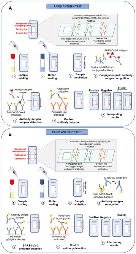
INTERNATIONAL JOURNAL OF MOlecular medicine 47: 100, 2021                                                     3

            Figure 1. Overview of the available clinical, diagnostic and research strategies for the effective diagnosis of COVID‑19 infection.

on nasopharyngeal swabs or bronchoalveolar lavage fluid                        Indeed, these markers increase in the early stages of infection
(BALF) samples will have a greater chance of being positive,                   and can be used for the early diagnosis of COVID‑19 infec‑
as the virus is actively replicating. However, this probability is             tion (36). In summary, all these data suggest that for a correct
reduced over time, as a result of the elimination of the virus                 diagnosis of COVID‑19 infection to be made, it is necessary to
and the remission of the disease. In the case of molecular tests,              use the right test, at the right time.
the probability of a positive or negative result should be consid‑                 As already mentioned, the selection and the correct
ered, as no commercial diagnostic tools have a sensitivity of                  handling of samples for both molecular and serological anal‑
100%, particularly in cases of patients with low viral load as                 yses are fundamental for the diagnosis of COVID‑19 infection.
asymptomatic or paucisymptomatic individuals (32,34).                          In particular, the collection of samples and the pre‑analytical
    As regards serological tests, the search for IgM and IgG                   phases are crucial for the positive outcome of the diagnostic
antibodies begins to yield positivity at approximately one                     procedure (37,38). Different studies have demonstrated that
month after the presumed infection and the levels of these                     the positivity rate of molecular tests significantly depends
immunoglobulins remain high for long periods of time. Of                       on the quality of the starting sample influenced by sample
note, the IgM serum levels decrease significantly after six                    collection, poor quality material, wrong transport or storage,
weeks from the onset of symptoms (35). The detection of viral                  the presence of inhibitors, etc. (38). Technical troubleshooting
proteins and mucosal IgA antibodies is also very important.                    will be discussed in the following chapters.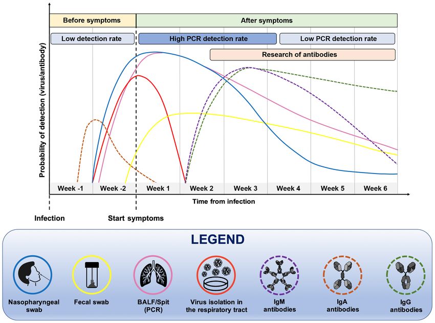
4                                       FALZONE et al: DIAGNOSTIC METHODS FOR COVID-19

Table I. Biological samples and methods used for an effective diagnosis of COVID‑19 infection.

	Diagnostic methods
                                                              ‑‑‑‑‑‑‑‑‑‑‑‑‑‑‑‑‑‑‑‑‑‑‑‑‑‑‑‑‑‑‑‑‑‑‑‑‑‑‑‑‑‑‑‑‑‑‑‑‑‑‑‑‑‑‑‑‑‑‑‑‑‑‑‑‑‑‑‑‑‑‑‑‑‑‑‑‑‑‑‑‑‑‑‑‑‑‑‑‑‑‑‑‑‑‑‑‑‑‑‑‑
	Collection                                  Transport            Approved                                           Research‑used
Type of specimens devices                    conditions       diagnostic methods                                  diagnostic methods	Comments

NP swab, OP swab, Dacron or VTM Within 5 days, 4˚C; Real‑time PCR      NGS ‑ CRISPR/Cas PCR ‑
and NP aspirate    flocked swabs >5 days, ‑70˚C		                      isothermal amplification
				                                                                   techniques ‑ddPCR ‑
				                                                                   viral culture
Sputum             Sterile vial      Within 48 h, 4˚C; Real‑time PCR   NGS ‑ CRISPR/Cas PCR ‑
		                                   >48 h, ‑70˚C		                    isothermal amplification
				                                                                   techniques ‑ ddPCR ‑
				                                                                   viral culture
Bronchial washing Sterile vial       Within 48 h, 4˚C; Real‑time PCR   NGS ‑ CRISPR/Cas PCR ‑                                                                         Pathogens
		                                   >48 h, ‑70˚C		                    isothermal amplification                                                                       may be diluted;
				                                                                   techniques ‑ ddPCR ‑                                                                           however, the
				                                                                   viral culture                                                                                  specimen can
					                                                                                                                                                                 be used for
					                                                                                                                                                                 diagnostic testing
Tracheal aspirate  Sterile vial      Within 48 h, 4˚C; Real‑time PCR   NGS ‑ CRISPR/Cas PCR ‑
and transtracheal		                   >48 h, ‑70˚C		                   isothermal amplification
aspirate				                                                           techniques ‑ ddPCR ‑
				                                                                   viral culture
Lung biopsy        Sterile vial      Within 48 h, 4˚C; Real‑time PCR   NGS ‑ CRISPR/Cas PCR ‑
		                                   >48 h, ‑70˚C		                    isothermal amplification
				                                                                   techniques ‑ ddPCR ‑
				                                                                   viral culture
Serum, plasma      Serum/plasma Within 5 days, 4˚C; Rapid serological Biosensors                                                                                      For the immune
                   collection tube: >5 days, ‑70˚C     test; immune		                                                                                                 enzymatic test,
                   Adults, 3‑5 ml; 		                  enzymatic test		                                                                                               two samples are
                   infants, 1 ml				                                                                                                                                  collected. The
					                                                                                                                                                                 first sample is
					                                                                                                                                                                 collected between
					                                                                                                                                                                 1‑7 days after
					                                                                                                                                                                 symptom onset
					                                                                                                                                                                 and the second is
					                                                                                                                                                                 collected 14 days
					                                                                                                                                                                 after the onset
					                                                                                                                                                                 of symptoms

    The chief samples used for molecular analyses are                     represent the optimal material to evaluate the permanence of
obtained from the respiratory tract. In particular, both                  viral RNA in patients with negative nasopharyngeal swabs,
oropharyngeal and nasopharyngeal swabs represent good                     but with clinical symptoms attributable to COVID‑19 infec‑
samples for viral RNA extraction and amplification through                tion (43). Indeed, a greater persistence of SARS‑CoV‑2 in the
RT‑PCR; however, previous studies have highlighted that                   gastrointestinal tract has been demonstrated, allowing the
BALF is the most appropriate sample for SARS‑CoV‑2                        detection of SARS‑CoV‑2 RNA even after more than a month
molecular detection (39,40). Apart from these commonly                    from infection (44).
analyzed respiratory tract samples, other specimens may also                  Other studies have proposed diagnostic screening based on
be collected, including nasal mid‑turbinate swabs and nasal or            the analysis of saliva and urine samples through serological
nasopharyngeal wash/aspirate (41). Of note, the permanence                and molecular tests. However, the collection of these samples
of the virus in the respiratory tract is only temporary, with a           is generally carried out by the patients themselves without
peak of positivity in the first three days of infection, followed         the surveillance of a healthcare professional, resulting in a
by a constant decrement of the rate of positivity for molecular           possible non‑representative sample. In addition, the presence
analyses until the 10th week following symptom onset (42). To             of interfering substances or substances that degrade viral RNA
ascertain SARS‑CoV‑2 positivity after a long period of time,              or human antibodies represents a considerable bias that signifi‑
other biological samples are used. Among these, fecal samples             cantly limits the use of these samples in clinical practice (30).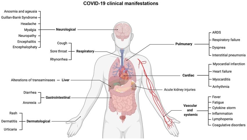
INTERNATIONAL JOURNAL OF MOlecular medicine 47: 100, 2021 5
Figure 2. Timing and type of samples that should be analyzed for the effective detection of SARS‑CoV‑2 RNA or anti‑SARS‑CoV‑2 human antibodies.
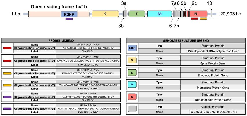
All the types of biological samples used for diagnostic the spike proteins, the envelope proteins, the membrane and
purposes are presented in Table I. A detailed description of nucleocapsid proteins, and a poly‑A tail of 30 bp (49).
the timing of use and the technique for which specific samples At present, several portions of the SARS‑CoV‑2 genome
are collected is also provided (Table I). are used for the design of specific primers and probes,
Overall, the type of sample should be collected considering including the genomic portions encoding for the RdRP gene,
the timing of the infection. Collecting different samples from for proteins constituting the nucleocapsid (N gene) and spike
different sites may be useful to avoid misdiagnosis of asymp‑ molecules (S gene), for proteins of the envelope (E gene), for
tomatic patients negative for molecular tests. Otherwise, it is the membrane, etc. (Fig. 3).
possible to carry out repeated nasopharyngeal swabs in two The CDC has made available a list of approved and
or three consecutive days in order to overcome the window validated kits and reagents to clinically diagnose COVID‑19
period of SARS‑CoV‑2 incubation thus being able to correctly infection (50). Similarly, the CDC has also made available a
diagnose a patient infected with SARS‑CoV‑2 (45). list of three primer pairs that can be used for research purposes
only, each working with two different molecular probes,
3. RT‑PCR‑based molecular tests specific for two portions of the N gene and the gene coding for
viral RdRP, respectively (51).
RT‑PCR‑based molecular methods represent the gold standard RT‑PCR‑based molecular tests are considered the optimal
techniques used worldwide to make a confirmatory diagnosis diagnostic option for wide surveillance strategies due to the
of COVID‑19 infection (46). Since the complete sequencing relatively low costs of the entire viral RNA extraction, reverse
of the SARS‑CoV‑2 genome (26), researchers of different transcription and amplification procedure, and the availability
countries have begun to design molecular primers and probes of RT‑PCR thermal cyclers in hospitals, research institutes and
specific to SARS‑CoV‑2 RNA sequences in order to perform private laboratories (52). Other advantages of RT‑PCR methods
differential diagnosis between COVID‑19 infections and compared to other diagnostic techniques are the timesaving
other pathologies with similar symptoms, such as seasonal of the procedure, the easy execution of the technique and the
flu or bacterial infections (47,48). Of note, the whole genome non‑necessity of highly trained personnel (53). In addition, a
sequence of SARS‑CoV‑2 is 29,903 bp in length containing number of the available RT‑PCR kits are based on one‑step
the following functional elements: A poly‑A cap of 50 bases, amplification methods, where the buffer of the nasopharyngeal
the open reading frame 1/ab (ORF1/ab) containing the coding swab is inserted into the plate and the machine autonomously
sequences for the RNA‑dependent RNA polymerase (RdRP), provides for the extraction, reverse transcription, amplification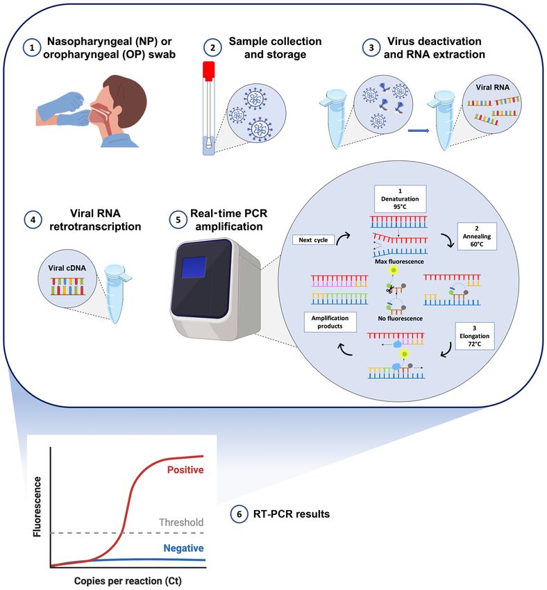
6                                        FALZONE et al: DIAGNOSTIC METHODS FOR COVID-19

              Figure 3. SARS‑CoV‑2 genome structure and probes for the molecular detection of SARS‑CoV‑2 RNA in human samples.

and analysis of the samples. These procedures ensure fast              procedural errors that may occur by analyzing the samples
results guaranteeing excellent reproducibility and standardiza‑        individually (60).
tion of the data obtained which are less influenced by operator            RT‑PCR‑based methods ensure also a low limit of detec‑
bias (54).                                                             tion (LoD) of SARS‑CoV‑2 RNA (61). Specifically, during
    As already mentioned, the EMA has approved 192 PCR-                the early stages of the pandemic, when diagnostic techniques
based methods while the FDA has approved 235 different                 had not yet been optimized and standardized, a significant
molecular tests for both RT‑PCR and the rapid detection of             fraction of COVID‑19‑positive patients were identified as
SARS‑CoV‑2 RNA (17,55).                                                false‑negative due to the low sensitivity of the primers and
    All these approved molecular tests detect two or three             probes used or the inaccuracy of the whole RT‑PCR procedure
fragments of SARS‑CoV‑2 RNA mainly using multiplex                     (false‑negative rates ranging from 38% at the day of symptoms
assays based on standard RT‑PCR protocols. Briefly,                    onset to 67% before one day from the onset of symptoms or to
following the collection of nasopharyngeal swabs from indi‑            66% after 16 days from the onset of symptoms) (62). As will
viduals with suspected COVID‑19 infection, the viral RNA               be better described at the end of this chapter, in the case of
is extracted using commercial kits and lysing solutions with           asymptomatic or paucisymptomatic patients, generally char‑
manual or automated extraction protocols. Subsequently, the            acterized by a low viral load, the RT‑PCR investigations could
obtained RNA, containing both human and viral RNA, can                 mistakenly yield negative results. Currently, the commercially
be directly analyzed by RT‑PCR in the one‑step protocols or            available RT‑PCR kits have partially solved the problem of
must be retrotranscribed into complementary DNA (cDNA)                 the low sensitivity of RT‑PCR. Indeed, the methods available
before RT‑PCR amplification. After obtaining the cDNA, the             today have a theoretical LoD that varies from 0.3 copies/µl to
SARS‑CoV‑2 targets are amplified through RT‑PCR using                  100 copies/µl, depending on the diagnostic system used (63).
TaqMan probes specific for the two‑three viral targets. In             However, it should be considered that this limit is only theo‑
particular, the exonuclease activity of the Taq polymerase             retical; therefore, in clinical practice, procedural errors or
(5'→3' direction) cleaves the probes annealed with the viral           reaction interferers raise the LoD by 10‑fold (64).
targets allowing the emission and real‑time detection of fluo‑             Although RT‑PCR represents the gold standard method for
rescent signals. In this manner, the intensity of the fluorescent      the diagnosis of COVID‑19 infection, this method is subject to
signal is proportional to the total amount of the amplified            several limitations and criticisms that can lead to false‑positive
targets. However, all the existing RT‑PCR protocols are only           or false‑negative results, thus affecting the correct manage‑
qualitative and not quantitative (Fig. 4) (56,57).                     ment of the pandemic. As already mentioned, one of the main
    The entire analytical procedure is completed in 4‑8 h,             limits of RT‑PCR is its low sensitivity in correctly diagnosing
based on the type of RT‑PCR protocol used (one‑step or                 samples with low viral load, including swabs taken incorrectly
two‑steps). This makes it possible to establish the positivity         or obtained from asymptomatic or paucisymptomatic indi‑
of an individual in a relatively short amount of time, allowing        viduals (65). In addition, RT‑PCR is affected by contaminants
the initiation of quarantine protocols that limit the spread of        and interferers contained in the sample or introduced by the
infections (58,59). In addition, the majority of the automated or      operator capable of inhibiting the reaction (65). Other limits
semi‑automated systems available are based on 48‑ or 96‑well           also concern the execution time of the analysis, which in
platforms in order to process a series of samples, thus reducing       non‑automated systems, can take up to 24 h to obtain a result
the execution times, the costs of plastics and consumables and         that can be communicated to the patient. Finally, RT‑PCR is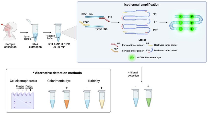
INTERNATIONAL JOURNAL OF MOlecular medicine 47: 100, 2021                                                         7

Figure 4. Schematic workflow of RT‑PCR‑based diagnostic methods. 1) Collection of samples from suspected COVID‑19 patient through nasopharyngeal
or oropharyngeal swab; 2) storage and handling of the swab to preserve viral RNA integrity; 3) SARS‑CoV‑2 heat inactivation and RNA extraction through
custom or commercial protocols; 4) viral RNA retro‑transcription into double strand cDNA; 5) RT‑PCR amplification and real‑time fluorescent signal detec‑
tion; 6) interpretation of amplification signals and setting of the positivity threshold.

a method that is profoundly affected by pre‑analytical and                    the availability of instruments in public and private hospitals
analytical bias regarding the collection, storage and handling                and laboratories. In addition, the RT‑PCR tests currently avail‑
of samples, therefore, careful attention should be paid during                able on the market ensure good sensitivity and specificity rates
the collection and management of samples (66).                                for the diagnosis of COVID‑19 infection. The main RT‑PCR
    In order to reduce the errors in the interpretation of molec‑             diagnostic systems currently available on the market, illus‑
ular tests, the WHO has published specific recommendations                    trating their main technical features are presented in Table SI.
useful to make a correct diagnosis of COVID‑19. In particular,
a molecular test must be conducted in two different biological                4. Rapid antigen and rapid antibody tests
matrices, for example, a nasopharyngeal swab and a fecal
swab, or be performed on two consecutive nasopharyngeal                       With the increase in the number of individuals with a suspected
swabs in order to obtain reliable results. The approved tests                 COVID‑19 infection, it became necessary to adopt more
include the analysis of three different viral genes or two viral              rapid and low‑cost diagnostic strategies to carry out extensive
genes and a human control gene and the use of specific nega‑                  surveillance campaigns (68‑70). To cope with this emergency,
tive and positive controls useful to ascertain the presence of                various rapid tests have been developed to detect viral anti‑
contamination or evaluate the inhibition of the reaction (67).                gens or anti‑SARS‑CoV‑2 human antibodies in salivary, nasal
    Overall, RT‑PCR represents the gold standard and most                     or oropharyngeal swabs and blood samples. These tests are
widely used method worldwide to make an accurate diagnosis                    currently adopted for the frequent monitoring of personnel
of COVID‑19 infection due to the rapidity of the method and                   operating in at‑risk environments such as schools or hospitals
8                                        FALZONE et al: DIAGNOSTIC METHODS FOR COVID-19

or to carry out extensive screening strategies on populations         infection without assessing the viral load. In addition, in the
where a new outbreak of infection is suspected (70,71).               case of rapid antibody tests, it is possible to establish whether
    Compared to RT‑PCR‑based methods, rapid antigenic                 the patient is carrying anti‑SARS‑CoV‑2 antibodies; however,
and rapid antibody tests are characterized by more rapid              it is not possible to establish whether the patient has an active
execution times of ~15‑30 min, a lower cost and an easier             SARS‑CoV‑2 infection or already resolved disease (77,78).
procedure that does not require the presence of highly trained             Overall, among the main advantages of rapid antigen and
personnel (72). These tests are mainly built on platforms             rapid antibody tests are the low cost, the possibility of carrying
based on the principle of lateral flow immunoassay (LFIA)             out the test directly at the point‑of‑care and the high speed
for the direct detection of viral proteins (rapid antigen tests)      and easy execution that ensure a positive or negative result in
or human antibodies against SARS‑CoV‑2 antigens (rapid                ~30 min. However, these tests suffer from important limita‑
antibody tests). As regards rapid antigen tests, these allow          tions mainly related to a low sensitivity and specificity of 56.2
the identification of COVID‑19‑positive individuals through           and 99.5%, respectively (79). Indeed, a recent review of the
the detection of SARS‑CoV‑2 nucleocapsid or Spike proteins            literature demonstrated that rapid antigen tests had a false
(viral antigens) in swabs collected from the upper airways of         negative rate of 27.9%, while no false‑positive results were
the subject with suspected infection. The principle behind            observed (68). As regards rapid antibody tests, a recent study
these LFIA rapid antigen tests is very simple and based on            comparing three different kits demonstrated a false‑positive
the bond between antigens and antibodies that occurs on the           rate ranging from 51.6 to 28.1%, and a false‑negative rate
surface of a porous membrane where the swab buffer flows              ranging from 0 to 4.2% (80).
by capillarity. Briefly, the swab buffer is loaded in the sample           As regards rapid antibody tests, these are very straight‑
well of the cartridge and flows by capillarity at the level of        forward and can be quickly performed, and provide useful
the conjugation pad containing control rabbit antibodies              information about the stage of infection. Indeed, in the case of
and specific antibodies against SARS‑CoV‑2 antigens both              negativity, only the control band is colored. In the case of an
linked to detector molecules (conjugated antibodies). In the          ongoing infection, the IgM and control bands are stained. In
case of a positive sample, the link between viral antigens and        the case of a recent infection, both the IgM and IgG bands are
conjugated antibodies take place at the level of the conjuga‑         stained, since both antibodies are present in the bloodstream.
tion pad. Subsequently, the buffer containing conjugated              Following the remission of the disease, for a period of time
control rabbit antibodies and the antigen‑conjugated antibody         that varies from patient to patient, only the IgG band is positive
complex flows to the test line where other antibodies specific        together with the control one, and in the case of re‑infection,
for viral antigens are immobilized. In the case of a positive         all the three bands are positive again (Fig. S1). The test is
sample, the binding between antibodies immobilized in the             considered invalid when none of the lines stain or when the
test line and the antigen‑conjugated antibody complex gives           test lines are stained but the control line is not (81).
rise to a colorimetric reaction indicating the positivity of the           The limited sensitivity and the related high false‑negative
sample. Finally, the buffer flows further to the control line         results are mainly related to the low viral load and the low
where anti‑antibodies specific for the conjugated control rabbit      antibody response observed in some patients; however, as
antibodies are immobilized; if the test is performed correctly,       already mentioned, the probability of obtaining a positive test
the reaction between these two molecules also gives rise to a         depends also on the time of the presumed infection and on
colorimetric reaction. To be trusted, the test control line must      the test execution time. Indeed, although the viral antigens
always be positive (73,74) (Fig. 5A).                                 are found in the samples after a short time from infection,
    Similarly, rapid antibody tests use the same LFIA                 their permanence and stability in the biological sample are
principle; however, human IgA or IgG and IgM against                  limited; therefore, it is not always possible to correctly identify
SARS‑CoV‑2 antigens are searched for. In particular, the              these proteins (82). Similarly, rapid antibody tests are mainly
blood (or saliva) sample is loaded inside the sample well             designed to identify IgM and IgG antibodies, which are not
and flows to the conjugated pad containing gold‑tagged viral          produced by the body immediately, but begin to be found in
antigens and gold‑tagged control antibodies. In this test,            the bloodstream after the third week of the suspected infec‑
anti‑SARS‑CoV‑2 human antibodies (IgA, IgG or IgM) bind               tion. Therefore, it is important to use the most appropriate test
the gold‑tagged antigens and the sample flows to the test line        considering the time of the presumed infection. More recently,
where anti‑human IgA, IgG or IgM antibodies are pre‑attached          the use of rapid tests for the detection of IgA to accelerate the
to the membrane. In the case of a positive sample, the IgA,           diagnosis of COVID‑19 infection has been proposed (83).
IgG or IgM‑gold‑tagged antigen complex binds to the human                  Overall, rapid antigen and rapid antibody tests are widely
anti‑antibodies immobilized in the test line determining              used for screening strategies on large portions of the popula‑
a colorimetric reaction which indicates the positivity of the         tion (70,84); however, they do not ensure a precise diagnosis of
sample. Finally, the gold‑tagged control rabbit antibodies flow       COVID‑19. Therefore, these tests should be always confirmed
to the control line binding anti‑rabbit antibodies thus giving        by RT‑PCR analyses. Details of rapid antigen and rapid anti‑
rise to a confirmatory colorimetric reaction (75,76) (Fig. 5B).       body tests currently approved by international agencies are
    Both rapid antigen and rapid antibody tests yield results         reported in Table SII.
readable to the naked eye in a very short period of time and can
be performed at the ‘point‑of‑care’ without any specific instru‑      5. Immunoenzymatic serological tests
ments or sample processing. Of note, these tests yield qualitative,
but not quantitative results; therefore, it is only possible to       Most of the immunoenzymatic serological tests used for
establish whether the individual is positive or not for COVID‑19      COVID‑19 investigations are based on the principle of indirect
INTERNATIONAL JOURNAL OF MOlecular medicine 47: 100, 2021                                                            9

Figure 5. Rapid antigen and rapid antibody tests. (A) Analytical workflow of rapid antigen test for the rapid detection of SARS‑CoV‑2 viral antigens through
lateral flow immunoassay. (B) Analytical workflow of rapid antibody test for the rapid detection of human IgA, IgG or IgM antibodies against SARS‑CoV‑2
antigens through lateral flow immunoassay.
10 FALZONE et al: DIAGNOSTIC METHODS FOR COVID-19
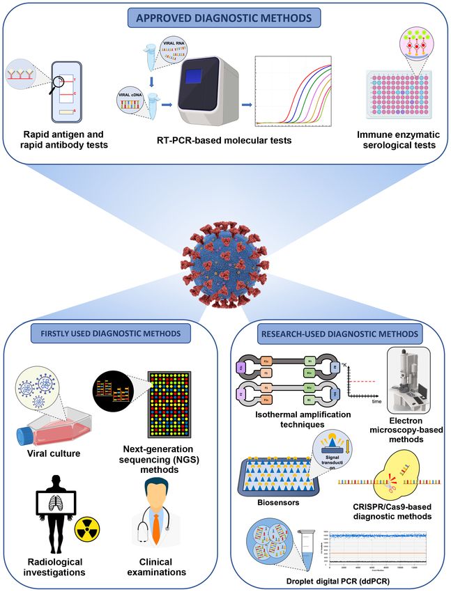
Figure 6. ELISA‑based immune enzymatic serological tests. (A) Analytical workflow of indirect ELISA for the effective detection of human IgA, IgG or IgM
antibodies against SARS‑CoV‑2 antigens. (B) Analytical workflow of sandwich ELISA for the effective detection of SARS‑CoV‑2 antigens.
enzyme‑linked immunosorbent assay (ELISA). Of note, ELISA body specific for human immunoglobulins is added to each
is a colorimetric, chemiluminescent or fluorescent microwell well. Following further washing, a chromogenic (or fluorescent
plate‑based assay used for the quantitation and detection of or chemiluminescent) substrate is finally added and metabo‑
human proteins, immunoglobulins, antigens and other peptides lized in the presence of antibodies against SARS‑CoV‑2 and
through the binding between the target protein and a specific enzyme‑conjugated anti‑human antibodies. This process gives
antibody that results in a detectable signal (85). This technique rise to a colorimetric reaction easily detectable by optical
allows researchers to obtain highly specific and sensitive results densitometry (or fluorescence or chemiluminescence) whose
in a relatively short time ranging from 1 to 5 h (85). intensity is indicative of the quantity of IgG, IgM or IgA anti‑
Briefly, 96‑well commercial COVID‑19 ELISA indirect body presents in the sample (Fig. 6A).
tests contain immobilized viral antigens at the bottom of each Similarly, 96‑well commercial COVID‑19 ELISA
well that are recognized and bound by anti‑SARS‑CoV‑2 sandwich tests contain immobilized antibodies against
antibodies present in the serum of patients following proper SARS‑CoV‑2 antigens at the bottom of each well able to
dilutions. A series of washes is then made to remove the serum bind antigens contained in the serum samples of patients.
and unbounded antibodies; subsequently, a conjugated anti‑ Subsequently, a series of washes is performed to remove theINTERNATIONAL JOURNAL OF MOlecular medicine 47: 100, 2021 11
serum and unbounded antigens and a conjugated antibody COVID‑19 infection or to readapt existing diagnostic systems
specific for SARS‑CoV‑2 antigens is added to each well. according to the characteristics of the new SARS‑CoV‑2 virus.
Finally, following further washing, a chromogenic substrate is When the infection had not yet assumed the dimension of
added and metabolized in the case of positive samples giving a global pandemic, traditional culture methods, clinical inves‑
rise to a colorimetric reaction (Fig. 6B). tigations and NGS techniques were among the first methods
As regards the management of the COVID‑19 pandemic, used to diagnose COVID‑19 infection. Apart from these
ELISAs are currently used for the detection of IgM and IgG approaches, other methods have been developed, including
antibodies (86) specific for SARS‑CoV‑2 antigens or for the biosensors, CRISPR/Cas‑based tests, nucleic acid isothermal
identification of viral Spike proteins (87). More recently, other amplification methods, electron microscopy, etc. (25,95).
ELISAs have been developed for the detection of human IgA
antibodies whose determination is of fundamental importance Viral culture and electron microscopy. Viral culture has
as they are the first antibodies to be produced following expo‑ represented the fundamental method that allows the identifi‑
sure to the virus (86). cation of SARS‑CoV‑2 as a novel causative agent of human
In a very short period of time, several COVID‑19 ELISAs pneumonia (96). Despite the difficult realization and the long
have been developed and some of these have been approved period of times necessary to obtain a viral culture in vitro,
by international agencies (Table SIII). These tests are mainly viral isolates represent a milestone for the discovery of novel
adopted to monitor the immunological status of patients viral infections (96,97). As regards SARS‑CoV‑2 infection,
or for the immunosurveillance of specific work categories, viral culture was fundamental in the initial phase of the
such as healthcare workers or school personnel (88,89). In outbreak prior to the development of other diagnostic assays.
particular, ELISA tests for the detection of IgG and IgM are Zhu et al (96) (2020) were the first to obtain SARS‑CoV‑2 viral
often performed on COVID‑19 patients who obtained a nega‑ isolates from clinical specimens and to observe cytopathic
tive result in molecular tests conducted on nasopharyngeal effects using transmission electron microscopy. Briefly, the
swabs in order to ascertain the seroconversion of patients and authors of that study inoculated 150 µl of BALF supernatant
the acquisition of immunocompetence against COVID‑19 obtained from a COVID‑19 patient into pathogen‑free human
infection (90). Contrariwise, ELISAs for the detection of viral airway epithelial cells. Following 2 h of incubation at 37˚C,
proteins and anti‑SARS‑CoV‑2 human IgA can be used for infected epithelial cells were washed with phosphate‑buffered
diagnostic purposes or for large screening strategies, as these saline and incubated at 37˚C for a long time period. To assess
molecules are rapidly founded in clinical samples (83,91). Of the efficacy of infection and the production of novel viral
note, ELISAs for the detection of viral antigens are based on particles, the authors of that study collected cells supernatant
sandwich ELISA instead of indirect ELISA (92). every two days for molecular analyses and observed cyto‑
Several ELISAs have been approved for the management of pathic effects under light microscopy. Finally, infected cells
COVID‑19 infection, allowing the identification of individuals were prepared for electron microscopy observation (96).
exposed to the SARS‑CoV‑2 virus (Table SIII); however, these Subsequently, following the study by Zhu et al (96), other
tests are not able to confirm the infectious status of the subject. research groups isolated SARS‑CoV‑2 with an aim to study
Indeed, in the case of an ELISA‑positive result (either IgA, the structural features and molecular interaction with infected
IgM, IgG or viral antigens), it is necessary to perform molec‑ cells (98,99). For these purposes, other cell lines have been
ular confirmatory analyses on nasopharyngeal swabs (93). used, including the Vero and LLC‑MK2 cell lines; by using
However, ELISA‑based serological tests are much more reli‑ electron microscopy and cells infected with clinical specimens
able than rapid antigen or antibody tests; the sensitivity and obtained from COVID‑19 patients, it was possible to identify
specificity values of these tests range from 75.6 to 100%, and the ultrastructural details of the virus, the interaction between
85.7 to 100%, respectively, albeit important variations in these virus and cells and the resulting cytopathic effects (100).
values may be related to both the manufacturer or the human Of note, electron microscopy is one of the pioneering
antibody or viral antigen tested (94). Indeed, generally, the methods for the discovery of novel pathogens, allowing the
search for IgG is more accurate compared to that of IgM or identification of their structural features. As regards viral
IgA (this latter is less sensitive) (94). Of note, both sensitivity infections, two main applications of electron microscopy
and specificity depend on the timing of infection and the exist: Solid‑phase immune electron microscopy (SPIEM) and
timing of test execution. immunolabeling electron microscopy (IEM), which are based
Overall, SARS‑CoV‑2 ELISAs represent a good clinical on the observation of cells blocked in the surface of a grid and
option for large screening and surveillance campaigns mainly on the observation of antibody‑antigen complex occurring in
adopted for specific work categories due to the rapidity of this infected cells, respectively (101,102).
method, the possibility of analyzing multiple samples in one Overall, viral culture and electron microscopy are impor‑
round and the availability of automated or semi‑automated tant techniques used to observe the main characteristics of
systems that allow a precise quantitation of viral antigens or the virus. In the case of SARS‑CoV‑2, these two methods
human antibodies. allowed the identification of the typical structure of corona‑
viruses characterized by a nucleocapsid enclosed within a
6. Alternative methods for the effective diagnosis of crown‑like envelope composed of spike proteins. As regards
COVID‑19 infection the cytopathic effects, both methods displayed a broad range
of cellular alterations mainly represented by the formation
The COVID‑19 pandemic has prompted research groups of plaques characterized by a net‑like structure or fused
worldwide to develop novel technologies for the diagnosis of cells. These plaques, composed of multinucleated syncytial12 FALZONE et al: DIAGNOSTIC METHODS FOR COVID-19
cells, also present deformed cilia with a granular formation amplicon‑based sequencing allows the amplification and the
and a disordered polarity. SARS‑CoV‑2‑infected cells also subsequent sequencing of SARS‑CoV‑2 viral RNA. Together,
exhibited double‑membrane vesicles and degraded mitochon‑ amplicon‑based sequencing and metagenomic sequencing
dria. Finally, viral infections also led to the expansion of the are able to correctly diagnose COVID‑19 infection, thus also
endoplasmic reticulum and an increased number of secretory identifying secondary infections due to other pathogens aggra‑
vesicles (96). vating the health status of patients (109).
Overall, these techniques have made it possible to estab‑ As already mentioned, amplicon‑based and metagenomic
lish the main characteristics of the novel SARS‑CoV‑2 virus, approaches based on the sequence‑independent single primer
which then allowed researchers to develop the diagnostic amplification (SISPA) method are able to detect mutations
systems currently used, as well as to propose the first thera‑ occurring in the sequence of SARS‑CoV‑2 and potentially
peutic approaches for the treatment of COVID‑19 infection. associated with vaccine inefficacy or resistance to antiviral
Despite the importance of both viral culture and electron therapies (110,111). Examples of this type of sequencing
microscopy, both methods present some issues that limit their technique are the MinION and IDbyDNA platforms produced
use in clinical settings. Indeed, viral culture is time‑consuming by Oxford Nanopore Technologies and Illumina, respec‑
and requires specific equipment and high biosafety levels. tively (112). As regards the IDbyDNA platform, it ensures the
For these reasons, the CDC recommends SARS‑CoV‑2 viral collection of >13 million reads of which >8 million are unique
culture only for research studies carried out in laboratories reads with an average length of ~75 bp and an in‑depth coverage.
equipped with level 3 biosafety cabinets (103). On the other This shotgun sequencing allows the generation of high‑quality
hand, electron microscopy is not widely used as it requires library score and Q score ensuring the correct identification of
costly instruments and highly trained personnel with specific single variants in the SARS‑CoV‑2 sequence (113).
skills in sample preparation and electron microscopy image Similarly, MinION technology ensures the collection of
analysis. In addition, this technique is characterized by low millions of short and ultra‑long reads (4,000 bp in length)
diagnostic sensitivity and specificity and optimal results can be obtaining output data up to 30 Gb. This technology is based
obtained only if appropriate viral cultures are available (104). on a portable platform that allows the real‑time analysis of
clinical samples with limited costs (114). This technology
NGS. Apart from viral culture and electron microscopy, was effectively used for the analysis of the SARS‑CoV‑2
NGS has represented a key method for the identification of genome by using primers for 16 conserved binding sites of
SARS‑CoV‑2 and for the development of almost all of the coronavirus allowing the reconstruction of the whole genome
currently adopted molecular diagnostic methods. Through of SARS‑CoV‑2 through the generation of 1,000 bp reads with
NGS, it was possible to fully characterize the entire overlapping regions each other (75).
genome of SARS‑CoV‑2, thus establishing that it belongs Overall, NGS whole‑genome sequencing is the most
to the β‑coronavirus genus (49). The de novo sequencing of powerful method for the molecular characterization of
SARS‑CoV‑2 was performed using the nanopore technology SARS‑CoV‑2, for the identification of novel variants during
through a sequence‑independent single‑primer amplification genomic surveillance screening and for the development of
approach (105,106). genome‑based therapeutic approaches (115‑117).
At present, NGS is not used for diagnostic purposes, but
for molecular epidemiology and for the discovery of novel Clinical investigations and imaging techniques. During the
molecular variants. Indeed, its diagnostic application is early stages of the infection, when the causative agent was
limited due to the high costs of the analysis, the requirement still unknown, the diagnosis of COVID‑19 was predominantly
of expensive technologies and the need for highly trained clinical based on the observation of the patient's respiratory
personnel with molecular and bioinformatics skills (25). and extra‑respiratory symptoms and on the use of radiological
Despite the high procedural costs, some companies have imaging techniques (118,119).
proposed commercial tests for the sequencing of SARS‑CoV‑2 Of note, a significant fraction (~50‑75%) of COVID‑19
through NGS platforms. In particular, the commercial kits patients is asymptomatic or paucisymptomatic, presenting
available are mainly based on NGS techniques coupled with mild symptoms for a limited period of time, while other
hybrid capture methods (BioCat, Arbor Biosciences and Swift patients (~10%) present severe respiratory symptoms resulting
tests). These platforms are built with biotinylated RNA probes in acute respiratory distress syndrome (ARDS) responsible for
that hybridize SARS‑CoV‑2 RNA fragments. Subsequently, dyspnea, interstitial pneumonitis, multiorgan dysfunctions and
the biotinylated probes are amplified through PCR using in some cases, even death (120,121). Such severe manifestations
streptavidin‑coated beads (107,108). are often observed in patients with pre‑existing comorbidities,
Apart from these platforms, more complex NGS tools such as diabetes, cardiovascular diseases, hypertension and
have been developed to detect mutations in the sequence cancer, and are mediated by specific host cell entry media‑
of SARS‑CoV‑2 genome, thus identifying novel emerging tors (122‑126).
strains important from an epidemiological point of view Among the COVID‑19 symptoms, there are not only
and for the development of novel vaccines. Among these respiratory manifestations, but also other systemic symp‑
tools, amplicon‑based metagenomic sequencing represents toms. According to a recent comprehensive review, the main
the most powerful approach with which to rapidly identify respiratory symptoms include, but are not limited to dyspnea
and comprehensively characterize SARS‑CoV‑2 and other (19‑64%), cough (69‑82%), rhinorrhea (4‑24%) and a sore
pathogens. In particular, metagenomic sequencing allows the throat (5‑14%). Other common symptoms are fever (44‑98%),
identification of the normal microbiome of patients, while headaches (5‑14%) and diarrhea (2‑5%) (127). However, theINTERNATIONAL JOURNAL OF MOlecular medicine 47: 100, 2021 13
Figure 7. Respiratory and extra‑respiratory clinical manifestations of COVID‑19 infection.
most common symptoms reported by patients with COVID‑19 Overall, COVID‑19 symptoms are mild in 80‑90% of
are anosmia and ageusia, together with fatigue (128,129). positive individuals; however, a small fraction of patients
Apart from these commonly reported symptoms, a plethora experiences severe symptoms that require hospitalization.
of clinical manifestations has been reported, ranging from Approximately 5% of patients develop interstitial pneumonia
cardiovascular to neurological disorders and from gastrointes‑ associated with respiratory failure, cytokine storm and multi‑
tinal to systemic symptoms (Fig. 7). In particular, SARS‑CoV‑2 organ failure that could lead to patient death (127,136).
exhibits a neuroinvasive behavior via the retrograde trans‑ Therefore, the careful clinical evaluation of all these symp‑
synaptic invasion of the central nervous system. SARS‑CoV‑2 toms, together with radiological and laboratory data, helps the
is able to bind specific receptors in the cells constituting the clinicians to correctly diagnose COVID‑19 infection, and thus
olfactory bulb or pulmonary and airways sensorial receptors. to the timely commencement of effective therapeutic proto‑
Among the most frequent neurological symptoms, there are cols (24).
headaches, neuropathy, myalgia, encephalitis, encephalopathy, As already mentioned, radiological investigations help
etc. (130). clinicians to correctly diagnose COVID‑19 infection in the
Other common symptoms are related to systemic inflam‑ case of a suspicious case of pneumonia. Among radiological
mation responsible for the alteration of coagulative and imaging techniques, chest X‑ray (CXR) and computed tomog‑
hematological parameters and for the so‑called ‘cytokine raphy (CT) are the most powerful methods for the diagnosis of
storm’ observed in patients with COVID‑19 with severe COVID‑19 pneumonia (137,138).
respiratory symptoms. In fact, inflammation leads to the CXR is usually used for the detection of pulmonary abnor‑
increase of fibrin/fibrinogen debris and D‑dimer associated malities following lung injuries due to infectious or neoplastic
with coagulopathy. In rare cases, such alterations may induce diseases (139). During the first phase of the COVID‑19
disseminated intravascular coagulopathy that requires antico‑ outbreak, CXR was widely used to detect multifocal opacities
agulant prophylactic or curative treatments (131). In addition, affecting mainly lung interstitial space and alveoli in patients
inflammation is also responsible for lymphopenia and T‑cell with COVID‑19‑related symptomatology (140). In particular,
exhaustion (132). Strictly related to these hematological altera‑ CXR is mainly adopted for patients suspected of COVID‑19
tions are cardiovascular disorders, such as acute myocardial infection with moderate or severe symptomatology who
injury, coronary syndrome, cardiomyopathy, myocarditis, usually exhibit interstitial opacities (71.7%), or alveolar opaci‑
arrhythmias, etc. (133). ties (60.5%) frequently affecting both lungs (64.5%) (141).
Among gastrointestinal disorders, COVID‑19 infection These radiological findings become more severe over time
is responsible for nausea, vomiting, weight loss, anorexia and with the progression of symptoms and are mostly found in
more frequently, diarrhea. Among these symptoms, hepatic elderly patients with previous pulmonary parenchyma altera‑
manifestations mainly represented by increasing levels of tions (such as patients with chronic obstructive pulmonary
transaminases are also included (134). Finally, other symptoms disease) who exhibit both bilateral interstitial and alveolar
affect the kidneys, skin, endocrine system, etc. (135). opacities (142).14 FALZONE et al: DIAGNOSTIC METHODS FOR COVID-19
Despite the low cost and the rapid radiological findings As regards electrochemical systems for viral RNA detec‑
obtained through CRX, some pulmonary abnormalities are tion, Zhao et al (155) (2020) developed an ultrasensitive
not clearly detectable through this technique. Therefore, electrochemical biosensor built with calixarene functionalized
besides CRX, CT scan is frequently adopted to better display graphene oxide containing specific probes for SARS‑CoV‑2
lung abnormalities mainly represented by bilateral interstitial RNA. This system allows the detection of viral RNA
ground‑glass opacities (143). In particular, the CT scan has a without nucleic acid amplification systems through the use
great resolution power with a sensitivity of ~95‑100%, although of capture probes and label probes specific for SARS‑CoV‑2
the specificity is very low, as this method does not allow for RNA that through a calixarene substrate are able to detect
the discrimination of pulmonary alterations associated with electrochemical mediators, including toluidine blue and gold
other etiological agents other than SARS‑CoV‑2 (144). nanoparticles, generated by the binding between probes and
The CT scan has been demonstrated to be an important the viral RNA. The device is portable and the analysis of
technique for the diagnosis of asymptomatic patients with data can be performed on smartphone apps as a point‑of‑care
COVID‑19 negative for molecular tests due to low viral testing (155).
load (145,146). In fact, the CT scan is able to discriminate the Several other types of biosensors have been produced;
presence of morphological abnormalities in the lung already however, their detailed description is beyond the scope of
during the early stages of infection; however, this technique the present review. Overall, these biosensors exhibited a
is not effective in correctly diagnosing a COVID‑19 infec‑ sensitivity ranging from 86.43 to 93.75%, and a specificity of
tion characterized by the absence of respiratory symptoms or 90.63‑100% depending on the platform used (156). As already
where there is no involvement of the lung parenchyma (147). mentioned, biosensors present several advantages mainly
Other limits of the CT scan are represented by the needs represented by high sensitivity and specificity, low costs of
of two radiologists' evaluations and the risk of cumulative analysis, rapid execution time, the optimal LoD and the possi‑
radiations. This latter issue is addressed through the use of bility of developing miniaturized platforms used directly in
low‑dose CT scan or ultra‑low‑dose CT scan often used for the point‑of‑care.
the long‑term monitoring of COVID‑19 patients with severe
respiratory symptoms (148). Loop‑mediated isothermal amplification (LAMP) COVID‑19
Overall, the radiological hallmarks of COVID‑19 infection testing methods. LAMP systems are among the most commonly
are the bilateral interstitial ground‑glass opacities that may used alternative nucleic acid amplification methods for the
also affect the peripheral areas and the alveolar parenchyma. diagnosis of infectious diseases. These techniques are charac‑
These alterations together with clinical symptoms, lead clini‑ terized by an easy execution of the assay that does not require
cians to perform a diagnosis of COVID‑19 infection. specific equipment, a rapid and highly sensitive detection of
targets and an easy interpretation of results (157). As regards
Biosensor COVID‑19 testing techniques. To effectively SARS‑CoV‑2, several RT‑LAMP systems have been developed
diagnose COVID‑19 infection rapidly and directly at the for the effective detection of viral RNA, highlighting how
point‑of‑care, several electrochemical biosensing systems these methods can be coupled with other diagnostic techniques
have been developed. These biosensor‑based tools use including NGS, digital detection systems, biosensors, etc. (158).
the principle of impedance and electrochemical reactions The SARS‑CoV‑2 RT‑LAMP detection systems are based
occurring when viral RNA or proteins bind specific probes on autocycling strand displacement DNA synthesis‑mediated
or antibodies. Different types of biosensor platforms are Bst DNA polymerase and six primers, which work at isothermal
currently available for the diagnosis of COVID‑19. These temperatures ranging from 60 to 65˚C, thus avoiding the use of
include electrochemical biosensors, colorimetric biosensors, a costly PCR thermocycler. RT‑LAMP amplification is able to
fluorescence‑based biosensors, surface‑enhanced Raman scat‑ amplify RNA fragments up to 106‑109 copies in ~30‑60 min.
tering (SERS) biosensors, quartz crystal microbalance (QCM) The final amplification products can be visualized by agarose
biosensors, localized surface plasmon resonance (LSPR) and gel electrophoresis, by fluorescence labeling, turbidity, or colo‑
other platforms that ensure the accurate and fast detection of rimetry for an immediate readout of the data obtained and a
SARS‑CoV‑2 particles (149‑152). prompt diagnosis of COVID‑19 infection (Fig. 8).
Among these platforms, the most commonly used are elec‑ Overall, SARS‑CoV‑2 RT‑LAMP tests have a high sensi‑
trochemical biosensors and SERS adopted as point‑of‑care tivity and specificity and some of these have been approved
platforms due to the limited size of instruments, the low cost for diagnostic purposes by different national and international
and the easy execution of the procedure (153). agencies. Some studies have reported a sensitivity of ~100%
Electrochemical biosensors can be used for the detection for the detection of ORF1ab SARS‑CoV‑2 gene, highlighting
of SARS‑CoV‑2 proteins or for the detection of viral RNA. a higher diagnostic accuracy compared to other validated
Seo et al (154) (2020) described an innovative biosensor for the RT‑PCR diagnostic methods. In addition, the specificity rate
detection of a low concentration of SARS‑CoV‑2 spike protein is higher than that obtained through RT‑PCR, as the use of
(LoD, 1 fg/ml) built with a graphene sensor with immobilized more than six primers in the RT‑LAMP ensures the correct
anti‑SARS‑CoV‑2 spike antibodies. This type of field‑effect diagnosis of SARS‑CoV‑2 infection (25).
transistor biosensor (FET) allowed the effective identification Among the most commonly used RT‑LAMP methods there
of SARS‑CoV‑2 in different types of samples (PBS, transport is The ID NOW™ COVID‑19 assay (Abbott Laboratories),
medium, viral culture medium, etc.) through the evaluation which has been approved by FDA with Emergency Use
of electrical performance after S protein‑antibody interaction Authorization (EUA). This RT‑LAMP‑based system ensures
suggesting its application in clinical settings (154). high‑sensitive results in ~5 min through the identification ofYou can also read