Scoliosis Round Table: What are Your Optimal Surgical Strategies for a Double Major Curve in Adolescent Idiopathic Scoliosis? - jposna
←
→
Page content transcription
If your browser does not render page correctly, please read the page content below
Panel Discussion

Scoliosis Round Table:
What are Your Optimal Surgical Strategies for a
Double Major Curve in Adolescent Idiopathic Scoliosis?

Matthew E. Oetgen, MD, MBA1; Stuart L. Weinstein, MD2; Lindsay Andras, MD3; Suken Shah, MD4;
Daniel J. Sucato, MD, MS5
1
 George Washington University School of Medicine, Washington DC; 2University of Iowa Hospitals and Clinics, Iowa City,
Iowa; Keck School of Medicine, University of Southern California, Children’s Orthopaedic Center, Children’s Hospital
Los Angeles, Los Angeles, CA; Nemours/Alfred I duPont Hospital for Children, Wilmington, DE; Texas Scottish Rite
Hospital, University of Texas Southwestern Medical Center, Dallas, TX

Introduction                                                    Additionally, while understanding that the technique of
Adolescent idiopathic scoliosis (AIS) is a common               deformity correction is important, recent investigations
condition seen by pediatric orthopaedic surgeons. While         into achieving maximum quality and safety in AIS
the general indications for surgical treatment have             surgery have shown standardization of the care pathway
changed very little over the past few decades, the              is likely of equal importance in achieving the best
methodology has. This is due to a greater understanding         outcomes for patients.1-3 Yet one technique may not be
of the biomechanics, advances in surgical technique and         optimal for all types of deformity correction and
instrumentation, and a more robust understanding of the         selective implementation of different methods defines
goals and outcomes of surgical intervention.                    the “art” of surgery.

When surgical intervention is indicated, posterior spinal       The discussion and sharing of differences in surgical
fusion remains the gold standard for the treatment of           planning, approach, and technique by experts is a
adolescent idiopathic scoliosis with the aims:                  powerful way to learn new insights into methods of
                                                                treatment. The goal of this roundtable is to present a case
1. Correction of deformity while maintaining good               of a patient with AIS and to discuss different surgical
coronal and sagittal balance with as much flexibility as        approaches from a group of experts and to learn from
possible.                                                       their experience in treating AIS.*
2. Fusion of the spine to prevent future deformity
progression in a safe, complication free process.               *This report summarizes key points from each panel
                                                                member, and where similar concepts were discussed by
Despite these relatively simple objectives, the optimal         multiple panel members, this is noted. A complete
technique to achieve these goals is hard to define.             transcript of these valuable pearls and pitfalls are
Different strategies exist for preoperative planning,           provided in Appendix 1. This discussion is extremely
instrumentation, and deformity correction.                      thorough and valuable for those desiring a nuanced
                                                                description.

Copyright @ 2020 JPOSNA                                     1                                                 www.jposna
JPOSNA
Volume 2, Number 1, May 2020
Invited Experts
Stuart L. Weinstein, MD Lindsay Andras, MD Suken Shah, MD Daniel J. Sucato, MD, MS
Ignacio V. Ponseti Chair Assistant Professor of Division Chief, Spine and Chief of Staff, Texas
and Professor of Orthopaedic Surgery Scoliosis Center, Clinical Scottish Rite Hospital
Orthopaedic Keck School of Medicine, Fellowship Director, Professor, Department of
Surgery/Professor of University of Southern Nemours/Alfred I duPont Orthopaedic Surgery,
Pediatrics, University of California Children’s Hospital for Children, University of Texas
Iowa Hospitals and Clinics, Orthopaedic Center, Wilmington, DE Southwestern Medical
Iowa City, Iowa Children's Hospital Los Center, Dallas, TX
Angeles, Los Angeles, CA
Case
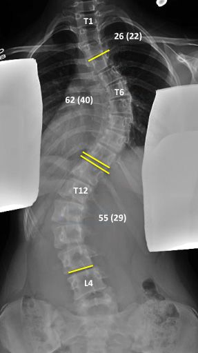
13+9-year-old girl who is 4 months post-menarchal who presents for a second opinion regarding her spinal
deformity. She was originally diagnosed at age 11 with a right thoracic deformity of 29° and a left
thoracolumbar deformity of 21°. She was noncompliant with a brace and now presents with progression of
her deformity. Current radiographs show an upper left thoracic curve of 26° that bends to 22°, 62° right
thoracic that bends to 40°, and 55° left lumbar curve that bends to 29°. She is a Risser 2. (Figures 1-2)
Figure 1a, 1b and 1c. Presenting PA and Lateral Radiographs Figure 2. Unbending Films
Copyright @ 2020 JPOSNA 2 www.jposna
JPOSNA
Volume 2, Number 1, May 2020

Case Questions and Panel Discussion                            3. What type of preoperative imaging do you
                                                               typically obtain?
1. Has this child met indications for surgical
                                                               All our panel members agree that MRIs are not routinely
treatment?
                                                               obtained. Instead, they are reserved for specific
Drs. Shah and Sucato summarize the consensus from the          indications such as: neurological findings, kyphosis at
panel.                                                         the apex, atypical curve, pain, rapid progression, early
                                                               onset, and left sided curve (SS).
SS: Yes. She has progressive scoliosis >50 degrees, is
skeletally immature, and is mildly symptomatic.                All our panel members have similar thoughts on what
                                                               images to obtain as Dr. Sucato writes.
DS: Yes. The absolute indications for surgery in AIS are
those patients whose curve will continue to progress           DS: All surgical patients get an AP and lateral
despite skeletal maturity. This generally means thoracic       radiograph using the EOS technology to decrease the
curves greater than 50 degrees and                             amount of radiation as well as to have the potential to
thoracolumbar/lumbar curves greater than 40 or 45              measure the three-dimensional deformity. Two-view
degrees. For the case presented here, the risk for curve       supine best-bend radiographs are also obtained and are
progression is exceptionally high, and surgery is              used to help classify the patient using the Lenke
indicated.                                                     classification which ultimately predicts those curves,
                                                               which potentially should be included in the fusion.8
2. How would you approach this patient in terms of a
preoperative evaluation?                                       LA: I agree, but we have noted if it doesn’t look as
                                                               flexible on XR as what you appreciate clinically, then
All our panel members have a similar basic approach to         consider assisting on a repeat film.
preop evaluation as summarized by Dr. Weinstein.
                                                               SS: I would add traction/pull films for all curves ≥ 80
SW: Our preoperative workup would include a complete           degrees. I would like to do all imaging in the EOS, but
physical examination and blood work, as well as a type         I’m not convinced erect benders give you a reliable
and screen. Patients are given the opportunity to              picture.
complete standardized health-related quality of life
(HRQOL) surveys prior to their initial visit to our            4. How do you classify these deformities, and how
service, either through our online patient portal or           do you use this classification to begin your surgical
during the check-in process.                                   planning?

LA: I agree but would add that for curves over 70              All our panel members use the Lenke System with some
degrees, they have a cardiology evaluation/echo to             caveats.
evaluate for pulmonary hypertension and a pulmonary
                                                               LA: The Lenke classification remains the preeminent
evaluation, which includes PFTs. Patients with
                                                               classification system both in our practice and
significant preoperative pain concerns are referred to
                                                               worldwide. I think some of the work on 3D modeling and
psychology or our pain team. (This pain and
                                                               classification is exciting but not quite to where I am
psychological evaluation are described in the Appendix
                                                               using it in everyday practice.12
by Dr. Sucato.)

Copyright @ 2020 JPOSNA                                    3                                               www.jposna
JPOSNA
Volume 2, Number 1, May 2020

DS: AIS curves that are indicated for surgical treatment         2. Look at the sagittal profile and determine if that
should be assessed using the Lenke classification.               makes any of the minor curves structural (this is one of
Ultimately, the radiographic classification of each              the points of the Lenke classification that people
patient needs to be reconciled with the physical                 sometimes forget to pay attention to; for example, if the
examination of the patient to ensure that the appropriate        T2 to T5 kyphosis is more than 20 degrees then the
curves are included in the fusion. In general, it is             proximal thoracic curve is structural and you will
important to look for opportunities to preserve motion           develop imbalance if you don’t include it).
segments since the long-term health of the spine is
dependent in general, on two aspects: balance and                3. I draw the posterior sacral vertical line to determine
motion.                                                          the stable sagittal vertebra line and know I shouldn’t
                                                                 plan to end my construct at a more proximal vertebral
5. How do you do your surgical planning for this type            body based on the PA radiographs.
of surgery?
                                                                 Then I switch over to the PA and bending views. From
Our panel emphasizes the importance of sagittal plane            these, we can determine that both the main thoracic
assessment, shoulder balance on radiographs and                  (major) and lumbar curves are structural (this should
physical examination, and preservation of lumbar                 also coincide with our clinical exam), but the upper
motion.                                                          thoracic is not (bends out to less than 25), so from that I
                                                                 conclude that I would include both main thoracic and
SW: I start by looking at the sagittal plane to determine        lumbar curves. Since the upper thoracic curve is not
whether the patient has hypokyphosis and what I ideally          structural, then I usually go by T4 for high right
would like to achieve in sagittal plane correction or            shoulder (which this is by clinical description though it
restoration. On both the standing PA and supine AP, I            is subtle on radiographs), T3 for level shoulders, T2 if
identify a perpendicular to the sacrum to identify the           the left is high. For the LIV, typically, you would use the
center sacral line to determine the touched, substantially       vertebrae just touched by the center sacral line, but this
touched, neutral, and end vertebra. I next draw a line           is one area where we frequently “break the rules” and
connecting the superior aspects of the acromion to get           especially with L3 vs. L4. We will “work hard” to end
an idea of shoulder tilt and also the angle of the T1            at L3 and may have some significant potential benefit
superior endplate and superior aspect of the first ribs. I       from doing that.
do the same measurements on the supine film. My goals
of surgery are to level the shoulders, correct as much of        SS: I start with a detailed exam of the radiograph, with
the rotational deformity as possible, and balance the            special attention to the lateral and 3D views reproduced
spine both in the coronal and sagittal plane. Distally, my       using the EOS system. The areas I concentrate on are
goal is to spare as many lumbar segments as possible in          the rib hump offset, the need for thoracic kyphosis
achieving correction and balance.                                correction, and preservation of lumbar lordosis based
                                                                 on radiographic pelvic incidence. Once that is done,
LA: “Start with the lateral” has been a mantra at our            make sure you compare the radiographic deformity and
institution. This way, you make sure you don’t forget to         that of the patient’s clinical appearance (often photos of
consider it. For me, this has three components:                  the patient from the clinic are helpful). I then determine
1. Check for spondylolysis/spondylolisthesis, which can          the UIV based on the shoulder appearance. Similar to
be seen in 10-15% of AIS patients.                               Dr. Andras, I include T3 if a large main thoracic
                                                                 correction is planned. I try to pick the LIV as the last

Copyright @ 2020 JPOSNA                                      4                                                 www.jposna
JPOSNA
Volume 2, Number 1, May 2020

touched by center sacral vertical line (LTCSVL), but            DS: The use of posterior column (Ponte) osteotomies in
almost never to L4.                                             AIS is somewhat controversial and, in general, I employ
                                                                the same strategy as screws in that they are only
DS: The initial steps to surgical planning are first to         necessary to properly dose the amount of correction
determine which curves require inclusion in the                 needed for the deformity.
arthrodesis and then to decide which specific vertebral
levels to instrument and fuse. The physical examination         7. How do you optimize patient positioning in the OR
is the most important aspect of determining which curves        to help with correction?
require surgical treatment and includes an assessment of
shoulder elevation, coronal balance, rotational                 SW: The key for me is to position the patient in a way
deformity of the potential curves, and waistline                that promotes normal standing posture. This helps me
asymmetry. The radiographs should then be assessed,             ensure that I have good coronal and sagittal balance
and a comparison of these images to the physical                and that my films are taken in the appropriate position.
appearance of the patient is then made. In general, the         DS: The hip pads should be at the level of the anterior
physical examination of the patient supersedes the              superior iliac spine, and I most often place them more
radiographs if there are any discrepancies noted. For           distal, especially for those patients in which we are
example, if the left shoulder is elevated despite the           instrumenting into the lumbar spine. In this way, the hips
radiographs not demonstrating a structural PT curve             can be extended to improve lumbar lordosis when more
(because the curve bends to less than 25 degrees), it is        is desired.
important to include the PT curve to ensure that
shoulder balance will be achieved following surgery.            SS: We use cranial tongs for bigger curves and traction
                                                                when necessary. I will adjust the chest pad to aid with
6. How do you determine the need for osteotomies,               thoracic kyphosis restoration, sometimes even adjusting
and how do you decide where to place these if                   intraoperatively before rods go in. (Dr. Andras
needed?                                                         describes a similar approach to positioning.)
SW: I do complete inferior facet joint excision, remove         8. What are your tips and tricks for
the spinous processes to the level of my inferior facet         blood management in the operating room?
excision, and thin the ligamentum flavum significantly at
each level to allow maximal mobility. If, however, the          Drs. Andras, Shah, and Sucato employ controlled
curve is extremely rigid or very large (greater than 75         hypotensive protocols of 60 mmHg for exposure, ~75
degrees), then I consider using osteotomies. Ponte              mmHg for correction through completion.
osteotomies add to the potential blood loss and
hematoma formation and the increased risk of                    SW: Over the years, I have gone away from using any
neurologic deficit, so hence, I do them when necessary          hypotensive anesthesia and currently just keep the
but not routinely.                                              patient’s normotensive, normothermic, and rely on
                                                                meticulous surgical dissection techniques and as short
LA: For me, the debate of whether or not to do Ponte            as possible surgical times to prevent blood loss. We use
osteotomies is a “when and how” not “if” question.              tranexamic acid (TXA) preoperatively with a 50 mg/kg
                                                                bolus and then 10 mg/kg continuous infusion until the
SS: Always, as the principle is to mobilize the spine.          wound is closed and dressings were applied.22 (Drs.
(Drs. Andras and Shah use ultrasonic bone scalpel for           Andras and Shah also note TXA, SS with 30mg/kg bolus
facetectomies and Pontes to limit blood loss.) 23               and a 10mg/kg infusion.) I have not used the cell saver
                                                                in over 25 years.

Copyright @ 2020 JPOSNA                                     5                                                www.jposna
JPOSNA
Volume 2, Number 1, May 2020
SS: Cell saver is used for all cases except anterior and SS: With differential rod bending, only one set screw is
selective thoracic fusions. tightened to keep the rod properly oriented in the
sagittal plane while the other rod is implanted. Then, set
9. What is your standard deformity reduction screws are tightened segmentally as axial plane is
technique for this type of deformity? corrected via segmental direct vertebral rotation. I also
Our panel emphasizes the importance of differential rod work hard to balance the LIV with
bending as described by Dr. Weinstein and the use of compression/distraction while simultaneously de-
pedicle screw derotation described by Drs. Andras and rotating the LIV to neutral if it is not spontaneously so.
Sucato. Look at LIV+1 – does it look perfect? If not, it won’t
look any better when she stands up, so get it right in the
SW: I would be placing the left-sided rod first, OR!
contouring the rod as I would like to see it in the sagittal
plane, restoring kyphosis in the thoracic spine. I would DS: I prefer to start this process by placing derotators
be capturing the rod at every level that is instrumented. I on the convex side of the lumbar spine to correct and
would first derotate the lumbar spine with the uniplanar derotate the lumbar curve followed by a temporary
screws, which I use in this region. I then would put right-sided lumbar rod. Now the lumbar curve is
multiple screwdrivers in the apical thoracic vertebrae partially corrected, and the left rod can be placed
screwheads and derotate the spine and use the reduction engaging the rod partially in the left lumbar spine while
screws on the concave side to help derotate the spine engaging only the top screw(s) of the thoracic curve
and pull the spine to the rod to create additional leaving the overcontoured rod posterior to the apex of
kyphosis in the thoracic spine. In the right-sided rod, I the thoracic spine. The temporary right lumbar rod is
tend to contour the thoracic spine with minimal kyphosis removed, and correction of the spine using the left rod
as I tend to use this rod to “push down” and derotate the begins with the apex of the thoracic curve pulled to the
ribs on the curve convexity. Similarly, I bend the lumbar rod with reducers and the lumbar curve corrected as
segment a bit less with lordosis to help derotate the partial rod rotation to complete the axial plane
lumbar spine further. Once the main thoracic and correction. In-situ bending of the rods in the coronal
lumbar curves are stabilized, I do my shoulder leveling plane provides opportunities to improve correction and
by distraction across T3 on the left with a T2 hook generally are performed at the apex and prior to
loosened to allow me to push up on the left shoulder. compression-distraction maneuvers.
Then I distract at T2 on the right relying on 10. How do you judge your correction in the
ligamentotaxis to balance the shoulder on that side. operating room?
Finally, I compress the T2-3 “claw” on the left to secure
these hooks. I check balance with the fluoroscopy SW: I judge my correction through intraoperative
looking for horizontalization of all the cervical vertebrae fluoroscopy as mentioned above, or on occasion 72-inch
and the upper thoracic vertebrae, and then I check with films taken in the operating room, but again, I find this
fluoroscopy distally looking for horizontalization of the rarely necessary. I make my decisions about whether I
distal portion of the spine. need to do more or less well before this point in the
surgery. I make continual assessments along the way,
LA: In addition to aggressive differential rod bend never at the end. I may take a quick fluoroscopic view if
techniques and vertebral column rotation at the apex of I have any concerns.
the thoracic and lumbar curves, I follow that by fine-
tuning with compression and distraction to balance the
UIV and LIV.
Copyright @ 2020 JPOSNA 6 www.jposnaJPOSNA Volume 2, Number 1, May 2020 LA: We have a T-square that I center first on the hips to make sure the upper portion is traveling through the center of T2, so I know coronal balance has been achieved.27 Then I flip it around and center it on the coracoid processes to judge the shoulder balance. DS: Every spine deformity surgery at our institution has a 3-foot film obtained from an overhead-mounted X-ray machine in the OR. 11. What is your immediate postoperative patient protocol? Our panel has worked to develop institutional rapid recovery protocols that get patients home 2-4 days postoperatively (Please note the CHLA Intrathecal Injection Technique in this edition of JPOSNA). SS: We were among the first to use gabapentin and Toradol to decrease morphine equivalents, so we have lots of experience with rapid recovery pathway.28 Now, on top of that, we use a clonidine patch, get on oral pain Figure 3. Postoperative radiographs of T4 to L3 medicine on POD 1 with oxycodone, Tylenol, and reveal excellent sagittal and coronal balance with level valium. The patient sits up in bed in PACU and typically shoulders and a horizontal LIV within the stable zone. is admitted to the floor the first night, with something to drink. To advance mobility, we expect the patient to be 12. What is your longer-term activity protocol? out of bed to the chair twice on POD 1 and walk in the SW: We release them to full unrestricted activities at 6 hall on POD 2, with stairs should be cleared by the end months postop. For male patients I generally do not of POD 2 or 3. With this protocol, we have been able to recommend tackle football or competitive wrestling (no achieve an average length of stay of 2.7 days. data to support these restrictions just my intuitive feeling DS: Our patients have an epidural catheter placed at the of too much risk). time of surgery with administration of rupivicaine, LA: We agree that there is likely some increased risk of together with continuous intravenous dexmedetomidine spine injury with participation in contact sports. In the (Precedex®) without narcotics except for Dilaudid prn. absence of level 1 data on this subject, we all have to The patient is given oral meds and, if tolerated, the share that theoretical risk and balance it against the epidural is removed at 11 am, together with the arterial known benefits of sports participation. line and Foley catheter. The patient is in a chair for 1 hour, back to bed, and then up walking laps 2 hours DS: We restrict contact sports for 6 months for any later. Walking is done three times per day, and in fusion into the lumbar spine. For our selective thoracic patients with a thoracic fusion, only the patient is fusions, we allow full activities without restrictions at 6 usually discharged the second postoperative day. If the weeks. Patients are seen back at 1 year from surgery fusion extends into the lumbar spine, the patient is unless there are concerns by the family. usually discharged on POD 2 or 3. Copyright @ 2020 JPOSNA 7 www.jposna
JPOSNA
Volume 2, Number 1, May 2020

Conclusion                                                         accomplish my ultimate goals if I can’t get a pedicle
As can be appreciated by the panel discussion, many                screw in place, how I can use an occasional hook or even
different successful techniques exist for performing               go back to a more “ancient technique” called the three-
posterior spinal fusion for AIS. While differences exist           rod technique popular for big curves in the Cotrel-
in the details (the need for osteotomies, implant density,         Dubousset days. I think it is very important in children
implant type, reduction maneuver, even follow up                   for spine surgery to be facile with the use of corrective
imaging and schedules), what can be appreciated in the             techniques e.g. using hooks as a fall back for some
comments of all of these experts is that the goals of              uncomfortable situations. I never plan to extend my
spinal balance, achieving a lasting fusion, and avoiding           levels because of blown pedicles so particular care must
complications during and after surgery are universal.              be taken with screws distally.
Detailed preoperative planning, meticulous surgical                4. Shoulder balance is key. I think it is critical for all
technique, and open and honest communication with                  pediatric spinal deformity surgeons to develop
families, are the keys to success in pediatric spinal              techniques and have an understanding of the spine such
deformity surgery.                                                 that one is always able to achieve shoulder balance.
                                                                   Shoulder imbalance is, in my experience, the one
The panel provides their                                           deformity that patients and families are most unhappy
Keys to Success                                                    about, much more so than residual rib prominence.

                                                                   5. Know your implant system. Each of the implant
Dr. Weinstein                                                      companies patent their tools and implants. Rods and
                                                                   screws vary from company to company, and just because
1. Loosen the spine. I always do complete facetectomy
                                                                   you are an expert with one system does not mean you
in the lumbar spine and 90% removal of the inferior
                                                                   can rapidly gain that expertise using another company’s
facet in the thoracic spine. I also remove the spinous
                                                                   system. Rods may have different modulus of elasticity,
process back to the level of resection of the inferior facet
                                                                   even within the same company. Screws have different
in addition to thinning the ligamentum flavum with my
                                                                   thread pitches, and different pull out strengths, and
“fluted” Midus Rex burr to get as much mobility
                                                                   patients are different with respect to bone quality.
between segmental levels.

2. Maximize screw size. I try and use the largest pedicle
                                                                   Dr. Andras
screw size that I think the patient can tolerate, as I
                                                                   1. Keep your team informed. Email your team the
believe this gives better control during correction of the
                                                                   week/weekend before and include your surgical plan for
deformity.
                                                                   levels, implants, and any other equipment needed, as
3. Be flexible with your implant plan. I tend not to               well as any pertinent information about the patient (i.e.
spend too much time trying to cannulate pedicles, which            MRI negative for intraspinal pathology, no pulmonary
are extremely small and thin. As I do all my screw                 hypertension on echo).
placement by the freehand technique, if I cannot
                                                                   2. Think power. Power pedicle screw placement (and
penetrate and cannulate the pedicle quickly, then I tend
                                                                   tract preparation) is really helpful for both patient and
to skip it and move to the next level proximally. I also,
                                                                   surgeon preservation.
as mentioned above, place my screws distal to proximal
always thinking about “plan B” so I am very cognizant              3. Spread the force. Aggressive differential rod bend
of the fact by viewing the preoperative X-rays how I can           and lots of serial reducers to share the load.

Copyright @ 2020 JPOSNA                                        8                                                 www.jposna
JPOSNA
Volume 2, Number 1, May 2020
4. Avoid “shoulder shame”. No one is happy with a 5. Develop a team - OR teams for spine surgery
high left shoulder (patients, parents or surgeons). improve efficiency and outcomes.
Understand this has become much more prevalent now
that we have more powerful corrections and more rigid Dr. Sucato
fixation. Focus on getting this right in the operating
room. 1. Share the plan. Share the preoperative plan with the
entire operative team, including the anesthesia team, the
5. Develop a preoperative class. Work with your scrub tech, circulating nurse, spinal cord monitoring
hospital, nursing staff, and child life to develop a preop team, and assistant surgeon (fellow or resident). This
class that allows patients and parents to raise their gets everyone on the same page, provides opportunities
concerns without worrying about how it will be for discussion, and makes everyone feel part of the
perceived by their surgeon. I think it really helps patients operative team.
and families prepare for the upcoming surgery.
2. Be efficient. This includes doing as much as you can
Dr. Shah with the instrument in your hand, transitioning between
steps in as seamless a way as possible and always
1. Proper preop planning. Deliberately classify EVERY communicating with the operative team members to
curve, look for proximal thoracic kyphosis, thoracic anticipate the upcoming steps.
lordosis and seek to match pelvic incidence with lumbar
lordosis and thoracic kyphosis (better neck alignment 3. Be at your best physically and mentally. There is a
also). Make sure to look for rib/vertebra numbering physical and emotional aspect to these surgeries, and
anomalies and the Lenke 1 subtypes (1AR, 1AL) to you need to be ready to perform at a high level.
avoid making mistakes that will lead to adding on. 4. Understand intraoperative neuromonitoring. I
2. Manage patient/family expectations. Frankly would recommend you have the team set up a monitor so
discuss complications but frame them in the proper way you can see the waveforms and recognize the subtle
that families understand (severity, odds ratio, plan of changes that are occurring in real time–a pattern
action for treatment). recognition process that provides an improved and more
rapid response to IONM changes when they occur.
3. Maximize available technology. Bone scalpel for
facetectomies and osteotomies reduces blood loss, poly- 5. Stay until the completion of the surgery and
directional reduction screws placed proximally and built- debrief. The closure may be as important to avoiding
in retractor where soft tissue preservation is key, complications as anything that we do. It also
differential rod contouring for severe curves, and demonstrates your commitment to the patient, to the
sublaminar bands at the apex for translation in patients team, and gives you time to solidify relationships with
with poor bone avoids screw pullout. your valuable team members. Provide an opportunity for
a good debrief to highlight things done well and where
4. Optimize your bone graft. Bone marrow aspiration there are opportunities. I have never seen a perfect
prior to screw insertion gives stem cells, growth factors, operation, and the operative team will benefit, and
and nutrients that make osteoconductive bone grafts ultimately, the patient will benefit from this “deliberate
(allograft and synthetics) osteoinductive. learning.”
Copyright @ 2020 JPOSNA 9 www.jposnaJPOSNA
Volume 2, Number 1, May 2020
References a new classification to determine extent of spinal
arthrodesis. J Bone Joint Surg Am. 2001 Aug;83(8):1169-
1. Fletcher ND, Andras LM, Lazarus DE, Owen RJ,
81.
Geddes BJ, Cao J, Skaggs DL, Oswald TS, Bruce RW Jr.
Use of a Novel Pathway for Early Discharge Was 9. Ouellet JA, LaPlaza J, Erickson MA, Birch JG, Burke S,
Associated With a 48% Shorter Length of Stay After Browne R. Sagittal plane deformity in the thoracic spine: a
Posterior Spinal Fusion for Adolescent Idiopathic Scoliosis. clue to the presence of syringomyelia as a cause of
J Pediatr Orthop. 2017 Mar;37(2):92-97. scoliosis. Spine (Phila Pa 1976). 2003 Sep 15;28(18):2147-
51.
2. Gornitzky AL, Flynn JM, Muhly WT, Sankar WN. A
Rapid Recovery Pathway for Adolescent Idiopathic 10. Richards BS, Sucato DJ, Johnston CE, Diab M,
Scoliosis That Improves Pain Control and Reduces Time to Sarwark JF, Lenke LG, Parent S; Spinal Deformity Study
Inpatient Recovery After Posterior Spinal Fusion. Spine Group. Right thoracic curves in presumed adolescent
Deform. 2016 Jul;4(4):288-295. idiopathic scoliosis: which clinical and radiographic
findings correlate with a preoperative abnormal magnetic
3.Oetgen ME, Martin BD, Gordish-Dressman H, Cronin J,
resonance image? Spine (Phila Pa 1976). 2010 Sep
Pestieau SR. Effectiveness and Sustainability of a
15;35(20):1855-60.
Standardized Care Pathway Developed with Use of Lean
Process Mapping for the Treatment of Patients Undergoing 11. Rothenfluh DA, Mueller DA, Rothenfluh E, Min K.
Posterior Spinal Fusion for Adolescent Idiopathic Scoliosis. Pelvic incidence-lumbar lordosis mismatch predisposes to
J Bone Joint Surg Am. 2018 Nov 7;100(21):1864-1870. adjacent segment disease after lumbar spinal fusion. Eur
Spine J. 2015 Jun;24(6):1251-8.
4. Dolan LA, Weinstein SL, Abel MF, Bosch PP, Dobbs
MB, Farber TO, Halsey MF, Hresko MT, Krengel WF, 12. Nault ML, Mac-Thiong JM, Roy-Beaudry M, Turgeon
Mehlman CT, Sanders JO, Schwend RM, Shah SA, Verma I, Deguise J, Labelle H, Parent S. Three-dimensional spinal
K. Bracing in Adolescent Idiopathic Scoliosis Trial morphology can differentiate between progressive and
(BrAIST): Development and Validation of a Prognostic nonprogressive patients with adolescent idiopathic scoliosis
Model in Untreated Adolescent Idiopathic Scoliosis Using at the initial presentation: a prospective study. Spine (Phila
the Simplified Skeletal Maturity System. Spine Deform. Pa 1976). 2014 May 1;39(10):E601-6.
2019 Nov;7(6):890-898.
13. Richards BS, Sucato DJ, Konigsberg DE, Ouellet JA.
5. Weinstein SL, Dolan LA, Wright JG, Dobbs MB. Effects Comparison of reliability between the Lenke and King
of bracing in adolescents with idiopathic scoliosis. N Engl J classification systems for adolescent idiopathic scoliosis
Med. 2013 Oct 17;369(16):1512-21. using radiographs that were not premeasured. Spine (Phila
Pa 1976). 2003 Jun 1;28(11):1148-56.
6. Mange TR, Sucato DJ, Poppino KF, Jo CH, Ramo BR.
The incidence and risk factors for perioperative allogeneic 14. Larson AN, Fletcher ND, Daniel C, Richards BS.
blood transfusion in primary idiopathic scoliosis surgery. Lumbar curve is stable after selective thoracic fusion for
Spine Deform. 2020 Mar 9. adolescent idiopathic scoliosis: a 20-year follow-up. Spine
(Phila Pa 1976). 2012 May 1;37(10):833-9.
7. Zebracki K, Thawrani D, Oswald TS, Anadio JM, Sturm
PF; Spine Deformity Study Group. Predictors of emotional 15. Louer C Jr, Yaszay B, Cross M, Bartley CE, Bastrom
functioning in youth after surgical correction of idiopathic TP, Shah SA, Lonner B, Cahill PJ, Samdani A, Upasani
scoliosis. J Pediatr Orthop. 2013 Sep;33(6):624-7. VV, Newton PO. Ten-Year Outcomes of Selective Fusions
for Adolescent Idiopathic Scoliosis. J Bone Joint Surg Am.
8. Lenke LG, Betz RR, Harms J, Bridwell KH, Clements
2019 May 1;101(9):761-770.
DH, Lowe TG, Blanke K. Adolescent idiopathic scoliosis:
Copyright @ 2020 JPOSNA 10 www.jposnaJPOSNA
Volume 2, Number 1, May 2020
16. Shah SA, Dhawale AA, Oda JE, Yorgova P, Neiss GI, Decreasing the Rate of Blood Loss in Adolescent Scoliosis
Holmes L Jr, Gabos PG. Ponte Osteotomies With Pedicle Surgery: A Randomized Placebo-Controlled Trial. J Bone
Screw Instrumentation in the Treatment of Adolescent Joint Surg Am. 2018 Dec 5;100(23):2024-2032.
Idiopathic Scoliosis. Spine Deform. 2013 May;1(3):196-
204. 23. Bartley CE, Bastrom TP, Newton PO. Blood Loss
Reduction During Surgical Correction of Adolescent
17. Holewijn RM, Schlösser TP, Bisschop A, van der Veen Idiopathic Scoliosis Utilizing an Ultrasonic Bone Scalpel.
AJ, Stadhouder A, van Royen BJ, Castelein RM, de Spine Deform. 2014 Jul;2(4):285-290.
Kleuver M. How Does Spinal Release and Ponte
Osteotomy Improve Spinal Flexibility? The Law of 24. Murgai RR, Andras LM, Nielsen E, Scott G, Gould H,
Diminishing Returns. Spine Deform. 2015 Sep;3(5):489- Skaggs DL. Dedicated spine nurses and scrub technicians
495. improve intraoperative efficiency of surgery for adolescent
idiopathic scoliosis. Spine Deform. 2020 Apr;8(2):171-176.
18. Halanski MA, Cassidy JA. Do multilevel Ponte
osteotomies in thoracic idiopathic scoliosis surgery 25. Borden TC, Bellaire LL, Fletcher ND. Improving
improve curve correction and restore thoracic kyphosis? J perioperative care for adolescent idiopathic scoliosis
Spinal Disord Tech. 2013 Jul;26(5):252-5. patients: the impact of a multidisciplinary care approach. J
Multidiscip Healthc. 2016 Sep 14;9:435-445.
19. Koerner JD, Patel A, Zhao C, Schoenberg C, Mishra A,
Vives MJ, Sabharwal S. Blood loss during posterior spinal 26. Mankin KP, Moore CA, Miller LE, Block JE.
fusion for adolescent idiopathic scoliosis. Spine (Phila Pa Hemostasis with a bipolar sealer during surgical correction
1976). 2014 Aug 15;39(18):1479-87. of adolescent idiopathic scoliosis. J Spinal Disord Tech.
2012 Jul;25(5):259-63.
20. Samdani AF, Bennett JT, Singla AR, Marks MC,
Pahys JM, Lonner BS, Miyanji F, Shah SA, Shufflebarger 27. Andras L, Yamaguchi KT Jr, Skaggs DL, Tolo VT.
HL, Newton PO, Asghar J, Betz RR, Cahill PJ. Do Ponte Surgical technique for balancing posterior spinal fusions to
Osteotomies Enhance Correction in Adolescent Idiopathic the pelvis using the T square of Tolo. J Pediatr Orthop.
Scoliosis? An Analysis of 191 Lenke 1A and 1B Curves. 2012 Dec;32(8):e63-6.
Spine Deform. 2015 Sep;3(5):483-488.
28. Choudhry DK, Brenn BR, Sacks K, Shah S. Evaluation
21. Floccari, L. Ponte Osteotomies in a Matched Series of of Gabapentin and Clonidine Use in Children Following
Large AIS Curves Increase Surgical Risk Without Spinal Fusion Surgery for Idiopathic Scoliosis: A
Improving Outcomes in Pediatric Orthopaedic Society of Retrospective Review. J Pediatr Orthop. 2019
North America. 2019. Oct;39(9):e687-e693.
22. Goobie SM, Zurakowski D, Glotzbecker MP, McCann 29. Shau DN, Bible JE, Gadomski SP, Samade R,
ME, Hedequist D, Brustowicz RM, Sethna NF, Karlin LI, Armaghani S, Mencio GA, Devin CJ. Utility of
Emans JB, Hresko MT. Tranexamic Acid Is Efficacious at Postoperative Radiographs for Pediatric Scoliosis:
Association Between History and Physical Examination
Findings and Radiographic Findings. J Bone Joint Surg
Am. 2014 Jul 2;96(13):1127-1134.
Copyright @ 2020 JPOSNA 11 www.jposnaJPOSNA
Volume 2, Number 1, May 2020
Appendix 1 SS: Progressive scoliosis >50 degrees, skeletally
immature female and mildly symptomatic.
Case Questions and Panel Discussion DS: The indications for surgery in AIS should take into
1. Indications for surgery? account the clinical appearance and radiographic
analysis of the patient as well as their perception and the
SW: The indications for surgery in this patient include parent’s perception of deformity. The absolute
progression of her curvature to 62 degrees from the indications for surgery in AIS are those patients whose
initial curve of 29 degrees and not having reached curve will continue to progress despite skeletal maturity.
skeletal maturity. She is currently Risser 2 and only 4 This generally means thoracic curves greater than 50
months post-monarchal. Firstly, I want to comment on degrees and thoracolumbar/lumbar curves greater than
her initial treatment. She was first seen at 11 years, 40 or 45 degrees. There are additional factors that go
premenarchal, with a right thoracic curve was 29 degrees into deciding whether surgery is indicated and that
and a left thoracolumbar curve was 21 degrees. In my especially includes coronal/trunk balance. For example,
clinic I place great reliance on digital skeletal age for a well-balanced double curve in a skeletally mature
prognostication as I feel it is much more accurate in patient whose magnitudes are 50 degrees and would
assessing maturity than the Risser sign. In addition, include fusion to L4 may be someone that can be
even though she was premenarchal at that stage and no observed over time since the procedure is most likely
doubt Risser 0, I would also like to know what her going to limit some mobility and may not progress with
triradiate cartilage status was (open or closed) as another time and the functional outcome of the patient may be
indicator of maturity. In our clinic we usually use a better without surgery. On the other hand, a 45-degree
patient decision aid (https://uichildrens.org/ais- “single overhang” thoracic curve with a 3cm trunk shift
prognosis-calculator-simplified) to discuss prognosis is better off having surgical treatment to normalize the
with the patient and their family.4 We know from the balance and improve the cosmetic appearance of the
Braist clinical trial that bracing is effective in preventing patient. For the case presented here, with the largest
the need for surgery and high-risk patients with AIS.5 curve being 62 degrees, while still Risser 2, the risk for
The use of the brace for only 10 hours a day would not curve progression is exceptionally high and surgery is
be sufficient to get the optimal benefit of the orthotic. indicated. In this particular case, significant curve
Not that bracing works in every case but effective progression of the lumbar curve would place at risk the
treatment would mandate much greater wear; 18 hours a ability to stop at L3 and the need to go to L4 with time,
day per the Braist trial. this is something to consider and provides more reason
to move forward with surgery.
LA: In a patient whose thoracic major curve has
exceeded 50 degrees such as this, the natural history 2. How would you approach this patient in terms of a
studies predict continued progression even after skeletal preoperative evaluation?
maturity. Consequently, even in the asymptomatic
patient with a 60-degree curve in adolescence, I would SW: Our preoperative work up would include a
recommend surgical intervention. We know that complete physical examination, blood work to include a
addressing this as a teenager is a far better option than type and screen and CBC w/diff, PT/INR, PTT, Sodium,
addressing the deformity as an adult later in life. Potassium, Chloride, CO2, BUN, Creatinine, Glucose,
Urine Analysis, Type and Screen (we also do an
Albumin if neuromuscular and a pregnancy screen if
patient is over 11 years of age). Our clinic system has
Copyright @ 2020 JPOSNA 12 www.jposnaJPOSNA
Volume 2, Number 1, May 2020
automated patient data entry of standard patient reported DS: The preoperative evaluation is critically important
outcomes (PRO) forms. Patients are given the for any patient undergoing surgery and that is certainly
opportunity to complete standardized health related true for patients with AIS. We should never forget the
quality of life (HRQOL) surveys prior to their initial basics of obtaining a good history and performing a very
visit to our service, either through our online patient good physical examination. Any history of cardiac or
portal or during the check-in process. These include: respiratory issues should be evaluated by those specific
PROMIS Health and Function and SRS-30 (at baseline specialties. We perform a risk assessment questionnaire
and 1 year postop). If the patient has a very thin body including personal history of bleeding disorders as well
habitus or very low BMI, we might consider nutritional as any history of a family history of these conditions. A
evaluation and possibly some preop nutritional nutritional assessment is performed when the patient has
counseling but would certainly discuss the symptoms of a low BMI (85 percentile) as
Superior Mesenteric Artery Syndrome with the patient these are associated with a significant risk of
and family preoperatively and in the pre-discharge complications including infection and poor wound
discussions. healing. At the time of admission, electrolytes are also
drawn. For the underweight patient, they get a CBC with
LA: Our current protocol is CBC, Chem 8 and coags in differential, albumin, prealbumin, iron profile and a
addition to a type and cross for two units. However, we Vitamin D-25 hydroxy. For the overweight patient, they
are currently in the process of evaluating whether that is get the same with an additional hemoglobin A1c and
necessary as it seems to be pretty low yield and some liver function tests. For all other patients (80%) who
other centers are considering eliminating this as well. have normal weight and are healthy, the only lab that is
Although we order a preoperative nutrition evaluation on drawn is a CBC. We have recently demonstrated that
all of our early onset and neuromuscular patients, we the risk of receiving a blood transfusion(s) in our AIS
have not typically done that for idiopathic patients unless patients are: lower BMI percentile (48% vs 61%), larger
there was a markedly low or high BMI that would preoperative curve (69° vs 61°), lower preop hemoglobin
potentially increase risk of surgical complications. For (13.1 vs 13.7), use of osteotomies (Ponte), greater fusion
otherwise healthy adolescents with curves less than 70 levels (11.8 vs 10.3). So, for patients with an average
degrees, we do not routinely order any additional BMIT (and therefore, larger blood volume) with a single
medical evaluation. For curves over 70 degrees, they thoracic curve in the 50-degree range in which
have an cardiology evaluation/echo to evaluate for osteotomies are not performed the likelihood for
pulmonary hypertension and a pulmonary evaluation intraoperative or postoperatrive blood transfusions is
which includes PFTs. Patients with significant small. Postoperatively, a single CBC is ordered on POD
preoperative pain concerns are referred to psychology or 1 and if the hemogloblin is greater than 9.6 mg/dl the
our pain team, having them meet those physicians likelihood of transfusion during the hospital stay is very
preoperatively and establish a relationship I think is low and a repeat CBC on POD 2 is not ordered.6 In this
really beneficial. In terms of PRO, we are using the adolescent age group, a low threshold should be utilized
SRS-22. for an evaluation by a psychologist or counselor to
SS: We obtain a preop CBC and type and screen only assess their “readiness” for surgery, including any excess
after screening questions for bleeding disorders and stressors in their life, their interest in carrying out the
anesthesia/surgical issues in family. A nutrition postoperative activities necessary for success, their
evaluation is obtained only if the BMI indicates the expectations with respect to outcome, etc. In a recent
patient is significantly underweight. We use the SRS study, postoperative pain following AIS surgery was
questionnaire for preop PRO. predicted by preoperative assessment of anxiety and
Copyright @ 2020 JPOSNA 13 www.jposnaJPOSNA
Volume 2, Number 1, May 2020
perioperative pain. In a large series of AIS patients, For most idiopathic curves we do supine bending films.
mental health preoperatively predicted postoperative There can be some variability with their accuracy based
mental health and also predicted self-image scores at 2 on patient effort/skill of the radiology technician. If it
years.7 We use a number of patient-reported outcome doesn’t look as flexible on XR as what you appreciate
scores to assess for these issues. clinically, then consider assisting on a repeat film. This
is particularly true when clinically the lumbar curve is
3. What type of preoperative imaging do you minimal on Adams forward bending and the bending
typically obtain? film measurements are borderline, its worth taking a few
SW: Our preoperative imaging needs may be altered more minutes and another film to see if they are
should the patient have any unusual features such as candidates for a selective thoracic fusion.
atypical curve pattern, an unusual amount of pain (takes SS: We do not routinely obtain MRIs for AIS, but the
patient out of pleasurable activities, frequent school indications for this would be neurological findings,
absences, pain that awakens from sleep, etc.) neurologic kyphosis at the apex, atypical curve, pain, rapid
deficit (particularly asymmetrical abdominal reflex), or progression, early onset, and left-sided curve. The
if their history included the onset of the curvature under preoperative bending radiographs we obtain are fulcrum
10 years of age. In these circumstances we generally benders over apex thoracic curve and supine left bender
order a preoperative MRI. What is often not discussed is for lumbar curve. We get traction/pull films for all
the sagittal plane. If the patient with assumed AIS has a curves ≥ 80 degrees. I would like to do all imaging in
kyphotic thoracic spine, then that too would be unusual the EOS, but I’m not convinced erect benders give you a
and warrant a preoperative MRI. With respect to reliable picture.
radiographs, our standard preoperative films include a
standing PA, standing lateral (done in the EOS machine) DS: All surgical patients get an AP and lateral
a supine AP and supine maximum right and left side radiograph using the EOS technology to decrease the
bending films. Over the last several years I have placed amount of radiation as well as to have the potential to
great reliability on determination of instrumentation measure the three-dimensional deformity. Supine best-
levels by comparing the standing PA to the supine AP bend radiographs are also obtained, one to the right and
films augmented by the side bending films. Side one to the left, and are used to help classify the patient
bending films are notoriously unreliable and I have done using the Lenke classification which ultimately predicts
them in many ways including standing, sitting, and those curves which potentially should be included in the
fulcrum bending. In our clinic there are too many fusion.8 The fulcrum bend test seems to be better for
variabilities including patient effort and technician thoracic curves relative to lumbar curves, however, we
proficiency in positioning patients positioning bolsters, have stopped doing it to keep the imaging efficient and
so I have been very comfortable over the last several reproducible for our radiology technicians. The
years in making decisions based on the aforementioned indications for an MRI seem to vary from region to
films. In neuromuscular patients, we do use traction region and may be related to the environment and the
films. cost of the scan. We continue to think based on the risk
of identifying an abnormality of the neural axis and
LA: We are not routinely ordering MRI scans on therefore our indications are asked on the history, the
idiopathic patients preoperatively. However, it is worth physical exam and the radiographic picture which
noting that “idiopathic” is a diagnosis of exclusion and a include: history: If the patient complains of dysesthesias
thorough history and physical exam is a prerequisite of in the upper or lower extremities, or uncharacteristic
reaching that conclusion. back pain (pain that wakes them from sleep, constant
Copyright @ 2020 JPOSNA 14 www.jposnaJPOSNA
Volume 2, Number 1, May 2020
pain not relieved with NSAIDS); physical examination: LA: The Lenke classification remains the preeminent
asymmetric abdominal reflexes, foot deformities classification system both in our practice and worldwide.
(cavovarus foot); radiographs: left thoracic curve, lack I think some of the work on 3D modeling and
of thoracic hypokyphosis when a thoracic curve is classification is exciting but not quite to where I am
present9 or hyperkyphosis measured on the lateral using it in everyday practice.12
radiograph.10 In addition to the traditional measurements
on the AP and lateral radiograph, it is important to SS: In regards to the current case, this deformity would
measure the pelvic parameters especially pelvic be classified as a Lenke 3CN, which implies both the
incidence as this is important when dialing in the sagittal thoracic and lumbar curves need to be included in the
plane correction for each patient. Studies in the adult fusion. There is no significant kyphosis of the proximal
spine literature suggest that when a pelvic incidence- thoracic curve, so I don’t feel there is a need to include
lumbar lordosis mismatch exists, the incidence of the entire curve as it is non-structural.
degenerative changes is greater.11 DS: AIS curves that are indicated for surgical treatment
4. How do you classify these deformities and how should be assessed using the Lenke classification which
do you use this classification to begin your surgical provides the best framework to identify those curves
planning? which require surgical treatment. The classification is
easy to use and reliable, however, there is some
SW: With respect to classifications, I use them as a variability in determining whether the proximal thoracic
framework for discussion with residents and fellows, but (PT) curve is structural as the criteria of bending to less
do not rely in any of them in particular to make actual than 25 degrees is applied to these very stiff curves.13
decisions. I have developed my own way of arriving at Ultimately, the radiographic classification of each
surgical decision making based on the above-mentioned patient needs to be reconciled with the physical
films, in conjunction with careful examination of the examination of the patient to ensure that the appropriate
patient noting their rib prominence or paraspinous curves are included in the fusion. The clinical
muscle prominence and their respective flexibility on appearance should be assessed for overall coronal
clinical assessment. As the Lenke classification8 is the balance with the understanding that right curves result in
most commonly used, I start with it in our case planning a trunk shift to the right, while left curves result in
exercises with the residents. I do think it is a good coronal trunk shift to the left. This is important in
framework for starting discussions but as the readers will general and may be critically important when deciding
know there have been many modifications and add-ons whether a selective fusion is appropriate in the setting of
to the original classification scheme which are important a radiographic double curve. In this example, if the
also in considering instrumentation levels. The patient has a clinical examination indicating a right trunk
“structurality” of a curve dependent on side bending shift with radiographs demonstrating a large right
films, as mentioned above, is heavily dependent on thoracic and left lumbar curve, the clinical examination
patient effort and the format used to acquire the film. indicates the right curve is dominant over the lumbar
Hence, currently, I place greater stock in the supine film curve and helps feeling confident that a selective
and then looking at the flexibility of each individual thoracic fusion is indicated and will lead to an excellent
curve and how it affects the pedicle rotation to help me result. Similarly, in a primary lumbar curve, if there is
decide if I include the secondary curve in the construct. significant waistline asymmetry with a trunk shift to the
left then a selective lumbar fusion is appropriate. In
general, it is important to look for opportunities to
preserve motion segments since the long-term health of
Copyright @ 2020 JPOSNA 15 www.jposnaJPOSNA
Volume 2, Number 1, May 2020
the spine is dependent in general, on two aspects: have tended to use a hook construct at the top two levels
balance and motion. relying on ligamentotaxis to achieve shoulder correction
so in this particular case I would use a supralaminar
5. How do you do your surgical planning for this type hook at T2 on the left side, an upgoing pedicle hook at
of surgery? T3 on the left side, and an upgoing pedicle hook at T2
SW: In surgical planning for an AIS case, I start by on the right side. After I had achieved my correction
looking at the sagittal plane to determine whether the distally, my final maneuvers would involve distraction
patient has hypokyphosis and what I ideally would like across the pedicle hook at T3 (T2 hook loosened) then
to achieve in sagittal plane correction or restoration. On distraction at T2 on the right side and finally
both the standing PA and supine AP, I identify a compression T2-3 on the left. This method is a
perpendicular to the sacrum to identify the center sacral carryover from the days when hooks were used, and I
line to determine the touched, substantially touched, continue to find it a very reliable method to ensure
neutral and end vertebra. I next draw a line connecting shoulder balancing with all pedicle screw constructs. I
the superior aspects of the acromion to get an idea of also have not seen problems with proximal junctional
shoulder tilt and also the angle of the T1 superior kyphosis using hooks at these levels as opposed to
endplate and superior aspect of the first ribs. I do the screws. In general, my upper instrumented vertebrae in
same measurements on the supine film. My goals of curves like our index patient is either T2 or T4
surgery are to level the shoulders, correct as much of the depending on the above side bender film caveats.
rotational deformity as possible, and balance the spine The lowest instrumented vertebrae are a much more
both in the coronal and sagittal plane. Distally, my goal difficult decision for me even after more than 40 years of
is to spare as many lumbar segments as possible in doing deformity surgery. If I have a structural lumbar
achieving correction and balance. In our index patient, curve that on the standing film is more than 45 degrees,
the upper curve goes from 26 to 22 which fits within the even if it has significant flexibility, I tend to include the
Lenke classification as nonstructural. The clinical exam curve in the fusion area particularly if the patient is
shows the right shoulder as slightly elevated which is skeletally immature. In the index patient, there is
also noted on the standing AP radiograph by the significant rotation of the lumbar curve even on the side
interacromial line and the line of the first ribs. I have bending films despite it correcting just under 50%. In
concerns that on the left side bending film the curve this scenario L4 is substantially touched with the center
there is still pedicle rotation which must be considered to sacral line passing just medial to the pedicle on the right
achieve the goal of level shoulders. In this scenario, I side. In idiopathic patients (children), I never extend
am always concerned that ending the construct at T4 fusion below L4 and in this case try to stop but L3 if
may push-up the left shoulder proximally beyond the possible. I make my final decision in this case in the
ability of the fractional curve to compensate and level operating room with the patient anesthetized and prone
the shoulders. While this patient has an excellent doing a push prone image. If I feel I can completely
radiograph result stopping at T4, the left shoulder is now derotate L3 and horizontalize it to the sacrum I will stop
slightly elevated. Hence, if I have any similar concerns, I the construct L3. As I have chosen to fuse the lumbar
carry the proximal extent of the instrumentation fusion curve in this scenario and my decision is whether to stop
to T2. Regardless of my reasoning in this case, each at L3 or L4, I feel somewhat comfortable knowing that if
surgeon must develop a method of instrumentation that coronal and sagittal balance are restored, long term
makes the patient’s shoulders level at the end of the results will be acceptable. With that said, intuitively one
procedure. Any shoulder imbalance generally leads to would like to fuse as few segments as possible, so I
lower patient and parental satisfaction. In my practice, I
Copyright @ 2020 JPOSNA 16 www.jposnaYou can also read