STABILISATION DYNAMIQUE - REVUE BIBLIOGRAPHIQUE - BACKBONE
←
→
Page content transcription
If your browser does not render page correctly, please read the page content below

REVUE BIBLIOGRAPHIQUE

STABILISATION DYNAMIQUE

SOMMAIRE

1°) Does Wallis implant reduce adjacent segment degeneration above lumbosacral
instrumented fusion?,"Korovessis P., Repantis T., Zacharatos S., Zafiropoulos A.","European
Spine Journal (2009) 18:6 (830-840). Date of Publication: June 2009"
2°) Adjacent segmental degeneration following Wallis interspinous stabilization
implantation,"Zhou Z., Xiong W., Li L., Li F.","Medicine (United States) (2017) 96:22 Article
Number: e7056. Date of Publication: 1 Jun 2017"

3°) Middle-period curative effect of posterior lumbar intervertebral fusion (PLIF) and
interspinous dynamic fixation (Wallis) for treatment of L45 degenerative disease and its
influence on adjacent segment degeneration,"Yue Z.-J., Liu R.-Y., Lu Y., Dong L.-L., Li Y.-Q., Lu
E.B.","European Review for Medical and Pharmacological Sciences (2015) 19:23 (4481-4487).
Date of Publication: 2015"

4°) A prospective randomised controlled trial to assess the efficacy of dynamic stabilisation
of the lumbar spine with the Wallis ligament,"Marsh G.D.J., Mahir S., Leyte A.","European
Spine Journal (2014) 23:10 (2156-2160). Date of Publication: 27 Sep 2014"

5°) Mechanical supplementation by non-rigid fixation in degenerative intervertebral lumbar
segments: The wallis system,"Sénégas J.","European Spine Journal (2002) 11:SUPPL. 2 (S164-
S169). Date of Publication: 2002"

6°) Posterior dynamic stabilization in the lumbar spine - 24 months results of a prospective
clinical and radiological study with an interspinous distraction device,"Daentzer D., Hurschler
C., Seehaus F., Noll C., Schwarze M.","BMC Musculoskeletal Disorders (2016) 17:1 Article
Number: 90. Date of Publication: 18 Feb 2016"

7°) Minimum 5-year follow-up study on the effects of the wallis dynamic stabilization system
in the treatment of lumbar degenerative disease,"Chen Z., Peng B.G., Li D.M., Pang X.D.,
Yang H.","Chinese Medical Journal (2014) 127:20 (3587-3591). Date of Publication: 2014"

8°) Preliminary evaluation of posterior dynamic lumbar stabilization in lumbar degenerative
disease in Chinese patients,"Jia Y.-H., Sun P.-F.","Chinese Medical Journal (2012) 125:2 (253-
256). Date of Publication: January 2012"

9°) Minimum 5 year follow-up of multi-segmental lumbar degenerative disease treated with
discectomy and the Wallis interspinous device,"Jiang Y.-Q., Che W., Wang H.-R., Li R.-Y., Li X.-
L., Dong J.","Journal of Clinical Neuroscience (2015) 22:7 (1144-1149). Date of Publication: 1
Jul 2015"

10°) Experience with the second-generation Wallis interspinous dynamic stabilization device
implanted in degenerative lumbar disease: a case series of 50 patients,"Pan B., Zhang Z.-J.,
Lu Y.-S., Xu W.-G., Fu C.-D.","Turkish neurosurgery (2014) 24:5 (713-719). Date of
Publication: 2014"
11°) Clinical evaluation of a lumbar interspinous dynamic stabilization device (the Wallis
system) with a 13-year mean follow-up,"Sénégas J., Vital J.-M., Pointillart V., Mangione
P.","Neurosurgical Review (2009) 32:3 (335-341). Date of Publication: July 2009"

12°) Interspinous implants (X Stop(®), Wallis(®), Diam(®)) for the treatment of LSS: Is there a
correlation between radiological parameters and clinical outcome?,"Sobottke R., Schlüter-
Brust K., Kaulhausen T., Röllinghoff M., Joswig B., Stützer H., Eysel P., Simons P., Kuchta
J.","European Spine Journal (2009) 18:10 (1494-1503). Date of Publication: 2009"

13°) A systematic review of interspinous dynamic stabilization,"Lee S.-H., Seol A., Cho T.-Y.,
Kim S.-Y., Kim D.-J., Lim H.-M.","CiOS Clinics in Orthopedic Surgery (2015) 7:3 (323-329).
Date of Publication: 1 Sep 2015"
Eur Spine J (2009) 18:830–840
DOI 10.1007/s00586-009-0976-y

 ORIGINAL ARTICLE

Does Wallis implant reduce adjacent segment degeneration
above lumbosacral instrumented fusion?
Panagiotis Korovessis Æ Thomas Repantis Æ
Spyros Zacharatos Æ Andreas Zafiropoulos

Received: 26 July 2008 / Revised: 1 February 2009 / Accepted: 28 March 2009 / Published online: 23 April 2009
! Springer-Verlag 2009

Abstract Delayed complications following lumbar spine                    of group W developed pseudarthrosis: two patients of group
fusion may occur amongst which is adjacent segment                       C deep infection and one patient of group C ASD in the
degeneration (ASD). Although interspinous implants have                  segment below instrumentation and were excluded from the
been successfully used in spinal stenosis to authors’                    final evaluation. Thus, 24 patients of group W and 21 in
knowledge such implants have not been previously used to                 group C aged 65? 13 and 64? 11 years, respectively were
reduce ASD in instrumented lumbar fusion. This prospec-                  included in the final analysis. The follow-up averaged
tive controlled study was designed to investigate if the                 60 ± 6 months. The instrumented levels averaged 2.5 ? 1
implantation of an interspinous implant cephalad to short                vertebra for both groups. All 45 spines showed radiological
lumbar and lumbosacral instrumented fusion could elimi-                  fusion 8–12 months postoperatively. Lumbar lordosis did
nate the incidence of ASD and subsequently the related re-               not change postoperatively. Postoperatively at the first
operation rate. Groups W and C enrolled initially each 25                cephalad adjacent segment: DH increased in the group W
consecutive selected patients. Group W included patients,                (P = 0.042); ROM significantly increased only in group C
who received the Wallis interspinous implant in the unfused              (ANOVA, P \ 0.02); olisthesis decreased both in flexion
vertebral segment cephalad to instrumentation and the group              (P = 0.0024) and extension (P = 0.012) in group W. The
C selected age-, diagnosis-, level-, and instrumentation-                degeneration or deterioration of already existed ASD in the
matched to W group patients without interspinous implant                 two cephalad segments was shown in 1 (4.1%) and 6
(controls). The inclusion criterion for Wallis implantation              (28.6%) spines in W and C groups, respectively. Physical
was UCLA arthritic grade \II, while the exclusion criteria               function (SF-36) and ODI improved postoperatively
were previous lumbar surgery, severe osteoporosis or                     (P \ 0.001), but in favour of the patients of group W
degeneration[UCLA grade II in the adjacent two segments                  (P \ 0.05) at the final evaluation. Symptomatic ASD
cephalad to instrumentation. All patients suffered from                  required surgical intervention was in 3 (14%) patients of
symptomatic spinal stenosis and underwent decompression                  group C and none in group W. ASD remains a significant
and 2–4 levels stabilization with rigid pedicle screw fixation           problem and accounts for a big portion of revision surgery
and posterolateral fusion by a single surgeon. Lumbar lor-               following instrumented lumbar fusion. In this series, the
dosis, disc height (DH), segmental range of motion (ROM),                Wallis interspinous implant changed the natural history of
and percent olisthesis in the adjacent two cephalad to                   ASD and saved the two cephalad adjacent unfused vertebra
instrumentation segments were measured preoperatively,                   from fusion, while it lowered the radiographic ASD inci-
and postoperatively until the final evaluation. VAS, SF-36,              dence until to 5 years postoperatively. Longer prospective
and Oswestry Disability Index (ODI) were used. One patient               randomized studies are necessary to prove the beneficial
                                                                         effect of the interspinous implant cephalad and caudal to
                                                                         instrumented fusion. We recommend Wallis device for
P. Korovessis (&) ! T. Repantis ! S. Zacharatos !                        UCLA degeneration I and II.
A. Zafiropoulos
Orthopaedic Department, General Hospital ‘‘Agios Andreas’’,
1 Tsertidou str., 26224 Patras, Greece                                   Keywords Adjacent segment degeneration ! Wallis !
e-mail: korovess@otenet.gr                                               Lumbar fusion ! Disc degeneration

123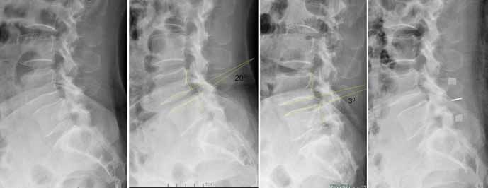
Eur Spine J (2009) 18:830–840                                                                                           831

Introduction                                                   reduces motion without suppressing it and lowers stress in
                                                               the disc fibres and annulus matrix [32].
Lumbar fusion has been used to reduce persistent conser-          The hypothesis of this prospective randomized com-
vative treatment axial back pain and/or segmental insta-       parative study was if the Wallis interspinous implant,
bility. Decompression and fusion in symptomatic                inserted in the unfused segment cephalad to instrumented
degenerative spondylolisthesis (grades I, II) has been fol-    lumbar fusion, could reduce the incidence of ASD.
lowed by very good functional results [49]. Furthermore,
wide decompression for significant symptomatic spinal
stenosis often associated with the loss of segmental lumbar    Materials and methods
lordosis may jeopardise segmental stability of the lumbar
spine that makes an additional stabilization mandatory.        From May 2001 to March 2002, we carried out a pro-
   Although instrumented lumbar spine fusion is a com-         spective controlled study comparing two consecutive
monly performed procedure, its role remains debated, and       homogenous groups of 25 consecutive patients each, who
moreover, delayed complications may occur, amongst             underwent surgery for degenerative spinal stenosis,
which is adjacent segment degeneration (ASD).                  spondylolisthesis, loss of segmental lordosis or combined
   Adjacent segment degeneration describes nearly any          in the same period. Group W included patients who
abnormal process that develops in the mobile segment           received the Wallis implant in the unfused segment ceph-
next to a spinal fusion and although the exact mechanism       alad to pedicle screw instrumentation and group C patients
remains uncertain, altered biomechanical stresses (hyper-      without interspinous implant, who were selected subse-
mobility, olisthesis, disc height (DH) loss, instability)      quently to match the characteristics of the patients of group
appear to play a key role in its development [4, 16, 19,       W and were used as controls. All 50 patients, who were
30, 33, 34, 39, 47]. Although several clinical and radio-      initially enrolled in this study were treated with the wide
logical criteria have been introduced to define segmental      decompression and posterior transpedicular rigid fixation
spinal instability there is no consensus as regard its         and fusion. All surgeries were performed by the first author
definition.                                                    who is a senior spine surgeon. The surgeon was unaware
   Although most often the criteria to determine ASD are       preoperatively that patient was going to be included in each
based solely on radiographic findings [2, 7, 19, 28, 30, 34,   group to avoid bias in patient’s selection. The ethics
39, 41, 59] reporting an ASD incidence ranging from 8 to       committee of this institution approved this study.
100%, the symptomatic incidence of ASD is significantly           The inclusion criteria were the following degenerative
lower ranging from 5.2 to 18.5% [7, 28, 31], while the rate    spine disease (spinal stenosis, spondylolisthesis, loss of
of re-operation rate for symptomatic ASD ranges from 2.7       segmental lordosis), 2–4 instrumented vertebrae and fusion
to 20% [16, 58].                                               in the lumbar and lumbosacral spine, modified arthritic
   There is a controversy regarding the risk factors           UCLA scale grade BII [15] without olisthesis or lytic
involved in the development of ASD [2, 11, 12, 22, 28,         lesion in the cephalad the instrumentation segment, and
40, 41, 47, 57–59]. Non-rigid, dynamic or flexible             informed consensus.
instrumentations for lumbar spine have been developed to          The exclusion criteria were the following: severe oste-
reduce ASD [17, 18, 27]. These implants are either fixed       oporosis, loss of lumbar lordosis [28], previous lumbar
in the pedicles, or secured between the spinous processes      surgery–fracture, lack of motion (ankylosis), UCLA [ II
of adjacent vertebrae [38, 48]. Long-term results showed       arthritic grade in the adjacent segment cephalad to instru-
several significant drawbacks and implant-related com-         mentation, spondylolisthesis, and acquired spinous process
plications in the non-rigid pedicle fixed instrumentations     insufficiency.
[18, 54].                                                         The radiographic criteria for ASD in the cephalad
   The interspinous process implants, that are currently       segment above to instrumentation were the development of
used for the treatment of neurogenic Claudicatio, reduce       olisthesis, disc collapse, increased segmental range of
pathologic extension at the symptomatic spinal levels          motion (ROM), deterioration ([grade II) of modified
and intradiscal pressure and facet load, preventing nar-       UCLA arthritic grade (Table 1) [15, 19, 30, 33, 47].
rowing of the spinal canal and neural foramens [36, 46,           The clinical criteria for ASD were the worsening of low
59, 61].                                                       back pain, despite radiographic solid fusion in the instru-
   Amongst these interspinous process implants a ‘‘sec-        mentation area and the absence of any surgery-related
ond’’ generation implant for non-rigid stabilization of        complication. In this study, the vertebral segment cephalad
lumbar segments, called Wallis system, has been devel-         to instrumented fusion was selected for several docu-
oped [48]. A recent in vitro biomechanical and finite-ele-     mented reasons: (1) this is the most frequent localisation of
ment analysis of the Wallis showed that this implant           ASD [3]; (2) symptomatic ASD in the lower lumbar and

                                                                                                                 123
832                                                                                                     Eur Spine J (2009) 18:830–840

Table 1 Arthritic grade for intervertebral disc degeneration
       UCLA grading for intervertebral space degeneration
       Disc space narrowing      Osteophytes      Endplate sclerosis

I      (-)                       (-)              (-)
II     (?)                       (-)              (-)
III    (?/-)                     (?/-)            (-)
IV     (?/-)                     (?/-)            (?)
Grade is based upon the most severe radiographic evident on plain
radiographs
(? present, - absent, ?/- either present or absent)

lumbosacral instrumented fusion is very rare (\3%) [15];
and (3) with exception of few studies, all clinical and                Fig. 1 The Wallis implant
biomechanical studies address cranial segment degenera-
tion following the rigid lumbar/lumbosacral instrumenta-               titanium implant. In addition, the implant includes two
tion [39].                                                             ligaments made of woven Dacron that are wrapped around
   All patients were clinically assessed preoperatively and            the spinous processes and fixed under tension to the
postoperatively with the SF-36 (physical function domain)              blocker. Wallis (Abbot, USA) is fixed to the spine by two
and Oswestry Disability Index (ODI) score and the pain                 polyester bands looped around the proximal and distal
magnitude with Visual Analogue Scale (0–10 scale, VAS).                spinous processes of the instrumented level and reattached
The preoperative radiological work-up included conven-                 to the spacer by means of two clips that are visible on plain
tional standing whole spine roentgenograms (lumbar lor-                radiographs. Four implant sizes (10, 12, 14, and 16 mm)
dosis, and DH and UCLA grading in the cephalad                         are available to fit individual interspinous distances. While
segment), sitting lateral dynamic bending films to measure             during the surgical procedure, the smallest size that had
olisthesis and ROM and supine oblique views for spinal                 sufficient stability on the two laminae is chosen to avoid
fusion determination. The radiological parameters that                 reduction of lumbar lordosis [42]. Wallis confers sub-
were measured preoperatively to the latest evaluation                  stantial mechanical advantages [32]: when the spinal col-
included: T12–S1 lumbar lordosis, ROM (flexion and                     umn is submitted to loading, the interspinous blocker
extension) in degrees of the vertebral segment immediately             displaces the mechanical constraints dorsally and reduces
cephalad to instrumentation, olisthesis of the vertebra                the load upon the disc and the facet joint system. The
cephalad instrumentation (in flexion and extension), ante-             Dacron ligaments resist traction of 200 daN and stretch
rior and posterior standing DH.                                        approximately 20% before failure by overloading. The
   As instability was defined as any sagittal translation of           overall implant constitutes a ‘‘floating’’ system with no
the adjacent vertebral body above fusion greater than 3 mm             permanent fixation in the vertebral bone, which might
and/or angle change greater than 10" between two adjacent              otherwise expose in the risk of loosening. Mechanical
vertebrae.                                                             human cadaver studies [48] have shown that Wallis permits
   CT scans, MRI were made in most, but not in all cases               a reduction in the mobility of intervertebral segments
and thus were not included in the evaluation of the ASD                previously destabilized by discectomy and that it achieves
changes as others also quite recently did [52]. The radio-             an increase in the rigidity of the destabilized segment
graphic changes were evaluated by a senior orthopaedic                 beyond normal values. There is no implant for the L5/S1
radiologist and spine surgeon who did not participate in               segment and thus it cannot be used below a L4/L5 fusion
surgery and thus did not know to which group each patient              [63].
belong.                                                                    Rigid pedicle screw instrumentation was used in this
                                                                       series for both groups. Care was taken to avoid to harm the
Surgical technique and Wallis interspinous implant                     facet joints adjacent to instrumentation (avoidance opening
                                                                       of facet joint capsule and lateral insertion of the pedicle
The second generation Wallis implant, that was used in this            screws. For technical reasons (too narrow space between
study is a interspinous blocker, which is made of PEEK                 spinous process and pedicle screw tulips), the Wallis
(polyetheretherketone). Due to its shape (Fig. 1) and the              device was inserted immediately after pedicle screw
properties of PEEK, the implant has much greater elasticity            insertion and decompression; then the longitudinal rods
(30 times less rigid than titanium) than the first generation          were assembled after appropriate contouring. In all patients

123
Eur Spine J (2009) 18:830–840                                                                                                      833

were used autogenous local bone derived from decom-
pression and decortication mixed with coralline HA
(50:50) for posterior and intertransverse fusion.
   One-way ANOVA was used to show the changes of
each parameter in a group and t test for differences in a
parameter between the two groups.

Results

Four patients were excluded from the final evaluation for
different reasons: one patient of group W for pseudar-
                                                                      Fig. 3 Changes of anterior disc height (mm) preoperatively and
throsis; two patients of group C for deep infection that              postoperatively to the latest evaluation
required re-operation and one patient of group C for ASD
in the segment below instrumentation.
   Twenty-four patients of group W and 21 patients of
group C, who showed radiological fusion 8–12 months
postoperatively were included in the final evaluation. The
age of the patients of groups W and C averaged
65 ± 13 years (range 32–72 years) and 64 ± 11 years
(range 33–71 years), respectively.
   The instrumented levels in both groups averaged
2.5 ± 1 (range 2–4).
   The Wallis was most often inserted in the L3/L4 segment
in 14/24 cases of group W, while the adjacent segment
cephalad to instrumentation was the L3/L4 in 15/21 cases of
C group.                                                              Fig. 4 Changes of posterior disc height (mm) preoperatively and
   The follow-up observation averaged 54 ± 6 months.                  postoperatively to the latest evaluation
   T12–S1 lordosis (Fig. 2) did not postoperatively change
until the final evaluation in any group, while no difference
between individuals of different groups was shown in all
periods of observation (W group, ANOVA, P = 0.35 vs. C
group, ANOVA, P = 0.41).
   Standing anterior DH (Fig. 3) did not postoperatively
change in W group (ANOVA, P = 0.26) and C group
(ANOVA, P = 0.69).

                                                                      Fig. 5 Range of    motion   in   the   cephalad   segment   above
                                                                      instrumentation

                                                                         Standing posterior DH (Fig. 4) increased immediately
                                                                      postoperatively (P = 0.042) in the W group, while it did
                                                                      not change in the C group (P = 0.44).
                                                                         The ROM (Fig. 5) at the cephalad to the instrumentation
                                                                      segment did not significantly postoperatively change
                                                                      (ANOVA, P = 0.5) in the W group, while it significantly
                                                                      increased at final evaluation (ANOVA, P \ 0.02) in the
                                                                      spines of the group C.
                                                                         Wallis decreased significantly (P = 0.0024) the percent
Fig. 2 Comparative plotting of T12–S1 lordosis changes preoperative   amount of olisthesis in flexion of the vertebra cephalad to
to the last evaluation                                                instrumented spinal segments a year postoperatively, while

                                                                                                                            123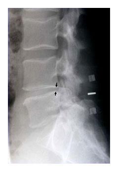
834                                                                                                             Eur Spine J (2009) 18:830–840

Fig. 6 Percent amount of olisthesis in flexion preoperatively to the     Fig. 8 SF-36 (physical function domain) changes preoperative until
last evaluation. Wallis decreased significantly (P = 0.0024) the         the final evaluation
percent amount of olisthesis

                                                                         Fig. 9 ODI changes preoperatively to the last evaluation

Fig. 7 Percent amount of olisthesis in extension preoperatively to the   (28.6%) spines in W and C groups, respectively. The levels
last evaluation. Wallis decreased (P = 0.012) the amount of olisthesis
in extension
                                                                         involved in ASD are shown in Table 2.
                                                                            Symptomatic ASD required surgical intervention was
it insignificantly (P = 0.31) decreased at the final evalua-             shown in 3 (14%) patients of group C with UCLA grade-IV
tion (Fig. 6).                                                           degeneration (disc space narrowing, osteophytes and end-
    Wallis     decreased     postoperatively    significantly            plate sclerosis) (Table 1; Figs. 10, 11, 12) in the first
(P = 0.012) the amount of olisthesis in extension (retro-                cephalad segment, while no patient from group W needed
listhesis) of the cephalad instrumentation vertebra one-year             intervention (Figs. 13, 14, 15) (Table 3).
follow-up postoperatively (Fig. 7), while it remained
unchanged at the final evaluation (P = 0.28).                            Complications
    Physical function domain (SF-36) improved (P \0.001)
postoperative (1 year) in an equal amount in the patients of             In one patient of group W and two patients of group C
both groups (Fig. 8). At the last evaluation, there was a                accidentally occurred intraoperatively dural violation that
statistically significant (P = 0.05) difference in favour of             was immediately sutured without further problems.
the patients of W group.                                                     Two patients of group C developed deep infection in the
    Oswestry Disability Index decreased significantly                    early postoperative phase (6–12 days postoperation) that
(P \ 0.005) in an equal amount in the patients of both                   required re-operation (wound drainage and continuous
groups a year postoperatively (Fig. 9). At the final obser-              irrigation plus intravenous antibiotics).
vation, there was a significant difference (P \ 0.05) in ODI             Table 2 Levels of ASD in above instrumentation segments
score in favour of the patients of W group.
    Visual Analogue Scale score (lumbar spine) averaged                  Group                 Segment
preoperatively 7.2 ± 2.1 and 7.4 ± 3 in the patients of                                        L2–L3                L3–L4             L4–L5
groups W and C, respectively, and improved postoperatively
                                                                         Wallis                0a                   1a, 1b            0a
to 3 ± 2 and 3.6 ± 3 in groups W and C, respectively.                                            a   b               a    b
                                                                         Controls              1,1                  4,4               1a, 1b
    Degeneration or deterioration of already existed low-
                                                                         a
grade degeneration (UCLA B II) in the adjacent segment                       One level above instrumentation
                                                                         b
cephalad to instrumentation was shown in 1 (4.1%) and 6                      Two levels above instrumentation

123
Eur Spine J (2009) 18:830–840 835
Fig. 10 Standing lateral roentgenogram of a 60-year-old female
patient suffering from degenerative disc disease L3/L4
Fig. 12 Lateral roentgenogram of the patient of Figs. 10 and 11,
34 months postoperatively showing collapse of this disc. No inter-
spinous implant was inserted at the L2/L3 segment. This patient was
revised because of intractable pain 38 months postoperatively
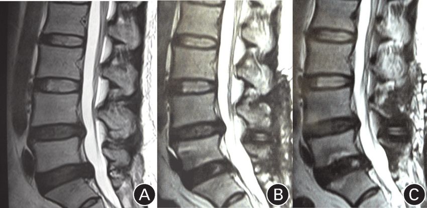
Fig. 11 Lateral MRI view showing severe degeneration in the
segment L3/L4 of the patient of Fig. 10
In one patient of group W and one of group C, there
were observed incidentally at the final observation remote
simple osteoporotic compression fractures (AO type
A1.1.1) 4 and 5 levels cephalad to instrumentation. These
Fig. 13 Lateral roentgenogram of a 58-year-old female patient with
fractures were not linked to any known trauma and were degenerative disc disease and disc herniation at the segment L4/L5.
clinically silent. UCLA degeneration II at the L3/L4 segment
123836 Eur Spine J (2009) 18:830–840
eliminating segmental instability, which is recognised as a
cause of low back pain. Indications comprise degenerative
disorders and spondylolisthesis (grades I, II) [20, 24, 49].
During the last years, lumbar fusion has been increasingly
criticised [6], while clinical studies have shown similar
long-term follow-up results with conservative treatment [8,
14]. Side effects of lumbar fusion include ASD, pseudar-
throsis, bone-graft morbidity, high rates of re-operation,
implant failure, and sagittal spinal imbalancing. More
specifically, spinal fusion alters the biomechanics of the
spine and the loss of motion at the fused levels is at least
theoretically compensated by increased motion at other
unfused segments resulting in ASD [33].
Adjacent segment degeneration is a common long-term
sequela or complication of spinal fusion surgery. The exact
aetiology is uncertain but alterations in facet loading,
hypermobility, and increased intradiscal pressure at the
segments adjacent to fusion mass is believed to play a key
role [15, 21, 35, 44, 51, 60]. Superior segment facet vio-
lation or laminectomy has recently shown in vitro to
destabilize the adjacent level in transpedicular fixation [9].
Radiological disc degeneration (ASD) is all too common
(5.2–100%) complication that the initial good results fol-
lowing a posterior spinal fusion degrade as adjacent mobile
Fig. 14 Lateral roentgenogram of the patient of Fig. 13, 57 months
segments proximal to the fusion degenerate over time
after L4/L5 laminectomy and discectomy plus instrumented fusion. compromising the late outcome of many short and mid-
At the segment L3/L4, a Wallis has been inserted (arrow). Note the term successes [1, 16, 26, 33, 43, 53]. There is an
normal height of the disc L3/L4 increasing concern, regarding the long-term consequences
of these asymptomatic changes; however, correlation of
ASD and clinical outcome is still unclear [46, 52].
The reported incidence of symptomatic ASD is signifi-
cantly lower (5.2–18.5%) than the radiologic ASD. ASD
incidence is higher in patients with transpedicular instru-
mentation (12.2–18.5%) compared with patients fused with
other forms of instrumentation or with no instrumentation
(5.2–5.6%). Evidence of radiographic degeneration, how-
ever, does not necessarily lead to a poor clinical outcome
of surgery [21, 52]. A recent clinical study [52] showed
that the incidence of ASD (DH reduction) in the first
cephalad adjacent segment 10 years following 360"
instrumented lumbar fusion averaged 21%, while in the
second adjacent level averaged 16%. In our series, the
incidence of ASD both in the first and second cephalad
Fig. 15 Axial CT view of the patient of Figs. 13 and 14 showing the
adjacent segment following posterolateral transpedicular
correct position of the Wallis implant close to the spinous process fixation without Wallis averaged 28.6%, while in the
Wallis group it was 4.1%. Thus, although different fusion
methods and imaging techniques were used in Schulte’s
Discussion and ours series it seems that the addition of Wallis pro-
tected not only the first but also the second cephalad seg-
Lumbar spine fusion is a common procedure in spine ment from ASD. In our series, the incidence of
surgery to improve the pain and clinical outcome of radiographic ASD cephalad to instrumentation was 4.1% in
patients with failed conservative treatment for lumbar the patients who received interspinous implant, signifi-
degenerative disease and degenerative spondylolisthesis by cantly lower compared with patients without spacer in
123Eur Spine J (2009) 18:830–840 837
Table 3 Arthritic grade for
UCLA? Wallis groupa Control groupa
intervertebral disc degeneration
in first cephalad segment Grading Preoperatively Postoperatively Preoperatively Postoperatively
I 17 17 16 15
Grade is based upon the most II 7 6 5 2
severe radiographic evident on III 0 1 0 1
plain radiographs IV 0 0 0 3
a
Number of patients listed 24 24 21 21
according to UCLA grading
which it was 28.6%. Although the most common segment osteophytes, etc) with clinical symptoms [3, 37, 45]. Others
involved in ASD was the L3/L4 in 5/7 spines, no conclu- have additionally used advanced imaging techniques as
sion can be drawn regarding correlation between levels computed tomography (CT), magnetic resonance imaging
involved in ASD and clinical outcome because of small (MRI) [13, 25, 53, 63]. In this study, we used the ‘‘statis-
sample of patients. In the present study, the incidence of tic’’ radiographic criteria (UCLA arthritic grading system)
symptomatic ASD in the cephalad segment that required that has been successfully used by others [15] along with
surgical intervention was 14% and was limited only in the dynamic motion parameters for the cephalad ASD (ROM,
patients without Wallis (group C). olisthesis). Schulte recently used DH reduction on plain
However, to solve the complication of ASD several roentgenograms as a measure of ASD. In the present study,
flexible or even dynamic devices have been used with we were able to show that the increased ROM and olis-
controversial results [17, 43, 50, 52]. It has been proposed thesis in the control group should be responsible for the
that non-fusion motion preservation surgery may prevent higher incidence of radiological incidence of ASD in this
accelerated ASD because of the protective effects of group when compared with the Wallis group.
continuing segmental motion. Dynesys have been used for To reduce the incidence of ASD by preserving motion,
motion preservation since 1994 to allow mobile load several implants of non-rigid or even dynamic stabilization
transfer, and provide controlled motion, thereby off-load- of lumbar intervertebral segments have been developed.
ing the facet joints and posterior disc [51]. Because of the Some of them (Graf, Dynesys) were secured to the spine by
rigidity of Dynesys some authors have doubted the pro- pedicle screw fixation systems [17], while other implants
tective effect of this construct on adjacent segment [23, 51, are secured in the interspinous space [38, 48]. Although
55, 56]. A recent prospective clinical study with 2 years of early results of pedicle-screw systems of flexible interver-
follow-up showed with the use of MRI that disc degener- tebral stabilization have been encouraging [17] some long-
ation at the bridged and cranial adjacent segment continue term results have revealed possible drawbacks [18, 54],
(20%) despite Dynesys dynamic stabilization [29]. Others including increased lumbar lordosis, stretching of the
[27] compared three posterior pedicle-screw instrumenta- Dacron parts, and malpositioning and loosening of pedicle
tions (rigid, semi-rigid and dynamic) and found no differ- screws leading to increased re-operation rate.
ences in the incidence of ASD after a follow-up of 4 years. Recently, several implants have been developed with
With the exception of a few studies, all of the biome- non-bony fixation, some connecting spinous processes, and
chanical and clinical studies address cranial segment laminae [5, 42] other connecting two adjacent spinous
degeneration [15, 39], because ASD caudal to a fusion is processes [38].
significantly less common [10]. The explanation for this is Amongst these implants is the Wallis, a ‘‘second’’
that in the adjacent segment cephalad to a fusion there is generation PEEK implant for non-rigid interspinous sta-
increased mobility compared with the adjacent caudal bilization of lumbar segments, which was used in our series
segment [5]. A recent clinical study showed that ASD cephalad to the uppermost instrumented lumbar vertebra to
occurred in 89% of the cases cephalad to lumbar fusion, preserve motion and reduce ASD incidence in this transi-
3.7% of the cases caudal and combined cephalad and tional unfused segment. A recent comparative biome-
caudal in 7.5% of the cases [10]. For these reasons, in our chanical study showed that Wallis reduced the ROM and
study, we investigated the mobility and associated degen- load on the disc and articular processes stresses, while it
erative signs only the vertebral segment cephalad to increased loads transmitted through the spinous processes
instrumented lumbar fusion. In our series of 45 followed up [43]. In our series, Wallis implant controlled the ROM of
patients only one (2.2%) developed ASD caudal to the cephalad not fused vertebra and restored the DH at this
instrumentation. segment without reduction of the global lumbar lordosis
Most of the previous relative studies have correlated and sagittal balance until the latest observation 60 months
‘‘static’’ radiographic criteria (DH, traction spurs, after index surgery.
123838 Eur Spine J (2009) 18:830–840
Using an MRI-based classification, some investigators and longer follow-up are necessary to definitively support
inserted the Wallis implant to treat disc degeneration the conclusions of this study and to determine the useful-
grades II–IV [33, 62]. In our study, we used the Wallis ness of the Wallis to protect adjacent unfused mobile
implant also in patients with UCLA I and II degeneration in lumbar segments.
the segment cephalad to instrumentation.
Senegas reported 7% re-operation rate, within 3 months
postoperatively due to the loosening of the previous gen-
eration implant in a discectomy population that was treated References
with the Wallis implant because of persistent low back
pain. No loosening or re-operation of the second generation 1. Adams MA (1996) Biomechanics of spinal implants. In: Szpalski
M, Gunzburg R, Spengler DM et al (eds) Instrumented fusion of
Wallis was shown in the present series. the degenerative lumbar spine: state of the art, questions, and
There are limitations to our study, as there are several controversies. Lippincott-Raven, Philadelphia
inherent difficulties in studying ASD. First, the definition 2. Aota Y, Kumano K, Hirabayashi S (1995) Postfusion instability
of ASD, not to mention the specific definition of radio- at the adjacent segments after rigid pedicle screw fixation for
degenerative lumbar spinal disorders. J Spinal Disord 8:464–473.
graphic ASD and clinical ASD, differs from study to study. doi:10.1097/00002517-199512000-00008
In this study, we studied clear static radiographic along 3. Axelsson P, Johnsson R, Stromqvist B (1997) The spondylolytic
with dynamic parameters. The latter seems to be in vertebra and its adjacent segment. Mobility measured before and
accordance with others (Schulte), who recently used plain after posterolateral fusion. Spine 22:414–422. doi:10.1097/
00007632-199702150-00012
radiological criteria (DH) to evaluate ASD. Second, MRI 4. Bastian L, Lange U, Knop C et al (2001) Evaluation of the
was not used to define the degree of ASD in the segment mobility of adjacent segments after posterior thoracolumbar fix-
cephalad instrumentation; however, there are no evidence- ation: a biomechanical study. Eur Spine J 10:295–300. doi:
based studies to support any link between pain and MR- 10.1007/s005860100278
5. Boden SD, Davis DO, Dina TS et al (1990) Abnormal magnetic-
positive signal. Third, ASD in the ‘‘unprotected’’ segment resonance scans of the lumbar spine in asymptomatic subjects. J
cephalad to rigid fixation may be a physiological process Bone Joint Surg Am 72:403–408
and not the results of stress concentration even after short 6. Bono CM, Lee CK (2004) Critical analysis of treads in fusion for
fusion? Finally, the outcome evaluation questionnaires degenerative disc disease over the past 20 years: influence of
technique on fusion rate and clinical outcome. Spine 29:455–463.
(VAS, SF-36 and ODI) specific enough to differentiate the doi:10.1097/01.BRS.0000090825.94611.28
origin of pain (ASD degeneration vs. other aetiology of 7. Booth KC, Bridwell KH, Eisenberg BA et al (1999) Minimum 5-
postoperative pain). year results of degenerative spondylolisthesis treated with
In accordance with previous observations, the incidence decompression and instrumented posterior fusion. Spine
24:1721–1727. doi:10.1097/00007632-199908150-00014
of clinically important ASD was significantly less than that 8. Brox IJ, Sorensen R, Friis A et al (2003) Randomized clinical
of the radiographic ASD. Thus, surgeons should be aware trial of lumbar instrumented fusion and cognitive intervention
that radiographic evidence of disc space narrowing and and exercises in patients with chronic low back pain and disc
degenerative changes do not necessarily correlate with degeneration. Spine 28:1913–1921. doi:10.1097/01.BRS.
0000083234.62751.7A
symptoms [43]. 9. Cardoso JM, Dmitriev EA, Helgeson M et al (2008) Does
In this series, the Wallis interspinous implant changed superior-segment facet violation or laminectomy destabilize the
the natural history of ASD in the free segment cephalad adjacent level in lumbar transpedicular fixation? Spine 26:2868–
to 2–4 levels instrumented rigid lumbar fusion and 2873. doi:10.1097/BRS.0b013e31818c63d3
10. Cheh G, Bridwell KH, Lenke L et al (2007) Adjacent segment
reduced until to 5 years postoperatively in an equal rate disease following lumbar/thoracolumbar fusion with pedicle
the incidence of the radiographic and symptomatic ASD screw instrumentation: a minimum 5-year follow-up. Spine
in the two adjacent segments cephalad to instrumented 32(20):2253–2257
lumbar fusion. The remote fractures in the thoracolumbar 11. Chen CS, Cheng CK, Liu CL et al (2001) Stress analysis of the
disc adjacent to interbody fusion in lumbar spine. Med Eng Phys
spine seem not to be related to spine surgery but to nat- 23:483–491. doi:10.1016/S1350-4533(01)00076-5
ural history of the degenerative disease and ageing 12. Esses SI, Doherty BJ, Crawford MJ et al (1996) Kinematic
process. evaluation of lumbar fusion techniques. Spine 21:676–684. doi:
We recommend the use of interspinous implants such as 10.1097/00007632-199603150-00003
13. Farfan HF (1973) Mechanical disaorders of the low back. Lea &
Wallis in UCLA I and II grades to protect the two adjacent Febiger, Philadelphia
cephalad to short (2–4 vertebrae) rigid fixation segments in 14. Fritzell P, Hagg, Nordwall A (2004) 5–10 years follow up in the
the lumbar spine. However, for more severe arthritic Swedish lumbar spine study. Spine Week Porto, Portugal, 30 May
changes (UCLA C II) we strongly recommend inclusion of 05 June 2004
15. Ghiselli G, Wang JC, Hsu WK, Dawson EG (2003) L5–S1 seg-
degenerated segments into the fusion. ment survivorship and clinical outcome analysis after L4–L5
Prospective randomized comparative studies with isolated fusion. Spine 12:1275–1280. doi:10.1097/00007632-
greater number of patients, more levels of instrumentation 200306150-00011
123Eur Spine J (2009) 18:830–840 839
16. Gillet P (2003) The fate of the adjacent motion segments after 34. Lee CK (1988) Accelerated degeneration of the segment adjacent
lumbar fusion. J Spinal Disord Tech 16:338–345 to a lumbar fusion. Spine 13:375–377. doi:10.1097/00007632-
17. Grevitt MP, Gardner AD, Spilsbury J et al (1995) The Graf sta- 198803000-00029
bilisation system: early results in 50 patients. Eur Spine J 4:169– 35. Lee CK, Langrana NA (1984) Lumbosacral spine fusion. A
175. doi:10.1007/BF00298241 discussion 35 biomechanical study. Spine 9:574–581. doi:10.1097/00007632-
18. Grob D, Benini A, Junge A et al (2005) Clinical experience with the 198409000-00007
Dynesys semirigid fixation system for the lumbar spine: surgical 36. Leong JC, Chun SY, Grange WJ et al (1983) Long-term results of
and patient-oriented outcome in 50 cases after an average of lumbar intervertebral disc prolapse. Spine 8:793–799. doi:
2 years. Spine 30:324–331. doi:10.1097/01.brs.0000152584. 10.1097/00007632-198310000-00018
46266.25 37. Lindsey DP, Swanson KE, Fuchs P et al (2003) The effects of an
19. Hambly MF, Wiltse LL, Raghavan N et al (1998) The transition interspinous implant on the kinematics of the instrumented and
zone above a lumbosacral fusion. Spine 23:1785–1792. doi: adjacent levels in the lumbar spine. Spine 28:2192–2197. doi:
10.1097/00007632-199808150-00012 10.1097/01.BRS.0000084877.88192.8E
20. Hanley EN Jr, David SM (1999) Lumbar arthrodesis for the 38. MacNab I (1971) The traction spur: an indicator of segmental
treatment of back pain. J Bone Joint Surg Am 81:716–730 instability. J Bone Joint Surg Am 53:663–670
21. Hilibrand AS, Robbins M (2004) Adjacent segment degeneration 39. Minns RJ, Walsh WK (1997) Preliminary design and experi-
and adjacent segment disease: the consequences of spinal fusion? mental studies of a novel soft implant for correcting sagittal plane
Spine J 4:190S–194S. doi:10.1016/j.spinee.2004.07.007 instability in the lumbar spine. Spine 22:1819–1825. doi:
22. Hsu K, Zucherman J, White A et al (1988) Deterioration of 10.1097/00007632-199708150-00004 discussion 1826–1827
motion segments adjacent to lumbar spine fusions. Ortho 40. Miyakoshi N, Abe E, Shimada Y et al (2000) Outcome of one-
Transact 12:605–606 level posterior lumbar interbody fusion for spondylolisthesis and
23. Huang RC, Wright TM, Panjabi MM et al (2005) Biomechanics postoperative intervertebral disc degeneration adjacent to the
of nonfusion implants. Orthop Clin North Am 36:271–280. doi: fusion. Spine 25:1837–1842. doi:10.1097/00007632-200007150-
10.1016/j.ocl.2005.02.010 00016
24. Ishihara H, Osada R, Kanamori M, Kawaguchi Y, Ohmori K, 41. Nagata H, Schendel MJ, Transfeldt EE et al (1993) The effects of
Kimura T, Matsui H, Tsuji H (2001) Minimum 10-year follow-up immobilization of long segments of the spine on the adjacent and
study of anterior lumbar interbody fusion for Isthmic spondylo- distal facet force and lumbosacral motion. Spine 18:2471–2479.
listhesis. J Spinal Disord 14:91–99. doi:10.1097/00002517- doi:10.1097/00007632-199312000-00017
200104000-00001 42. Nakai S, Yoshizawa H, Kobayashi S (1999) Long-term follow-up
25. Kimura S, Steinbach GC, Watenpaugh DE, Hargens AR (2001) study of posterior lumbar interbody fusion. J Spinal Disord
Lumbar spine disc height and curvature responses to an axial load 12:293–299. doi:10.1097/00002517-199908000-00004
generated by a compression device compatible with magnetic 43. Papp T, Porter RW, Aspden RM et al (1997) An in vitro study of the
resonance imaging. Spine 26:2596–2600. doi:10.1097/00007632- biomechanical effects of flexible stabilization on the lumbar spine.
200112010-00014 Spine 22:151–155. doi:10.1097/00007632-199701150-00005
26. Kleiner JB, Odom JA Jr, Moore MR et al (1995) The effect of 44. Park P, Garton HJ, Gala VC et al (2004) Adjacent segment dis-
instrumentation on human spinal fusion mass. Spine 20:90–97. ease after lumbar or lumbosacral fusion: review of the literature.
doi:10.1097/00007632-199501000-00016 Spine 29:1938–1944. doi:10.1097/01.brs.0000137069.88904.03
27. Korovessis P, Papazisis Z, Koureas G, Lambiris E (2004) Rigid, 45. Pfirrmann CWA, Metzdorf A, Zanetti M, Hodler J, Boos N
Semirigid versus dynamic instrumentation for degenerative (2001) Magnetic resonance classification of lumbar intervertebral
lumbar spinal stenosis. A correlative radiological and clinical disc degeneration. Spine 26:4873–4878. doi:10.1097/00007632-
analysis of short-term results. Spine 29:735–742. doi:10.1097/ 200109010-00011
01.BRS.0000112072.83196.0F 46. Pope MH, Hanley EN, Matteri RE et al (1977) Measurement of
28. Korovessis P, Stamatakis M, Baikousis A (1999) Segmental intervertebral disc space height. Spine 2:282–286. doi:
Roentgenographic analysis of vertebral inclination on sagittal 10.1097/00007632-197712000-00007
plane in asymptomatic versus chronic low back pain patients. J 47. Richards JC, Majumdar S, Lindsey DP et al (2005) The treatment
Spinal Disord Tech 12:131–137 mechanism of an interspinous process implant for lumbar neu-
29. Kumar A, Beastall J, Hughes J et al (2008) Disc changes in the rogenic intermittent claudication. Spine 30:744–749. doi:
bridged and adjacent segments after dynamic stabilization system 10.1097/01.brs.0000157483.28505.e3
after two years. Spine 33(26):2909–2914. doi:10.1097/BRS. 48. Rigby MC, Selmon GP, Foy MA et al (2001) Graft ligament
0b013e31818bdca7 stabilisation: mid- to long-term follow-up. Eur Spine J 10:234–
30. Kumar MN, Baklanov A, Chopin D (2001) Correlation between 236. doi:10.1007/s005860100254
sagittal plane changes and adjacent segment degeneration fol- 49. Rousseau MA, Lazennec JY, Saillant G (2006) Predictors of
lowing lumbar spine fusion. Eur Spine J 10:314–319. doi: outcomes after posterior decompression and fusion in degenera-
10.1007/s005860000239 tive spondylolisthesis. Eur Spine J 15:8–15. doi:10.1007/s00586-
31. Kumar MN, Jacquot F, Hall H (2001) Long-term follow-up of 005-0935-1
functional outcomes and radiographic changes at adjacent levels 50. Schlegel JD, Smith JA, Schleusener RL (1996) Lumbar motion
following lumbar spine fusion for degenerative disc disease. Eur segment pathology adjacent to thoracolumbar, lumbar, and lum-
Spine J 10:309–313. doi:10.1007/s005860000207 bosacral fusions. Spine 21:970–981. doi:10.1097/00007632-
32. Kuslich SD, Danielson G, Dowdle JD et al (2000) Four-year 199604150-00013
follow-up results of lumbar spine arthrodesis using the Bagby and 51. Schnake KJ, Schaeren S, Jeanneret B (2006) Dynamic stabiliza-
Kuslich lumbar fusion cage. Spine 25:2656–2662. doi:10.1097/ tion in addition to decompression for lumbar stenosis with
00007632-200010150-00018 degenerative spondylolisthesis. Spine 31:442–449. doi:10.1097/
33. Lafage V, Gangnet N, Senegas J et al (2007) New interspinous 01.brs.0000200092.49001.6e
implant evaluation using an in vitro biomechanical study com- 52. Schulte LT, Leistra F, Bullmann V, Osada N, Vieth V, Marquardt
bined with a finite-element analysis. Spine 32(16):1706–1713. B, Lerner T, Liljenqvist U, Hachenberg L (2007) Disc height
doi:10.1097/BRS.0b013e3180b9f429 reduction in adjacent segments and clinical outcome 10 years
123840 Eur Spine J (2009) 18:830–840
after lumbar 360" fusion. Eur Spine J 16:2152–2158. doi: 60. Weihofer SL, Guyer RD, Herbert M et al (1995) Intradiscal pres-
10.1007/s00586-007-0515-7 sure measurements above an instrumented fusion. A cadaveric
53. Senegas J (2002) Mechanical supplementation by non-rigid fix- study. Spine 20:526–531. doi:10.1097/00007632-199503010-
ation in degenerative intervertebral lumbar segments: the Wallis 00004
system. Eur Spine J 11(Suppl 2):164–169 61. Wiltse LL, Radecki SE, Biel HM et al (1999) Comparative study
54. Sénégas J, Vital JM, Guerin J et al (1997) Stabilisation lombaire of the incidence and severity of degenerative change in the
souple, instabilite vertebrales lombaires. Expans Sci Fr 12:4–32 transition zones after instrumented versus noninstrumented
55. Sengupta DK (2006) Point of view: dynamic stabilization in fusions of the lumbar spine. J Spinal Disord 12:27–33. doi:
addition to decompression for lumbar spine stenosis with 10.1097/00002517-199902000-00004
degenerative spondylolisthesis. Spine 31:450. doi:10.1097/01.brs. 62. Wiseman C, Lindsey DP, Fredrick AD et al (2005) The effect of
0000200051.24623.33 an interspinous process implant on facet loading during exten-
56. Singh K, An HS (2006) Motion preservation technologies: sion. Spine 15(30):903–907. doi:10.1097/01.brs.0000158876.
alternatives to spinal fusion. Am J Orthop 35:411–416 51771.f8
57. Swanson KE, Lindsey DP, Hsu KY et al (2003) The effects of an 63. Zhu SH, McCarthy ID, McGregor AH, Coombs RR, Hughes SP
interspinous implant on intervertebral disc pressures. Spine (2000) Geometrical dimensions of the lower lumbar vertebra. Eur
28:26–32. doi:10.1097/00007632-200301010-00008 Spine J 9(3):242–248
58. Umehara S, Zindrick MR, Patwardhan AG et al (2000) The 64. Zucherman JF, Hsu KY, Hartjen CA et al (2004) A prospective
biomechanical effect of postoperative hypolordosis in instru- randomized multi-center study for the treatment of lumbar spinal
mented lumbar fusion on instrumented and adjacent spinal seg- stenosis with the X STOP interspinous implant: 1-year results.
ments. Spine 25:1617–1624. doi:10.1097/00007632-200007010- Eur Spine J 13:22–31. doi:10.1007/s00586-003-0581-4
00004
59. Whitecloud TSIII, Davis JM, Olive PM (1994) Operative treat-
ment of the degenerated segment adjacent to a lumbar fusion.
Spine 19:531–536
123Observational Study Medicine ®
OPEN
Adjacent segmental degeneration following Wallis
interspinous stabilization implantation
Biomechanical explanations and the value of magnetic
resonance imaging
∗ ∗
Zhiguo Zhou, MSa, Wei Xiong, MDb, Li Li, MSc, , Feng Li, MDb,
Abstract
Adjacent segmental degeneration (ASD) is a major issue after pedicular fixation. This study examined the degeneration of the adjacent
levels due to the insertion of the Wallis interspinous stabilization system compared with discectomy, using magnetic resonance
imaging (MRI).
Thirty-eight patients diagnosed with lumbar degeneration disorders at L4-L5 were reviewed: 19 patients underwent discectomy
and Wallis system implantation (group A), and 19 patients underwent discectomy (group B). The Visual Analog Scale (VAS) and
Oswestry Disability Index (ODI) were assessed preoperatively and postoperatively. ASD was evaluated by MRI.
There was no difference in the preoperative ODI scores between the 2 groups (non-normal distribution, median, 50 (40, 50) vs 50 (50,
50), P = .331), but the postoperative ODI scores were different (non-normal distribution, median, 0 (0, 32) vs 20 (20, 30), P < .005). Similar
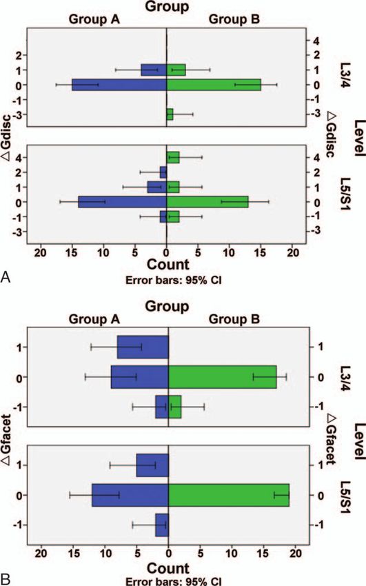
results were observed for VAS. In group A, ASD occurred in 4 patients (21.1%) in the disc and 8 (42.1%) in the facet joint at L3/4, and in 4
(21.1%) in the disc and 5 (26.3%) in the facet joint at L5/S1. In Group B, ASD occurred in 3 patients (15.8%) in the disc at L3/4, and in 4
(21.1%) in the disc at L5/S1. In general, there was no difference between the 2 groups (P > .05), except at L3/4 (P = .015).
ASD of the facet joint in the cranial segment occurred after Wallis system implantation, suggesting that the Wallis system cannot
prevent ASD of the facet joint, but could have some other benefits for the discs.
Abbreviations: ASD = adjacent segmental degeneration, BMI = body mass index, FOV = field of view, MRI = magnetic
resonance imaging, ODI = Oswestry Disability Index, TE/TR = echo time and repetition time, VAS = visual analog scale.
Keywords: adjacent segmental degeneration, disc, facet joint, pedicular fixation, Wallis system implantation
1. Introduction Several reports revealed that ASD could be accelerated due to the
relative immobility of fused spinal segments transferring stress to
Acute or progressive disc lesions lead to instability of the spinal
adjacent segments after fusion.[5–7] Symptoms and signs of ASD
segments.[1,2] Currently, pedicular fixation (fusion) is the gold
include pain, stenotic lesions, and instability, leading to
standard treatment in terms of increasing the biomechanical
additional surgeries such as extended fusion and neural
rigidity and clinical fusion rates because pedicle screws are the
decompression.[8] Unfortunately, there is currently no relevant
strongest component of spinal implants.[3] Adjacent segment
literature about the prevention of ASD.
degeneration (ASD) is the development of a pathology at the
To reduce the incidence of fusion-related morbidity, non-
mobile segment next to a lumbar or lumbosacral spinal fusion.[4]
fusion technologies have been developed, such as the Wallis
interspinous stabilization system.[9] Although the implant offers
Editor: Kavindra Nath.
some advantages over fusion (e.g., motion of the involved levels
The authors have no funding and conflicts of interest to disclose.
a
and small operation wound), the efficacy of non-fusion implants
Department of Orthopaedics, Wuhan Children’s Hospital (Wuhan Maternal and in the prevention of ASD is now well established.[3,8]
Child Healthcare Hospital), Tongji Medical College, Huazhong University of
Science and Technology, Wuhan, b Department of Orthopaedics, c Department of
ASD was first described using x-ray indexes such as disc height
Radiology, Tongji Hospital, Tongji Medical College, Huazhong University of and segmental range of motion, [10] but a previous animal study
Science and Technology, Wuhan, China. suggested that the changes in x-ray indexes were less sensible than
those extracted from magnetic resonance imaging (MRI),[11] as
∗
Correspondence: Li Li, Department of Radiology, Tongji Hospital, Tongji Medical
College, Huazhong University of Science and Technology, Wuhan, China supported by a study in humans. [12]
(e-mail: chen_lili1985@126.com); Feng Li, Department of Orthopaedics, Tongji Nevertheless, it is poorly known whether the use of the Wallis
Hospital, Tongji Medical College, Huazhong University of Science and
Technology, Wuhan, China (e-mail: lifeng_lili@126.com).
system could prevent ASD. Therefore, the aim of the present
study was to compare the patients who underwent discectomy
Copyright © 2017 the Author(s). Published by Wolters Kluwer Health, Inc.
This is an open access article distributed under the terms of the Creative and Wallis system implantation with the patients who underwent
Commons Attribution-Non Commercial-No Derivatives License 4.0 (CCBY-NC- discectomy only, based on MRI examinations.
ND), where it is permissible to download and share the work provided it is
properly cited. The work cannot be changed in any way or used commercially
without permission from the journal. 2. Methods
Medicine (2017) 96:22(e7056)
2.1. Study design and patients
Received: 19 December 2016 / Received in final form: 10 May 2017 / Accepted:
11 May 2017 Patients diagnosed with lumbar disc herniation at L4-L5 and
http://dx.doi.org/10.1097/MD.0000000000007056 operated (by the same surgeon) at the Department of Orthopedic
1Zhou et al. Medicine (2017) 96:22 Medicine
Table 1
Grading of intervertebral disc degeneration.
Distinction between inner and outer fibers of
Grade Signal from nucleus and inner fibers of annulus annulus at posterior aspect of the disc Height of the disc
1 Uniformly hyperintense, equal to CSF Distinct Normal
2 Hyperintense (>presacral fat and 5 and/or facet degeneration grade to impairments such as pain and abilities such as personal care,
>2 according to MRI (Table 1[13] and Table 2[14]); (4) history of lifting, walking, sitting, standing, sleeping, sex life, social life, and
cardiovascular or cerebrovascular diseases, trauma, or cancer; (5) traveling. In each section, the patient chose the statement that best
lost to follow-up; or (6) missing data. described his/her status. If the limitation fell between 2 levels, the
During the study period, 100 patients were treated at our higher point value was selected. The chosen statements received
center, but after excluding patients lost to follow-up and those scores 0 to 5 corresponding to the level indicated. The total scores
with missing data, and after matching the 2 groups for age, could range from 0 (the highest level of function) to 50 (the lowest
gender, and occupation, only 38 patients remained. level of function).
2.2. Surgery 2.4. MRI
The treatment approach was decided by the surgeon in All patients had undergone magnetic resonance imaging (MRI)
consultation with patients. After oral and written explanations before and 6 months after operation. The lumbar spine MRI
on the details of the surgery, all participants signed a written examination of each participant was done by the same clinical
surgical informed consent. After discussion, the patients under- 1.5T system (Signa 1.5 T HD, GE Healthcare, Waukesha, WI)
went either discectomy and Wallis implantation (n = 19, group A) using a 4-channel Phased Array CTL Spine Coil. T1-weighted fast
or discectomy only (n = 19, group B). spin-echo sagittal images with effective echo time and repetition
The indications for discectomy were: (1) symptoms of lumber times (TE/TR) of 10/400 ms, T2-weighted fast spin-echo sagittal
spinal cord or nerve root compression; (2) conservative treatment images with TE/TR of 102/3000 ms and T2-weighted fast spin-
did not produce satisfactory outcomes; and (3) willing to undergo echo axial images with TE/TR of 120/3000 ms were included in
Table 2
Grading of the facet joint degeneration.
Grade Criteria
1 Uniformly thick cartilage covers the articular surfaces completely. Articular processes have a thin layer of cortical bone. No osteophyte.
2 Cartilage covers the entire surface of the articular processes but with erosion of the irregular region evident. Cortical bone of the articular processes is focally thickened.
Possible or small osteophyte.
3 Cartilage incompletely covers the articular surfaces, with regions of the underlying bone exposed to the joint. Thickened cortical bone covers less than half of the articular
processes. Definite and moderate osteophyte.
4 Cartilage is absent except for traces on the articular surfaces; dense cortical bone covers greater than half the articular process. Large osteophyte.
2You can also read