KOELIS BEST PAPERS 4 Edition-2009-2020 - See Beyond Prostate Biopsy.
←
→
Page content transcription
If your browser does not render page correctly, please read the page content below

See Beyond
Prostate Biopsy.

      KOELIS BEST PAPERS
       4th Edition - 2009 - 2020

            www.koelis.com
CONTENTS

DIAGNOSTIC ACCURACY................................................................................             4

  MRI/US TARGETED BIOPSY..................................................................................4

  3D ULTRASOUND GUIDED BIOPSY................................................................. 14

 PRECISION OF THE OBT* FUSION®................................................................ 16

CLINICAL PRACTICE.....................................................................................          20

 TRANSPERINEAL BIOPSY UNDER LOCAL ANESTHESIA.............................. 20

BEYOND PROSTATE BIOPSY........................................................................                  25

  DIAGNOSTIC ACCURACY: PET/US TARGETED BIOPSY.............................25

  REPEAT BIOPSY FOR ACTIVE SURVEILLANCE STRATEGY..........................28

 FOCAL TREATMENT..............................................................................................32

BIBLIOGRAPHY.................................................................................................   36

                                                                      *ORGAN BASED TRACKING®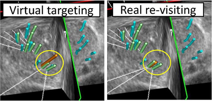
DIAGNOSTIC ACCURACY
            MRI/US TARGETED BIOPSY

    IMPROVEMENT OF THE INTERMEDIATE RISK PROSTATE
    CANCER SUB-CLASSIFICATION BY INTEGRATING MRI
    AND FUSION BIOPSY FEATURES
    Roumiguie M¹, Lesourd M, Zgheib J, Tollon C, Salin A, Alméras C, Doumerc N, Thoulouzan M, Soulié M, Gautier
    J-R, Loison G, Assoun J, Vacher A, Aziza R, Bernard Malavaud B, Beauval J-B, Ploussard G.

    ¹ Department of Urology, CHU Toulouse, Toulouse, France. Department of Urology, La Croix du Sud Hospital, Quint Fonsegrives, France. Depart-
    ment of Urology, Institut Universitaire du Cancer Toulouse - Oncopole, Toulouse, France. Department of Radiology, La Croix du Sud Hospital,
    Quint Fonsegrives, France. Department of Radiology, Institut Universitaire du Cancer Toulouse - Oncopole, Toulouse, France.

                                                                                                                  Urol Oncol. 2020

    INTRODUCTION: Treatment decision-making for intermediate-risk prostate cancer (CaP) is mainly based on
    grade and tumor involvement on systematic biopsy. We aimed to assess the added value of multi-parametric
    magnetic resonance imaging (mpMRI) and targeted biopsy (TB) features for predicting final pathology and for
    improving the well-established favourable/unfavourable systematic biopsy-based sub-classification.

    MATERIALS AND METHODS: From a prospective database of 377 intermediate risk CaP cases, we evaluated the
    performance of the standard intermediate risk classification (IRC), and the predictive factors for unfavourable
    disease on final pathology aiming to build a new model. Overall unfavourable disease (OUD) was defined by
    any pT3-4 and/or pN1 and/or grade group (GG) ≥ 3.

    RESULTS: The standard IRC was found to be predictive for unfavourable disease in this population. However,
    in multivariable analysis
    regression, ECE on mpMRI and GG ≥3 on TB remained the 2 independent predictive factors for OUD disease
    (HR = 2.7, P = 0.032, and
    HR = 2.41, P = 0.01, respectively). By using the new IRC in which unfavorable risk was defined by ECE on mpMRI
    and/or GG ≥3 on TB,
    the proportion of unfavorable cases decreased from 62.3% to 34.1% while better predicting unfavorable
    disease in RP speciments. The new
    model displayed a better accuracy than the standard IRC for predicting OUD (AUC: 0.66 vs. 0.55).

    CONCLUSIONS: The integration of imaging and TB features drastically improves the intermediate risk sub-
    classification performance and better discriminates the unfavourable risk group that could benefit from more
    aggressive therapy such as neo-adjuvant and/or adjuvant treatment, and the favourable group that could
    avoid over-treatment. External validation in other datasets is needed.

4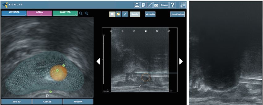
MRI/US TARGETED BIOPSY
DIAGNOSTIC ACCURACY
                      ACCURACY OF ELASTIC FUSION BIOPSY IN DAILY
                      PRACTICE: RESULTS OF A MULTICENTER STUDY OF 2115
                      PATIENTS
                      Oderda M, Marra G, Albisinni S, Altobelli E, Baco E, Beatrici V, Cantiani A, Carbone A, Ciccariello M, Descotes JL,
                      Dubreuil-Chambardel M, Eldred-Evans D, Fasolis G, Ferriero M, Fiard G, Forte V, Giacobbe A, Kumar P, Lacetera
                      V, Mozer P, Muto G, Papalia R, Pastore A, Peltier A, Piechaud T, Simone G, Roche JB, Roupret M, Rouviere O, Van
                      Velthoven R, Gontero P.

                                                                                                                          Int J Urol. 2018

                      OBJECTIVES: To assess the accuracy of Koelis fusion biopsy for the detection of prostate cancer and clinically
                      significant prostate cancer in the everyday practice.

                      METHODS: We retrospectively enrolled 2115 patients from 15 institutions in four European countries
                      undergoing transrectal Koelis fusion biopsy from 2010 to 2017. A variable number of target (usually 2-4) and
                      random cores (usually 10-14) were carried out, depending on the clinical case and institution habits. The
                      overall and clinically significant prostate cancer detection rates were assessed, evaluating the diagnostic
                      role of additional random biopsies. The cancer detection rate was correlated to multiparametric magnetic
                      resonance imaging features and clinical variables.

                      RESULTS:The mean number of targeted and random cores taken
                      were 3.9 (standard deviation 2.1) and 10.5 (standard deviation 5.0),
                      respectively. The cancer detection rate of Koelis biopsies was 58%
                      for all cancers and 43% for clinically significant prostate cancer.
                      The performance of additional, random cores improved the cancer
                      detection rate of 13% for all cancers (P < 0.001) and 9% for clinically
                      significant prostate cancer (P < 0.001). Prostate cancer was detected
                      in 31%, 66% and 89% of patients with lesions scored as Prostate
                      Imaging Reporting and Data System 3, 4 and 5, respectively. Clinical
                      stage and Prostate Imaging Reporting and Data System score were
                      predictors of prostate cancer detection in multivariate analyses.
                      Prostate-specific antigen was associated with prostate cancer
                      detection only for clinically significant prostate cancer.

                      CONCLUSIONS: Koelis fusion biopsy offers a good cancer
                      detection rate, which is increased in patients with a high Prostate
                      Imaging Reporting and Data System score and clinical stage. The
                      performance of additional, random cores seems unavoidable for
                      correct sampling. In our experience, the Prostate Imaging Reporting
                      and Data System score and clinical stage are predictors of prostate
                      cancer and clinically significant prostate cancer detection; prostate-
                      specific antigen is associated only with clinically significant
                      prostate cancer detection, and a higher number of biopsy cores are        Figure 1. Biopsy results in terms of PCa and clinically
                                                                                                significant PCa detection, comparing target biopsies
                      not associated with a higher cancer detection rate.
                                                                                                only with target + random biopsies

                                                                                                                                                          5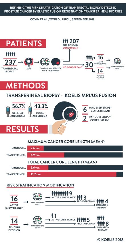
ACCURACY OF ELASTIC FUSION BIOPSY IN DAILY PRACTICE

                                                                            DIAGNOSTIC ACCURACY
      RESULTS OF A MULTICENTER STUDY OF 2115 PATIENTS
                   ODERDA ET AL., INT J UROL, AUGUST 2018

    PATIENTS
                           66
                           years

                           8.4
                                                                  4
                           ng/mL

    2115                                 15
                     PSA

                           52cc

    METHODS
    KOELIS PROSTATE MRI/US FUSION

                            2-4 TARGETED
                           10-14 RANDOM

     RESULTS
           DETECTION RATES ACCORDING TO PIRADS SCORE
    100%
     80%                                                    89%
                                                   79%
     60%
                                         66%
     40%                           47%

     20%               31%
             17%
     0%
             PIRADS 3              PIRADS 4         PIRADS 5
             CLINICALLY SIGNIFICANT CANCER
             ANY CANCER

                                                            © KOELIS 2018

6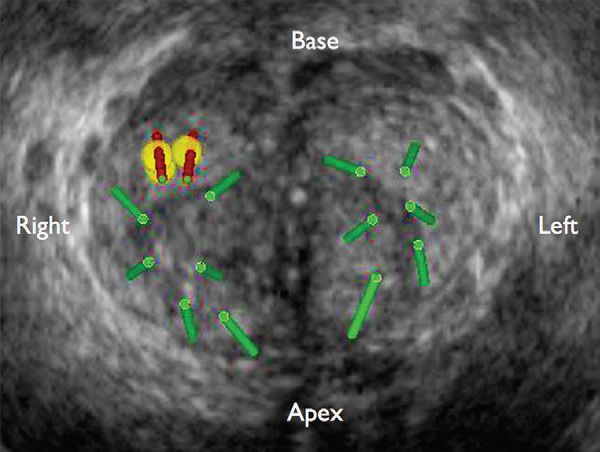
DIAGNOSTIC ACCURACY      MRI/US TARGETED BIOPSY

                      USE OF PROSTATE SYSTEMATIC AND TARGETED
                      BIOPSY ON THE BASIS OF MULTIPARAMETRIC MRI IN
                      BIOPSY-NAIVE PATIENTS (MRI-FIRST): A PROSPECTIVE,
                      MULTICENTRE, PAIRED DIAGNOSTIC STUDY
                      Rouvière O, Puech P, Renard-Penna R, Claudon M, Roy C, Mège-Lechevallier F, Decaussin-Petrucci M, Dubreuil-
                      Chambardel M, Magaud L, Remontet L, Ruffion A, Colombel M, Crouzet S, Schott A, Lemaitre L, Rabilloud M,
                      Grenier N, for the MRI-FIRST Investigators*

                                                                                                           Lancet Oncol 2018

                      BACKGROUND: Whether multiparametric MRI improves the detection of clinically significant prostate
                      cancer and avoids the need for systematic biopsy in biopsy-naive patients remains controversial. We aimed
                      to investigate whether using this approach before biopsy would improve detection of clinically significant
                      prostate cancer in biopsy-naive patients.

                      METHODS: In this prospective, multicentre, paired diagnostic study, done at 16 centres in France, we enrolled
                      patients aged 18–75 years with prostate-specific antigen concentrations of 20 ng/mL or less, and with stage
                      T2c or lower prostate cancer. Eligible patients had been referred for prostate multiparametric MRI before a
                      first set of prostate biopsies, with a planned interval of less than 3 months between MRI and biopsies. An
                      operator masked to multiparametric MRI results did a systematic biopsy by obtaining 12 systematic cores
                      and up to two cores targeting hypoechoic lesions. In the same patient, another operator targeted up to two
                      lesions seen on MRI with a Likert score of 3 or higher (three cores per lesion) using targeted biopsy based on
                      multiparametric MRI findings. Patients with negative multiparametric MRI (Likert score ≤2) had systematic
                      biopsy only. The primary outcome was the detection of clinically significant prostate cancer of International
                      Society of Urological Pathology grade group 2 or higher (csPCa-A), analysed in all patients who received both
                      systematic and targeted biopsies and whose results from both were available for pathological central review,
                      including patients who had protocol deviations. This study is registered with ClinicalTrials.gov, number
                      NCT02485379, and is closed to new participants.

                      FINDINGS: Between July 15, 2015, and Aug 11, 2016, we enrolled 275 patients. 24 (9%) were excluded from
                      the analysis. 53 (21%) of 251 analysed patients had negative (Likert ≤2) multiparametric MRI. csPCa-A was
                      detected in 94 (37%) of 251 patients. 13 (14%) of these 94 patients were diagnosed by systematic biopsy only,
                      19 (20%) by targeted biopsy only, and 62 (66%) by both techniques. Detection of csPCa-A by systematic biopsy
                      (29·9%, 95% CI 24·3–36·0) and targeted biopsy (32·3%, 26·5–38·4) did not differ significantly (p=0·38). csPCa-A
                      would have been missed in 5·2% (95% CI 2·8–8·7) of patients had systematic biopsy not been done, and in
                      7·6% (4·6–11·6) of patients had targeted biopsy not been done. Four grade 3 post-biopsy adverse events were
                      reported (3 cases of prostatitis, and 1 case of urinary retention with haematuria).

                      INTERPRETATION: There was no difference between systematic biopsy and targeted biopsy in the detection
                      of ISUP grade group 2 or higher prostate cancer; however, this detection was improved by combining both
                      techniques and both techniques showed substantial added value. Thus, obtaining a multiparametric MRI
                      before biopsy in biopsy-naive patients can improve the detection of clinically significant prostate cancer but
                      does not seem to avoid the need for systematic biopsy.

                                                                                                                                        7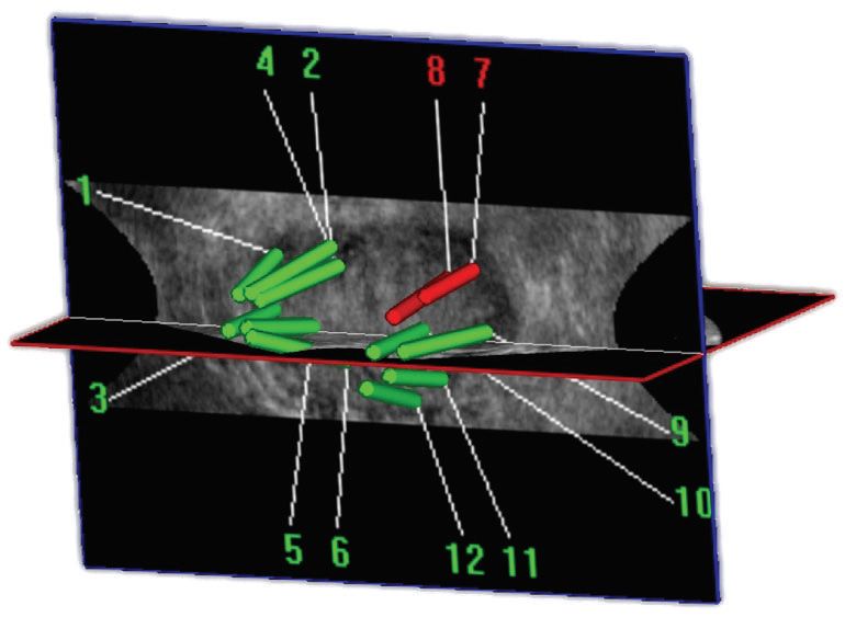
DIAGNOSTIC ACCURACY
       MRI/US TARGETED BIOPSY

    MRI-TARGETED OR STANDARD BIOPSY FOR
    PROSTATE-CANCER DIAGNOSIS
    Kasivisvanathan V, M.R.C.S., Rannikko A.S, Borghi M, Panebianco V, Mynderse L.A,Vaarala M.H, Briganti A, Budäus
    L, Hellawell G, F.R.C.S.(Urol.), Hindley R.G, F.R.C.S.(Urol.), Monique J. Roobol M.J, Scott Eggener S, et al., for the
    PRECISION Study Group Collaborators*

                                                                                                   NEJM 2018

    BACKGROUND: Multiparametric magnetic resonance imaging (MRI), with or without targeted biopsy, is an
    alternative to standard transrectal ultrasonography–guided biopsy for prostate-cancer detection in men with
    a raised prostate-specific antigen level who have not undergone biopsy. However, comparative evidence is
    limited.

    METHODS: In a multicenter, randomized, noninferiority trial, we assigned men with a clinical suspicion of
    prostate cancer who had not undergone biopsy previously to undergo MRI, with or without targeted biopsy,
    or standard transrectal ultrasonography–guided biopsy. Men in the MRI-targeted biopsy group underwent a
    targeted biopsy (without standard biopsy cores) if the MRI was suggestive of prostate cancer; men whose MRI
    results were not suggestive of prostate cancer were not offered biopsy. Standard biopsy was a 10-to-12–core,
    transrectal ultrasonography–guided biopsy. The primary outcome was the proportion of men who received a
    diagnosis of clinically significant cancer. Secondary outcomes included the proportion of men who received a
    diagnosis of clinically insignificant cancer.

    RESULTS: A total of 500 men underwent randomization. In the MRI-targeted biopsy group, 71 of 252 men
    (28%) had MRI results that were not suggestive of prostate cancer, so they did not undergo biopsy. Clinically
    significant cancer was detected in 95 men (38%) in the MRI-targeted biopsy group, as compared with 64 of
    248 (26%) in the standard-biopsy group (adjusted difference, 12 percentage points; 95% confidence interval
    [CI], 4 to 20; P=0.005). MRI, with or without targeted biopsy, was noninferior to standard biopsy, and the 95%
    confidence interval indicated the superiority of this strategy over standard biopsy. Fewer men in the MRI-
    targeted biopsy group than in the standard-biopsy group received a diagnosis of clinically insignificant cancer
    (adjusted difference, −13 percentage points; 95% CI, −19 to −7; P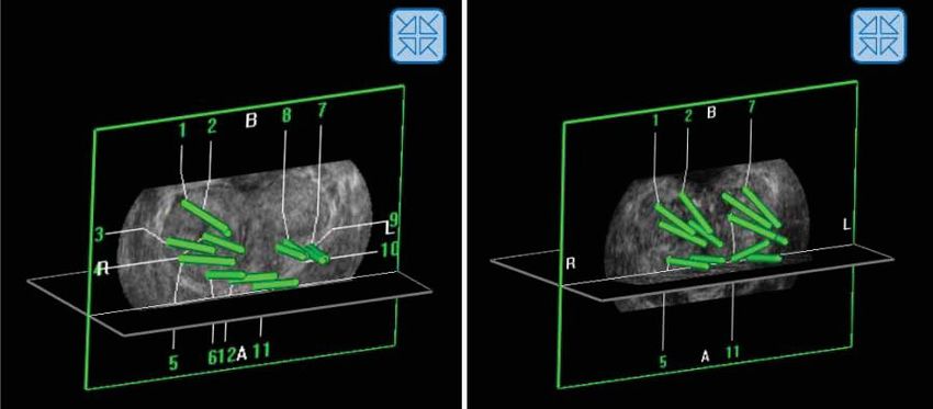
MRI/US TARGETED BIOPSY
DIAGNOSTIC ACCURACY
                      DETECTION OF PROSTATE CANCER USING MRI-
                      ULTRASONOGRAPHY IMAGE-FUSION TARGETED BIOPSY
                      IN AFRICAN-AMERICAN MEN
                      Shin T¹ ², Smyth T.B, Ukimura O, Ahmadi N, Abreu A.L.d.C, Oishi M, Mimata H, Gill I.S.

                      ¹ USC Institute of Urology, Keck School of Medicine, University of Southern California, Los Angeles, CA, USA.
                      ² Department of Urology, Oita University, Oita, Japan.

                                                                                                                                      BJU Int. 2017

                      OBJECIVE: To assess the diagnostic yield of targeted prostate biopsy in African-American (A-A) men using image
                      fusion of multi-parametric magnetic resonance imaging (mpMRI) with real-time transrectal ultrasonography
                      (US).

                      MATERIALS AND METHODS: We retrospectively analysed 661 patients (117 A-A and 544 Caucasian) who had
                      mpMRI before biopsy and then underwent MRI/US image-fusion targeted biopsy (FTB) between October
                      2012 and August 2015. The mpMRIs were reported on a 5-point Likert scale of suspicion. Clinically significant
                      prostate cancer (CSPC) was defined as biopsy Gleason score ≥7.

                      RESULTS: After controlling for age, prostate-specific antigen level and prostate volume, there were no
                      significant differences between A-A and Caucasian men in the detection rate of overall cancer (35.0% vs 34.2%,
                      P = 0.9) and CSPC (18.8% vs 21.7%, P = 0.3) with MRI/US FTB. There were no significant differences between
                      the races in the location of dominant lesions on mpMRI, and in the proportion of 5-point Likert scoring. In
                      A-A men, MRI/US FTB from the grade 4-5 lesions outperformed random biopsy in the detection rate of overall
                      cancer (70.6% vs 37.2%, P = 0.003) and CSPC (52.9% vs 12.4%, P < 0.001). MRI/US FTB outperformed random
                      biopsy in cancer core length (5.0 vs 2.4 mm, P = 0.001), in cancer rate per core (24.9% vs 6.8%, P < 0.001),
                      and in efficiency for detecting one patient with CSPC (mean number of cores needed 13.3 vs 81.9, P < 0.001),
                      respectively.

                      CONCLUSIONS: Our key finding confirms a lack of racial difference in the detection rate of overall prostate
                      cancers and CSPC with MRI/US FTB between A-A and Caucasian men. MRI/US FTB detected more CSPC using
                      fewer cores compared with random biopsy.

                                                                                                                                                      9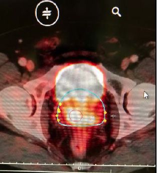
DIAGNOSTIC ACCURACY
        MRI/US TARGETED BIOPSY

     A RANDOMIZED CONTROLLED TRIAL TO ASSESS AND
     COMPARE THE OUTCOMES OF TWO-CORE PROSTATE
     BIOPSY GUIDED BY FUSED MAGNETIC RESONANCE
     AND TRANSRECTAL ULTRASOUND IMAGES AND                                                                                                  IENTIFIC P
                                                                                                                                          SC          A

     TRADITIONAL 12-CORE SYSTEMATIC BIOPSY

                                                                                                                                      T

                                                                                                                                                            PE
                                                                                                                                   BES

                                                                                                                                                              R
                                                                                                                                          2017
     Baco E¹, Rud E, Eri LM, Moen G, Vlatkovic L, Svindland A, Eggesbø HB, Ukimura O.

                                                                                                                                                                    GY
                                                                                                                                   PUB

                                                                                                                                                                LO
                                                                                                                                    LIS
                                                                                                                                         HE

                                                                                                                                                            RO
                                                                                                                                              DI                U
                                                                                                                                                   N EUROPEAN

     ¹Department of Urology, Division for Cancer Medicine, Surgery and Transplantation, Oslo University Hospital

                                                                                                                         Eur Urol. 2016

     PURPOSE: Prostate biopsy guided by computer-assisted fusion of magnetic resonance imaging (MRI) and
     transrectal ultrasound (TRUS) images (MRI group) has not yet been compared with 12-core random biopsy
     (RB; control group) in a randomized controlled trial (RCT).

     OBJECTIVE: To compare the rate of detection of clinically significant prostate cancer (csPCa) between the two
     groups.

     DESIGN, SETTING, AND PARTICIPANTS: This RCT included 175 biopsy-naïve patients with suspicion for
     prostate cancer, randomized to an MRI group (n=86) and a control group (n=89) between September 2011 and
     June 2013.

     INTERVENTION: In the MRI group, two-core
     targeted biopsy (TB) guided by computer-
     assisted fusion of MRI/TRUS images of MRI-
     suspicious lesions was followed by 12-core
     RB. In the control group, both two-core TB
     for abnormal digital rectal examination
     (DRE) and/or TRUS-suspicious lesions and
     12-core RB were performed. In patients
     with normal MRI or DRE/TRUS, only 12-core
     RB was performed.

     OUTCOMES        MEASUREMENTS         AND
     STATISTICAL ANALYSIS: The detection rates                 Figure 1. Results for a 67-yr-old man with prostate-specific antigen of 7.2 ng/ml,
                                                               a normal digital rectal examination, and a prostate volume of 75 ml. Prebiopsy
     for any cancer and csPCa were compared                    magnetic resonance imaging (MRI) suggested anterior prostate cancer visible on (A)
     between the two groups and between TB                     axial T2-weighted images and (B) an apparent diffusion coefficient map with color
     and RB.                                                   overlay (arrows). MRI/transrectal ultrasound (TRUS)-targeted biopsy (red bars), as
                                                               demonstrated by (C) axial and (D) sagittal MRI/TRUS fused images, revealed Gleason
                                                               3 + 4 prostate cancer. The cancer core length was 9 and 5 mm (53% and 45% cancer
     RESULTS AND LIMITATIONS: Detection                        core invasion). The patient was treated with radical prostatectomy. (E) A step-sec-
     rates for any cancer (MRI group 51/86,                    tioned prostate specimen confirmed pT2 Gleason 3 + 4 prostate cancer. The tumor
     59%; control group 48/89, 54%; p=0.4) and                 dimensions were 20 mm T 17 mm T 12 mm (2.2 ml) in the right anterior mid-gland
     csPCa (38/86, 44% vs 44/89, 49%; p=0.5)                   region. (F) Positive biopsies (red bars) and the targeted region (yellow circle) shown
                                                               in (C) and (D) corresponded to the three-dimensional tumor location in segment 10p.
     did not significantly differ between the
     groups. Detection of csPCa was comparable between two-core MRI/TRUS-TB (33/86, 38%) and 12-core RB in
     the control group (44/89, 49%; p=0.2). In a subset analysis of patients with normal DRE, csPCa detection was
     similar between two-core MRI/TRUS-TB (14/66, 21%) and 12-core RB in the control group (15/60, 25%; p=0.7).
     Among biopsy-proven csPCas in MRI group, 87% (33/38) were detected by MRI/TRUS-TB. The definition of
     csPCa was only based on biopsy outcomes.

     CONCLUSION: Overall csPCa detection was similar between the MRI and control groups. Two-core MRI/TRUS-
     TB was comparable to 12-core RB for csPCa detection.

     PATIENT SUMMARY: Our randomized controlled trial revealed a similar rate of prostate cancer detection
     between targeted biopsy guided by magnetic resonance imaging (MRI) and transrectal ultrasound (TRUS)
     and 12-core random biopsy. The traditional 12-core random biopsy may be replaced by two-core MRI/TRUS
     targeted biopsy for detection of clinically significant prostate cancer.

10
MRI/US TARGETED BIOPSY
DIAGNOSTIC ACCURACY
TRANSRECTAL ULTRASOUND-GUIDED PROSTATE
BIOPSY FOR CANCER DETECTION: PERFORMANCE OF
2D-, 3D- AND 3D-MRI FUSION TARGETED TECHNIQUES
Klein J¹, De Górski A, Benamran D, Vallee J-P, De Perrot T, Wirth G.J, Iselin C.E.
¹ Division of Urologic Surgery, Department of Surgery, Geneva University Hospital, Geneva, Switzerland.
Urol Int. 2016
INTRODUCTION: The study aimed to evaluate 3 different modalities of transrectal ultrasound (TRUS)-guided
prostate biopsies (PBs; 2D-, 3D- and targeted 3D-TRUS with fusion to MRI – T3D). Primary end point was the
detection rate of prostate cancer (PC). Secondary end point was the detection rate of insignificant PC according
to the Epstein criteria.
MATERIALS AND METHODS: Inclusion of 284 subsequent patients who underwent 2D-, 3D- or T3D PB from
2011 to 2015. All patients having PB for initial PC detection with a serum prostate-specific antigen value ≤20
ng/ml were included. Patients with T4 and/or clinical and/or radiological metastatic disease, so as these under
active surveillance were excluded.
RESULTS: Patients with T3D PB had a significantly higher detection rate of PC (58 vs. 19% for 2D and 38%
for 3D biopsies; p = 0.001), with no difference in Gleason score distribution (p = 0.644), as well as detection
rate of low-risk cancers (p = 0.914). Main predictive factor for positive biopsies was the technique used, with
respectively a 3- and 8-fold higher detection rate in the 3D- and T3D group. For T3D-PB, there was a significant
correlation between radiological cancer suspicion (Prostate Imaging Reporting and Data System Score) and
cancer detection rate (p = 0.02).
CONCLUSIONS: T3D PB should be preferred over 2D PB and 3D PB in patients with suspected PC as it improves
the cancer detection rate.
11DIAGNOSTIC ACCURACY
MRI/US TARGETED BIOPSY
PROSTATE IMAGING REPORTING AND DATA SYSTEM
AND LIKERT SCORING SYSTEM: MULTIPARAMETRIC MR
IMAGING VALIDATION STUDY TO SCREEN PATIENTS
FOR INITIAL BIOPSY
Renard-Penna R¹, Mozer P, Cornud F, Barry-Delongchamps N, Bruguière E, Portalez D, Malavaud B.
¹From the Departments of Radiology (R.R.) and Urology (P.M.), Hôpital Pitié-Salpétrière, Paris, France; Department of Radiology, Hôpital Cochin,
Paris, France (F.C., N.B.); and Departments of Radiology (E.B., D.P.) and Urology (B.M.), Institut Universitaire du Cancer
Radiology. 2015
PURPOSE: To compare the diagnostic performance
of the magnetic resonance (MR) imaging-based
Prostate Imaging Reporting and Data System (PI-
RADS) and a Likert scale in the detection of prostate
cancer in a cohort of patients undergoing initial
prostate biopsy.
MATERIALS AND METHODS: This institutional review
board-approved two-center prospective study
included 118 patients with normal digital rectal
examination (DRE) results but elevated prostate-
specific antigen (PSA) levels (4-20 ng/mL) who were
referred for initial prostate biopsies and had one
suspicious (Likert scale score, ≥3) focus at prebiopsy
1.5-T multiparametric MR imaging performed with
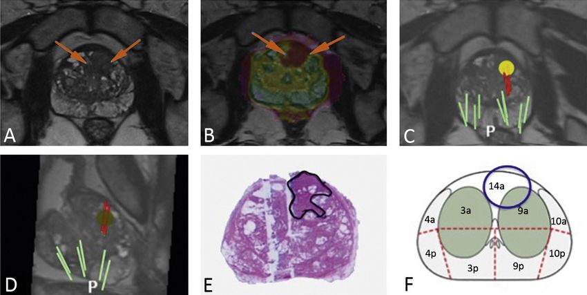
T2-weighted, diffusion-weighted [DW], and dynamic Figure 1. Three-dimensional transrectal US fused image shows the
location in the 1a sector of a suspicious focus (yellow; summed PI-RADS
contrast material-enhanced imaging. Targeted core score at multiparametric MR imaging, 14) that was targeted by two
biopsies and random systematic core biopsies were targeted (red) cores in complement to 12 random (green) systematic
performed. The elementary unit for analysis was the cores in a 61-mL prostate.
core. Relationships were assessed by using the Mann-
Whitney U test. Yates corrected and Pearson X(2) tests were used to evaluate categoric variables. A training
set was randomly drawn to construct the receiver operating characteristic curves for the summed PI-RADS
scores and for the Likert scale scores. The thresholds to recommend biopsy were obtained from the Youden J
statistics and were tested in the remaining validation set in terms of predictive characteristics. Interobserver
variability was analyzed by using weighed k statistics in a random set of 50 patients.
RESULTS: Higher T2-weighted, DW, and dynamic contrast-enhanced imaging PI-RADS scores were observed
in areas that yielded cancer-positive cores. The percentage of positive cores increased with the sum of scores
aggregated in five classes as follows: For summed PI-RADS scores of 3-5, the percentage of positive cores was
2.3%; for scores of 6-8, it was 5.8%; for scores of 9 or 10, it was 24.7%; for scores of 11 or 12, it was 51.8%; and
for scores of 13-15, it was 72.1% (P for trend,MRI/US TARGETED BIOPSY
DIAGNOSTIC ACCURACY
FIRST ROUND OF TARGETED BIOPSIES USING
MAGNETIC RESONANCE IMAGING/ULTRASONOGRAPHY
FUSION COMPARED WITH CONVENTIONAL
TRANSRECTAL ULTRASONOGRAPHY-GUIDED BIOPSIES
FOR THE DIAGNOSIS OF LOCALISED PROSTATE CANCER
Mozer P¹, Rouprêt M, Le Cossec C, Granger B, Comperat E, de Gorski A, Cussenot O, Renard-Penna R.
¹Academic Department of Urology, AP-HP, Hopital Pitié-Salpétrière, Paris, France; UPMC University of Paris 06, Institut des
Systèmes Intelligents et de Robotique.
BJUI. 2014
OBJECTIVES: To assess the accuracy of magnetic resonance imaging (MRI)/transrectal ultrasonography
(TRUS) fusion to guide first-round biopsies in the diagnosis of localised prostate cancer (PCa) in men with a
prostate-specific antigen (PSA) ≤10 ng/mL.
PATIENTS AND METHODS: A prospective study was conducted on men who met the following criteria: first-
round biopsy, multiparametric MRI (mpMRI) showing a lesion with a Likert score ≥2 and a PSADIAGNOSTIC ACCURACY
3D ULTRASOUND GUIDED BIOPSY
3D VERSUS 2D SYSTEMATIC TRANSRECTAL
ULTRASOUND-GUIDED PROSTATE BIOPSY: HIGHER
CANCER DETECTION RATE IN CLINICAL PRACTICE
Alexandre Peltier12, Fouad Aoun1&2 Fouad El-Khoury,1 Eric Hawaux,1 Ksenija Limani,1
Krishna Narahari,1 Nicolas Sirtaine,3 and Roland Van Velthoven1,2
¹ Department of Urology, Jules Bordet Institute, 1 Rue Héger-Bordet, 1000 Brussels, Belgium
² Université Libre de Bruxelles, 50 Franklin Roosevelt Avenue, 1050 Brussels, Belgium
³Department of Anatomopathology, Jules Bordet Institute, 1 Rue Héger-Bordet, 1000 Brussels, Belgium
Hindawi. 2013
PURPOSE: To compare prostate cancer detection rates of extended 2D versus 3D biopsies and to further assess
the clinical impact of this method in day-to-day practice.
MATERIALS AND METHODS: We analyzed the data of a
cohort of 220 consecutive patients with no prior history
of prostate cancer who underwent an initial prostate
biopsy in daily practice due to an abnormal PSA and/
or DRE using, respectively, the classical 2D and the
new 3D systems. All the biopsies were done by a single
experienced operator using the same standardized
protocol.
RESULTS: There was no significant difference in terms
of age, total PSA, or prostate volume between the two
groups. However, cancer detection rate was significantly
higher using the 3D versus the 2D system, 50% versus 34%
(P < 0.05). There was no statistically significant difference
while comparing the 2 groups in term of nonsignificant Figure 1. 3D trajectory visualization after biopsy
cancer detection. along with mapping and cartography.
CONCLUSIONS: There is reasonable evidence demonstrating the superiority of the 3D-guided biopsies in
detecting prostate cancers that would have been missed using the 2D extended protocol.
14DIAGNOSTIC ACCURACY 3D ULTRASOUND GUIDED BIOPSY
MAPPING OF TRANSRECTAL ULTRASONOGRAPHIC
PROSTATE BIOPSIES: QUALITY CONTROL AND
LEARNING CURVE ASSESSMENT BY IMAGE PROCESSING
Mozer P¹, Baumann M, Chevreau G, Moreau-Gaudry A, Bart S, Renard-Penna R, Comperat E, Conort P, Bitker
MO, Chartier-Kastler E, Richard F, Troccaz J.
Department of Urology, Assistance Publique-Hôpitaux de Paris, Paris, France
1
J US Med. 2009
PURPOSE: Mapping of transrectal ultrasonographic (TRUS) prostate biopsies is of fundamental importance for
either diagnostic purposes or the management and treatment of prostate cancer, but the localization of the
cores seems inaccurate. Our objective was to evaluate the capacities of an operator to plan transrectal prostate
biopsies under 2-dimensional TRUS guidance using a registration algorithm to represent the localization of
biopsies in a reference 3-dimensional ultrasonographic volume.
Thirty-two patients underwent a series of 12 prostate biopsies under local anesthesia performed by 1 operator
using a TRUS probe combined with specific third-party software to verify that the biopsies were indeed
conducted within the planned targets.
RESULTS: The operator reached 71% of the planned targets with substantial variability that depended on
their localization (100% success rate for targets in the middle and right parasagittal parts versus 53% for
targets in the left lateral base). Feedback from this system after each series of biopsies enabled the operator
to significantly improve his dexterity over the course of time (first 16 patients: median score, 7 of 10 and
cumulated median biopsy length in targets of 90 mm; last 16 patients, median score, 9 of 10 and a cumulated
median length of 121 mm; P = .046).
CONCLUSIONS: In addition to being a useful tool to improve the distribution of prostate biopsies, the potential
of this system is above all the preparation of a detailed «map» of each patient showing biopsy zones without
substantial changes in routine clinical practices.
Figure 1. Examples of biopsy distributions in the coronal plane for 2 different patients. Left, Patient from the first group, left base not sampled.
Right, Patient from the second group, good sampling. A indicates apex; B, base; L, left; and R, right.
15DIAGNOSTIC ACCURACY
PRECISION OF THE OBT FUSION®
PRECISION MATTERS IN MR IMAGING-TARGETED
PROSTATE BIOPSIES: EVIDENCE FROM A PROSPECTIVE
STUDY OF COGNITIVE AND ELASTIC FUSION
REGISTRATION TRANSRECTAL BIOPSIES.
Cornud F1, Roumiguié M1, Barry de Longchamps N1, Ploussard G1, Bruguière E2, Portalez D2, Malavaud B2.
¹Departments of Radiology (F.C.) and Urology (N.B.d.L.), Hôpital Cochin, Université Paris Descartes, Paris, France
2 Institut Universitaire du Cancer, Toulouse
Radiology 2018
PURPOSE: To measure the precision in placement of a biopsy needle in a magnetic resonance (MR) imaging–
detected target with transrectal ultrasonography (US), to document the clinical relevance of precision, and to
report on the precision of cognitive and software-based registrations.
PATIENTS AND METHODS: This prospective study was approved by the institutional review board and
performed between June 2013 and September 2013. Patients provided informed verbal consent. Two cores
each were obtained with cognitive and fusion techniques in 88 patients with a Prostate Imaging Reporting and
Data System version 1 score of at least 3. Precision was measured with Euclidian geometry by using the Digital
Imaging and Communications in Medicine archives of the biopsy as the distance from the core to the center
(dCC) and the distance from the core to the surface of the target modeled as a sphere. To address clustering of
data from multiple cores in the same patients, analyses of precision focused on the best shot for a patient or a
technique. The Welch unequal variance t test and Yates corrected x2 test were used as appropriate.
RESULTS: Mean precision was 2.5 mm (95% confidence interval: 1.8 mm, 3.3 mm). Positive cores were closer
to the center than were negative cores (dCC: 1.7 mm vs 3.1 mm, respectively; P = .025). More cancers were
detected with on-target than off-target cores (33 of 71 cores [46.5%] vs three of 17 cores [17.6%]; P = .03).
Cores obtained with the fusion technique achieved a higher precision than did cores obtained with the
cognitive technique (dCC: 2.8 mm vs 7.1 mm, respectively; P < .0001). Targeted cores demonstrated cancer in
44 patients. Fewer cancers were detected with the cognitive technique than with the fusion technique (31 of
44 patients [70.5%] vs 40 of 44 patients [90.9%]; P = .03).
CONCLUSION: A deformable MR imaging/transrectal US image registration system achieved a higher precision
and depicted cancer in more patients than did the cognitive freehand technique. was probably best than PET
choline for detecting prostate cancer but it could be complementary.
16DIAGNOSTIC ACCURACY PRECISION MATTERS IN
MR IMAGING–TARGETED PROSTATE BIOPSIES
CORNUD ET AL., RADIOLOGY , MAY 2018
PATIENTS UROLOGISTS
63
years >10
years experience
8.2
ng/mL COGNITIVE MRI FUSION &
88
PSA
ELASTIC MRI FUSION
40cc
METHODS
TRANSRECTAL BIOPSY 2 COGNITIVE CORES 2 ELASTIC FUSION CORES
ON-TARGET vs OFF-TARGET
CANCER TISSU SAMPLE
OFF-TARGET
2.5mm
ON-TARGET
7mm
CHANCE OF DEMONSTRATING CANCER
46.5% 17.6%
ON-TARGET OFF-TARGET
COGNITIVE vs ELASTIC FUSION
% OF POSITIVE CORES
70.5% 90.9%
COGNITIVE ELASTIC FUSION
PRECISION INFORMATION
Distance from the core to Distance from the core to
the center of the target the surface of the target
COGNITIVE FUSION COGNITIVE FUSION
LOCATION OF TARGETS
CORES (n=88) CORES (n=88) CORES (n=88) CORES (n=88)
BASE
8.4mm 3.6mm 4.0mm -0.8mm
MID 6.6mm 2.5mm 2.6mm -1.5mm
6.3mm 2.3mm 1.6mm -2.4mm
APEX
© KOELIS 2018
17DIAGNOSTIC ACCURACY
PRECISION OF THE OBT FUSION®
TRUS-MRI IMAGE REGISTRATION: A PARADIGM SHIFT IN
THE DIAGNOSIS OF SIGNIFICANT PROSTATE CANCER
Cornud F¹, Brolis L, Delongchamps NB, Portalez D, Malavaud B, Renard-Penna R, Mozer P.
¹Department of Radiology, Hôpital Cochin, Paris, France
Abdom Imaging. Dec 2013
ABSTRACT: Accuracy of multiparametric MRI has greatly improved the ability of localizing tumor foci of prostate
cancer. This property can be used to perform a TRUS–MR image registration, new technological advance,
which allows for an overlay of an MRI onto a TRUS image to target a prostate biopsy toward a suspicious
area Three types of registration have been developed: cognitive-based, sensor-based, and organ-based
registration. Cognitive registration consists of aiming a suspicious area during biopsy with the knowledge of
the lesion location identified on multiparametric MRI. Sensor-based registration consists of tracking in real
time the TRUS probe with a magnetic device, achieving a global positioning system which overlays in real-
time prostate image on both modalities. Its main limitation is that it does not take into account prostate and
patient motion during biopsy. Two systems (Artemis and Uronav) have been developed to partially circumvent
this drawback. Organ-based registration (Koelis) does not aim to track the TRUS probe, but the prostate itself
to compute in a 3D acquisition the TRUS prostate shape, allowing for a registration with the corresponding 3D
MRI shape. This system is not limited by prostate/patient motion and allows for a deformation of the organ
during registration. Pros and cons of each technique and the rationale for a targeted biopsy only policy are
discussed.
Figure 1. Diagram explaining differences between rigid (non defor-
mable) and elastic (deformable) registration. (b) Demonstration of the efficiency of elastic deformation. A The original
(a) Rigid registration. A Three points have been placed on the MRI (1) shape of the prostate with its correspondent 3D shape (red image, lower
and on TRUS prostate contour (2). Differences in prostate shape and row). B Induction with a mathematical model of a posterior deformation
deformation do not allow for an accurate prostate overlay after rigid of the prostate (10-mm-diameter sphere to simulate TRUS probe inser-
registration (3). B Elastic deformation with surface-based registration tion). C After activation of the elastic registration software, the original
and organ deformation. Multiple points have been placed on MRI and 3D shape has been rebuilt.
TRUS prostate contour (1, 2). This first step is a rigid registration which
still lacks accuracy owing to the differences in prostate shape (3). An
algorithm allows for a deformation of the MRI prostate shape to allow
for an accurate registration (4).
18CLINICAL PRACTICE
TRANSPERINEAL BIOPSY UNDER LOCAL ANESTHESIA
MULTICENTER TRANSPERINEAL MRI-TRUS FUSION
GUIDED OUTPATIENT CLINIC PROSTATE BIOPSIES
UNDER LOCAL ANESTHESIA
Jacewicz M¹, Günzel K, Rud E, Lauritzen P.M, Galtung K.F, Hinz S, Baco E.
Department of Urology, Oslo University Hospital, Oslo. Norway
1
Urol Oncol. 2020
INTRODUCTION: Transperineal Prostate biopsies (TPBx) are usually performed under general anesthesia
without image fusion. This study aimed to evaluate prostate cancer (Pca) detection rates (CDR), pain, and
adverse events using a novel, free-hand TPBx technique, based on elastic fusion of magnetic resonance
imaging (MRI) and transrectal ultrasound (TRUS) under local anesthesia.
MATERIALS AND METHODS: This multicenter retrospective study included all consecutive patients scheduled
for a TPBx. All had clinical suspicion of Pca, active surveillance scheduled for a re-biopsy, or suspicion of
local recurrence after previous treatment. Bi-parametric or multiparametric MRI was performed in all patients
and classified as positive in the case of Prostate Imaging-Reporting and Data System (PIRADS) suspicion ≥3.
At least 1 targeted TPBx was realized from each PIRADS ≥3 index lesion. Six to 12 systematic random TPBx
were done in patients with negative MRI. All biopsies were performed under local anesthesia in an outpatient
clinic with MRI-TRUS fusion and the 3D navigation system Trinity Perine (Koelis, France). Any- and clinically
significant Pca (csPca) (ISUP gr. ≥2) was recorded. Biopsy-related pain and adverse events were reported
according to a visual analogue score of 0–10.
RESULTS: In total, 377 patients were included for analyses. The mean age was 67 years (95% Confidence
Interval: 66–68) and the median prostate-specific antigen was 7.2 ng/ml (interquartile range [IQR] 4.8–11.0).
MRI was negative in 6% and positive in 94%. The median MRI prostate volume was 43 ml (IQR 31–60) and the
median MRI index tumor volume was 0.9 ml (IQR 0.5–2.1). The median number of TPBx was 4 (IQR 3–4). The
overall detection of any- and csPca was 64% and 52%, respectively. The overall CDR according to PIRADS 3,
4, and 5 was 30%, 70%, and 94%, respectively. In patients with negative MRI, any- and csPca was detected in
23% and 9%, respectively. The median visual analogue score score was 2 (IQR 1–3, range 0–7). Two patients
(0.5%) developed postbiopsy infection, of which one developed urosepsis. Treatment requiring haematuria
or urinary retention did not occur.
CONCLUSION: Free-hand MRI/TRUS fusion-guided and systematic random TPBx in LA is a feasible, safe, and
well-tolerated technique for diagnosing Pca.
20CLINICAL PRACTICE 21
CLINICAL PRACTICE
            TRANSPERINEAL BIOPSY UNDER LOCAL ANESTHESIA

     REFINING THE RISK-STRATIFICATION OF TRANSRECTAL
     BIOPSY-DETECTED PROSTATE CANCER BY ELASTIC
     FUSION REGISTRATION TRANSPERINEAL BIOPSIES
 Covin B1, Roumiguié M1, Quintyn-Ranty ML, Graff P, Khalifa J, Aziza R, Ploussard G, Portalez D, Malavaud B.
 1
     Department of Urology, Institut Universitaire du Cancer, Toulouse, France.

                                                                                           World J Urol 2018

 PURPOSE: To evaluate image-guided Transperineal Elastic-Registration biopsy (TPER-B) in the risk-
 stratification of low-intermediate risk prostate cancer detected by Transrectal-ultrasound biopsy (TRUS-B)
 when estimates of cancer grade and volume discorded with multiparametric Magnetic Resonance Imaging
 (MRI).

 METHODS: All patients referred for active surveillance or organ-conservative management were collegially
 reviewed for consistency between TRUS-B results and MRI. Image-guided TPER-B of the index target (IT) defined
 as the largest Prostate Imaging-Reporting Data System-v2 ≥ 3 abnormality was organized for discordant cases.
 Pathology reported Gleason grade, maximum cancer core length (MCCL) and total CCL (TCCL).

 RESULTS: Of 237 prostate cancer patients (1-4/2018), 30 were required TPER-B for risk-stratification. Eight
 cores were obtained [Median and IQR: 8 (6-9)] including six (IQR: 4-6) in the IT. TPER-B of the IT yielded longer
 MCCL [Mean and (95%CI): 6.9 (5.0-8.8) vs. 2.6 mm (1.9-3.3), p < 0.0001] and TCCL [19.7 (11.6-27.8) vs. 3.6 mm
 (2.6-4.5), p = 0.0002] than TRUS-B of the gland. On TPER-B cores, longer MCCL [Mean and (95%CI): 8.7 mm
 (6.7-10.7) vs. 4.1 mm (0.6-7.6), p = 0.002] were measured in Gleason score-7 cancers. TPER-B cores upgraded
 13/30 (43.3%) patients. 14/30 (46.7%) met University College London-definition 1 and 18/30 (60.0%) definition
 2, which correlate with clinically significant cancers > 0.5 mL and > 0.2 mL, respectively. 7/16 (43.8%) patients
 under active surveillance were re-allocated toward prostatectomy (n = 5) or radiation therapy (n = 2). In 14
 patients not yet assigned, TPER-B risk-stratification spurred the selection (13/14, 92.9%) of treatments with
 curative intent.

 CONCLUSION: Image-guided TPER-B of the index target provided more cancer material for pathology.
 Subsequent re-evaluation of cancer volume and grade switched a majority of patients towards higher-risk
 groups and treatments with curative intent.

22
CLINICAL PRACTICE 23
DIAGNOSTIC ACCURACY : PET/US TARGETED BIOPSY
BEYOND PROSTATE BIOPSY
                         INCIDENTALLY DETECTED 18 F-FDG-AVID PROSTATE
                         CANCER DIAGNOSED USING A NOVEL FUSION BIOPSY
                         PLATFORM
                         Meyer A.R¹, Leroy A², Allaf M.E, Rowe S.P, Gorin M.A.
                         1
                          The James Buchanan Brady Urological Institute and Department of Urology,Johns Hopkins University School of Medicine, Baltimore, Maryland
                         ² KOELIS, Inc., Cambridge, Massachusetts

                                                                                                                          J Endourol Case Rep. 2019

                         BACKGROUND: Localized prostate cancer rarely undergoes a shift in metabolism towards aerobic glycolysis, a
                         process known as the Warburg Effect. Because of this, positron emission tomography (PET)/CT imaging using
                         2-deoxy-2-[18F]fluoro-d-glucose (18F-FDG) is uncommonly used to evaluate patients with early-stage prostate
                         cancer. However, men undergoing an 18F-FDG PET/CT for unrelated reasons will on occasion be found to have
                         radiotracer uptake within the prostate gland. The appropriate work-up of these patients is poorly defined

                         CASE PRESENTATION: We present the case of a 61-year-old man with a history of tonsillar squamous cell
                         carcinoma who was incidentally found on 18F-FDG PET/CT to have a hypermetabolic nodule within the
                         prostate. The patient's prostate-specific antigen level was 2.1 ng/cc and digital rectal examination revealed no
                         abnormalities. The patient underwent a targeted prostate biopsy of the lesion using the KOELIS Trinity biopsy
                         platform, which uniquely allows for the real-time overlay of transrectal ultrasonography and PET/CT images.
                         Targeted biopsy revealed Gleason score 4 + 3 = 7 (grade group 3) prostate cancer.

                         CONCLUSION: Although the incidental detection of 18F-FDG uptake within the prostate is uncommon,
                         more than half of all patients will be found to have prostate cancer. Based on this case and our review of the
                         available medical literature, it is our belief that men with incidentally detected uptake of 18F-FDG within the
                         prostate should undergo further evaluation with a prostate biopsy. This recommendation is supported by
                         data suggesting that 18F-FDG-avid prostate cancer represents a more aggressive clinical phenotype.

                                                                                                                                                                     25
BEYOND PROSTATE BIOPSY
DIAGNOSTIC ACCURACY : PET/US TARGETED BIOPSY
FUSION TARGETED BIOPSY USING PSMA-PET/CT FOR
PROSTATE CANCER DIAGNOSIS IN PATIENTS WITH A
PREVIOUS NEGATIVE BIOPSY
Olivares R¹, Jofre B²,González P², Velasco A, Franco C, Román C.
¹Urology Department of Clínica Santa María, Santiago, Chili
²Imaging Department of Clínica Santa María, Santiago
CAU 2018
INTRODUCTION: For patients with a previous negative biopsy but with maintained clinical suspicion for
prostate cancer, Multiparametric Magnetic Resonance (MRI)-guided biopsy has demonstrated its usefulness
and efficiency, especially, for the diagnosis of a clinically significant disease.
Approximately 20% of patients have lesions that are “invisible” to resonance. In these cases, PET-CT could
have diagnostic usefulness with the definition of limits and guidance of the sampling during the medical
procedure.
METHODS:
A 57-year-old patient
PSA level of 10ng/ml
Non suspicious DRE
Biopsy: Two previous negative biopsies
PET/CT PSMA: Two lesions with increased uptake
RESULTS:
45-minute procedure
US/PET-CT Elastic Fusion using KOELIS Trinity® cartographer
Figure 1: Two radiotracer uptakes on
5 targeted core samples obtained from the suspect lesions PSMA-PET/CT
18 additional, random cores using sextant scheme as the reference
Anatomical Pathology confirmed a Gleason score of 3+4 in 3/5 of targeted biopsy cores and in 1 among
the aleatory sextant biopsy sampling
CONCLUSIONS: First report in Latin America about the usefulness of US/PET-CT PSMA Fusion Biopsy for
diagnosis of prostate cancer in a patient with previous negative biopsy and no evidence of malignancy in MRI
Series involving a higher number of patients will make possible to evaluate the usefulness and the cost-
effectiveness in clinical practice.
Figure 2: Targets' contour for the targeted biopsy on the US/PET-CR Fusion Figure 3: 3D map displaying sample cores
26BEYOND PROSTATE BIOPSY DIAGNOSTIC ACCURACY : PET/US TARGETED BIOPSY
TRIMODAL (18) F-CHOLINE-PET/MPMRI/TRUS
TARGETED PROSTATE BIOPSIES: FIRST CLINICAL
EXPERIENCE
J.L. Bonnal¹, A. Marien, A. Rock, K. El Maadarani, C. Francois, A. Delebarre, D. Berssard, B. Mauroy, P. Gosset, T.
Blaire
¹Hôpital Saint Philibert, Lomme, Groupement des Hôpitaux de l'Institut Catolique de Lille
EAU 2017
PURPOSE: In this preliminary study ,the feasibility of PET choline compared mpMRI was studied, to define
target prostate biopsy. The fusion of these two modalities with 3D echography was to compare the diagnostic
performance for primary localization of PCa with mpMRI and the latest generation of PET.
PATIENTS AND METHODS: In a prospective single-center study, fromDecember 2014 to October 2016, all
patients with PSA above 10ng/ml or patient with medical history of negative prostate biopsy were included.
3D biopsy with KOELIS system , mpMRI and PET scan Choline were done for each patients. The biopsy targets
were defined with both modalities and merging was done in real time during prostate biopsy sessions with the
3D echography. A review has been done to exclude patients with missed targets.The results were compared to
anatomopathological outcome of the biopies.Biopsy was done twice for each target at least and randomized
biopsy was done outside the target.
RESULTS: 31 patients were included, mean PSA was 13.01 (5.32-73). Mean number of biopsy was 16 (13-21)
and mean prostate volume was 63.41 cc (25-169). During our learning curve, 4 patients with several negative
targets but 1 missed target were excluded for global analysis.However,3 patients were detected as positive
while all targets were not biopsied. Furthermore, the PET fusion analysis failed for one patient. The cancer
detection rate was 69%. If the biopsy came back positive for cancer, the PET,th mpMRI or both targets were
respectively positive in 72%, 94%, 100%. On average in this population the number of biopsies by target
with TEP or mpMRI were respectively 1.77 (1-7) ,2.74 (3-11).The TEP and IRM by target were associated with
positives biopsies respectively in 43% and 62% .Compared to mpMRI ,for one patient only TEP gave a positive
target but fail with four other patients. mpMRI was probably best than PET choline for detecting prostate
cancer but it could be complementary.
CONCLUSIONS: We demonstrate the feasibility of multiple imagery fusion with echography 3D to define
localization of prostate cancer. It was very interesting to observe sometimes a great difference in the
distribution of PET choline target and mpMRI target in prostate. A new study with the novel ligands targeting
prostate specific membrane antigen (PSMA) could improve our clinical results.
Figure 1: The biopsy procedure was performed after registration of real-time TRUS with mpMRI and choline-PET by the same
operator, using 3D TRUS-tracking system. At the time of biopsy, volume data of the mpMRI and PET 18-ch was elastically fused
with TRUS. Each target was biopsied twice.
27BEYOND PROSTATE BIOPSY
REPEAT BIOPSY FOR ACTIVE SURVEILLANCE STRATEGY
MULTIPARAMETRIC MAGNETIC RESONANCE IMAGING
FACILITATES RECLASSIFICATION DURING ACTIVE
SURVEILLANCE FOR PROSTATE CANCER
Fujihara A¹, Iwata T, Shakir A, Tafuri A, Cacciamani G, Gill K, Ashrafi A, Ukimura O, Desai M, Duddalwar V, Stern
M, Aron M, Palmer S, Gill I, Abreu A.L.
1
USC Institute of Urology and Catherine & Joseph Aresty Department of Urology, University of Southern California, Los Angeles, CA, USA
BJUI Inter. 2020
OBJECTIVE: To investigate the utility of multiparametric magnetic resonance imaging (mpMRI) in the
reassessment and monitoring of patients on active surveillance (AS) for Grade Group (GG) 1 prostate cancer
(PCa).
PATIENTS AND METHODS: We identified, from our prospectively maintained institutional review board‐
approved database, 181 consecutive men enrolled on AS for GG 1 PCa who underwent at least one surveillance
mpMRI followed by MRI/prostate biopsy (PBx). A subset analysis was performed among 68 patients who
underwent serial (at least two) mpMRI/PBx during AS. Pathological progression (PP) was defined as upgrade
to GG ≥2 on follow up biopsy.
RESULTS: Baseline MRI was performed in 34 patients (19%). At a median follow‐up of 2.2 years for the overall
cohort, the PP was 12% (6/49) for Prostate Imaging Reporting and Data System (PI‐RADS) 1–2 lesions and
37% (48/129) for the PI‐RADS ≥3 lesions. The 2‐year PP‐free survival rate was 84%. Surveillance prostate‐
specific antigen density (P < 0.001) and surveillance PI‐RADS ≥3 (P = 0.002) were independent predictors of
PP on reassessment MRI/PBx. In the serial MRI cohort, the 2‐year PP‐free survival was 95% for the No‐MRI‐
progression group vs 85% for the MRI‐progression group (P = 0.02). MRI progression was significantly higher in
the PP (62%) than in the No‐PP (31%) group (P = 0.04). If serial MRI were used for PCa surveillance and biopsy
were triggered based only on MRI progression, 63% of PBx might be postponed at the cost of missing 12% of
GG ≥2 PCa in those with stable MRI. Conversely, this strategy would miss 38% of those with upgrading to GG ≥2
PCa on biopsy. Stable serial mpMRI correlates with no reclassification to GG ≥3 PCa during AS.
CONCLUSION: On surveillance mpMRI, PI‐RADS ≥3 was associated with increased risk of PCa reclassification.
Surveillance biopsy based only on MRI progression may avoid a large number of biopsies at the cost of missing
many PCa reclassifications.
28BEYOND PROSTATE BIOPSY REPEAT BIOPSY FOR ACTIVE SURVEILLANCE STRATEGY
PERFORMANCE OF SYSTEMATIC, MRI-TARGETED
BIOPSIES ALONE OR IN COMBINATION FOR THE
PREDICTION OF UNFAVOURABLE DISEASE IN MRI-
POSITIVE LOW-RISK PROSTATE CANCER PATIENTS
ELIGIBLE FOR ACTIVE SURVEILLANCE
Ploussard G¹, Beauval J-B, Lesourd M, Almeras C, Assoun J, Aziza R, Gautier J-R, Loison G, Portalez D, Salin A,
Tollon C, Soulié M, Malavaud B, Roumiguié M.
Department of Urology, La Croix du Sud Hospital, IUCT-O, Toulouse, France
1
World Journ Urol. 2019
PURPOSE: To assess the upstaging/upgrading rates of low-risk prostate cancer (PCa) according to the biopsy
scheme used (systematic (SB), targeted biopsies (TB), or both) in the setting of positive pre-biopsy MRI.
PATIENTS AND METHODS: We included 143 consecutive men fulfilling the Toronto University active
surveillance (AS) criteria who underwent a pre-biopsy positive MRI, a combination of SB and software-based
fusion TB, and a radical prostatectomy, in two expert centres. The primary endpoints were the pathological
upgrading and upstaging rates. Overall unfavourable disease (OUD) was defined by any pT3-4 and/or pN1
and/or ≥ GG 3.
RESULTS: Using TB alone would have missed 21.7% of cancers including 16.7% of ≥ GG 3. The use of TB
was significantly associated with a lower risk of ≥ Grade Group (GG) 3 disease (p < 0.006) in RP specimens.
Combination of SB and TB lowered this risk by 39%, compared with TB alone. The biopsy scheme did not
affect the upstaging rates which were substantial even in case of combination scheme (from 37 to 46%). OUD
was detected in approximately 50% of cases. The presence of high grade on TB was the only independent
predictive factor for both ≥ GG 2 (p = 0.015) and ≥ GG 3 (p = 0.023) in RP specimens.
CONCLUSION: High grade on TB biopsies represented the major predictor of upgrading. Combination of SB
and TB better defined the sub-group of patients having the lowest risk of reclassification, compared with TB
or SB alone. The risk of non-organ-confined disease remained high, and could not be accurately predicted by
MRI or systematic/targeted biopsy features.
29BEYOND PROSTATE BIOPSY
REPEAT BIOPSY FOR ACTIVE SURVEILLANCE STRATEGY
A NOVEL TECHNIQUE USING THREE-DIMENSIONALLY
DOCUMENTED BIOPSY MAPPING ALLOWS PRECISE
RE-VISITING OF PROSTATE CANCER FOCI WITH SERIAL
SURVEILLANCE OF CELL CYCLE PROGRESSION GENE
PANEL
Ukimura O1, Gross ME, de Castro Abreu AL, Azhar RA, Matsugasumi T, Ushijima S, Kanazawa M, Aron M, Gill IS.
1USC Institute of Urology, Keck School of Medicine, University of Southern California, Los Angeles, California.
The Prostate. 2015
BACKGROUND: Conventional systematic biopsy
has the shortcoming of sampling error and reveals
«no evidence of cancer» with a rate of >50% on
active surveillance (AS). The objective of this study
is to report our initial experience of applying a
3D-documented biopsy-mapping technology to
precisely re-visit geographically documented low-
risk prostate cancer and to perform serial analysis of
cell-cycle-progression (CCP) gene-panel.
METHODS: Over a period of 40 months (1/2010- Figure. 1. The re-visiting biopsy technique using the pair of ‘’virtual’’ and
4/2013), the 3D-biopsy-mapping technique, in ‘’real’’ targeting. The re-visiting biopsy technique is demonstrated in Figure
2 by the pair of ‘’virtual’’ (long orange trajectory, circled in yellow in the
which the spatial location of biopsy-trajectory was left of Fig. 3) and ‘’real’’ biopsies (short orange trajectory, circled in yellow
digitally recorded (Koelis), was carried out. A pair in the right of Fig. 2), intentionally sampling from the previously positive
of diagnostic (1st-look) and surveillance (2nd-look) targets in the location of the right-apex-medial. Because the virtual tar-
biopsy were performed per subject (n = 25), with geting seemed 3–4 mm too lateral to reach the target, the direction of the
real biopsy was further corrected to bring it closer to the target. Note that
median interval of 12 months. The documented one core (green trajectory) had already been sampled during the current
biopsy-trajectory was used as a target to guide the 2nd-look biopsy from the same positive target with the same re-visiting
re-visiting biopsy from the documented cancer technique. These two re-visiting cores in the 2nd look biopsy were positive
focus, as well as the targeted field-biopsy from the for cancer. The overlaid image demonstrates that the pair of green and
orange trajectories of the re-visiting technique almost corresponded (or
un-sampled prostatic field adjacent to negative were located within 1–2 mm) with the blue trajectory of the positive core
diagnostic biopsies. The accuracy of re-visiting at the right-apex-medial on the 1st-look biopsy.
biopsy and biopsy-derived CCP signatures were
evaluated in the pair of the serial biopsy-cores.
RESULTS: The 1st-look-biopsy revealed a total of 43 cancer lesions (1.7 per patient). The accuracy of re-visiting
cancer was 86% (37/43) per lesion, 76% (65/86) per core, and 80% (20/25) per patient. This technology also
provided an opportunity for 3D-targeted field-biopsy in order to potentially minimize sampling errors. The CCP
gene-panel of the 1st-look (-0.59) versus 2nd-look (-0.37) samples had no significant difference (P = 0.4); which
suggested consistency in the molecular signature of the known cancer foci during the short-time interval of
median 12 months. Any change in CCP of the same cancer foci would be likely due to change in sampling
location from the less to more significant portion in the cancer foci rather than true molecular progression.
The study limitations include a small number of the patients.
CONCLUSION: The 3D-documented biopsy-mapping technology achieved an encouraging re-sampling
accuracy of 86% from the known prostate cancer foci, allowing the serial analysis of biopsy-derived CCP
signatures.
30BEYOND PROSTATE BIOPSY REPEAT BIOPSY FOR ACTIVE SURVEILLANCE STRATEGY
VALIDATION OF THE EUROPEAN SOCIETY OF
UROGENITAL RADIOLOGY SCORING SYSTEM FOR
PROSTATE CANCER DIAGNOSIS ON MULTIPARAMETRIC
MAGNETIC RESONANCE IMAGING IN A COHORT OF
REPEAT BIOPSY PATIENTS
Portalez D¹, Mozer P, Cornud F, Renard-Penna R, Misrai V, Thoulouzan M, Malavaud B.
Department of Radiology, Clinique Pasteur, Toulouse, France
1
World Journ Urol. 2019
BACKGROUND: Wide variations in acquisition protocols and the lack of robust diagnostic criteria make
magnetic resonance imaging (MRI) detection of prostate cancer (PCa) one of the most challenging fields in
radiology and urology.
DESIGN, SETTING, AND PARTICIPANTS: An institutional review board-approved multicentric prospective
study; 129 consecutive patients (1514 cores) referred for mpMRI after at least one set of negative biopsies.
INTERVENTION: Transfer of mpMRI-suspicious areas on three-dimensional (3D) transrectal ultrasound
images by 3D elastic surface registration; random systematic and targeted cores followed by core-by-core
analysis of pathology and mpMRI characteristics of the core locations. The ESUR scores were assigned after
the procedure on annotated Digital Imaging and Communications in Medicine archives.
IOUTCOME MEASUREMENTS AND STATISTICAL ANALYSIS: Relationships between ESUR scores and biopsy
results were assessed by the Mann-Whitney U test. The Yates correction and Pearson χ(2) tests evaluated
the association between categorical variables. A teaching set was randomly drawn to construct the receiver
operating characteristic curve of the ESUR score sum (ESUR-S). The threshold to recommend biopsy was
obtained from the Youden J statistics and tested in the remaining validation set in terms of sensitivity,
specificity, positive predictive value, negative predictive value, and accuracy.
RESULTS AND LIMITATIONS: Higher T2-weighted, dynamic weighted imaging and dynamic contrast-enhanced
ESUR scores were observed in areas yielding cancer-positive cores. The proportion of positive cores increased
with the ESUR-S aggregated in five increments (ESUR-S 3-5: 2.9%; ESUR-S 6-8: 11.1%; ESUR-S 9-10: 38.2%;
ESUR-S 11-12: 63.4%; and ESUR-S 13-15: 83.3%; pYou can also read