Efficient rational modification of non-ribosomal peptides by adenylation domain substitution - Nature
←
→
Page content transcription
If your browser does not render page correctly, please read the page content below
ARTICLE
                  https://doi.org/10.1038/s41467-020-18365-0                OPEN

                  Efficient rational modification of non-ribosomal
                  peptides by adenylation domain substitution
                  Mark J. Calcott          1,2,   Jeremy G. Owen1,2 & David F. Ackerley                     1,2 ✉

                  Non-ribosomal peptide synthetase (NRPS) enzymes form modular assembly-lines, wherein
                  each module governs the incorporation of a specific monomer into a short peptide product.
1234567890():,;

                  Modules are comprised of one or more key domains, including adenylation (A) domains,
                  which recognise and activate the monomer substrate; condensation (C) domains, which
                  catalyse amide bond formation; and thiolation (T) domains, which shuttle reaction inter-
                  mediates between catalytic domains. This arrangement offers prospects for rational peptide
                  modification via substitution of substrate-specifying domains. For over 20 years, it has been
                  considered that C domains play key roles in proof-reading the substrate; a presumption that
                  has greatly complicated rational NRPS redesign. Here we present evidence from both directed
                  and natural evolution studies that any substrate-specifying role for C domains is likely to be
                  the exception rather than the rule, and that novel non-ribosomal peptides can be generated
                  by substitution of A domains alone. We identify permissive A domain recombination
                  boundaries and show that these allow us to efficiently generate modified pyoverdine peptides
                  at high yields. We further demonstrate the transferability of our approach in the PheATE-
                  ProCAT model system originally used to infer C domain substrate specificity, generating
                  modified dipeptide products at yields that are inconsistent with the prevailing dogma.

                  1 School of Biological Sciences, Victoria University of Wellington, Wellington, New Zealand. 2 Centre for Biodiscovery and Maurice Wilkins Centre for

                  Molecular Biodiscovery, Victoria University of Wellington, Wellington, New Zealand. ✉email: david.ackerley@vuw.ac.nz

                  NATURE COMMUNICATIONS | (2020)11:4554 | https://doi.org/10.1038/s41467-020-18365-0 | www.nature.com/naturecommunications                                1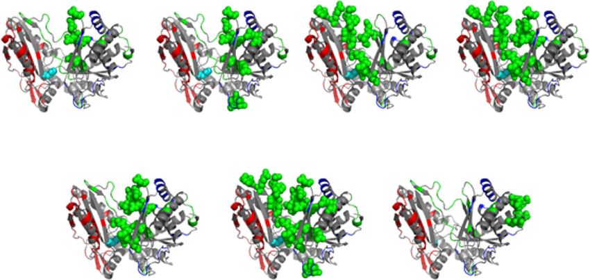
ARTICLE NATURE COMMUNICATIONS | https://doi.org/10.1038/s41467-020-18365-0
T
he earliest reported attempts to create artificial non- versus zero of nine substitutions of A domains specifying alternative
ribosomal peptide synthetase (NRPS) enzymes were sub- residues9,15. In contrast, non-synonymous C-A domain substitu-
stitutions of A-T domains into the second and seventh tions generated modified pyoverdines in three out of ten cases,
modules of the NRPSs involved in the biosynthesis of the lipo- suggesting that a C domain with compatible acceptor site specificity
peptide surfactin1,2. Although modified lipopeptides were detected was required for functionality9,15,16.
using mass spectrometry, in each case the yield of modified lipo- We now show that our previous failure to generate modified
peptide was substantially diminished, to only trace levels2. Evidence pyoverdines by A domain substitution is surmountable by use of
that C domains exhibit stringent specificity toward the acceptor more effective recombination boundaries, and that the C domains
substrate activated by their cognate (downstream) A domains in the pyoverdine NRPS system do not impose stringent proof-
offered an explanation for the reduced yields3,4. This evidence was reading constraints. We perform comprehensive evolutionary
based on NRPS enzymes artificially loaded with amino-acyl CoA or analyses across three different bacterial genera that are consistent
aminoacyl-N-acetylcysteamine thioesters, which mimic an amino with this conclusion, indicating that non-ribosomal peptide
acid attached to a T domain. Soon after, a substrate binding pocket divergence is primarily driven by recombination of A domains or
of the C domain was suggested to play a role in controlling the sub-domains, independent of partner C domains. Tellingly, we
direction of biosynthesis5. Subsequent efforts at substituting cognate show that we can also incorporate leucine as the acceptor residue
C-A domains together enjoyed modest success, reinforcing the in the tyrocidine PheATE/ProCAT dimodular NRPS model
belief that this was necessary to bypass C domain substrate (Fig. 1b, d), which was previously found not to accept artificially-
specificity6,7. Since then the most successful engineering attempts loaded leucine thioesters at this position—a key observation
have focused on substituting C-A domains together8,9 or A-T-C underpinning the original hypothesis that C domains exhibit
domains with the condition of not disrupting C domain acceptor stringent selectivity for the acceptor substrate3.
site specificity10–12. It is now widely accepted in the field that
successful domain substitution requires working within the con- Results
straints imposed by C domain specificity8,10,13. Investigating C domain proofreading via semi-rational DNA
Until now, our own work using pyoverdine as a model system shuffling. Previous structural biology and bioinformatics
has been consistent with this dogma. Pyoverdine from Pseudomo- approaches to identify residues involved in the presumed sub-
nas aeruginosa PAO1 is a UV-fluorescent siderophore formed from strate specificity of C domains have been unsuccessful17–19. We
an 11-membered peptide, encoded by NRPS modules that will here instead adopted a semi-rational shuffling strategy, by combining
be referred to as Pa1-Pa11 (Fig. 1a, c). We observed that five out of regions of DNA encoding C domains that incorporate different
five synonymous A domain substitutions into PvdD (an NRPS amino acid substrates, then seeking to retrospectively identify
comprised of modules Pa10 and Pa11, both of which specify L-Thr important substrate-defining residues. For this we selected the
as the native substrate)14 yielded detectable pyoverdine products, Lys-specific module Pa8 and the Thr-specific module Pa11, as we
a
PvdL PvdI PvdJ PvdD
E E E Te
ACL T C A T C A T C A T C A T C A T C A T C A T C A T C A T C A T C A T
FA L-Glu D-Tyr L-Dab D-Ser L-Arg D-Ser L-fhOrn L-Lys L-fhOrn L-Thr L-Thr
b
TycA TycB TycC
E E Te
A T C A T C A T C A T C A T C A T C A T C A T C A T C A T
D-Phe L-Pro L-Phe D-Phe L-Asn L-Gln L-Tyr L-Val L-Orn L-Leu
c d
OH
NH O O
H O
N
H2 N N N NH HN H
H N O
H O
O NH O
O HN OH N N
HN
O O H
HO HN H NH2
HO O
O
NH N N O
H 2N HN
NH
O O O O
HO O O
HO
HN N OH NH O
N NH
O NH2
H2 N O N
N OH O HN H
H OH
O
O
Fig. 1 NRPS assembly lines and products relevant to this work. Depicted are the biosynthetic gene clusters (blue arrows) and their encoded NRPS
assembly lines involved in the biosynthesis of a pyoverdine and b tyrocidine, and the corresponding chemical structures for c pyoverdine and d tyrocidine.
The NRPS module Pa11 is highlighted in blue in a, as is the corresponding L-Thr residue that it incorporates into pyoverdine in c. Likewise, the two modules
that were extracted from the tyrocidine NRPS system by Belshaw et al.3 and used to generate their PheATE/ProCAT model system are highlighted in
orange in b, as are the D-Phe and L-Pro residues these incorporate into tyrocidine in d. NRPS domains are labelled as follow: ACL acyl-CoA ligase, C
condensation, A adenylation, T thiolation, E epimerisation, and Te thioesterase. The following non-standard amino acid abbreviations are used: Dab, 2,4-
diaminobutyric acid; fhOrn, N5-formyl-N5-hydroxyornithine; Orn, ornithine.
2 NATURE COMMUNICATIONS | (2020)11:4554 | https://doi.org/10.1038/s41467-020-18365-0 | www.nature.com/naturecommunications
NATURE COMMUNICATIONS | https://doi.org/10.1038/s41467-020-18365-0                                                                  ARTICLE

had previously found a pyoverdine with Lys at position 11 could                 proof-reading residues21 was inconsistent with the hypothesis
be generated by substituting the C-A domains (but not the A                     that C domain acceptor site specificity had caused our previous A
domain) from Pa8 into Pa11 of PvdD9. At the time, we inter-                     domain substitutions to be non-functional9,15.
preted this result as showing that the Pa8 A domain can function
within the PvdD environment, but only when paired with a Lys-
                                                                                Assessing the efficiency of A domain substitutions. Our pre-
specifying C domain. An added attraction of the model is that the
                                                                                vious C-A substitutions in pyoverdine module Pa11 had a com-
Pa8 and Pa11 C domains appear to be paralogs, as they are nearly
                                                                                bined success rate of only 3/10 constructs yielding a detectable
identical apart from three stretches of low sequence identity
                                                                                pyoverdine product, with two of these being in very low
(Supplementary Fig. S1a). We reasoned that these low-identity
                                                                                yield9,15,16. To test whether A domain substitution using our new
regions were likely to contain substrate-specifying residues, and a
                                                                                upstream recombination boundary was a more efficient strategy,
homology model based on the C domain from TycC (pdb:
                                                                                we generated the equivalent linker + A domain substitution
2JGP20) suggested the three regions could be shuffled effectively,
                                                                                constructs for each of our three previously successful C-A domain
with only a minimal number of amino acid perturbations intro-
                                                                                substitutions. In each case, the pyoverdine yield was increased by
duced (Fig. 2a; Supplementary Fig. S1b, c).
                                                                                substituting the linker and A domain together (Fig. 3a; Supple-
   DNA blocks spanning each of the three variable regions of the
                                                                                mentary Figs. S6, S7). We then tested whether we could efficiently
Pa8 (presumed K-specific) and Pa11 (presumed T-specific) C
                                                                                produce other modified pyoverdines, by randomly selecting nine
domains were shuffled to yield all eight possible combinations
                                                                                A domains that activate substrates other than Thr from Pseudo-
(TTT, KTT,…, KKK; Fig. 2b). Each shuffled C domain was then
                                                                                monas species within the antiSMASH database22, and substitut-
introduced into a pvdD gene construct, immediately upstream of
                                                                                ing them into module Pa11 (Supplementary Table 1;
either the native Pa11 or a substituted Pa8 A domain (Fig. 2b;
                                                                                Supplementary Fig. S8). Six of these A domain substitution var-
Supplementary Fig. S2). We observed that region 3 played a
                                                                                iants gave modified pyoverdines at high yields (Fig. 3b; Supple-
dominant role in defining the substrate compatibility of the
                                                                                mentary Fig. S9). Collectively, these results confirmed not only
recombinant C domains. With the exception of the recombinant
                                                                                that substitution of an A domain without the corresponding C
C domain KKT, which was not functional in association with
                                                                                domain is possible, but that it can result in improved success rates
either A domain, C domains that contained region 3 from Pa11
                                                                                and yields compared to C-A domain substitutions.
were substantially more active in partnership with the Pa11 A
domain, and only C domains that contained region 3 from Pa8
were active in partnership with the Pa8 A domain (Fig. 2c;                      A domain substitution has driven NRPS diversification in
Supplementary Fig. S3). These data suggested that region 3                      nature. Our success in generating modified pyoverdines via A
contains key specificity-determining residues. However, it is                    domain substitution led us to consider whether this approach
important to note that the recombination point between the C                    might be applicable to other pathways. Compared to natural
and A domains was near the A1 motif. This meant region 3 of the                 evolution, the number of substitutions that can be created in the
C domain was always substituted in association with the                         lab is infinitesimal, and we reasoned that we could gain valuable
corresponding loop that delineates C and A domains and is                       insight by investigating whether A domain substitution has
often referred to as a linker region10,21. The linker region is an              occurred frequently during natural NRPS diversification. The
approximately 36 residue sequence that begins at the C-terminus                 tight acceptor site specificity originally proposed by Belshaw
of the final helix of the C domain, and extends to the first helix of             et al.3 has fuelled speculation that C and A domains are likely to
the A domain. When designing this experiment we considered                      co-evolve23,24. This supposition is at odds with observations that
the linker was unlikely to be a significant factor, as there is no               complete or partial A domain substitution has driven diversifi-
structural basis for considering that the linker region could be                cation of the microcystin25, aeruginosin26, hormaomycin27 and
involved in acceptor site specificity, and the linker region has                 lipo-octapeptide28 biosynthetic pathways. The microcystins are a
appeared unimportant in previously successful synonymous                        particularly interesting example, as diversification by A domain
substitutions9,10,15.                                                           substitution has been especially prevalent25 despite in vitro assays
   Region 3 contains 38 non-identical residues between the Pa8                  suggesting the C domain may play an extended gatekeeper role in
and Pa11 C domains (Supplementary Fig. S1a). With the goal of                   controlling substrate specificity29. We consider it pertinent to
narrowing down the key substrate defining elements, proximal                     note that the latter study also demonstrated that A domains
clusters of residues within the C domain of the Thr-specific                     in vitro may adenylate a very different repertoire of amino acid
module Pa11 were substituted by the corresponding residues                      substrates if they are purified in isolation than when they are
within Pa8 (Fig. 2d; Supplementary Fig. S4). These substitutions                purified in association with their partner C domain (a second
focused on clusters of 6 (Shell 1, Fig. 2d) or 12 (Shell 1 and 2,               example of this in vitro inconsistency was reported soon after-
Fig. 2d) residues closest to the catalytic histidine and/or the loop            ward for SulM from the sulfazecin monobactam gene cluster30).
extending across the solvent channel. We also generated a control               As adenylation is independent of any catalytic role of the C
(Linker, Fig. 2d) in which the Pa11 C domain was fused to the                   domain, the inconsistency has been attributed to indirect con-
linker region from module Pa8. Surprisingly, modifications to the                formational effects30, which in turn suggests that isolated A
C domain had little effect on pyoverdine production, however                    domains may appear in vitro to be more promiscuous than they
changing the Pa11 linker region to that from Pa8 was sufficient to               actually are in vivo. Thus, while it is possible that diversification
allow the native PvdD C domain to function efficiently with the                  via A domain substitution in the microcystin, aeruginosin, hor-
Pa8 lysine-specifying A domain (Fig. 2e; Supplementary Fig. S5).                maomycin and lipo-octapeptide pathways has been enabled by
The resulting pyoverdine species was produced in high yield and                 unusually relaxed-specificity C domains, our experimental success
contained lysine at position 11. Conceptually, substituting the A               in generating modified pyoverdines led us to consider that these
domain together with its cognate linker is just an A domain                     examples might not be so unusual, and that A domain substitu-
substitution that uses a different recombination boundary (i.e. at              tion might be a more global phenomenon driving NRPS
the very C-terminal end of the C domain as opposed to the                       diversification.
recombination sites within the linker previously used by us9,15                    We first investigated this by constructing phylogenetic trees
and Bozhüyük et al.10). Identification of a more permissive                      based on the C and A domains from NRPS enzymes involved in
recombination boundary that did not exchange any plausible                      the biosynthesis of four different pyoverdines (from strains of P.

NATURE COMMUNICATIONS | (2020)11:4554 | https://doi.org/10.1038/s41467-020-18365-0 | www.nature.com/naturecommunications                          3
ARTICLE                                                                                 NATURE COMMUNICATIONS | https://doi.org/10.1038/s41467-020-18365-0

          a   Highlighted regions of the C domains
                                                                    C domain
                                                                                                                                                                    A domain
                     Region 1

                     Region 2

                     Region 3

                                                  C                                                                                                                                                   N

          b   Semi-rational shuffling approach                                             c                                      Shuffling the three regions
                                                                                                                            100
                                              Shuffled C-domains                                                                                                                                                                                              Thr A domain

                                                                                           Yield relative to wildtype (%)
                                                                                                                                                                                                                                                              Lys A domain
                Thr         C                 C            C               C                                                 75

                Lys         C                 C            C               C                                                 50

                                                                                                                             25

                                         PvdD A-domain variants
                                                                                                                             0
                 C      A       T         A    T Te       C     A   T        A   T Te                                             TTT             KTT               TKT      TTK                      KKK                  TKK            KTK                      KKT
                                     Thr A-domain                       Lys A-domain
                                                                                                                                              C             C         C       C                       C                             C   C                         C

          d   Modifications to region 3                                                    e                                      Testing modifications to region 3
                                                                                                                            100
                                                                                           Yield relative to wildtype (%)

                                                                                                                             75

              Shell 1               Shell 1 and 2          Loop         Shell 1 and loop
                                                                                                                             50

                                                                                                                             25

                   Shell 1 and                 Shell 1, 2 and       Linker
                  edges of loop                     loop
                                                                                                                              0
                                                                                                                                    Shell 1

                                                                                                                                                    Shell 1 and 2

                                                                                                                                                                      Loop

                                                                                                                                                                                   Shell 1 and loop

                                                                                                                                                                                                            Shell 1 and edges of loop

                                                                                                                                                                                                                                        Shell 1, 2 and loop

                                                                                                                                                                                                                                                                      Linker

aeruginosa, Pseudomonas putida, Pseudomonas fluorescens and                                            NRPS gene clusters available within the antiSMASH database22
Pseudomonas syringae). This revealed strong clustering of A                                           for the genera Pseudomonas, Streptomyces and Bacillus. The
domains by substrate specificity, providing evidence that C and A                                      sequences were extracted, clustered at 95% identity and aligned to
domains had evolved independently (Supplementary Fig. S10).                                           give a total of 437, 370 and 213 LCL-A-T tri-domain sequences for
To perform a more global analysis of whether C and A domains                                          Pseudomonas, Bacillus and Streptomyces species, respectively.
have evolved independently, we downloaded the sequences of all                                        Analysis with TreeOrderScan31,32, which assesses 400 bp

4                NATURE COMMUNICATIONS | (2020)11:4554 | https://doi.org/10.1038/s41467-020-18365-0 | www.nature.com/naturecommunications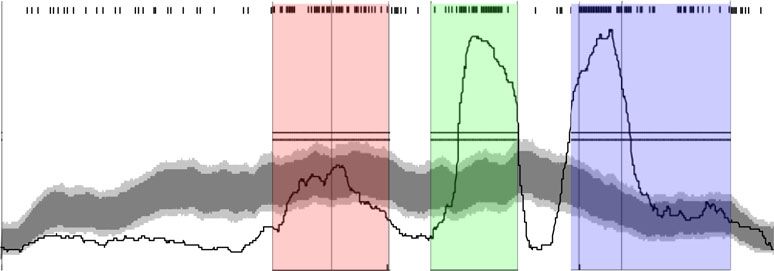
NATURE COMMUNICATIONS | https://doi.org/10.1038/s41467-020-18365-0 ARTICLE
Fig. 2 DNA shuffling reveals a permissive recombination boundary for A domain substitution. a The three variable regions (coloured red, blue and green
in accordance with shading used in Supplementary Fig. S1) of the Pa8 and Pa11 C domains mapped onto the structure of the C-A domains derived from PDB:
2VSQ21. Residues differing between the Pa8 and Pa11 C domains are shown as spheres. b Overview of the semi-rational shuffling approach used to narrow
down substrate specifying regions. The three variable regions of the C domains from modules Pa8 (green) and Pa11 (purple) were shuffled in every
combination to create eight variant C domains. Each of these was inserted into a plasmid containing a pvdD gene lacking the Pa11 C domain (left-hand
construct), and a plasmid containing a pvdD gene lacking the Pa11 C domain, and in which the Pa11 A domain had additionally been replaced by the Lys-
specific A domain from Pa8 (right-hand construct). c Pyoverdine production from pvdD deletion strains transformed by the variant pvdD genes from b was
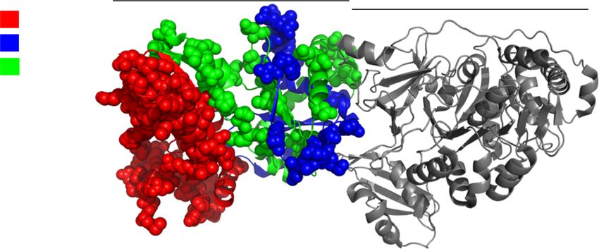
assessed by measuring absorbance at 400 nm relative to a wild-type P. aeruginosa strain. d Homology models highlighting (in light green) clusters of
residues that were substituted as groups within the third region of Pa11 together with the linker-only substituted control. e Pyoverdine production from pvdD
deletion strains transformed by the variant pvdD genes from panel d was assessed by measuring absorbance at 400 nm relative to a wild-type P. aeruginosa
strain. For c and e, n = 6 independent experiments and data are presented as mean values ± SD. Source data are provided as a Source Data file.
a Comparison of C-A with linker plus A domains b Additional linker plus A domain substitutions in PvdD
100 100
C-A
Yield relative to wildtype (%)
Yield relative to wildtype (%)
Linker + A
75 75
50 50
25 25
0 0
Lys Ser fhOrn
a
p
n
rn
rn
lu
ly
r
r
Se
Se
Al
As
As
G
G
O
O
fh
fh
1:
6:
3:
5:
2:
7:
9:
8:
Fig. 3 Efficient generation of modified pyoverdines by linker + A domain substitutions. a Pyoverdine production for C-A domain substitution strains 4:
compared with the corresponding linker + A domain substitution strains. b Pyoverdine production for nine additional A + linker domain substitution
variants. Pyoverdine production was assessed by measuring absorbance at 400 nm relative to a wild-type P. aeruginosa strain. In all cases, n = 6
independent experiments and data are presented as mean values ± SD. Source data are provided as a Source Data file. The abbreviation
fhOrn represents N5-formyl-N5-hydroxyornithine.
subalignments at 50 bp intervals, revealed increased phylogenetic Partial A domain substitution has previously been attempted in
incompatibility between A domains and the surrounding two key laboratory studies34,35. However, only one of these
domains (Fig. 4a). Segregation analysis to examine whether any (working in an initiation module that lacks a C domain)
of the 400 bp subalignments cluster by substrate specificity described the formation of a modified peptide, the rate of
identified the region of the A domain that exhibited the greatest formation of which was greatly reduced in vitro and not tested
phylogenetic incompatibility as clustering strongly by substrate further in vivo35. We tested partial A domain substitution in
specificity (Fig. 4b). PvdD using boundaries suggested by our recombination analysis,
The TreeOrderScan analysis is consistent with A domain but did not achieve visible production of pyoverdine in any
substitution driving NRPS evolution but does not identify the instance (Supplementary Figs. 11, 12). Reasoning that this might
recombination points that have arisen most commonly during be due to structural clashes restricting efficient recombination of
natural substitution events. To identify hotspots of recombina- NRPS enzymes within certain domain regions, we used
tion, sequence analysis was performed using RDP4, an ensemble SCHEMA36 to predict the number of perturbations generated
of tools that collectively identify regions at which DNA sequences by recombination of the C-A-T domains from PvdD with the C-
are likely to have recombined33. The breakpoint distribution A-T domains from alternative pyoverdine NRPS modules
identified recombination hotspots located between C and A (Fig. 4d). Whereas the recombination sites employed in our
domains (Fig. 4c, red shading), upstream to the A domain successful linker + A domain substitutions were within regions
substrate binding pocket between the A2 and A4 motifs (Fig. 4c, with low potential to cause structural perturbations during
green shading), and downstream to the binding pocket starting substitution, the recombination hotspot between the A2 and A4
from close to the A5 motif (Fig. 4c, blue shading). The largest motifs (Fig. 4c, green shading), was in a region with high
hotspots were located immediately on either side of the binding potential for perturbations. Testing of additional partial A
pocket, flanking the region that segregates most strongly by domain substitution constructs that used alternative upstream
substrate specificity (Fig. 4a–c). These data were consistent for recombination points in conjunction with the downstream site
each of the Pseudomonas, Bacillus and Streptomyces genera, and used in our A domain experiments was also unsuccessful
inconsistent with the hypothesis that C and A domains co-evolve. (Supplementary Figs. S11, S12). We believe that the SCHEMA
We conclude that complete or partial A domain substitution analysis explains why our (and previous34,35) experimental
appears to play a primary role in diversification of NRPS attempts at partial A domain substitution were generally
pathways in nature, rather than being an exception. unsuccessful, or at best highly inefficient35. In contrast, because
NATURE COMMUNICATIONS | (2020)11:4554 | https://doi.org/10.1038/s41467-020-18365-0 | www.nature.com/naturecommunications 5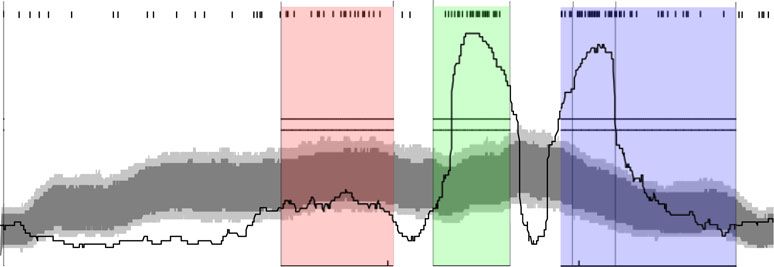
ARTICLE NATURE COMMUNICATIONS | https://doi.org/10.1038/s41467-020-18365-0
a Phylogenetic incompatibility per sequence c Recombination hotspot analysis
(kbp) Pseudomonas (kbp) Bacillus (kbp) Streptomyces Pseudomonas
1.0 C1 C7 X A1 A2 A4 A5 A6 A10
2.5 2.1 2.5
Violation frequency
2.0 0.8 100.0
2.0
Breakpoints per 200 nt
1.5 0.6
1.5 1.5 75.0
1.0 0.9 1.0 0.4
window
0.2 50.0
0.5 0.5
0.3
0.0 25.0
0.5 1.5 2.5 (kbp) 0.3 0.9 1.5 2.1 (kbp) 0.5 1.5 2.5 (kbp)
0.0
1 640 1281 1922 2562
b Segregation by consensus substrate specificity Nucleotide position in relation to CP000438.1.cluster007_CA6
C1 C7 A1 A2 A4 A5 A6 A10 Bacillus
C1 C7 A1 A2 A4 A5 A6 A10
Segregation score
0.6 Pseudomonas
60.0
Breakpoints per 200 nt
0.4 45.0
window
0.2 30.0
0 15.0
0 200 400 600 800 1000 1200 1400 1600 1800 2000 2200 2400 (bp)
0.0
C1 C7 A1 A2 A4 A5 A6 A10 1 569 1138 1708 2277
0.8 Nucleotide position in relation to CP000560.1.cluster006_CA5
Bacillus
Segregation score
Streptomyces
0.6
C1 C7 A1 A2 A4 A5 A6 A10
0.4 63.0
Breakpoints per 200 nt
47.3
0.2
window
31.5
0
0 200 400 600 800 1000 1200 1400 1600 1800 2000 2200 15.8
C1 C7 A1 A2 A4 A5 A6 A10
0.0
1 644 1288 1933 2577
0.8 Streptomyces
Segregation score
Nucleotide position in relation to CP014485.1.cluster015_CA1
0.6 d Clashes at each recombination point
C1 C7 X A1 A2 A4 A5 A6 A10
0.4 80
Average number of clashes
70
0.2 60
50
0 40
0 200 400 600 800 1000 1200 1400 1600 1800 2000 2200 2400 (bp)
30
20
Bootstrap value 0 Bootstrap value 50 Bootstrap value 70 10
0
0 200 400 600 800 1000
Alignment position
Fig. 4 Natural NRPS diversification is driven primarily by A domain and subdomain substitution. a Phylogenetic compatibility matrices of alignments of
C-A-T domains from Pseudomonas, Bacillus and Streptomyces species showing frequencies of phylogeny violations for each pairwise comparison of
sequence fragments. Analysis was performed using TreeOrder Scan31,32, considering alignments of 400 bp at 50 bp intervals and using a bootstrap value
of 70% to calculate phylogenetic violations. b Segregation of alignments by consensus substrate specificity predictions from antiSMASH22. Segregation
scores were calculated using a 0% (red line), 50% (blue line) and 70% (green line) bootstrap cut off. The locations of key conserved motifs are indicated
along the top of the graph. For this analysis, 400 bp alignments were compared for segregation into groups based on consensus substrate specificity
predictions from antiSMASH22. A segregation score of 0 means perfect segregation by substrate specificity and a score of 1.0 means no segregation on the
basis of substrate specificity. Shaded blocks have been added to aid comparison between the corresponding regions of interest in b–d. c Recombination
hotspot analysis of C-A-T domains from Pseudomonas, Bacillus and Streptomyces species. ‘X’ marks the location of the recombination point used for the
successful (linker control) Lys A domain substitution in Fig. 2e. Dark and light grey areas indicate local breakpoint hotspots at the 95 and 99% confidence
level, respectively, and the two horizontal lines indicate cut-offs for global breakpoint hotspots at the 95 and 99% confidence level. d The light blue shaded
plot indicates the mean number of clashes calculated using SCHEMA that would be introduced by recombination of the nine alternative pyoverdine NRPS
modules depicted in Fig. 3b with the domains from Pa11 (n = 9 C-A-T domains and the dark blue region indicates ± SD). Source data are available via the
links provided in the “Methods”.
natural recombination processes favour short sequences37,38, this domain acceptor substrate specificity is less of a barrier to NRPS
appears to be a preferred region of recombination during natural engineering than previously proposed by several groups, including
NRPS evolution, with the low success rate presumably offset by us3,4,10–13,39. However, the possibility remained that the Pa8 and
the high frequency of recombination events. Irrespective, our Pa11 C domains are more relaxed than C domains such as that in
diverse analyses provide unanimous support for partial or module 2 of tyrocidine biosynthesis—the system that Belshaw et al.3
complete A domain substitution being a primary mechanism used to develop the original hypothesis of C domains exhibiting
for NRPS diversification. strong acceptor substrate specificity. We therefore considered it
important to test whether we could create novel dipeptides by
A domain substitution in the PheATE-ProCAT model system. performing A domain substitution within the same system.
Our high success rates in modifying pyoverdine via A domain Belshaw et al showed that tyrocidine module one (PheATE,
substitution and our bioinformatics analyses suggested that C incorporating D-Phe) and module two (ProCAT, incorporating
6 NATURE COMMUNICATIONS | (2020)11:4554 | https://doi.org/10.1038/s41467-020-18365-0 | www.nature.com/naturecommunications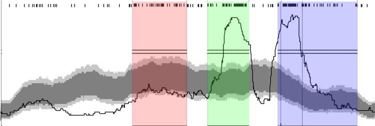
NATURE COMMUNICATIONS | https://doi.org/10.1038/s41467-020-18365-0                                                                              ARTICLE

L-Pro) could be artificially loaded with different amino acids                   specificity C domains, and other subtypes of C domains might
in vitro, and the resulting dipeptide purified and analysed3.                    exist that demonstrate stringent acceptor site specificity. Never-
Artificial loading with L-Pro resulted in the expected product, but              theless, we have also substantially expanded upon previous work
no product resulted when ProCAT was loaded with L-Leu,                          that has examined NRPS evolution in individual pathways by
suggesting stringent substrate specificity during condensation. If               providing evidence across diverse genera that NRPS diversifica-
this hypothesis is true, the C-domain of ProCAT will not accept                 tion occurs predominantly by A domain (or subdomain) sub-
L-Leu. We therefore considered that efficient production of D-                   stitution. This observation is inconsistent with widespread
Phe-L-Leu dipeptides via A domain substitution in the ProCAT                    acceptor site specificity existing as a barrier to successful A
module would disprove the C domain substrate specificity                         domain substitution. Further work will be needed to understand
hypothesis in this foundational model. To avoid potential for                   how broadly these findings can be applied, but we consider they
in vitro inconsistencies and facilitate construct generation and                hold considerable potential for efficient rational and combina-
analysis we used a similar two-plasmid in vivo system to previous               torial improvement of medically and industrially relevant
researchers40,41, with the replacement of the T domain from                     peptides.
ProCAT with the T-Te domains from SrfC, to enable release of
linear D-Phe-L-Leu dipeptides42,43. The genetic regions encoding
four different Leu-specifying A domains were selected for                       Methods
                                                                                DNA manipulation. All plasmids, primers and sequences used in this study are
substitution into the ProCATTe plasmid and compared to an                       provided in the Supplementary file Supplementary Data 1 - Plasmids Primers and
unsubstituted control (Supplementary Table S2; Supplementary                    Gene Sequences.xlsx.
Fig. S13). All A domains shared relatively low amino acid identity                  To create vectors for substituting domains into pvdD, the PBAD promoter was
to the A domain from ProCAT (40.4% to 47.6%; Supplementary                      excised from pSW196 using the restriction sites NsiI and SacI and ligated into
Table 2). The Leu-specifying A domain from SrfC was of                          pUCP22 using the restriction sites PstI and SacI. The resulting plasmid was named
                                                                                pUCBAD. Next, the pvdD gene lacking the C-A domains from the second module
particular interest because the crystal structure of this module                (module Pa11) was excised from the plasmid pSMC9 using NheI and SacI
fuelled speculation that C-A domains form a tight interface,                    restriction sites and annealed into the pUCBAD vector using the restriction sites
which may further restrict A domain substitution21. As such, this               NheI and SacI to create the vector pUCBAD-SMC. The Thr-specific A domain
experiment combined all the main factors that have been                         from the second module of PvdD and the Lys-specific A domain from the first
                                                                                module of PvdJ (module Pa8) were PCR amplified and separately ligated into the
suggested to prohibit effective A domain substitution, i.e. a C                 pUCBAD-SMC vector. This resulted in the creation of the vectors pDEC-Thr and
domain believed to exhibit tight acceptor site specificity, the                  pDEC-Lys, which contained a Thr-specific A domain and a Lys- specific A domain
substitution of distantly related A domains, and substitution of an             variant of the pvdD gene, respectively. For modifying the third variable region of
A domain believed to depend on a tight C-A domain interface                     the C domain from pvdD, the first two variable regions of the pvdD C domain from
                                                                                module Pa11 were PCR amplified. The resulting fragment was ligated into the
with its cognate C domain partner.                                              pDEC-Thr vector using a 5′ SpeI/XbaI and a 3′ SalI/SalI ligation to create the
   The recombinant ProCATTe plasmids and a second plasmid                       plasmid pTRN. The destruction of the SpeI restriction site within the vector by
containing PheATE were used to co-transform a BAP1 strain of                    SpeI/XbaI ligation meant the introduced SpeI site downstream to region 2 of the C
E. coli44. The strains were grown for 18 h, after which the                     domain was unique within the plasmid, allowing insertion of region 3 using SpeI
                                                                                and SalI restriction sites.
supernatant was extracted and dipeptides quantified using HPLC                       C domains were created by overlap PCR or synthesis (Twist Bioscience; San
and absorbance at 214 nm (Fig. 5). We detected production of the                Francisco, CA). C domain sequences were amplified using the appropriate forward
native D-Phe-L-Pro diketopiperazine at 7.8 mg/L by the control                  and reverse primers specific to the C-domain from modules Pa8-Lys, Pa11-Thr,
strain (Fig. 5a), a yield that compares favourably to previous                  Ps5-Ser or Pf6-Orn (Pa indicates Pseudomonas aeruginosa PA01; Ps5-Ser indicates
reports40,41. We also detected D-Phe-L-Leu dipeptides for three of              the fifth serine-incorporating pyoverdine NRPS module from Pseudomonas
                                                                                syringae 1448a; Pf6-Orn indicates the sixth ornithine-incorporating pyoverdine
the four strains containing Leu-specific A domain substitutions,                 NRPS module from Pseudomonas fluorescens SBW25; Supplementary Fig. S1). PCR
at yields ranging from ca. 25 to 40% of the D-Phe-L-Pro control                 products were digested using XbaI and XhoI, and ligated into the plasmids pDEC-
(Fig. 5b). Despite sharing the lowest amino acid identity with the              Thr and pDEC-Lys using compatible SpeI and SalI restriction sites. The partial C
ProCAT A domain, and its previously hypothesised requirement                    domain fragments containing region 3 of a C domain were amplified using the
                                                                                appropriate forward and reverse primers. PCR products were digested using SpeI
to maintain a native C domain interface, the strain containing the              and XhoI, and ligated into the plasmid pTRN at compatible SpeI and SalI
A domain from SrfC was found to produce D-Phe-L-Leu at 1.8                      restriction sites.
mg/L. We would not have expected these successful outcomes                          A domains were selected to activate a range of substrates, from modules
based on the previous work of Belshaw et al.3, and conclude                     exhibiting a range of amino acid sequence identities with Pa11-Thr
                                                                                (Supplementary Table S1). To enable cloning of A domains into the substitution
stringent acceptor site specificity against Leu as previously                    vector pTRN, inserts were designed to have an upstream region identical to the C-
inferred for this C domain is not a barrier to creating successful              domain from Pa11-Thr, fused to the linker and A domain from the selected
A domain substitutions. Rather, it appears that successful A                    modules. Recombination points for each substitution are shown in Supplementary
domain substitution relies greatly on the recombination bound-                  Fig. S9. To reduce the GC content to acceptable levels for synthesis, 5_AP013068.1.
                                                                                cluster003_CA1 was codon optimised for P. aeruginosa PAO1 using the guided
aries used.                                                                     random method in OPTIMIZER46.
                                                                                    Partial A domain substitutions were created for the Lys-specific A domain from
                                                                                module Pa8 and the Ser-specific A domain from number 2 in Fig. 2B. Substitutions
Discussion                                                                      were created by ligating synthetic DNA constructs into the vector pTRN.
While natural evolution has given rise to a large diversity of non-             Recombination points for partial A domain substitutions are shown in
                                                                                Supplementary Fig. S11, and are labelled in the Supplementary file Supplementary
ribosomal peptides, effective re-engineering of NRPS templates in               Data 1 - Plasmids Primers and Gene Sequences.xlsx using the GrsA nomenclature
the laboratory has proven difficult13,19,45. A primary focus in re-              from Kries et al.35. The recombination points tested were T221 and I352,
engineering NRPS enzymes has been to accommodate the pre-                       corresponding to those used previously by Kries et al.35, and K205 and A322,
sumed acceptor site specificity of the C domain. We have shown                   corresponding to those used by Crüsemann et al.34 Based on our SCHEMA
                                                                                analysis we also tested the promising recombination points S233 and A332 as well
this might not be as necessary as previously thought. Our iden-                 as upstream recombination points at A185, I167 and S233 in combination with the
tification of tolerant recombination points via an unbiased DNA                  downstream site used in the full A domain substitutions.
shuffling approach allowed us to subsequently produce modified                        C-A domains from modules Ps5-Ser, Pf6-Orn and Pa8-Lys were amplified by
products with high success rates in two diverse NRPS systems,                   PCR and ligated into the vector pUCBAD-SMC.
                                                                                    DNA encoding PheATE and ProC-TTe was artificially synthesised (Twist
one of which should have been intractable, as the cornerstone of                Bioscience; San Francisco, CA) following codon optimisation for E. coli using the
the C-domain acceptor site proof-reading hypothesis. The pos-                   guided random method in OPTIMIZER46. PheATE was cloned into pACYCDuet-1
sibility remains that both systems might contain relaxed-                       using NcoI and XhoI restriction sites, and ProC-TTe was cloned into pET28a+

NATURE COMMUNICATIONS | (2020)11:4554 | https://doi.org/10.1038/s41467-020-18365-0 | www.nature.com/naturecommunications                                         7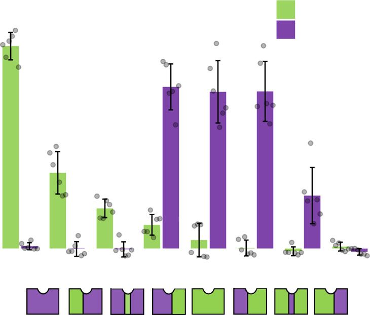
ARTICLE                                                                          NATURE COMMUNICATIONS | https://doi.org/10.1038/s41467-020-18365-0

              a

                  APhe     T     E        +       C       APro     T     Te                1.TycB m1                                    7.8 ± 0.7 mg/L

                                                                                                                                        D-Phe-L-Pro    DKP

                                                                                            6     8     10    12    14    16    18    min

              b
                                                                                           2.TycC m6                                    3.1 ± 0.1 mg/L

                                                                                           3. SrfC m1                                   1.8 ± 0.2 mg/L

                  APhe      T     E                C                T Te                                                                2.1 ± 0.1 mg/L
                                                                                           4. WP_094154864 m2

                                                                                           5. WP_000023653 m3                           ND

                                               ALeu                                                                                     D-Phe-L-Leu

                                                                                            6      8    10    12     14    16    18    min

Fig. 5 Successful substitution of Leu-specifying A domains in the PheATE-ProCAT model system. a Schematic showing the domain arrangement of the
PheATE/ProCATTe constructs used in this study and HPLC traces comparing the product made by an E. coli BAP1 strain expressing these constructs (1)
relative to an analytical standard of D-Phe-L-Pro DKP (dark blue trace). b Schematic and HPLC traces for the products generated by four strains (2–5)
bearing variants of ProCATTe in which the Pro-specifying A domain had been substituted by a Leu-specifying A domain. Strains 2, 3 and 4 show a peak
corresponding to an analytical standard of D-Phe-L-Leu (dark blue trace) (n = 3 independent experiments and data are presented as the mean yield ± SD).
Masses of the expected products were verified using HR-ESI-MS (Supplementary Fig. S14). Source data are provided as a Source Data file.

using NcoI and XhoI restriction sites. The Pro-specific A domain from ProCAT            and 100 bootstraps. The random seed was set to 1. To detect recombination hot
and four Leu-specific A domains were codon optimised and ligated into the SpeI          spots, the aligned sequences were analysed using RDP433. Default settings were
and NotI restriction sites of ProC-TTe using compatible NheI and NotI sites.           used except sequences were specified as linear, only recombination events detected
Alignments of A domains and sequence origins and identities are provided in            by at least three methods were considered and alignment consistency was
Supplementary Fig. S14 and Supplementary Table S2).                                    unchecked. A breakpoint distribution plot was created using a 200 bp window and
                                                                                       1000 permutations.
                                                                                           The structure 2VSQ was used as it was identified as the top template for
Data analysis. Structural models of the C domain from the second module of             modelling the CAT-domains from Pa11 using the Swiss-Model server63. Sequences
PvdD were created by submitting the C domain sequence to multiple automated            were aligned using MUSCLE68 and then SCHEMA36 was used to create a contact
servers47–62, and using the Swiss-Model server (http://swissmodel.expasy.org/)63       map for each structure. The python script Schema_profile.py (available at https://
and Modeller 9.1164. Models created from each method were submitted to the             github.com/MarkCalcott/NRPS_evolution/tree/master/Schema_Bar_Graph)
QMEAN server to obtain QMEAN6 and QMEANclust scores65,66. The model                    calculated the average number of clashes using SCHEMA for each recombination
RaptorXmsa was selected to work with as it scored best overall considering both        point between Pa11 and the modules used as a source of A-domains.
measurements. The model RaptorXmsa aligned well to the C domain structure
from TycC21 with a root mean-square deviation for the backbone α carbons of
0.381 Å.                                                                               Analysing pyoverdine production. Each strain to be analysed was first used to
   The antiSMASH database22 was queried for all NRPS biosynthetic gene clusters        inoculate 200 μL of low salt LB in a 96 well plate. After 24 h growth at 37 °C, 10 μL
from the genera Pseudomonas, Bacillus and Streptomyces. The genbank files for each      of each starter culture was used to inoculate 190 μL of M9 media amended with
cluster were downloaded, and the python script extractCATdomains_consensus.            0.1 % (w/v) L-arabinose and 4 g/L succinate (pH 7.0). Cultures were grown for
py (available at https://github.com/MarkCalcott/NRPS_evolution/tree/master/            37 °C for 24 h, centrifuged to pellet bacteria, and then 100 μL of supernatant
Raw_sequences) used to find, extract and save DNA encoding LCL-A-T tridomains           transferred to a fresh 96 well plate and diluted twofold in fresh M9 media to give a
into a separate FASTA file for each genera. The criteria for extracting tridomains      total volume of 200 μL. Absorbance (400 nm) was measured using an EnSpire 2300
were that the A domain had an antiSMASH consensus substrate specificity                 Multilabel Reader (PerkinElmer, Waltham, MA, USA). For mass spectrometry
prediction, and that C-A-T domains were located on the same protein in the correct     analysis, 1 μL of supernatant was mixed with 20 μL of matrix (500 μL acetonitrile,
order, with fewer than 250 amino acid residues between domains. Each extracted         500 μL ultrapure water, 1 μL trifluoroacetic acid, 10 μg α-cyano-4-hydroxycinnamic
DNA sequence was annotated with the consensus substrate specificity. Sequences          acid)16,70. Aliquots of 0.5 μL were spotted in triplicate onto an Opti-TOF® 384 well
were dereplicated using USEARCH 10.0.24067, and clustered at 95% identity              MALDI plate (Applied Biosystems, Foster City, CA) and allowed to dry at room
(Supplementary Table S3). A codon-alignment of the centroid nucleotide sequence        temperature. Spots were analysed using a MALDI TOF/TOF 5800 mass spectrometer
from each cluster was generated using MUSCLE68. Sequences were trimmed to the          (Applied Biosystems) in positive ion mode. Peaks were externally calibrated using cal2
C1 and T motifs inclusive, and any sequences not containing these motifs were          calibration mixture (Applied Biosystems). Peaks in spectra were labelled in Data
removed. Regions of ambiguous alignment were removed using GBLOCK version              Explorer (Applied Biosystems).
0.91b69. The default parameters were used for GBLOCK except the minimum
number of sequences for a flank position was set equal to 50% of the total sequences,
the minimum length of a block was five, and gap positions were allowed in half of       Analysing dipeptide production. The plasmids containing PheATE and Pro-
the sequences.                                                                         CATTe were transformed into the E. coli strain BAP144. A 100 µL aliquot from an
   Simple sequence editor version 1.3 was used for TreeOrder scan analysis31,32.       overnight culture was used to inoculate 10 mL M9 media amended with 0.1% (w/v)
Sequences were grouped by the antiSMASH consensus prediction of A domain               casamino acids and 0.4% (w/v) D-glucose. The culture was grown at 30 °C for 5 h
substrate specificity. Length of fragments were 400 bp with an increment of 50 bp,      and then incubated on ice for 20 min. IPTG was added to a final concentration of

8                    NATURE COMMUNICATIONS | (2020)11:4554 | https://doi.org/10.1038/s41467-020-18365-0 | www.nature.com/naturecommunications
NATURE COMMUNICATIONS | https://doi.org/10.1038/s41467-020-18365-0                                                                                          ARTICLE

1 mM, and the culture grown for a further 16 h at 18 °C. The cells were pelleted by        14. Ackerley, D. F., Caradoc-Davies, T. T. & Lamont, I. L. Substrate specificity of
centrifugation at 2000 g for 20 min. The supernatant was transferred to a fresh tube           the nonribosomal peptide synthetase PvdD from Pseudomonas aeruginosa. J.
and immediately extracted with 4:1 butanol–chloroform (vol/vol). The organic                   Bacteriol. 185, 2848–2855 (2003).
layer was washed with 1 volume of 0.1 M NaCl and the aqueous phase removed.                15. Ackerley, D. F. & Lamont, I. L. Characterization and genetic manipulation of
Solvent was removed under vacuum and residue dissolved in 150 µL of 10%                        peptide synthetases in Pseudomonas aeruginosa PAO1 in order to generate
CH3CN. HPLC separation was performed on an Agilent 1200 LC system using a                      novel pyoverdines. Chem. Biol. 11, 971–980 (2004).
C18 reverse-phase column (PhenoSphere-Next, 150 by 4.6 mm; pore size, 120 Å;               16. Calcott, M. J. & Ackerley, D. F. Portability of the thiolation domain in
particle size, 3 μm; Phenomenex, Torrance, CA). The column oven was set at 40 °C               recombinant pyoverdine non-ribosomal peptide synthetases. BMC Microbiol.
and injection volume at 30 µL. Elution was conducted with a mobile phase con-                  15, 162 (2015).
sisting of (A) water + 0.1% formic acid, and (B) CH3CN + 0.1% formic acid.                 17. Bloudoff, K., Alonzo, D. A. & Schmeing, T. M. Chemical probes allow
Following 1 min at 10% B, a gradient up to 25% B was performed in 17 min. The                  structural insight into the condensation reaction of nonribosomal peptide
flow rate was set at 1 mL/min. Concentrations were determined by comparison to                  synthetases. Cell Chem. Biol. 23, 331–339 (2016).
chemically synthesised standards with 98% purity by LifeTein (South Plainfield,             18. Rausch, C., Hoof, I., Weber, T., Wohlleben, W. & Huson, D. H. Phylogenetic
NJ). For HPLC-MS, separation was performed on an Agilent 6530 Accurate-Mass                    analysis of condensation domains in NRPS sheds light on their functional
Q-TOF LC/MS system equipped with an Agilent 1260 HPLC system (Agilent,                         evolution. BMC Evol. Biol. 7, 78 (2007).
Santa Clara, CA). The same HPLC conditions as previously were used except the
                                                                                           19. Sussmuth, R. D. & Mainz, A. Nonribosomal peptide synthesis-principles and
column oven was set at 45 °C and injection volume at 1 µL.
                                                                                               prospects. Angew. Chem. Int. Ed. 56, 3770–3821 (2017).
                                                                                           20. Samel, S. A., Schoenafinger, G., Knappe, T. A., Marahiel, M. A. & Essen, L. O.
Reporting summary. Further information on research design is available in the Nature           Structural and functional insights into a peptide bond-forming bidomain from
Research Reporting Summary linked to this article.                                             a nonribosomal peptide synthetase. Structure 15, 781–792 (2007).
                                                                                           21. Tanovic, A., Samel, S. A., Essen, L. O. & Marahiel, M. A. Crystal structure of
                                                                                               the termination module of a nonribosomal peptide synthetase. Science 321,
Data availability                                                                              659–663 (2008).
Source data are provided with this paper.
                                                                                           22. Blin, K., Medema, M. H., Kottmann, R., Lee, S. Y. & Weber, T. The antiSMASH
                                                                                               database, a comprehensive database of microbial secondary metabolite
Code availability                                                                              biosynthetic gene clusters. Nucleic Acids Res. 45, D555–D559 (2017).
All code generated during this study is available on GitHub via the following links: (1)   23. Baltz, R. H. Synthetic biology, genome mining, and combinatorial biosynthesis
python script extractCATdomains_consensus.py https://github.com/MarkCalcott/                   of NRPS-derived antibiotics: a perspective. J. Ind. Microbiol. Biotechnol. 45,
NRPS_evolution/tree/master/Raw_sequences (2) python script Schema_profile.py                    635–649 (2018).
https://github.com/MarkCalcott/NRPS_evolution/tree/master/Schema_Bar_Graph.                24. Lautru, S. & Challis, G. L. Substrate recognition by nonribosomal peptide
                                                                                               synthetase multi-enzymes. Microbiol-SGM 150, 1629–1636 (2004).
                                                                                           25. Fewer, D. P. et al. Recurrent adenylation domain replacement in the
Received: 5 June 2020; Accepted: 19 August 2020;                                               microcystin synthetase gene cluster. BMC Evol. Biol. 7, 183 (2007).
                                                                                           26. Ishida, K. et al. Plasticity and evolution of aeruginosin biosynthesis in
                                                                                               cyanobacteria. Appl. Environ. Microbiol. 75, 2017–2026 (2009).
                                                                                           27. Hofer, I. et al. Insights into the biosynthesis of hormaomycin, an exceptionally
                                                                                               complex bacterial signaling metabolite. Chem. Biol. 18, 381–391 (2011).
                                                                                           28. Götze, S. et al. Structure elucidation of the syringafactin lipopeptides provides
References                                                                                     insight in the evolution of nonribosomal peptide synthetases. Chem. Sci. 10,
1.    Stachelhaus, T., Schneider, A. & Marahiel, M. A. Rational design of peptide              10979–10990 (2019).
      antibiotics by targeted replacement of bacterial and fungal domains. Science         29. Meyer, S. et al. Biochemical dissection of the natural diversification of
      269, 69–72 (1995).                                                                       microcystin provides lessons for synthetic biology of NRPS. Cell Chem. Biol.
2.    Schneider, A., Stachelhaus, T. & Marahiel, M. A. Targeted alteration of the              23, 462–471 (2016).
      substrate specificity of peptide synthetases by rational module swapping. Mol.        30. Li, R. F., Oliver, R. A. & Townsend, C. A. Identification and characterization
      Gen. Genet. 257, 308–318 (1998).                                                         of the sulfazecin monobactam biosynthetic gene cluster. Cell Chem. Biol. 24,
3.    Belshaw, P. J., Walsh, C. T. & Stachelhaus, T. Aminoacyl-CoAs as probes of               24–34 (2017).
      condensation domain selectivity in nonribosomal peptide synthesis. Science           31. Simmonds, P. SSE: a nucleotide and amino acid sequence analysis platform.
      284, 486–489 (1999).                                                                     BMC Res. Notes 5, 50 (2012).
4.    Ehmann, D. E., Trauger, J. W., Stachelhaus, T. & Walsh, C. T. Aminoacyl-             32. Simmonds, P. Recombination and selection in the evolution of picornaviruses
      SNACs as small-molecule substrates for the condensation domains of                       and other mammalian positive-stranded RNA viruses. J. Virol. 80,
      nonribosomal peptide synthetases. Chem. Biol. 7, 765–772 (2000).                         11124–11140 (2006).
5.    Linne, U. & Marahiel, M. A. Control of directionality in nonribosomal peptide        33. Martin, D. P., Murrell, B., Golden, M., Khoosal, A. & Muhire, B. RDP4:
      synthesis: role of the condensation domain in preventing misinitiation and               Detection and analysis of recombination patterns in virus genomes. Virus
      timing of epimerization. Biochemistry 39, 10439–10447 (2000).                            Evol. 1, vev003 (2015).
6.    Mootz, H. D., Schwarzer, D. & Marahiel, M. A. Construction of hybrid peptide         34. Crusemann, M., Kohlhaas, C. & Piel, J. Evolution-guided engineering of
      synthetases by module and domain fusions. Proc. Natl Acad. Sci. U.S.A. 97,               nonribosomal peptide synthetase adenylation domains. Chem. Sci. 4,
      5848–5853 (2000).                                                                        1041–1045 (2013).
7.    Duerfahrt, T., Doekel, S., Sonke, T., Quaedflieg, P. J. & Marahiel, M. A.             35. Kries, H., Niquille, D. L. & Hilvert, D. A subdomain swap strategy for
      Construction of hybrid peptide synthetases for the production of alpha-l-                reengineering nonribosomal peptides. Chem. Biol. 22, 640–648 (2015).
      aspartyl-l-phenylalanine, a precursor for the high-intensity sweetener               36. Voigt, C. A., Martinez, C., Wang, Z. G., Mayo, S. L. & Arnold, F. H. Protein
      aspartame. Eur. J. Biochem. 270, 4555–4563 (2003).                                       building blocks preserved by recombination. Nat. Struct. Mol. Biol. 9, 553–558
8.    Baltz, R. H. Combinatorial biosynthesis of cyclic lipopeptide antibiotics: a             (2002).
      model for synthetic biology to accelerate the evolution of secondary metabolite      37. Gonzalez-Torres, P., Rodriguez-Mateos, F., Anton, J. & Gabaldon, T. Impact
      biosynthetic pathways. ACS Synth. Biol. 3, 748–758 (2014).                               of homologous recombination on the evolution of prokaryotic core genomes.
9.    Calcott, M. J., Owen, J. G., Lamont, I. L. & Ackerley, D. F. Biosynthesis of             Mbio 10, pii: ARTN e02494–18 (2019).
      novel pyoverdines by domain substitution in a nonribosomal peptide                   38. Didelot, X. & Maiden, M. C. J. Impact of recombination on bacterial
      synthetase of Pseudomonas aeruginosa. Appl. Environ. Microbiol. 80,                      evolution. Trends Microbiol. 18, 315–322 (2010).
      5723–5731 (2014).                                                                    39. Brown, A. S., Calcott, M. J., Owen, J. G. & Ackerley, D. F. Structural,
10.   Bozhuyuk, K. A. J. et al. De novo design and engineering of non-ribosomal                functional and evolutionary perspectives on effective re-engineering of non-
      peptide synthetases. Nat. Chem. 10, 275–281 (2018).                                      ribosomal peptide synthetase assembly lines. Nat. Prod. Rep. 35, 1210–1228
11.   Bozhuyuk, K. A. J. et al. Modification and de novo design of non-ribosomal                (2018).
      peptide synthetases using specific assembly points within condensation                40. Gruenewald, S., Mootz, H. D., Stehmeier, P. & Stachelhaus, T. In vivo
      domains. Nat. Chem. 11, 653–661 (2019).                                                  production of artificial nonribosomal peptide products in the heterologous
12.   Steiniger, C., Hoffmann, S. & Sussmuth, R. D. Desymmetrization of                        host Escherichia coli. Appl. Environ. Microbiol. 70, 3282–3291 (2004).
      cyclodepsipeptides by assembly mode switching of iterative nonribosomal              41. Kries, H. et al. Reprogramming nonribosomal peptide synthetases for
      peptide synthetases. ACS Synth. Biol. 8, 661–667 (2019).                                 “Clickable” amino acids. Angew. Chem. Int. Ed. 53, 10105–10108 (2014).
13.   Bozhuyuk, K. A., Micklefield, J. & Wilkinson, B. Engineering enzymatic                42. Schwarzer, D., Mootz, H. D. & Marahiel, M. A. Exploring the impact of
      assembly lines to produce new antibiotics. Curr. Opin. Microbiol. 51, 88–96              different thioesterase domains for the design of hybrid peptide synthetases.
      (2019).                                                                                  Chem. Biol. 8, 997–1010 (2001).

NATURE COMMUNICATIONS | (2020)11:4554 | https://doi.org/10.1038/s41467-020-18365-0 | www.nature.com/naturecommunications                                                      9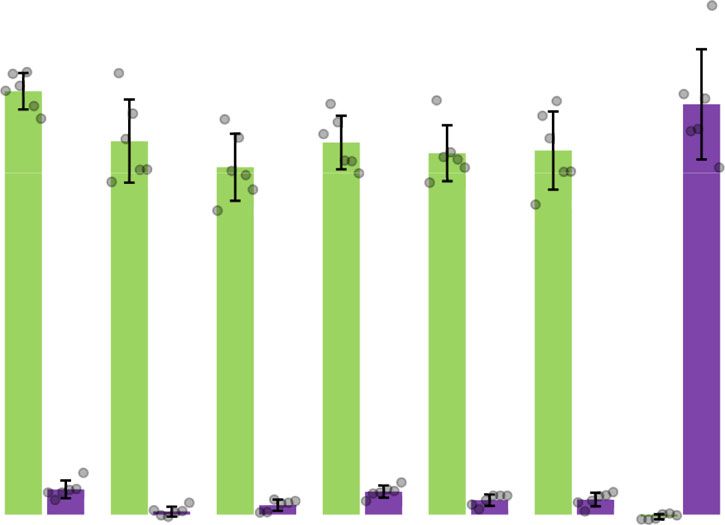
ARTICLE                                                                           NATURE COMMUNICATIONS | https://doi.org/10.1038/s41467-020-18365-0

43. Hahn, M. & Stachelhaus, T. Harnessing the potential of communication-              67. Edgar, R. C. Search and clustering orders of magnitude faster than BLAST.
    mediating domains for the biocombinatorial synthesis of nonribosomal                   Bioinformatics 26, 2460–2461 (2010).
    peptides. Proc. Natl Acad. Sci. USA 103, 275–280 (2006).                           68. Edgar, R. C. MUSCLE: multiple sequence alignment with high accuracy and
44. Pfeifer, B. A., Admiraal, S. J., Gramajo, H., Cane, D. E. & Khosla, C.                 high throughput. Nucleic Acids Res. 32, 1792–1797 (2004).
    Biosynthesis of complex polyketides in a metabolically engineered strain of E.     69. Castresana, J. Selection of conserved blocks from multiple alignments for their
    coli. Science 291, 1790–1792 (2001).                                                   use in phylogenetic analysis. Mol. Biol. Evol. 17, 540–552 (2000).
45. Winn, M., Fyans, J. K., Zhuo, Y. & Micklefield, J. Recent advances in engineering   70. Owen, J. G., Calcott, M. J., Robins, K. J. & Ackerley, D. F. Generating
    nonribosomal peptide assembly lines. Nat. Prod. Rep. 33, 317–347 (2016).               functional recombinant NRPSenzymes in the laboratory setting via peptidyl
46. Puigbo, P., Guzman, E., Romeu, A. & Garcia-Vallve, S. OPTIMIZER: a web                 carrier protein engineering. Cell Chem. Biol. 23, 1395–1406 (2016).
    server for optimizing the codon usage of DNA sequences. Nucleic Acids Res.
    35, W126–W131 (2007).
47. Bates, P. A., Kelley, L. A., MacCallum, R. M. & Sternberg, M. J. E. Enhancement    Acknowledgements
    of protein modeling by human intervention in applying the automatic programs       This work was supported by the Royal Society of New Zealand Marsden Fund (grants 09-
    3D-JIGSAW and 3D-PSSM. Proteins Suppl 5, 39–46 (2001).                             VUW-01 to D.F.A. and 18-VUW-082 to M.J.C.) and the Health Research Council of
48. Nielsen, M., Lundegaard, C., Lund, O. & Petersen, T. N. CPHmodels-3.0-             New Zealand (grant 16/172 to D.F.A. and J.G.O.). We thank Prof Iain Lamont for his
    remote homology modeling using structure-guided sequence profiles. Nucleic          pioneering work in defining the genetics and regulation of pyoverdine biosynthesis and
    Acids Res. 38, W576–W581 (2010).                                                   for originally introducing us to this versatile model NRPS system.
49. Lambert, C., Leonard, N., De Bolle, X. & Depiereux, E. ESyPred3D: prediction
    of proteins 3D structures. Bioinformatics 18, 1250–1256 (2002).                    Author contributions
50. Yang, Y. D., Faraggi, E., Zhao, H. Y. & Zhou, Y. Q. Improving protein fold         D.F.A. and M.J.C. conceived the work, with additional ideas contributed by J.G.O. M.J.C.
    recognition and template-based modeling by employing probabilistic-based           performed the experiments, bioinformatics and prepared all figures. J.G.O. provided
    matching between predicted one-dimensional structural properties of query          bioinformatics support. M.J.C. and D.F.A. wrote the manuscript, and all authors
    and corresponding native properties of templates. Bioinformatics 27,               approved the final version.
    2076–2082 (2011).
51. Jones, D. T. et al. Prediction of novel and analogous folds using fragment
    assembly and fold recognition. Proteins 61, 143–151 (2005).                        Competing interests
52. Offman, M. N., Fitzjohn, P. W. & Bates, P. A. Developing a move-set for            Drs. Calcott and Ackerley have submitted provisional patent filing AU2019903420,
    protein model refinement. Bioinformatics 22, 1838–1845 (2006).                      teaching methods for re-engineering NRPS assembly lines based on the optimal A
53. Kallberg, M. et al. Template-based protein structure modeling using the            domain recombination sites identified in this research. Dr Owen declares no competing
    RaptorX web server. Nat. Protoc. 7, 1511–1522 (2012).                              interests.
54. Shi, J. Y., Blundell, T. L. & Mizuguchi, K. FUGUE: sequence-structure
    homology recognition using environment-specific substitution tables and
    structure-dependent gap penalties. J. Mol. Biol. 310, 243–257 (2001).
                                                                                       Additional information
                                                                                       Supplementary information is available for this paper at https://doi.org/10.1038/s41467-
55. Rykunov, D., Steinberger, E., Madrid-Aliste, C. J. & Fiser, A. Improved scoring
                                                                                       020-18365-0.
    function for comparative modeling using the M4T method. J. Struct. Funct.
    Genomics. 10, 95–99 (2009).
                                                                                       Correspondence and requests for materials should be addressed to D.F.A.
56. Zhang, Y. I-TASSER server for protein 3D structure prediction. BMC
    Bioinform. 9 https://doi.org/10.1186/1471-2105-9-40 (2008).
                                                                                       Peer review information: Nature Communications thanks the anonymous reviewers for
57. Zhou, H. Y. & Skolnick, J. Ab initio protein structure prediction using Chunk-
                                                                                       their contribution to the peer review of this work. Peer review reports are available.
    TASSER. Biophys. J. 93, 1510–1518 (2007).
58. Wang, Z., Eickholt, J. & Cheng, J. L. MULTICOM: a multi-level combination
                                                                                       Reprints and permission information is available at http://www.nature.com/reprints
    approach to protein structure prediction and its assessments in CASP8.
    Bioinformatics 26, 882–888 (2010).
                                                                                       Publisher’s note Springer Nature remains neutral with regard to jurisdictional claims in
59. Kelley, L. A. & Sternberg, M. J. E. Protein structure prediction on the Web: a
                                                                                       published maps and institutional affiliations.
    case study using the Phyre server. Nat. Protoc. 4, 363–371 (2009).
60. Zhang, J. F. et al. MUFOLD: a new solution for protein 3D structure
    prediction. Proteins 78, 1137–1152 (2010).
61. Zhou, H. & Skolnick, J. Template-based protein structure modeling using                              Open Access This article is licensed under a Creative Commons
    TASSERVMT. Proteins 80, 352–361 (2012).                                                              Attribution 4.0 International License, which permits use, sharing,
62. Zhou, H. & Skolnick, J. Protein structure prediction by pro-Sp3-TASSER.            adaptation, distribution and reproduction in any medium or format, as long as you give
    Biophys. J. 96, 2119–2127 (2009).                                                  appropriate credit to the original author(s) and the source, provide a link to the Creative
63. Arnold, K., Bordoli, L., Kopp, J. & Schwede, T. The SWISS-MODEL                    Commons license, and indicate if changes were made. The images or other third party
    workspace: a web-based environment for protein structure homology                  material in this article are included in the article’s Creative Commons license, unless
    modelling. Bioinformatics 22, 195–201 (2006).                                      indicated otherwise in a credit line to the material. If material is not included in the
64. Eswar, N. et al. Comparative protein structure modeling using modeller. Curr.      article’s Creative Commons license and your intended use is not permitted by statutory
    Protoc. Bioinforma. 15, 5.6.1–5.6.30 (2006).                                       regulation or exceeds the permitted use, you will need to obtain permission directly from
65. Benkert, P., Tosatto, S. C. E. & Schomburg, D. QMEAN: a comprehensive              the copyright holder. To view a copy of this license, visit http://creativecommons.org/
    scoring function for model quality assessment. Proteins 71, 261–277 (2008).        licenses/by/4.0/.
66. Benkert, P., Schwede, T. & Tosatto, S. C. E. QMEANclust: estimation of
    protein model quality by combining a composite scoring function with
    structural density information. BMC Struct. Biol. 9, 35 (2009).                    © The Author(s) 2020

10                   NATURE COMMUNICATIONS | (2020)11:4554 | https://doi.org/10.1038/s41467-020-18365-0 | www.nature.com/naturecommunications
You can also read