Variables Affecting Shoot Growth and Plantlet Recovery in Tissue Cultures of Drug-Type Cannabis sativa L.
←
→
Page content transcription
If your browser does not render page correctly, please read the page content below

ORIGINAL RESEARCH
                                                                                                                                     published: 21 September 2021
                                                                                                                                     doi: 10.3389/fpls.2021.732344

                                             Variables Affecting Shoot Growth and
                                             Plantlet Recovery in Tissue Cultures
                                             of Drug-Type Cannabis sativa L.
                                             Janesse E. Holmes, Samantha Lung, Danielle Collyer and Zamir K. Punja*
                                             Department of Biological Sciences, Simon Fraser University, Burnaby, BC, Canada

                                             Tissue culture approaches are widely used in crop plants for the purposes
                                             of micropropagation, regeneration of plants through organogenesis, obtaining
                                             pathogen-free plantlets from meristem culture, and developing genetically modified
                                             plants. In this research, we evaluated variables that can influence the success of
                                             shoot growth and plantlet production in tissue cultures of drug-type Cannabis sativa L.
                                             (marijuana). Various sterilization methods were tested to ensure shoot development from
                                             nodal explants by limiting the frequency of contaminating endophytes, which otherwise
                           Edited by:
                                             caused the death of explants. Seven commercially grown tetrahydrocannabinol (THC)-
                          David Meiri,
            Technion Israel Institute of     containing cannabis genotypes (strains) showed significant differences in response
                   Technology, Israel        to shoot growth from meristems and nodal explants on Murashige and Skoog (MS)
                      Reviewed by:           medium containing thidiazuron (1 µM) and naphthaleneacetic acid (0.5 µM) plus 1%
                        Laura Pistelli,
              University of Pisa, Italy
                                             activated charcoal. The effect of Driver and Kuniyuki Walnut (DKW) or MS basal
                 Alessandro Nicolia,         salts in media on shoot length and leaf numbers from nodal explants was compared
         Council for Agricultural and
                                             and showed genotype dependency with regard to the growth response. To obtain
    Economics Research (CREA), Italy
                                             rooted plantlets, shoots from meristems and nodal explants of genotype Moby Dick
                  *Correspondence:
                      Zamir K. Punja         were evaluated for rooting, following the addition of sodium metasilicate, silver nitrate,
                        punja@sfu.ca         indole-3-butyric acid (IBA), kinetin, or 2,4-D. Sodium metasilicate improved the visual
                                             appearance of the foliage and improved the rate of rooting. Silver nitrate also promoted
                   Specialty section:
         This article was submitted to       rooting. Following acclimatization, plantlet survival in hydroponic culture, peat plugs, and
        Crop and Product Physiology,         rockwool substrate was 57, 76, and 83%, respectively. The development of plantlets
               a section of the journal
            Frontiers in Plant Science
                                             from meristems is described for the first time in C. sativa and has potential for obtaining
                                             pathogen-free plants. The callogenesis response of leaf explants of 11 genotypes
            Received: 28 June 2021
         Accepted: 10 August 2021            on MS medium without activated charcoal was 35% to 100%, depending on the
      Published: 21 September 2021           genotype; organogenesis was not observed. The success in recovery of plantlets from
                             Citation:       meristems and nodal explants is influenced by cannabis genotype, degree of endophytic
   Holmes JE, Lung S, Collyer D and
  Punja ZK (2021) Variables Affecting        contamination of the explants, and frequency of rooting. The procedures described here
 Shoot Growth and Plantlet Recovery          have potential applications for research and commercial utility to obtain plantlets in stage
     in Tissue Cultures of Drug-Type
                                             1 tissue cultures of C. sativa.
                   Cannabis sativa L.
         Front. Plant Sci. 12:732344.        Keywords: meristems, nodal explants, shoot growth, rooting, plantlet recovery, micropropagation, callogenesis,
     doi: 10.3389/fpls.2021.732344           Cannabis sativa L.

Frontiers in Plant Science | www.frontiersin.org                                  1                                     September 2021 | Volume 12 | Article 732344
Holmes et al.                                                                                          Tissue Culture of Drug-Type Cannabis sativa

INTRODUCTION                                                             MATERIALS AND METHODS
Cannabis sativa L., a member of the Cannabaceae family, is               Genotypes
a dioecious, annual flowering plant that has been cultivated             The genotypes used were Moby Dick (MBD), Space Queen
for thousands of years for its fiber (as hemp) and medicinal             (SPQ), Copenhagen Kush (CPH), Cheesequake (CHQ),
and psychotropic properties (as cannabis or marijuana).                  Pennywise (PWE), Girl Scout Cookie (GSC), Death Bubba
Vegetative cuttings are the conventional method for commercial           (DEB), Afghan Kush (AFK), Island Honey (ISH), Pink
propagation of cannabis to ensure rapid propagation of                   Kush (PNK), Pure CBD (CBD), and White Rhino (WHR).
desired genotypes without the introduction of genetic variability        Various combinations of genotypes were used in experiments,
resulting from sexual reproduction as C. sativa is allogamous            depending on their availability. Donor plants of the various
(Punja and Holmes, 2020). However, vegetative cuttings can               genotypes were grown by a licensed producer according to
lose vigor, since donor (mother or stock) plants can be                  Health Canada requirements in a controlled-environment
affected by fungal pathogens and viruses that can reduce                 room or under commercial greenhouse conditions The
their growth and quality (Punja et al., 2019; Punja, 2021).              growing medium for the donor plants was either pure coco
However, maintaining donor plants to be used as a source                 fibers or a coco fiber:vermiculite mix (3:1). The plants in
of vegetative cuttings can be time-consuming and space-                  the controlled-environment room were placed under two
intensive. Hence, there is interest in using tissue culture              Sunblaster brand 54-watt 6400 k T5HO lights with a 24-
methods to propagate cannabis, as it is recognized as a means            h photoperiod. The plants were watered as needed with
to potentially increase plant numbers of desired genotypes               a solution of 1 ml/L Sensi Grow Coco pH Perfect A+B
(micropropagation), and maintain them in a controlled and                (Advanced Nutrients, Los Angeles, CA, Untied States) and
stable environment (preservation) (Monthony et al., 2021).               1 ml/L CALiMAGIc (General Hydroponics, Santa Rosa, CA,
In addition, callus production (callogenesis) from explants              United States) adjusted to a pH of 5.8–6.2 using pH-Down
in tissue culture can potentially be used to increase plant              (Advanced Nutrients, Los Angeles, CA, Untied States) (Scott
numbers through organogenesis (Page et al., 2021). However,              and Punja, 2021). The plants in the commercial greenhouse
there are still many challenges remaining in establishing a              were grown according to the standards for the industry
micropropagation and callusing system for cannabis plants                and kept under a 16-h photoperiod to maintain vegetative
(Monthony et al., 2021). The quality of source (donor) plants,           growth. Explants were taken as needed for the tissue culture
genotype or strain used, surface sterilization methods, explant          experiments described.
type, and tissue culture medium and growth regulators can
all influence the success rate of recovery of plantlets in stage
1 of tissue culture (the introduction of explant material for            Explants
establishment of cultures). Rooting and acclimatization of               The explants tested included meristems, nodal segments, leaves,
plantlets are challenging aspects to tissue culture of hemp              and petiole tissues. Meristems were dissected from terminal and
and cannabis as well. Lata et al. (2009) reported successful             lateral shoots from donor plants (Figure 1A). The external tissue
propagation of strain MX-1 of cannabis in tissue culture, but            was cut away with a scalpel, and two sets of primordial leaves
it is not known whether this method can be applied to other              were left to protect the meristem from excessive damage from
genotypes of cannabis. Monthony et al. (2021) described a                sterilization. Nodal segments with axillary buds were removed
procedure for the micropropagation of six cannabis genotypes,            from lateral stems of the plants and trimmed to a 1-cm length
but rooting and plantlet recovery were not addressed. The success        (Figure 2A). Leaves were trimmed from the plants and used for
of a tissue culture method for cannabis is contingent upon               leaf explants in callus induction experiments. They were cut into
obtaining a high frequency of plantlets growing independently            pieces measuring 0.5 to 1 cm2 . Petiole segments from young
in growth media.                                                         leaves were cut into 1-cm long pieces for callus induction.
    In this research, we investigated various factors that can
influence the recovery of plantlets of cannabis in tissue culture.
The objectives of this study were to (1) assess the efficacy of          Sterilization
sterilization methods to reduce the frequency of contaminants            The standard sterilization protocol involved placing explants in
originating from donor plants; (2) recover and identify the              a stainless steel tea strainer and immersing them in 70% EtOH
various microbes present as contaminants in tissue cultures; (3)         in a glass beaker for 1 min while stirring with a magnetic stir
evaluate shoot growth from meristems of five different genotypes;        bar. The explants were transferred to a 10% bleach solution
(4) determine the responses of seven different genotypes to shoot        (0.625% NaOCl) with 0.1% Tween 20 for 20 min, followed by
growth from nodal explants; (5) evaluate the response of 11              three rinses in sterile distilled water, 3 min each. In some of the
different genotypes to callus development; (6) develop a rooting         experiments, the bleach concentration, length of sterilization,
method for plantlets derived from tissue culture; and (7) evaluate       and length of rinsing were adjusted according to explant type
acclimatization responses in different growth substrates (peat,          and source. The explants were blotted dry on sterile filter paper
rockwool, and hydroponic system) to achieve a high frequency             placed in a laminar flow hood and used immediately for tissue
of plantlet recovery.                                                    culture experiments.

Frontiers in Plant Science | www.frontiersin.org                     2                                September 2021 | Volume 12 | Article 732344
Holmes et al.                                                                                                            Tissue Culture of Drug-Type Cannabis sativa

 FIGURE 1 | Various stages of growth of shoots derived from meristems of drug-type cannabis genotypes grown on a multiplication medium (MM) containing activated
 charcoal. (A) Meristem explants placed on an agar medium in a 90-mm Petri dish to show their small size. (B) Early shoot growth after 4 weeks in culture from a
 meristem. (C) Shoot growth after 8 weeks. (D,E) Shoot growth after 10 weeks from meristems. (F) Baby food jars containing meristem explants at different stages of
 development in a controlled environment growth chamber. A number of different strains are shown. Conditions of incubation are described in the Methods section.

Frontiers in Plant Science | www.frontiersin.org                                 3                                     September 2021 | Volume 12 | Article 732344
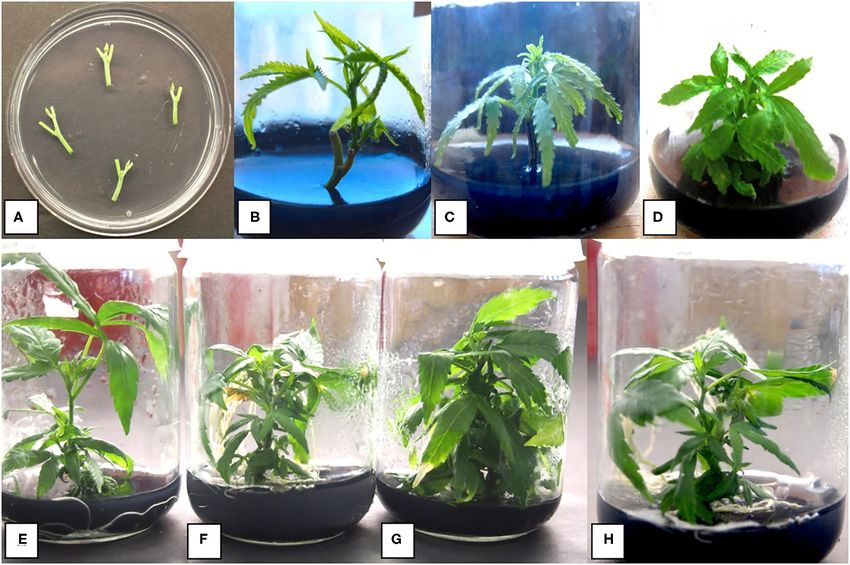
Holmes et al.                                                                                                             Tissue Culture of Drug-Type Cannabis sativa

 FIGURE 2 | Stages of growth of shoots derived from nodal segments of drug-type cannabis genotypes grown on MM containing activated charcoal. (A) Nodal stem
 explants placed on an agar medium in a 90-mm Petri dish to show their size. (B) Shoot growth after 4 weeks from a nodal stem explant. The rate of growth is greater
 than that from a meristem. (C) Shoot growth after 6 weeks. (D) Shoot growth after 8 weeks. (E) Shoot growth after 8 weeks of genotype Death Bubba (DEB) from
 nodal explants. (F) Shoot growth after 8 weeks of genotype Pink Kush (PNK) from nodal explants. (G) Shoot growth after 8 weeks of genotype White Rhino (WHR)
 from nodal explants. (H) Shoot growth after 8 weeks of genotype Moby Dick (MBD) from nodal explants.

Media                                                                                Meristem and Nodal Explant Growth
The medium containing Murashige and Skoog basal salts, as                            For meristems, genotypes Cheesequake, Pure CBD, Moby
described by Lata et al. (2009), was used in initial tissue culture                  Dick, Pennywise, and Space Queen were used. Jars containing
experiments. The medium was supplemented with myo-inositol                           meristem explants prepared as described above were placed
(0.1 g/L) and activated charcoal (1 g/L). The growth regulators                      inside a Conviron A1000 growth chamber (Conviron
added were thidiazuron (TDZ, 1 µM) and naphthaleneacetic acid                        Environments Ltd., Manitoba, Canada) under T5 fluorescent
(NAA, 0.5 µM). This combination of ingredients constitutes a                         lights with an 18-h photoperiod and a light intensity of
multiplication medium, referred to as MM. All the chemical                           102 µmoles m−2 s−1 (Figure 1). The temperature range
reagents were from Sigma-Aldrich (St. Louis, MO). The medium                         was 25 ± 2◦ C. The meristems were left in culture for 6
was adjusted to pH 6.6 ± 0.01 before autoclaving for 20 min                          weeks and transferred to fresh MM medium and incubated
at 121◦ C. Following autoclaving, the pH dropped to ∼5.8. The                        for another 4 weeks (Figure 1). At this time, shoot height,
medium was dispensed into 220 ml culture jars C1770 with                             number of axillary buds developing, and number of
Magenta B caps (Phytotechnology Laboratories R , Lenexa, KS,                         shoots that formed were evaluated (Table 1). There was a
United States), and each jar received ∼25 ml of the medium.                          minimum of 10 replicate jars, each containing a meristem
A single meristem or nodal explant was placed inside each                            for each genotype, and the experiment was conducted three
jar. For callus induction, MM without activated charcoal (MM-                        times (n = 30) using different sources of explants of the
AC) was used. Each 90-mm Petri dish received ∼25 ml of the                           same genotype. The shoots were maintained in culture
medium onto which four to five leaves or petiole segments                            for a maximum of 3 months by monthly transfer to fresh
were placed.                                                                         MM medium.

Frontiers in Plant Science | www.frontiersin.org                                 4                                      September 2021 | Volume 12 | Article 732344
Holmes et al.                                                                                                                  Tissue Culture of Drug-Type Cannabis sativa

TABLE 1 | Comparison of growth of shoots from meristems and nodal explants of five genotypes of drug-type Cannabis sativa.

                                                                               Explant type

                      Meristemsa                                                                                                 Nodal segmentsa

                Shoot height (cm)           No. of buds           No. of shoots                          Shoot height (cm)             No. of buds           No. of shoots
Genotypeb                                                                                  Genotypeb

CBD               3.6 (0.37) a,d             8.0 (1.38) a           1.9 (0.32) a           BLD               4.9 (0.51) a               4.6 (0.4) a            0.4 (0.13) a
CHQ               1.5 (0.08) b,c            6.2 (0.69) a,c          0.5 (0.18) c           SWD               3.8 (0.27) a             3.8 (0.38) a,b           0.4 (0.17) a
MBD                4.5 (0.42) a              6.9 (0.84) a           1.9 (0.27) a           MBD               2.2 (0.27) b              2.9 (0.28) b            0.3 (0.11) a
SPQ                1.9 (0.15) b              3.4 (0.37) c           0.2 (0.07) b           SPQ               1.5 (0.11) c              1.2 (0.11) c             0.0 (0.0) b
PWE               3.1 (0.25) c,d            5.6 (0.53) a,c          2.0 (0.31) a             –                    –                          –                      –

a Data for meristems were collected after 10 weeks in culture and for nodal explants after 6 weeks in culture.
b Within each explant type, genotypes were compared with each other for shoot height, number of buds produced, and number of shoots. Data presented are from 10 explants, and
the experiment was conducted three times (n = 30). Means were compared following ANOVA, and means separation was achieved by Tukey’s honestly significant difference (HSD)
test. Means within a column followed by the same letter are not significantly different at P < 0.05.

    For nodal segments, genotypes BLD, SPQ, MBD, and SWD                                 (Figure 3). The experiment was conducted twice. Only data
were used. Jars containing a segment each were incubated under                           from nodal stem segments that grew were included in the
T5 fluorescent lights with an 18-h photoperiod and a light                               statistical analysis. The shoot height data for CPH on both
intensity of 102 µmoles m−2 s−1 and a temperature range of                               media were compared by an unpaired two-sample t-test. For
21–27◦ C for 2 weeks. The shoots were transferred to fresh MM                            the data on leaf numbers for CPH and the shoot length data
medium and allowed to grow for another 4 weeks (Figure 2). The                           and number of leaves data for PWE, Wilcoxon Rank Sum
percentage of explants surviving, shoot height, number of shoots                         non-parametric tests were performed as the data did not meet
per explant, number of buds developing, and number of leaves                             the “normality” assumptions of the unpaired two-sample t-
were recorded (Table 1). For each genotype tested, there was a                           test.
minimum of 10 replicate jars, each containing a nodal explant,
and the experiment was conducted three times (n = 30) using                              Internal Contaminants in Nodal Explants
different explant sources of the same genotype. To determine                             In many nodal explants derived from donor plants used in
if there were differences between the genotypes, the data were                           tissue culture experiments, contamination by fungi, bacteria,
analyzed using Statplus version 2.21 and R systems (version                              and yeasts was frequently observed on the agar medium, in
3.3.3). ANOVA was performed followed by Tukey’s honestly                                 some cases up to 50%. This caused many of the explants to
significant difference (HSD) test to determine significance at P                         die (Figure 4). To reduce the level of microbial contamination,
< 0.05.                                                                                  the following treatments were assessed by adding them to MM
                                                                                         by filter sterilization after autoclaving: (1) the fungicide captan
Comparison of DKW and MS Salts                                                           (Maestro 8-DF) was added at 0.01 and 0.02 g/L; (2) streptomycin
Two basal salts media, Driver and Kuniyuki Walnut (cat.#D190;                            sulfate was added at 100 mg/L; (3) Plant Preservative Mixture
Phytotechnology Laboratories R , Lenexa, KS, United States) and                          (PPM) (Plant Cell Technology, Washington, DC, United States)
Murashige and Skoog (cat.#M5524; Sigma Aldrich, Saint Louis,                             was added at 2 mg/L. For all treatments, 10 nodal stem segments
MO, United States), were compared for nodal explant growth                               of genotype CHQ were used for each of the treatments, and
using genotypes CPH and PWE. The composition of DKW                                      the experiment was conducted twice (n = 20). Assessments of
medium, as used in this study, is as follows: 5.22 g/L DKW basal                         the extent of microbial contamination were made after 4 weeks.
salts, 20 g/L sucrose, 0.1 g/L myo-inositol, 1 ml/L Gamborg B5                           To assess the effect of fungicides on endophytic contamination,
vitamins, 0.5 uM NAA, 1 uM TDZ, 1 g/L activated charcoal,                                the fungicide Luna (fluopyram) was applied to donor plants of
and 3 g/L Phytagel. Explants were sterilized as described above                          genotypes CPH and PWE at 5 ml/L as a foliar spray until run-off.
but rinsed for 1 min instead of 3 min. They were transferred                             Plants were grown for 3 weeks before nodal stem segments were
to either DKW or MM. Twenty jars, each containing a nodal                                collected. Nodal stem segments from treated and control plants
explant, were placed under the lighting conditions described                             were dissected and sterilized with ethanol:bleach as described
above. The initial height of the nodal explants was measured 4                           previously and rinsed with sterile distilled three times for 1 min
days after placement on each medium to account for differences                           each. The nodal stem segments were transferred to MM and
between initial explant size. The explants were transferred to                           grown under a 16-h photoperiod for 4 weeks, after which the
the respective fresh media after 2 weeks, and 2 weeks later, the                         proportion of jars with microbial contamination was evaluated.
heights of the shoot from the base where it attached to the                              A total of 20 explants were included for each treatment group.
original explant to the top were measured using ImageJ. The                              The data obtained from the CPH genotype met the requirements
height differences from the start to the end of the experiment                           for a chi-square test, but the data from PWE did not, so Fisher’s
represented the shoot height for each explant. The number of                             Exact test was performed. The experiment was conducted once.
leaves on nodal explants on each medium was also assessed                                The sample size was n = 20 for each group.

Frontiers in Plant Science | www.frontiersin.org                                     5                                        September 2021 | Volume 12 | Article 732344
Holmes et al.                                                                                                                    Tissue Culture of Drug-Type Cannabis sativa

 FIGURE 3 | Comparison of shoot growth and leaf number from nodal stem explants of two cannabis genotypes placed on a basal salt medium containing either Driver
 and Kuniyuki Walnut (DKW) or Murashige and Skoog (MS) salts. (A) Shoot length of genotype Copenhagen Kush (CPH). (B) Shoot length of genotype Pennywise
 (PWE). (C) Leaf number of genotype Copenhagen Kush (CPH). (D) Leaf numbers of genotype Pennywise (PWE). The box of each dataset represents the interquartile
 range (IQR), which contains the third quartile (Q3–top side of the box), the median value of all of the data (the middle line), and the first quartile (Q1–bottom side of the
 box). The bars represent the maximum (Q3 + 1.5*IQR) and minimum (Q1–1.5*IQR) of the data. The numbers (n) above the bars depict explant numbers used.

Identification of Tissue Culture                                                          followed by 4◦ C hold. PCR products were cut and sent
Contaminants                                                                              to Eurofins Genomics (Eurofins MWG Operon LLC 2016,
Colonies representing the most common contaminants seen                                   Louisville, KY, United States) for sequencing. The resulting
in tissue culture were transferred to potato dextrose agar +                              sequences were compared with the corresponding ITS1-5.8S-
streptomycin (130 mg/L) for 2 weeks and then to potato                                    ITS2 sequences from the National Center for Biotechnology
dextrose broth placed on a shaker at 125 rpm at room                                      Information (NCBI) GenBank database to confirm species
temperature for 7 days. The mycelium was harvested, and                                   identity using only sequence identity values above 99%. Once
DNA was extracted using the DNeasy Plant Mini Kit (cat.                                   the identity of the cultured microbes was determined, random
No. 69104; QIAGEN, Hilden, Germany). For PCR, the internal                                samples of nodal explants were obtained from the same
transcribed spacer regions (ITS1 and ITS2) as well as the 5.8S                            donor plants (genotypes CPH and MBD), and 10–50 mg of
gene of ribosomal rDNA were amplified using the universal                                 fresh tissue was used for total DNA extraction and PCR
                                   ′
eukaryotic primers UN-UP18S42 (5 -CGTAACAAGGTTTCCG                                        following the conditions described above for fungal cultures. For
                 ′                          ′
TAGGTGAAC-3 ) and UN-LO28S576B (5 -GTTTCTTTTCC                                            examination of the nodal explants under a scanning electron
                           ′
TCCGCTTATTAATATG-3 ) to produce a DNA template for                                        microscope for potential microbes that could be present in
sequencing. PCR conditions were as follows: initial denaturation                          internal tissues, samples were taken from donor plants known
at 94◦ C for 3 min, followed by 40 cycles of denaturation at                              to be contaminated based on isolation in culture. They were
94◦ C for 30 s, annealing at 55◦ C for 45 s, and extension at                             processed following the procedure described by Punja et al.
72◦ C for 2 min, and a final extension at 72◦ C for 7 min,                                (2019).

Frontiers in Plant Science | www.frontiersin.org                                      6                                        September 2021 | Volume 12 | Article 732344
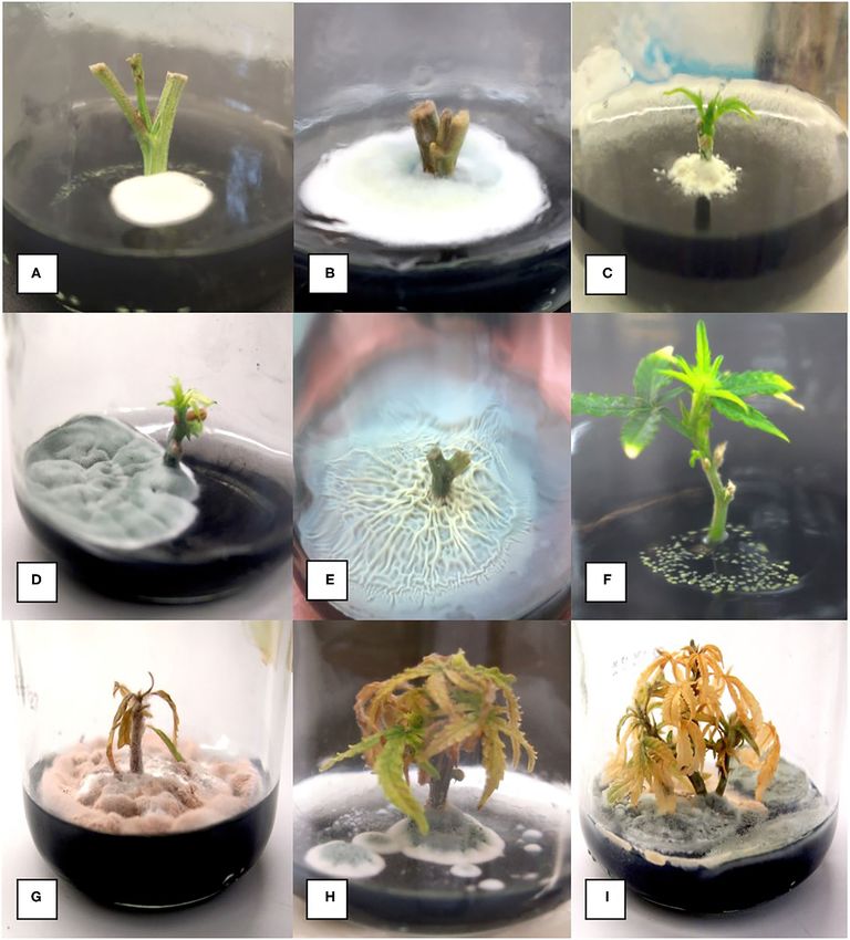
Holmes et al.                                                                                                            Tissue Culture of Drug-Type Cannabis sativa

 FIGURE 4 | Range of microbial contaminants emerging from surface-sterilized nodal stem explants at various times after placement on MM containing activated
 charcoal. (A) Early emergence of Penicillium species. (B) Large colony of Penicillium growing from a nodal explant. (C) Colony of Chaetomium emerging from a nodal
 explant. (D) Large colony of Penicillium growing out of a nodal stem explant. (E) Extensive Bacillus growth emerging from a nodal stem explant. (F) Growth of
 Pseudomonas from a stem explant. (G–I) Death of established shoots from nodal stem segments due to contamination by microbes appearing later during plantlet
 growth.

Rooting of Shoots From Nodal Explants                                                2 cm in height were transferred to MS basal salts medium with
Genotype Moby Dick was used for root induction. Following                            the following amendments to induce rooting. The amendments
the 8-week growth period for nodal segments, shoots measuring                        were silver nitrate (40 µM), sodium metasilicate (6 and 9

Frontiers in Plant Science | www.frontiersin.org                                 7                                      September 2021 | Volume 12 | Article 732344
Holmes et al.                                                                                           Tissue Culture of Drug-Type Cannabis sativa

mg/L), indole-3-butyric acid (5, 12.3, 37, and 42 µM), 2,4-               wrapped in Al foil and incubated at 21–23◦ C for a maximum of 8
dichlorophenoxyacetic acid (5 µM), and kinetin (1 µM). The                weeks. The callus area was measured using the “freehand” tool in
shoots were incubated for 4 weeks on media containing these               ImageJ from photos taken of the Petri dishes that included a ruler
additives and rated for rooting frequency. Each treatment had a           for measurement. If the data did not meet the assumptions of
minimum of 10 explants, and the experiment was repeated three             parametric tests (i.e., ANOVA, unpaired two-sample t-test, etc.),
times (n = 30). The data were analyzed using Statplus version             then non-parametric tests were used (i.e., Welch’s ANOVA, and
2.21 and R systems (version 3.3.3). ANOVA was performed                   Wilcoxon Rank Sum test). If the data met the assumptions of the
followed by Tukey’s HSD test to determine significance at P               ANOVA test, then they were used followed post-hoc by Tukey’s
< 0.05.                                                                   HSD test.

Acclimatization for Plantlet Recovery                                     RESULTS
Shoots of genotype Moby Dick derived from either meristems or
nodal explants that had formed roots were selected at random              Shoot Growth From Meristem and Nodal
and transferred to one of the following growing substrates: peat          Explants
plugs (Jiffy-7 R peat pellets), rockwool cubes (2.5 × 2.5 × 3.8 cm,       Placement of meristems on MM medium (with 1% activated
Grodan) or placed in a Turboklone (T-24 turbo-mini) (https://             charcoal) resulted in shoot growth after 4 weeks in culture
turboklone.com) containing a hydroponic nutrient solution with            (Figure 1B), which continued to elongate after 8–10 weeks
1 ml/L of pH Perfect R Sensi Grow A&B and CALiMAGIc                       (Figures 1C,D). Baby food jars containing meristem explants
(General Hydroponics, Santa Rosa, CA, United States) and 0.25             at different stages of development were placed in a controlled
ml/L of Rapid Start Rooting Enhancer (General Hydroponics,                environment growth chamber to allow for a comparison of
Santa Rosa, CA, United States) ∼pH 5.8. Every 3 days, a nutrient          growth of different genotypes (Figure 1E). Meristems of five
mixture (without Rapid Start) was added to top up the system.             different cannabis genotypes were assessed for their shoot
The peat plugs/rockwool cubes were soaked for a minimum of                regeneration response. After 10 weeks of growth, shoot height,
20 min in the same nutrient solution used for hydroponic growth.          number of axillary buds, and number of axillary shoots were
The plugs containing shoots were then placed in a sterilized              measured (Table 1). The genotype with the greatest shoot height
tray (28 × 56 cm) and covered with a plastic dome (http://                value was MBD, with an average height of 4.5 cm, which was
www.jiffypot.com/). The domes were misted with water every                significantly greater compared with genotypes CHQ, SPQ and
2 days, and the vents were opened halfway after 7 days and                PWE (P < 0.001). The average shoot growth across genotypes
fully opened after 9 days. Domes were removed 2 weeks post                was 2.92 cm (Table 1). The number of axillary buds present on
transfer. The percentage of survival of plantlets was assessed            the shoots from meristems ranged from 8.5 for CBD to 3.4 for
4 weeks after transfer. Each treatment had a minimum of 10                SPQ. The number of axillary buds produced per plantlet was
replicates, and the experiment was conducted twice. The data              not significantly different (P > 0.05) among CBD, CHQ, MBD,
were analyzed using Statplus version 2.21 and R systems (version          and PWE. SPQ had the lowest number of buds compared with
3.3.3). ANOVA was performed followed by Tukey’s HSD test to               the other genotypes (P < 0.05). Across genotypes, plantlets from
determine significance at P < 0.05.                                       meristems produced 6.38 axillary buds on average and 1.4 shoots
                                                                          (Table 1). Genotype SPQ showed poor growth for all parameters
Callus Growth                                                             compared with the four other genotypes. While CBD plantlets
Genotypes Girl Scout Cookie, Space Queen, Moby Dick, and                  were generally shorter, they had a higher number of buds that
Pennywise were tested. After sterilization, leaf and petiole              resulted in a bushier appearance. Genotypes MBD and PWE had
explants were placed on MM-AC. Petri dishes with leaf and                 similar shoot growth, but MBD was significantly taller than PWE
petiole explants were kept on wire rack shelves under ambient             (Table 1). Genotype SPQ generally showed the poorest growth
conditions (temperature range of 21–25◦ C). One-half of the               among all the genotypes.
dishes were wrapped in aluminum foil to exclude light, and the               Measurements of height, number of axillary buds, and number
remainder was left under the same conditions of supplemental              of axillary shoots were obtained from nodal stem explants after
lighting as for the nodal explants. Callus development was first          6 weeks (Table 1). The genotypes tested were BLD, SWD, MBD,
observed after 4 weeks. Dishes were incubated for 6–10 weeks              and SPQ. The average shoot height ranged from 1.5 to 4.9 cm
before measurements were taken. The percentage of explants                for SPQ and BLD, respectively (Table 1). BLD and SWD were
that developed callus as well as the surface area of callus was           significantly (P
Holmes et al. Tissue Culture of Drug-Type Cannabis sativa
explants on DKW and 89.2% on MM basal salts grew and Pseudomonas, yeasts that were not identified, and many fungi. In
were included in a statistical comparison by an unpaired two- an attempt to better categorize the fungal contaminants, which
sample t-test. For PWE, 70% of explants on DKW and 77.5% represented the majority of contaminants present in nodal stem
on MM grew and were included in a statistical comparison by tissues, a PCR-based method was utilized. Cultures of 11 fungi
the Wilcoxon Rank Sum test. The data showed that there was recovered from nodal explants each produced a band size of
a significant difference between DKW and MS basal salts in about 650 bp after PCR. After sequencing and comparing the
shoot length for CPH (p = 0.02947; Figure 3A). The average results in BlastN, a range of fungi were identified. This was
CPH shoot length on DKW and MM was 6.44 and 8.63 mm, followed by using naturally contaminated nodal explants, in
respectively. In comparing shoot length for PWE on DKW which the frequency of detection of fungal contamination was
and MM, there was no significant difference (p = 0.08354). 16/19 samples (Figures 5A,B). The plant DNA band was at 750
The average PWE shoot length on DKW and MS was 7.66 bp, as the universal primers used amplified both fungal and plant
and 9.2 mm, respectively (Figure 3B). For CPH leaf number, DNA. The genus and species of fungi present in cannabis stem
there was no difference between DKW and MM (p = 0.6882; tissues are shown in Figure 5. The PCR test could detect fungal
Figure 3C). The average number of leaves on DKW and MM contaminants present at DNA concentrations of 1 ng/ul (qPCR
was 3.31 and 3.12, respectively. For PWE leaf number, there was data not shown). Sections of stem segments when plated onto
a significant difference between DKW and MM (p = 0.04468; potato dextrose agar yielded colonies of Penicillium that emerged
Figure 3D). The average number of leaves on DKW and MM was directly from the pith tissues (Figure 5C). When pith tissues were
4.13 and 5.64, respectively. examined under a scanning electron microscope (Figure 5D), a
close-up showed that fungal spores could be seen growing in and
Internal Contaminants and Alternative around pith cells (Figures 5E,F).
Sterilization Methods Rooting of Shoots From Nodal Explants
In general, an average of 50% of nodal stem explants were
Representative plantlets from sodium metasilicate treatments of
found to be contaminated by microbes (a range of 10 to
0, 6, and 9 mg/L are shown in Figures 6A–C. The addition of
80%), despite the rigorous sterilization methods used, and
sodium metasilicate caused visible differences in plant growth
these had to be discarded. These contaminants included fungi,
and leaf morphology, recorded on a scale of 1–3, as shown in
bacteria, and yeasts (Figure 4). Due to the high frequency of
Figures 6D–F. A rating of 1 shows thin curled leaves, while a
microbial contaminants from nodal stem explants when grown
rating of 3 shows dark green, flat, and toothed margins. The
in a tissue culture medium, the addition of fungicides and
addition of sodium metasilicate at 6 mg/L significantly (p <
antibiotics was assessed. The addition of a fungicide (captan)
0.05) improved the leaf morphology rating compared with MM
at 0.01 and 0.02 g/L to the tissue culture medium post-
and MM + Na2 SiO3 at 9 mg/L according to Tukey’s HSD test
sterilization did not significantly reduce the contamination
(Figure 6G). On the MM + 6 mg/L Na2 SiO3, the leaf morphology
frequency on genotype CHQ (P = 0.05). The addition of
rating was comparable with the leaf rating of plants grown on
streptomycin sulfate at 100 mg/L caused stunting and chlorosis
the MM without Na2 SiO3 . The addition of Na2 SiO3 at 6 mg/L
of the tissue culture shoots (data not shown). Plant preservative
produced the greatest proportion of rooted plantlets according
mixture (PPM) at 2 ml/L appeared to delay the onset of
to Tukey’s HSD test (p < 0.05; Figure 6H). The proportion of
contamination, but microbial growth would appear 1 to 3
rooted plants was 0.4 (40%) for MM + Na2 SiO3 at 6 mg/L
weeks later. Nodal stem explants sterilized with 5% PPM +
compared with 0.1, 0.1, and 0.2 for treatments MM, MM + 9
3x basal salt solution or 70% EtOH and 10% bleach +0.1%
mg/L Na2 SiO3 , and MM without phytohormones (MMC). The
Tween 20 showed no differences in microbial contamination
addition of sodium metasilicate did not significantly affect any
on genotype CPH (60.5 vs. 60%, chi-square test p > 0.05).
of the growth parameters measured for nodal stem segments or
For CPH donor plants treated with Luna fungicide, a chi-
meristems: height, number of buds, and number of shoots (data
square test was significant at p < 0.05, which suggests that
not shown).
fungicide treatment of donor plants could reduce the endophytic
The addition of indole-3-butyric acid, 2,4-
contamination observed in tissue culture. The CPH control
dichlorophenoxyacetic acid (2,4-D), and kinetin (KIN) was
group showed 88.2% contamination, and the Luna treatment
tested as alternatives to TDZ and NAA in rooting media. KIN
group showed 31.8% contamination. However, the PWE plants
and 2,4-D were tested at 1 and 5 µM, respectively, alone and
treated with Luna did not differ significantly from the untreated
in combination. Neither hormone alone or in combination
control as overall contamination rates were much lower
performed significantly better than the MM (data not shown).
(12.5 vs. 16.7%, Fisher’s exact test p > 0.05). These results
The combination of KIN and 2,4-D produced an average of
indicate that genotypes may differ in terms of the extent of
63% rooted plants, while the MM produced an average of 44%
microbial contamination.
rooted plants. The MMC was used as the basal medium in the
indole-3-butyric acid experiments. IBA was tested at 5, 12.3,
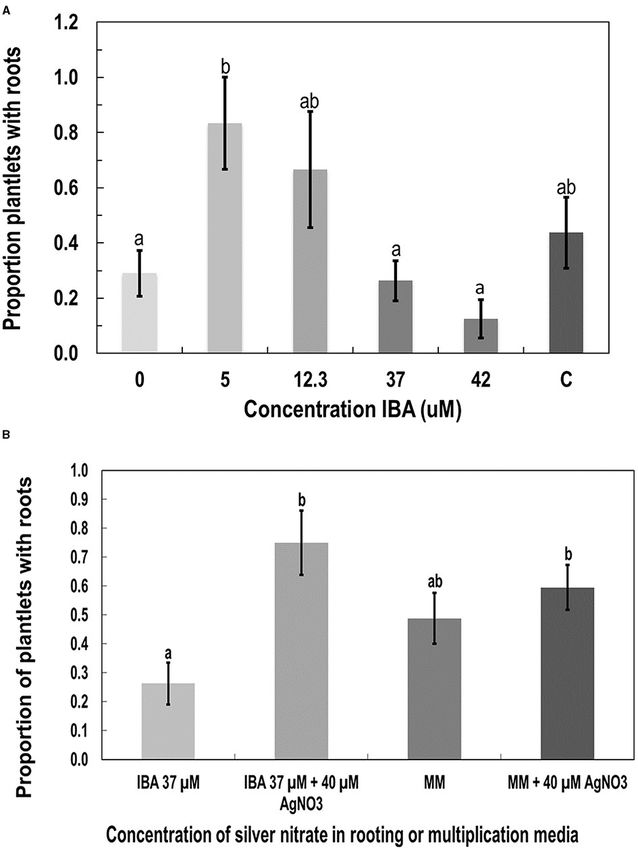
Identification of Tissue Culture 37, and 42 µM. While there was a trend toward decreased
Contaminants rooting as more IBA was added, the only statistically significant
The contaminants observed growing on agar media consisted difference (p < 0.05 by Tukey’s HSD test) was between 5 and
of bacteria, which were identified to genus level as Bacillus and 42 µM (Figure 7A). The MM (listed as C in Figure 7A) was
Frontiers in Plant Science | www.frontiersin.org 9 September 2021 | Volume 12 | Article 732344Holmes et al. Tissue Culture of Drug-Type Cannabis sativa
FIGURE 5 | Molecular detection and identification of tissue culture contaminants. (A,B) PCR detection of fungal DNA present in naturally contaminated nodal
explants. Upper bands at 750 bp size are plant DNA. Lower bands at ca. 650 bp are fungal DNA. These bands were cut and sequenced to determine the
corresponding fungus present. Lanes 1–13 (lower bands) are as follows: clean, Simplicillium lasoniveum, Trichoderma harzianum. Clean, Beauveria bassiana,
(Continued)
Frontiers in Plant Science | www.frontiersin.org 10 September 2021 | Volume 12 | Article 732344Holmes et al. Tissue Culture of Drug-Type Cannabis sativa
FIGURE 5 | Fusarium oxysporum. Clean, Trametes versicolor, Lecanicillium fungicola, Chaetomium globosum, F. oxysporum, F. oxysporum, L. fungicola. Lane 14 =
water control. L = molecular weight standards. Lanes 15–21 (lower bands) = Penicillium chrysogenum, Penicillium copticola, C. globosum, Penicillium olsonii, P.
olsonii, T. versicolor. Lane 22 = water control, L= molecular weight standards. (C) Growth of Penicillium colonies emerging from the center of pith tissues in cut nodal
segments. (D) Cross-section of a cannabis nodal stem explant showing the central pith surrounded by pith cells. (E) Close-up of pith cells as viewed in the scanning
electron microscope. (F) Magnified view of pith cells in the scanning electron microscope showing fungal sporulation inside pith cells, likely of Penicillium sp. (arrow).
not significantly different from any of the IBA concentrations; in culture, extensive callus growth could be observed from leaf
however, it averaged 44% rooted plants, while IBA at 5 µM segments (Figures 10A–D). The appearance of the callus was
produced 83% rooting. similar on leaf explants and petiole explants (Figures 10E,F).
Silver nitrate was added at 40 µM to the MM and MMC None of the calli showed evidence of differentiation toward shoot
containing IBA at 37 µM instead of TDZ and NAA. The IBA development or somatic embryo development. Further transfers
with added silver nitrate had significantly more roots than of calli to new media eventually resulted in browning and death.
IBA at 37 µM alone according to Tukey’s HSD test (p <
0.05; Figure 7B). The AgNO3 did not significantly increase the
proportion of roots produced when added to the MM. DISCUSSION
Rooting and Acclimatization for Plantlet The results from this study conducted on drug-type cannabis
Recovery (marijuana) show that the response to tissue culture conditions
Spontaneous rooting was observed on some of the nodal explants is first and foremost influenced by the genetic background
(Figures 8A,B). Rooted plantlets were carefully removed from (genotype) of the plants tested. The findings also indicated
the tissue culture jars, rinsed of excess medium (Figure 8C), and that both meristems and nodal explants were responsive to
transferred to a coco fiber growing medium. For comparison of the tissue culture conditions tested, and that measurements
rockwool, peat, or hydroponic system (Figures 8D,E), at least of shoot growth could be used to determine the response of
10 plantlets were used per acclimatization substrate, and the genotypes and quantify effects of media amendments on growth.
experiment was repeated twice for different batches of plants Lastly, the findings show that the recovery of rooted plantlets
(n = 20). The plantlets were of genotype MBD. Acclimatization is influenced by the degree of internal contamination of nodal
success was calculated based on the number of healthy surviving explants and the extent to which rooting and acclimatization
plants after 2 weeks divided by the total number of plants that had of the plantlets could be achieved using different treatments.
been transferred for acclimatization. The survival success rate Knowledge of these variables can enhance the successful recovery
after acclimatization was 57, 76, and 83% for hydroponic, peat, of plantlets from tissue cultures of C. sativa, which was the
and rockwool, respectively, which was not statistically different. main objective of this study. This study focused on stage 1 of
Figures 8F,G show plants grown in coco growing substrate where the micropropagation process as described by Page et al. (2021).
they grew normally and produced more shoots. However, plants This phase is equivalent to an initiation phase (establishment of
grown in rockwool appeared more vigorous and grew faster cultures) and did not involve repeated cycles of subcultures and
(Figure 8H). multiplication of shoots as observed in stage 2, the multiplication
phase that increases plant numbers through micropropagation
Callus Growth (Monthony et al., 2021; Page et al., 2021). Research on stage 1 is
Callus development from leaves, measured as callus diameter, valuable to establish the genotypic response of a range of cannabis
was genotype-dependent (Figure 9A). Genotypes GSC and SPQ strains to initial tissue culture conditions and to rapidly recover
readily developed callus, while for genotypes MBD and PWE, plantlets from meristems or nodal explants for a first cycle
callus diameter wasHolmes et al. Tissue Culture of Drug-Type Cannabis sativa FIGURE 6 | The effect of sodium metasilicate at 0 (A), 6 (B), and 9 (C) mg/L in the MM on shoot growth of genotype MBD from nodal segments. Optimal growth and rooting can be seen on 6 mg/L. (D–F) Leaf morphology rating scale applied to plantlets during growth in tissue culture as an indication of plantlet health. (D) A rating of 1 shows thin curled leaves, light green in color. (E) A rating of 2 shows leaves light green in color with some curling. (F) A rating of 3 shows dark green, flat leaves with serrated margins. (G) Average leaf morphology rating after 4 weeks of growth on MM with added sodium metasilicate. (H) Proportion of plantlets that developed roots on MM supplemented with sodium metasilicate at 6 mg/L was significantly different to the proportion of roots that developed from plantlets on MM, MM + sodium metasilicate 9 mg/L, and MMC. Bars followed by different letters indicate significant differences. the response of genotypes of interest to tissue culture conditions while the other genotypes (SPQ and CHQ) grew poorly. Strain would first need to be established before a full-scale tissue culture MBD was selected for a further study on plantlet recovery. A method could be developed for commercial use. In this study, higher frequency of shoots and buds can potentially give rise to some of the cannabis genotypes, e.g., MBD, produced shoots more plantlets during stage 2 micropropagation and can enhance over 4.5 cm in height and formed multiple shoots and buds, plantlet numbers. Frontiers in Plant Science | www.frontiersin.org 12 September 2021 | Volume 12 | Article 732344
Holmes et al.                                                                                                            Tissue Culture of Drug-Type Cannabis sativa

                                                                                          the meristems required a longer time in culture (10 weeks)
                                                                                          compared with the axillary buds (6 weeks).
                                                                                              Meristems have not been previously tested as an explant
                                                                                          source in either hemp or cannabis, although shoot tips were used
                                                                                          for direct regeneration of hemp (Wang et al., 2009), and stem tips
                                                                                          were used for micropropagation and retipping studies on hemp
                                                                                          (Lubell-Brand et al., 2021). Meristems are important starting
                                                                                          material, as they contain a lower frequency of internally-borne
                                                                                          microbes and viruses (Wang and Hu, 1980). Since C. sativa L. is
                                                                                          reported to harbor fungi and bacteria internally as endophytes
                                                                                          (Scott et al., 2018; Punja et al., 2019), consequently, explants
                                                                                          taken from mother plants that naturally carry endophytes or
                                                                                          pathogens have a higher risk of becoming contaminated after
                                                                                          transfer to tissue culture media, as observed in this study.
                                                                                          Monthony et al. (2021) circumvented this problem by first
                                                                                          establishing in vitro plants in stage 1 that were subsequently used
                                                                                          to provide an explant material for studies on micropropagation
                                                                                          and callogenesis in stage 2. While this approach is advantageous
                                                                                          to provide clean explants and maintain desired genotypes in
                                                                                          vitro, the explants used in the present study were derived from
                                                                                          donor plants grown under commercial greenhouse conditions,
                                                                                          as they provided unlimited quantities of tissues, and the plants
                                                                                          could be evaluated for commercially desired traits prior to
                                                                                          introduction into the tissue culture environment. To avoid higher
                                                                                          contamination rates from these tissues, we evaluated a range of
                                                                                          decontamination methods. Following reports of the addition of
                                                                                          fungicides to tissue culture media to reduce fungal contamination
                                                                                          (Nagy et al., 2005; Panathula et al., 2014), we added captan at 0.02
 FIGURE 7 | The effect of additives to the MS salts medium on root                        g/L, but it did not show any effect. We also tested a widely used
 development in plantlets of genotype MBD derived from nodal stem segments                broad spectrum product, Plant Presrvative MixtureTM (PPM;
 after 4 weeks of growth. Additives were (A) indole-3-butyric acid IBA, (B) silver
 nitrate (AgNO3 ), compared with MM as a control. Statistical analysis was
                                                                                          Plant Cell Technology, Washington, DC, United States), which
 performed by Tukey’s HSD test with significance at p < 0.05. Bars followed by            reduced initial contamination in tissue culture but not beyond
 different letters indicate significant differences.                                      2 weeks. Similarly, nodes that had been surface-sterilized in
                                                                                          5% PPM for 4 h showed no difference in contamination levels
                                                                                          compared with nodes sterilized with 10% bleach +0.1% Tween
                                                                                          20. Interestingly, the application of a systemic fungicide (Luna)
    Meristems and axillary buds have both been used as starting                           to the indoor-grown donor plants, followed 3 weeks later by the
explant sources for shoot induction in tissue culture experiments                         removal of nodal explants, showed reduced contamination levels
of various plant species. For example, axillary buds are commonly                         in tissue cultures of one strain by almost 3-fold.
used for the propagation of fruit and nut trees, while meristems                              The contaminating microbes in cannabis explants appear to
have been used for sweet potato and strawberry propagation                                reside within the central pith tissues, as shown in this study using
(Hussain et al., 2012). Growth of axillary buds in tissue culture                         scanning electron microscopy and reported elsewhere (Punja
has been studied in mint (Mentha species) (Rech and Pires, 1986),                         et al., 2019). This could explain the difficulty in eradicating
Cancer tree (Camptotheca acuminate) (Liu and Li, 2001), hops                              them with surface-sterilization methods. Donor plants of some
(Humulus lupus) (Roy et al., 2001), Andean blueberry (Vaccinium                           genotypes, e.g., CPH appeared to have a higher background
floribundum) (Cobo et al., 2018), and other woody plants (Sahoo                           level of contamination compared with other genotypes, e.g.,
and Chand, 1998). In order to obtain plants free from pathogens,                          PWE. Most of the contaminants emerged after several weeks in
particularly viruses, meristem tip culture is a preferred method.                         the tissue culture environment and originated from the central
Successful eradication of viruses using tissue culture techniques                         pith of the nodal explants (see Figure 5). Axillary buds may
alongside a secondary method, such as thermotherapy, has been                             contain pathogens or endophytes living internally, which can
demonstrated in sugarcane (Cheong et al., 2012), Lilium spp.                              easily be transferred into tissue culture (Wang and Hu, 1980).
(Chinestra et al., 2015), dahlias (Nerway et al., 2020), artichoke                        Internal contamination is less of a concern for meristems, as the
(Spanò et al., 2018), and many others. We compared meristem                               vascular dome and first primordial leaves are generally free of
and nodal explant types in this study and successfully obtained                           bacteria, fungi, and viruses (Ramgareeb et al., 2010). Previous
plantlets from both, with the meristems showing significantly                             studies have demonstrated that an extensive array of fungal and
lower microbial contamination rates compared with the nodal                               bacterial endophytes can colonize hemp and cannabis plants
explants bearing axillary buds. However, shoot production from                            (Kusari et al., 2013; Scott et al., 2018; Punja et al., 2019).

Frontiers in Plant Science | www.frontiersin.org                                     13                                 September 2021 | Volume 12 | Article 732344
Holmes et al. Tissue Culture of Drug-Type Cannabis sativa FIGURE 8 | Root development on shoots after 8–10 weeks of growth in tissue culture and acclimatization to produce plants. (A,B) Spontaneous development of roots on nodal explants. (C) Plantlets were removed from tissue culture and transferred to coco growth media and placed under high humidity conditions for 2 weeks. (D,E) Acclimatization of plantlets from meristems of genotype MBD in different growth substrates. Plantlets were removed from tissue culture jars after 12 weeks of growth on the MM and placed into rockwool or peat under humid conditions for 14 days. Rockwool or peat plugs were soaked in a fertilizer mix of 1 ml/L of pH Perfect® Sensi Grow A&B and CALiMAGIc (General Hydroponics, Santa Rosa, CA, United States) in ∼pH 5.8 water. (F,G) Growth of plants on a coco potting medium under ambient conditions following successful acclimatization. (H) Hydroponic system filled with 8 L of the fertilizer mix with vigorous growth of plants derived from meristem tissue cultures. Kusari et al. (2013) found 30 different species of fungal the most abundant bacterial genera were Pseudomonas, Pantoea, endophytes, of which Penicillium copticola was the most and Bacillus. Punja et al. (2019) showed that endophytic and prevalent. Scott et al. (2018) found 134 bacterial and 54 fungal pathogenic fungi, such as species of Chaetomium, Trametes, strains in three hemp cultivars. The most abundant fungal Trichoderma, Penicillium, and Fusarium, could colonize genera were Aureobasidium, Alternaria, and Cochliobolus, and cannabis plants internally. Previous tissue culture studies on Frontiers in Plant Science | www.frontiersin.org 14 September 2021 | Volume 12 | Article 732344
Holmes et al.                                                                                                              Tissue Culture of Drug-Type Cannabis sativa

 FIGURE 9 | Response of cannabis genotypes to callus development on MM without activated charcoal (MM-AC). (A) Callus diameter of four genotypes developing
 from 1 cm2 leaf explants. (B) Callus area from leaf explants of 8 cannabis genotypes compared with the mean callus area across all strains (represented by the dotted
 line). The “*”represents significance level and “ns” represents not significant relationships between each genotype and across all genotype means (dotted line). A
 Kruskal–Wallis test resulting in a p of 8 × 10⁻¹¹ and a Games–Howell post-hoc test were performed. Significant differences identified in the Games–Howell post-hoc
 test are shown using letters above the boxplots of each strain. (C) Growth of Cheesequake (CHQ) callus from leaf and petiole explants after 1 month under 24-h dark
 and 24-h light conditions on MM. Statistical analysis was performed by Tukey’s HSD test with significance at p < 0.05. Bars followed by different letters indicate
 significant differences. (D) Growth of callus from leaves and petioles of genotype Girl Scout Cookie (GSC) (left) and Space Queen (SPQ) (right). Measurements of
 diameter were made after 4 weeks. Statistical analysis was performed by Tukey’s HSD test with significance at p < 0.05. Bars followed by different letters indicate
 significant differences.

hemp and cannabis have not described problems with tissue                              bacteria and fungi (Moreno-Vázquez et al., 2014; University of
culture contaminants. It is likely that the coco fiber used as a                       California Davis, 2008). This approach can be applied to cannabis
substrate for growing plants in this study harbored microbes                           plants before they are deployed in tissue culture. In addition,
that eventually colonized the internal tissues of the stems and                        if meristem culture of cannabis is used to obtain pathogen-free
became established (Punja et al., 2019). Other growing media,                          plantlets, it would have to be accompanied by a similar PCR-
such as rockwool, may contain lower levels of contaminating                            based assay to test for the absence of these pathogens. Nodal
microbes. Donor plants grown in a coco fiber substrate over                            explant cultivation is unlikely to be free of pathogens given the
prolonged periods of time in indoor environments, e.g., for up                         high levels of internal contamination observed in this study.
to a year, such as CPH, showed much higher background levels                           Therefore, shoots derived from nodal cultures should be avoided
of contaminants. Recent microbiome studies have demonstrated                           because of the potential for contaminants. Meristems represent
that the bulk soil and rhizosphere of cannabis and hemp                                the explant of choice to obtain pathogen-free plantlets from tissue
plants are the most influential in determining the subsequent                          cultures of C. sativa.
composition of internal microbes in other regions of the plant                            The tissue culture medium used for growth of plant tissues
(Barnett et al., 2020; Comeau et al., 2021). Therefore, attention                      can influence the success in initiation and multiplication of shoot
should be given to the condition of donor plants with regard to                        growth and elongation. Following shooting, a second medium
the substrate they are grown in and their duration in the growing                      with a higher concentration of auxin can be used to induce
environment. Young plants grown in relatively sterile growth                           rooting (Lata et al., 2009; Wang et al., 2009; Chandra et al.,
substrates should be selected for tissue culture studies.                              2017). We used the multiplication medium described by Lata
    A polymerase chain reaction-based assay showed conclusively                        et al. (2009) containing Murashige and Skoog (MS) basal salts
that cannabis stem tissues contained a range of fungi. The method                      supplemented with myo-inositol and activated charcoal. The
allowed the detection of 1 ng/ml of genomic DNA and could be                           growth regulators added were thidiazuron (TDZ, 1.0 µM) and
used to screen donor plants to determine the background level of                       naphthaleneacetic acid (NAA, 0.5 µM). On this medium, both
microbial contamination. Similar PCR-based methods have been                           explant types responded favorably, and shoots were produced
used to screen mother plants and tissue-cultured plants such as                        in the initiation phase and could be transferred to subsequent
strawberries, sweet potatoes, and roses to ensure they are free of                     media of the same composition for measurements to be made.

Frontiers in Plant Science | www.frontiersin.org                                  15                                      September 2021 | Volume 12 | Article 732344
Holmes et al. Tissue Culture of Drug-Type Cannabis sativa FIGURE 10 | Callus development on leaf and petiole explants of different cannabis genotypes on MM-AC. (A) Leaf explants at the start of the experiment from a donor plant. (B) Callus from leaf explants of genotype SPQ after 8 weeks of growth. Variation in callus growth between explants can be seen. (C) Callus growth of genotype GSC from leaf explants after 8 weeks of growth. (D) Callus from leaf explants of genotype SPQ after 8 weeks of growth. (E) Callus from leaf explants of cannabis genotype Pink Kush after 8 weeks in the dark. (F) Callus from petiole explants of cannabis genotype Pure CBD after 8 weeks in the dark. No evidence of shoot development was observed in any of the callus cultures. However, continuous subcultures over extended time periods produced a larger canopy area and had a higher multiplication on MS salts medium tended to produce shoots that displayed rate than explants grown on MS. In this study, comparisons hyperhydricity and developed signs of nutrient deficiency with between the two basal salts media using two cannabis genotypes low multiplication rates (authors, unpublished observations). did not show consistent differences in shoot growth that could These symptoms were also described by Page et al. (2021) on be attributed to the effect of medium composition during stage MS salts medium. The addition of activated charcoal appeared 1 micropropagation. Wang et al. (2009) used an MS basal salts to improve growth on the MS medium; therefore, MS salts plus medium with 30 g/L sucrose, 6.8 g/L phytagel, and 1 µM of 1% activated charcoal was used in most of the experiments in this TDZ to produce axillary buds from shoot tips of hemp during study. Activated charcoal, when added to tissue culture media, micropropagation. Lubell-Brand et al. (2021) used an MS salts can absorb or bind some of the toxic waste compounds released medium described by Lata et al. (2016) in which TDZ was from growing plants, in particular phenolic compounds, thereby replaced with the growth regulator meta-topolin (mT). They improving in vitro plant tissue growth (Wang and Huang, 1976; reported that hyperhydricity was reduced by modifying the agar Thomas, 2008; Chandra et al., 2017). This would be particularly content of the medium, coupled with improved vessel ventilation useful in stage 1 micropropagation. Page et al. (2021) reported and enhanced nitrogen levels. Therefore, both DKW and MS that a tissue culture medium based on DWK basal salts supported salts media can support short-term growth in tissue culture better canopy growth than MS basal salts from nodal explant media during the initiation of cultures. However, continuous segments that were intended for stage 2 micropropagation. They subcultures and multiplication on a DKW-based medium appear compared five cannabis genotypes and used two-node explants to yield healthier and more vigorous plants (Page et al., 2021) originating from micro-propagated plantlets grown on a DWK or on an MS medium supplemented with enhanced levels of salts medium. Their results showed that explants grown on DKW nitrogen (Lubell-Brand et al., 2021). Frontiers in Plant Science | www.frontiersin.org 16 September 2021 | Volume 12 | Article 732344
Holmes et al.                                                                                              Tissue Culture of Drug-Type Cannabis sativa

    Rooting is often the most challenging step in                            improving acclimatization. Rooting can also be done ex vitro, i.e.,
micropropagation (equivalent to Stage 3 micropropagation                     outside the tissue culture environment. For example, a peat-based
according to Page et al., 2021), especially in woody plant species           medium and high humidity conditions were used successfully
(Ranaweera et al., 2013). IBA, a naturally occurring auxin,                  for tea plants (Camellia sinensis L.) (Ranaweera et al., 2013).
has previously been shown to induce rooting in cannabis                      When compared with conventionally propagated tea plants using
plants at 5 µM (Lata et al., 2009), which was confirmed in                   tissue culture, the ex vitro rooted micro shoots produced superior
this study. However, since the rooting frequency with IBA                    plants. Similarly, an in vitro–ex vitro micropropagation system
was not significantly different from that with MM, further                   was recently described for hemp (Lubell-Brand et al., 2021).
rooting experiments with sodium metasilicate and silver nitrate                  In addition to direct regeneration of shoots from axillary
were conducted on MM. KIN and 2,4-D were also tested for                     buds or meristems, efforts have been made toward indirect
promotion of rooting. In callus cultures, these hormones induced             regeneration of shoots from callus cultures of hemp and cannabis.
rooting (Feeney and Punja, 2003). When added to MMC, neither                 These have not been successful because of the recalcitrant
KIN nor 2,4-D alone or in combination induced rooting in                     nature of this species (Monthony et al., 2021). Differences
plantlets to the extent reported from callus. To promote root                in callus growth from petioles and leaves were attributed to
induction, sodium metasilicate (containing 22.9% silicon) was                different genetic backgrounds of the plants tested. Slusarkiewicz-
added to MM. Silicon was included because of its reported                    Jarzina et al. (2005) and Wielgus et al. (2008) studied the
positive impact on rooting seen in other plant species (Zhuo,                effect of plant growth regulators on the development of
1995; Soares et al., 2011). Previous research has also shown a               callus and subsequent regeneration in five hemp genotypes.
positive effect of the addition of silicon to tissue culture media on        Their results showed that genotype was an important and
leaf morphology of banana (Musa acuminata) (Luz et al., 2012).               determining factor for callus growth and regeneration. The
In this study, a significant increase in rooting and improved leaf           hemp cultivar Fibrimon-24 produced the most calluses (83%),
morphology were observed when sodium metasilicate was added                  while a different cultivar, Silesia, had a regeneration rate of
to MM at 6 mg/L. Sodium metasilicate has not been previously                 only 2.5%. In previous studies, callus formation has been
used in tissue culture of C. sativa. In this study, silver nitrate           induced from both hemp and cannabis explant tissues using
(AgNO3 ) increased the number of rooted plants when added                    combinations of the auxins 2,4-dichlorophenoxyacetic acid (2,4-
with IBA, but had no significant effect when added to MM.                    D), naphthaleneacetic acid (NAA), and indole-3-butyric acid
AgNO3 could be combined with a lower concentration of IBA                    (IBA), and the cytokinins kinetin (KIN) and thidiazuron (TDZ)
(5 instead of 37 µM) or with sodium metasilicate to determine                (Braemer and Paris, 1987; Feeney and Punja, 2003; Lata et al.,
the effects on plantlet recovery. However, the use of AgNO3 may              2009; Wahby et al., 2013; Movahedi et al., 2015). Various
alter the sex expression toward male flower formation (Punja                 explants have been studied for callus induction in hemp and
and Holmes, 2020).                                                           cannabis, such as cotyledons and epicotyls (Wielgus et al.,
    The final step in tissue culture propagation is acclimatization          2008; Movahedi et al., 2015), leaves (Mandolino and Ranalli,
of plantlets (Stage 4). At this stage, plantlets are removed from            1999; Page et al., 2021), and petioles (Slusarkiewicz-Jarzina
the jars/containers and acclimatized to external environmental               et al., 2005). In this study and in that of Page et al. (2021),
conditions. When removing plantlets from the medium, roots                   the genotype was shown to influence the extent of callus
should be carefully washed to avoid damage, and the agar should              formation. Page et al. (2021) found that the addition of 2,4-
be washed off to prevent fungal growth from the residual sucrose.            dichlorophenoxyacetic acid to media was required for callus
In this study, plantlets were transferred directly to rockwool,              production, and that media containing DWK salts yielded
peat, and hydroponic substrates for a comparison of survival                 healthier and faster-growing calluses than the MS medium.
following acclimatization. Rockwool, peat, and coco fiber are                We did not test callus growth on the DWK salts medium.
the most common soilless growing media used worldwide for                    Interestingly, genotype SPQ, which responded poorly for shoot
the production of fruits, vegetables, and cut flowers (Savaas                growth from meristems and nodal explants, responded well to
and Gruda, 2018). In the present study, the plantlets generally              callus production in this study. In contrast, genotype MDB,
grew better in rockwool during acclimatization, followed by peat             which responded very well to shoot growth, produced the
and the hydroponic system, although the substrate response                   least callus. The interest in deriving calluses from cannabis
was not statistically different. The plantlets exhibiting a bushy            explants followed by regeneration of shoots is to allow genetic
morphology with long thin curled leaves acclimatized poorly. The             transformation studies to succeed (Feeney and Punja, 2003).
addition of sodium metasilicate improved the morphology of the               In addition, there are numerous applications of tissue culture
plants and was a contributing factor to improved acclimatization.            methods for cannabis and hemp improvement, and these have
Lata et al. (2016) acclimatized and hardened well-rooted cannabis            been described elsewhere (Adhikary et al., 2021). To date,
plantlets with a 100% survival rate by 10-day pre-incubation                 however, there are few reports describing the successful utility
on a coconatural medium before transfer into potting mix-                    of tissue culture approaches for C. sativa on a large and cost-
fertilome. A mixed approach of using tissue culture medium                   effective scale.
with sterile rockwool cubes for multiplication and rooting of                    The interest in micropropagation through tissue culture is to
cannabis (Kodym and Leeb, 2019) may be a good option for                     produce genetically identical, pathogen-free plants year-round

Frontiers in Plant Science | www.frontiersin.org                        17                                September 2021 | Volume 12 | Article 732344
You can also read