REPORT Early-infantile onset epilepsy and developmental delay caused by bi-allelic GAD1 variants
←
→
Page content transcription
If your browser does not render page correctly, please read the page content below
doi:10.1093/brain/awaa178                                                                                                          BRAIN 2020: 143; 2388–2397                | 2388

REPORT
Early-infantile onset epilepsy and
developmental delay caused by bi-allelic
GAD1 variants

                                                                                                                                                                                          Downloaded from https://academic.oup.com/brain/article/143/8/2388/5875729 by guest on 24 December 2020
 Caroline Neuray,1,2,* Reza Maroofian,1,* Marcello Scala,1,3,4 Tipu Sultan,5 Gurpur S. Pai,6
Majid Mojarrad,7,8,9 Heba El Khashab,10,11 Leigh deHoll,6 Wyatt Yue,12 Hessa S. Alsaif,13
Maria N. Zanetti,14 Oscar Bello,14 Richard Person,15 Atieh Eslahi,7,13 Zaynab Khazaei,9
 Masoumeh H. Feizabadi,7 Stephanie Efthymiou,1 SYNaPS Study Group,1,#
 Hala T. El-Bassyouni,16 Doaa R. Soliman,17 Selahattin Tekes,18 Leyla Ozer,19
Volkan Baltaci,20 Suliman Khan,21 Christian Beetz,21 Khalda S. Amr,22 Vincenzo Salpietro,1
 Yalda Jamshidi,23 Fowzan S. Alkuraya24 and Henry Houlden1

*These authors contributed equally to this work.
#
 Appendix 1.

Gamma-aminobutyric acid (GABA) and glutamate are the most abundant amino acid neurotransmitters in the brain. GABA, an in-
hibitory neurotransmitter, is synthesized by glutamic acid decarboxylase (GAD). Its predominant isoform GAD67, contributes up
to 90% of base-level GABA in the CNS, and is encoded by the GAD1 gene. Disruption of GAD1 results in an imbalance of in-
hibitory and excitatory neurotransmitters, and as Gad1–/– mice die neonatally of severe cleft palate, it has not been possible to de-
termine any potential neurological dysfunction. Furthermore, little is known about the consequence of GAD1 disruption in
humans. Here we present six affected individuals from six unrelated families, carrying bi-allelic GAD1 variants, presenting with de-
velopmental and epileptic encephalopathy, characterized by early-infantile onset epilepsy and hypotonia with additional variable
non-CNS manifestations such as skeletal abnormalities, dysmorphic features and cleft palate. Our findings highlight an important
role for GAD1 in seizure induction, neuronal and extraneuronal development, and introduce GAD1 as a new gene associated with
developmental and epileptic encephalopathy.

 1 UCL Queen Square Institute of Neurology, University College London, London, UK
 2 Department of Neurology, Christian Doppler Klinik, Paracelsus Medical University, Salzburg, Austria
 3 Department of Neurosciences, Rehabilitation, Ophthalmology, Genetics, Maternal and Child Health, University of Genoa, Genoa,
   Italy
 4 IRCCS Istituto Giannina Gaslini, Genoa, Italy
 5 Department of Pediatric Neurology, Children’s Hospital and Institute of Child Health, Lahore, Pakistan
 6 Medical University of South Carolina, USA
 7 Department of Medical Genetics, Faculty of Medicine, Mashhad University of Medical Sciences, Mashhad, Iran
 8 Medical Genetics Research Center, Mashhad University of Medical Sciences, Mashhad, Iran
 9 Genetic Center of Khorasan Razavi, Mashhad, Iran
10 Department of Pediatrics, Children’s Hospital, Ain Shams University, Cairo, Egypt
11 Department of Pediatrics, Dr. Suliman Al Habib Medical Group, Riyadh, Saudi Arabia
12 Structural Genomics Consortium, Nuffield Department of Clinical Medicine, University of Oxford, UK
13 Student Research Committee, Faculty of Medicine, Mashhad University of Medical Sciences, Mashhad, Iran

Received November 29, 2019. Revised March 31, 2020. Accepted April 6, 2020. Advance access publication 23 July 2020
C The Author(s) (2020). Published by Oxford University Press on behalf of the Guarantors of Brain.
V
This is an Open Access article distributed under the terms of the Creative Commons Attribution License (http://creativecommons.org/licenses/by/4.0/), which permits unrestricted reuse,
distribution, and reproduction in any medium, provided the original work is properly cited.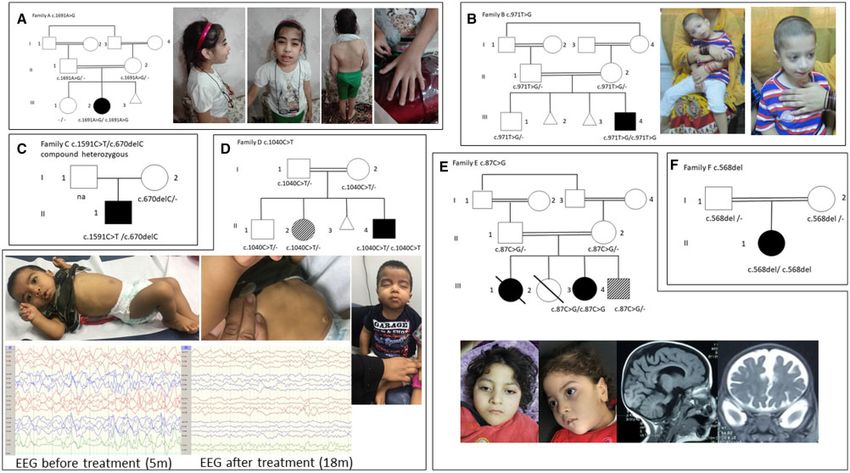
GAD1 causes epilepsy, developmental delay                                                      BRAIN 2020: 143; 2388–2397   | 2389

14   Department of Clinical and Experimental Epilepsy, University College London, London, UK
15   GeneDx, Gaithersburg, MD, USA
16   Clinical Genetics Department, National Research Centre, Cairo, Egypt
17   Department of Pediatrics, Faculty of Medicine, Benha University, Benha, Egypt
18   Dicle University, School of Medicine, Department of Medical Genetics, Diyarbakir, Turkey
19   Yuksek Ihtisas University, School of Medicine, Department of Medical Genetics, Ankara, Turkey
20   Mikrogen Genetic Diagnosis Center, Ankara, Turkey
21   CENTOGENE AG, Rostock
22   Molecular Genetics Department, National Research Centre, Cairo, Egypt
23   Molecular and Clinical Sciences Institute St George’s, University of London, UK
24   Department of Genetics, King Faisal Specialist Hospital and Research Center Riyadh, Saudi Arabia

Correspondence to: Prof. Henry Houlden
Department of Molecular Neuroscience, Institute of Neurology

                                                                                                                                      Downloaded from https://academic.oup.com/brain/article/143/8/2388/5875729 by guest on 24 December 2020
University College London, Gower Street, London, WC1E 6BT, London, UK
E-mail: h.houlden@ucl.ac.uk

Keywords: GAD1; epilepsy; neurodevelopmental delay; muscle weakness; cleft palate
Abbreviation: GAD = glutamate decarboxylase

                                                                     homozygous missense mutations, one carrying a homozy-
Introduction                                                         gous frameshift variant, two compound heterozygous var-
The neurotransmitter c-aminobutyric acid (GABA) is one of            iants, and one harbouring a homozygus stop gain variant).
the main inhibitory neurotransmitters deriving from glutam-          All affected individuals presented with seizures, strongly
ate (Cooper et al., 1996). It plays a critical signalling role in    impaired neurocognitive development, and reduced muscle
the nervous system and also in a number of non-neuronal              tone of variable severity. Interestingly only one presented
cell types. The enzyme responsible for the conversion of glu-        with cleft palate, which has been suggested to be one of the
tamate into GABA is glutamate decarboxylase (GAD), and               key features in the GAD1 animal models.
occurs in two isoforms GAD65 and GAD67, depending on
its molecular weight (Kaufman et al., 1991). These isoforms
are products of two different genes, GAD1 (encoding a 67
kDa molecular weight protein, GAD67) and GAD2 (encod-
                                                                     Material and methods
ing a 65 kDa molecular weight protein, GAD65). GAD67 is
constitutively active and produces 490% of the base level
                                                                     Patients and genetic analysis
GABA in the CNS, whilst GAD65 is transiently activated               Six patients from six unrelated families of Persian (Family
(Asada et al., 1997).                                                A), Pakistani (Family B), African American (Family C),
   Animal studies have also shown a distinct role for                Sudanese (Family D), Egyptian (Family E) and Turkish an-
GAD65 and GAD67. Gd65–/– mice are viable but show a                  cestry (Family F) were identified through GeneMatcher
higher susceptibility to seizure induction despite normal            (Sobreira et al., 2015) and enrolled in this study. The study
GABA levels (Asada et al., 1996), whereas Gad67–/– mice              was conducted according to the Declaration of Helsinki and
are characterized by neonatal death, severe cleft plate and          with the approval of the institutional review boards of
respiratory failure. GAD activity and GABA concentration             University College of London and participating centres.
are also drastically reduced in Gad67–/– mice (Asada                 Genetic testing through whole exome sequencing (WES) was
et al., 1997).                                                       carried out in different research centres after informed con-
   The severity of the Gad67–/– phenotype in animal models           sent was obtained from the parents or legal guardians of the
might suggest severe phenotypical manifestions in humans,            studied subjects. Genomic DNA was extracted from periph-
yet there have been few reported families with GAD1 muta-            eral blood obtained from the probands, parents, and un-
tions (Lynex et al., 2004; Saito et al., 2010; Curley et al.,        affected siblings (when available). Exome sequencing and
2011; Ruzicka et al., 2015; Magri et al., 2018). Previous            data analysis was performed as follows: Families A, B and D
reports have described seemingly unparalleled phenotypes,            in the according centres as previously described (Monies
which include schizophrenia, autism spectrum disease and             et al., 2019; Dias et al., 2019), Family C through GeneDx
cerebral palsy, and functional studies are missing to confirm        (Retterer et al., 2016), and Families E and F at Centogene
the pathogenicity of these reported mutations.                       (Bauer et al., 2018). Potential candidate causal variants were
   Here we report a series of six affected individuals with dis-     subsequently confirmed by independent bi-directional Sanger
tinct phenotypical features from six unrelated families with         sequencing. Detailed information is provided in the
bi-allelic mutations in the GAD1 gene (three carrying                Supplementary material.
2390 | BRAIN 2020: 143; 2388–2397 C. Neuray et al.
Data availability GAD1 were identified in all affected individuals. The segre-
gation of the variants with the clinical phenotype was con-
The data that support the findings of this report are avail-
firmed by Sanger sequencing, which showed a recessive
able from the corresponding author, upon reasonable
mode of inheritance. Detailed genetic results are provided in
request.
Table 2. All affected individuals carried ultrarare GAD1 var-
iants, which were predicted to result in impaired protein
function. Homozygous variants were identified in five fami-
Results lies (Families A, B, D, E and F), whereas compound hetero-
zygous variants were found in Family C (Table 2).
Clinical manifestation The affected proband from Family A carried a homozy-
Four of the affected individuals were born from consanguin- gous c.1691A4G, p.(Asn564Ser) variant, which is reported
eous parents (first or second cousins), and all were born at only once in the heterozygous state in gnomAD. This variant
Downloaded from https://academic.oup.com/brain/article/143/8/2388/5875729 by guest on 24 December 2020
term following a normal pregnancy. A key clinical feature has a combined annotation dependent depletion (CADD)
common to all affected individuals was early onset seizures score of 26.1 and is predicted to be pathogenic by several
(from 2 to 6 months), predominantly focal motor seizures bioinformatic prediction tools, including SIFT (score
with and without impaired awareness (two with additional 0.9125), MutationTaster, and PolyPhen-2 (score 1)
epileptic spasms, four with focal non-motor seizures, and (Table 2). The proband from Family B harboured a homozy-
five with bilateral motor seizures). Seizures were pharmaco- gous c.971T4G, p.(Phe324Cys) variant, which causes the
logically controlled in three of six affected individuals, with substitution of a phenylalanine residue at position 324 with
three reported as drug-resistant. Drug regimens differed cysteine. This position is not strictly conserved in other spe-
across the individuals. EEG at seizure onset showed a burst cies, where leucine is found in place of phenylalanine.
suppression pattern in two individuals, diffuse slowing with However, both phenylalanine and leucine belong to the class
multifocal as well as generalized sharp waves in two, and of the amino acids with long hydrophobic chains (Fig. 2A)
hypsarrhythmia in two. Follow-up EEGs showed diffuse and share several chemical features. This variant has been
slowing of background activity or persistent epileptic activity seen in the heterozygous state in gnomAD with a minor al-
(two of six). Cranial MRI was normal in all but two individ- lele frequency of 0.00000796. It has a CADD score of 28
uals, one showing slight ventricular enlargement and one and is predicted damaging by all the prediction tools used
moderate global atrophy. (scores of 0.9125 and 0.978 for SIFT and PolyPhen-2, re-
The second common clinical feature was severe develop- spectively). The proband from Family D carried a homozy-
mental delay. Most patients did not achieve any speech or gous c.1040C4T, p.(Thr347Met) variant, which was
non-verbal communication, only one was reported to have reported twice in the heterozygous state in the gnomAD
developed simple speech and basic perceptive language skills. database. It was predicted damaging by both SIFT and
This individual has remained seizure-free under medication. PolyPhen-2 with high scores (0.9125 and 1, respectively).
Of the more severely affected individuals, despite remaining The CADD score for this variant was 29.1. Further in silico
seizure-free following pharmacological intervention, they still analysis predicted a reduction in protein stability for
failed to accelerate in their intellectual development. p.(Phe324Cys) and p.(Asn564Ser), in association with a
The third key feature we observed was a reduced muscle break in H-bonds in the pyridoxal 50 -phosphate (PLP) bind-
strength (five of six individuals) of varying severity ranging ing domain for p.(Thr347Met). Both these changes might re-
from slight muscle tone (one of five) to limited head control, sult in an impairment of the protein function due to the
inability to sit or crawl (four of six) and nasogastric tube de- abnormal degradation or the decreased binding activity to-
pendence, due to dysphagia (two of six). While slight dys- wards PLP, leading to a likely loss-of-function effect (Fig. 2B
morphic facial features were seen in four of six individuals and Supplementary Table 1). In Family E, a homozygous
(Table 1), only one presented with a cleft palate. stop gain variant c.87C4G, p.(Tyr29Ter) was identified.
Other features that were observed without clear common This variant is absent in all the queried population datasets.
elements included hirsutism, kidney stones, urogenital mal- It is predicted damaging by SIFT (score not available) and
formations, diastasis recti, reduced head circumference, cli- disease-causing by Mutation Taster, with a CADD score of
nodactyly, short arms, arthrogryposis of the lower limbs and 35. This null variant likely causes a nonsense-mediated
congenital hip dislocation. Metabolic workups did not show mRNA decay (NMD), leading to a complete loss-of-func-
any abnormalities. A detailed clinical summary of all tion. The proband from Family F carries a homozygous
affected individuals can be found in Table 1 along with c.568delC p.(Gln190SerfsTer11) variant, which is absent in
images of key features and family pedigrees in Fig. 1. gnomAD and other population datasets queried. It is pre-
dicted damaging by MutationTaster (score 1) and classified
as pathogenic according to the ACMG guidelines. Family C
Genetic findings was the only family in which heterozygous variants were
Variants were prioritized in each family based on allele fre- found: c.1591C4T, p.(Arg531*) and c.670delC,
quency 50.01%, predicted impact on protein function, and p.(Leu224Serfs*5). This individual’s mother was a carrier
biological consistency. Potentially causal bi-allelic variants in for c.670delC; however, the individual’s father was notTable 1 Clinical features of GAD1 patients
Family A (Patient III-2) B (Patient III-4) C (Patient II-1) D (Patient II-4) E (Patient III-3) F (Patient II-2)
Sex/ ethnic origin Female/ Persian Male/ Pakistani Male/ African American Male/ Sudanese Female/ Egyptian Female/ Turkish
Consanguinity Yes (double first cousins) Yes – Yes (second cousins) Yes (first cousins) Yes (first cousins)
Age at first/last exam 5 y/10 y 3 m 6 m/7 y 2 m/22 m 2 m/18 m 6 m/4 y 1 m/3 y
Development
Milestones Delayed in all milestones, Delayed in all milestones, Delayed in all milestones, Severe delay, poor head Severe delay in all mile- Severe delay in all
simple speech at 4 y, sitting and crawling no head control, no control achieved at stones, bed ridden milestones, no sitting
walking delayed, no sitting, no speech 18m, no sitting or crawling
complex movements
ID Moderate Severe Severe Severe Severe Severe
GAD1 causes epilepsy, developmental delay
Vision/hearing High myopia Normal NA Normal Moderate hearing High myopia
impairment
Dysmorphic facial Yes Yes – Yes Yes –
features
Cleft palate – – – – Yes (surgical correction) –
Skeletal abnormalities Clindodactyly, pes planus, Arthrogryposis of lower – Short arms Congenital hip dislocation
scoliosis limbs and malformation
Neurological examination Mild hypotonia Brisk DTR, stereotypic Mild hypotonia, spasticity Severe hypotonia, dyspha- Severe hypotonia, hypore- Severe hypotonia
hand movements, oral in lower extremities, gia (floppy epiglottis) flexia, dysphagia
automatisms oropharyngeal
dysphagia
Epilepsy
Age at onset 2m 6m 2m 2m 6m 2m
Type(s) of seizure at Focal non/motor with Focal motor impaired Focal non/motor with Epileptic spasms Bilateral tonic clonic Focal ± to bilateral
onset impaired awareness awareness, bilateral impaired awareness, motor with impaired
tonic-clonic bilateral tonic clonic awareness
Seizure progression (age) Controlled (10 y), last Controlled (7 y), last Refractory (28 m) Spasms continue, seizures Refractory (4 y) Partial control, 1
seizure at age 7 y seizure at 5.5 y controlled (18 m) seizure/week
EEG at onset Burst suppression Multifocal and generalized Burst suppression Hypsarrhythmia Generalized epileptogenic Hypsarrhythmia
epileptogenic activity activity
Follow-up EEG (age) Normal (11 y) Normal (7 y) Slowing, multifocal No epileptic abnormal- NA Generalized epilepti-
epileptic discharges ities (18 m) form activity (4 m)
(28 m)
AEDs trialled PB, VPA, CBZ, CLB PB, VPA, CLZ CBD, vigabatrin, ketogenic steroids, TP, vigabatrin LEV, CLZ, TP PB, TP, LEV, CLZ,
diet, CLZ, PB vigabatrin, steroids
Cardiovascular MRI (age) Normal (5 y) Prominent ventricular Normal (2 m) Normal (6 m) Moderate global atrophy Normal (2 m)
space (6 m) (1 y)
Other features Hydronephrosis, – NG-tube dependent Diastasis recti Intermittent NG tube –
nephrocalcinosis, dependence
bilateral kidney stones
BRAIN 2020: 143; 2388–2397
Current antiepileptic drugs (AEDs) are highlighted in italics. CBD = cannabidiol; CBZ = carbamazepine; CLB = clobazam; CLZ = clonazepam; DTR = deep tendon reflexes; ID = intellectual disability; LEV = levetiracetam; NA = not applic-
able; NG = nasogastric; OXC = oxcarbazepine; PB = phenobarbital; TP = topiramate; VPA = valproic acid.
| 2391
Downloaded from https://academic.oup.com/brain/article/143/8/2388/5875729 by guest on 24 December 20202392 | BRAIN 2020: 143; 2388–2397 C. Neuray et al.
Downloaded from https://academic.oup.com/brain/article/143/8/2388/5875729 by guest on 24 December 2020
Figure 1 Pedigrees of the reported families and clinical pictures of GAD1 patients. (A) The female patient from Family A (Patient II-
2) carries the homozygous c.1691A4G p.(Asn564Ser) variant and shows dysmorphic features with thick eyebrows, protruding ears, scoliosis,
and long fingers with clinodactyly. (B) Patient from Family B (Patient III-4) harbours the c.971T4G p.(Phe324Cys) variant. He has slight dys-
morphic features (wide mouth, thin upper lips, bitemporal narrowing and retrognathia). (C) Pedigree showing the segregation of the compound
heterozygous variants c.1591C4T, c.1591C4T p.(Arg531*) and c.670delC p.(Leu224Serfs*5) in Family C. (D) Pedigree of Family D shows the
segregation of the c.1040C4T p.(Thr347Met) variant. Patient II-4 carries the variant in homozygous state. He is severely hypotonic and shows
severe dysmorphic features (infra-orbital creases, severely depressed nasal bridge, anteverted nares, prominent nasolabial folds). In addition, sig-
nificant diastasis recti can be observed. His sister (Patient II-2) is heterozygous for the same variant and suffers from a different neurodevelop-
mental condition without seizures. (E) Patient III-2 from Family E harbours the c.87C4G (Tyr29*) variant, severely affected with dysmorphic
facial features and global atrophy on cardiovascular MRI, one similarly affected sibling passed away without any genetic testing being performed,
another sibling passed away only a few hours after birth, no phenotypical or genetic assessment could be carried out, and one sibling is alive with
a different phenotype (sensoneural hearing loss, Hirschsprung disease). (F) Patient II-1 from Family F harbours the c.568del (Gln190Serfs*11)
variant. His parents are both heterozygous carriers of the same variant. Empty and full symbols represent healthy and affected individuals, re-
spectively. The symbol with diagonal lines indicates carrier status/different phenotype. The double line indicates consanguinity.
available for genetic testing. The c.1591C4T, p.(Arg531*) pivotal role in synaptogenesis and neuronal development
variant results in a stop gain with a very high likely impact (Asada et al., 1997; Soghomonian and Martin, 1998; Sgadò
on protein function, as highlighted by a CADD score of 43. et al., 2011).
This is only reported in heterozygous state in the gnomAD The 594 amino acid protein GAD67 is composed of a N-
database. Similarly, the frameshift variant c.670delC, terminal domain involved in the generation of GAD65–
p.(Leu224Serfs*5) is predicted to be disease-causing by GAD67 heterodimers and subcellular targeting, a C-terminal
MutationTaster and affects a very conserved residue, with a domain containing the catalytic site, and a central conserved
GERP score of 6.17. The null variants identified in Families domain binding PLP (Bosma et al., 1999; Martin et al.,
B, E, and F may cause a premature termination of the tran- 2000; Lynex et al., 2004). This pyridoxal-dependent decarb-
script leading to a truncated protein or, alternatively, oxylase domain is essential for GAD67 function as GAD
affected transcripts might be target of NMD. requires PLP as a cofactor to catalyze the generation of
GABA from glutamate (Lernmark, 1996). Four of the seven
variants identified in our families affect conserved residues in
Discussion the PLP-binding domain, likely leading to loss-of-function
(Fig. 1A). In particular, the two missense variants
In humans, GAD1 encodes a 67-kDa protein, GAD67, c.971T4G, p.(Phe324Cys) and c.1040C4T,
which is the major contributor to GABA production in the p.(Thr347Met) probably cause impaired PLP binding,
CNS. As the major embryonic GAD isoform, it also plays a whereas the null variants c.568delC, p.(Gln190SerfsTer11)GAD1 causes epilepsy, developmental delay BRAIN 2020: 143; 2388–2397 | 2393
Table 2 Frequency and predicted effect of the reported GAD1 variants
GAD1 variant c.87C4G c.568delC c.670delC c.971T4G c.1040C4T c.1591C4T c.1691A4G
[NM_000817.2] (p.Tyr29Ter) (p.Gln190Serfs p.(Leu224Serfs*5) p.(Phe324Cys) p.(Thr347Met) p.(Arg531*) p.(Asn564Ser)
(Family E) Ter11) (Family F) (Family C) (Family B) (Family D) (Family C) (Family A)
g. (hg19) g.171678601C4G g.171693323delC g.171700586delC g.171702542T4G g.171704223C4T g.171715383C4T g.171716298A4G
Internal database – – – – – – –
ExAC/GnomAD – – – 0.00000796 (2 het) 0.00000795 (2 het) 0.00000398 (1 het) 0.00000398 (1 het)
GME – – – – – – –
Iranome – – – – – – –
Ensembl – – – – – – –
SIFT D- N/A N/A N/A D (0.9125) D (0.9125) N/A D (0.9125)
MutationTaster DC (1) DC (1) DC (1) DC (0.9768) DC (1) DC (1) DC (1)
PolyPhen-2 N/A N/A N/A PD (0.978) PD (1) N/A PD (1)
GERP score 4.97 5.55 6.17 5.91 5.67 4.67 5.48
Downloaded from https://academic.oup.com/brain/article/143/8/2388/5875729 by guest on 24 December 2020
CADD score 35 N/A N/A 28 29.1 43 26.1
ACMG class 5 (PVS1, PM2, PP4) 5 (PVS1, PM2, PP3) 5 (PVS1, PM2, PP3) 3 (PM2, PP3) 3 (PM2, PP3) 5 (PVS1, PM2, PP3) 3 (PM2, PP3)
GeneDx 0 0 0/135 084 0 1/130 874 0/135 084 1/130 874
CADD = Combined Annotation Dependent Depletion; D = damaging; DC = disease causing; GeneDx = variant frequencies from the GeneDx database; GERP = Genomic
Evolutionary Rate Profiling; GnomAD = Genome Aggregation Database; GME = Greater Middle East (GME) Variome Project; het = heterozygous; N/A = not applicable; PD =
probably damaging; PM2 = Pathogenic Moderate 2; PP3 = Pathogenic Supporting 3; PVS1 = Pathogenic Very Strong 1; SIFT = Sorting Intolerant From Tolerant.
and c.670delC p.(Leu224Serfs*5) might result in a truncated shown that there is a reduction of GABAergic neurons in the
protein or NMD. Similarly, the c.87C4G, p.(Tyr29Ter) epileptic brain, independent of the seizure’s aetiology sup-
variant localized to the N-terminal domain results in a pre- porting a conclusion that seizures themselves further de-
mature stop codon, likely leading to NMD. The remaining crease GABA release within the brain, causing further
two variants, c.1591C4T, p.(Arg531*) and c.1691A4G, imbalance (Wang et al., 2011). This evidence suggests that
p.(Asn564Ser), are localized to the C-terminal domain of the there is a clear correlation between abnormalities within the
protein and are predicted to result in a complete and partial GABAergic system and seizure occurrence. The occurrence
loss-of-function of the catalytic activity, respectively of seizures early in life of our patients is therefore not a sur-
(Fig. 2B). According to gnomAD, GAD1 shows a moderate prising phenotypical finding. However, previous data sug-
intolerance to missense variants (Z-score = 2.32; observed gest that the intellectual development of newborns with
217 and expected 336.5) and predicted loss-of-function var- epileptic encephalopathies is strongly dependent on seizure
iants (expected 33.1, observed 9). The likely negative impact control, and seizure freedom usually leads to acceleration of
of the missense variants on PLP-binding and C-terminal intellectual development (Bombardieri et al., 2010).
catalytic domains, together with the finding of four null However, despite seizure control (seizure freedom achieved
pathogenic variants in our case series, supports the idea that in three of six individuals), all patients described here still
the clinical phenotypes observed are likely due to a loss-of- showed severe intellectual disability. This has been observed
function mechanism. previously in other genetic conditions (Weckhuysen et al.,
With regard to the possible genotype-phenotype correl- 2012; Berecki et al., 2019), leading to the assumption that
ation, there have been few clinical case reports of GAD1 genetic defects themselves are influencing development and
mutations, with little overlap in clinical features (Lynex cognition, independently from seizure control. This is
et al., 2004; Ruzicka et al., 2015; Magri et al., 2018). The reflected in the ILAE’s terminology of ‘developmental and
six patients reported here, however, do show a clear pheno- epileptic encephalopathies’, though a clear distinction be-
typical manifestation. tween epileptic and developmental encephalopathy has been
One of the main common features we observed was the advised (Scheffer et al., 2017; Scheffer and Liao, 2020).
occurrence of seizures at a young age (between 2 and 6 Furthermore, some EEG abnormalities continued to be
months of age). Since GAD1 is centrally involved in the pro- prominent after achieving seizure freedom. We therefore hy-
duction of GABA, GAD1 mutations likely lead to an imbal- pothesize that GAD1 mutations may also play an important
ance of GABA in the brain. Previous studies have shown role in intellectual development.
that abnormalities in GABAergic function play an important The reduced muscle tone and weakness, causing severe
role in seizure induction (Olsen and Avoli, 1997). disability in four of six affected individuals was a surprising
Dysfunction of the GABAergic system can be caused finding. There is a single case described in the literature of
through either abnormal GABA synthesis (e.g. GAD dys- an individual with cerebral palsy and a GAD1 variant
function) or abnormal signalling (e.g. GABA receptor mal- (Lynex et al., 2004). However, identification of the variant
function). Animal studies have shown that mutant mice was based on autozygosity mapping, in which they identified
lacking GAD or certain subunits of GABA-A receptors are a recessive locus of 5 cM located at 2q24-31.1. The investi-
prone to spontaneous epileptic seizures (Asada et al., 1996; gators subsequently investigated the most interesting candi-
Kash et al., 1997; DeLorey et al., 1998). It has also been date in that region by sequencing the exons of GAD1 and2394 | BRAIN 2020: 143; 2388–2397 C. Neuray et al.
Downloaded from https://academic.oup.com/brain/article/143/8/2388/5875729 by guest on 24 December 2020
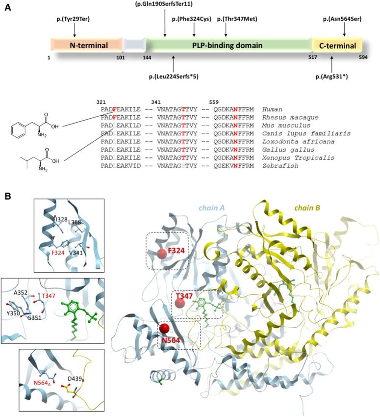
Figure 2 Schematic and cartoon representation of GAD1. (A) Schematic representation of the GAD1 isoform GAD67 (NP_000808.2)
with the pathogenic variants identified in this study. Of the six variants, four fall within the PLP-binding domain, a conserved region that is essen-
tial for the binding of the crucial cofactor pyridoxal 50 -phosphate (PLP). The remaining variants affect the C-terminal domain, which contains the
catalytic site of the enzyme. Conservation status among different species is shown for the missense variants. (B) Cartoon representation of
human GAD1 dimer (PDB: 2okj) with the two subunits in blue and yellow. Sites of the three missense mutations in this study are shown as red
spheres, and close-up views of their nearby atomic environment are shown as insets. The PLP cofactor is shown in green.
identified a homozygous missense variant c.35C4G gnomAD (exomes allele frequency of 0.0000239) and absent
(p.Ser12Cys) (NM_013445.3) in the N-terminal domain of in the homozygous state. However, the predictions on its
the protein (Lynex et al., 2004). This variant is rare in pathogenicity are conflicting as it is predicted benign byGAD1 causes epilepsy, developmental delay BRAIN 2020: 143; 2388–2397 | 2395
MutationAssessor, DEOGEN2, and MetaLR. Furthermore, studies and larger clinical series will be necessary to further as-
the 2q24-31.1 locus encompasses several other possible sess genotype-phenotype correlations for GAD1 variants.
genes of interest (e.g. DYNC1I2, responsible for a neurode-
velopmental disorder characterized by intellectual disability,
spasticity, and neuroradiological anomalies), which were not Acknowledgements
investigated. The N-terminal domain of GAD67 is involved
in the generation of GAD65–GAD67 heterodimers and sub- We thank all families and collaborators for providing
cellular targeting, and whilst we cannot rule out a possible detailed clinical data and samples to conduct this study.
impairment of the interaction of GAD67 with GAD65 or an
alteration in GAD67 subcellular targeting as a result of this
variant, we suggest that variants affecting the PLP-binding Funding
and C-terminal domain cause a more severe deficiency in
This study was supported by grants from The MRC (MR/
Downloaded from https://academic.oup.com/brain/article/143/8/2388/5875729 by guest on 24 December 2020
GAD67 activity.
S01165X/1, MR/S005021/1, G0601943), The National
In addition, we also identified four null variants likely
Institute for Health Research University College London
resulting in a loss-of-function. According to these observa-
Hospitals Biomedical Research Centre, Rosetree Trust,
tions and the consistency of the phenotype in our case series,
Ataxia UK, MSA Trust, Brain Research UK, Sparks GOSH
we emphasize that GAD1 pathogenic variants should be
Charity, Muscular Dystrophy UK (MDUK), Muscular
considered the cause of a distinctive neurodevelopmental dis-
Dystrophy Association (MDA USA). The families were col-
order instead of spastic cerebral palsy (Lynex et al., 2004).
lected as part of the SYNaPS Study Group collaboration
The clinical observation of muscle weakness is particularly
funded by The Wellcome Trust and strategic award
interesting as it has not been described previously. Other
(Synaptopathies) funding (WT093205 MA and
GAD1-related diseases, such as antibody-mediated syn-
WT104033AIA). This research was conducted as part of the
dromes, have been associated with motor symptoms (Dayalu
Queen Square Genomics group at University College
and Teener, 2012). However, these motor phenomena are
London, supported by the National Institute for Health
usually linked to a hyperexcitability (increased muscle tone
Research University College London Hospitals Biomedical
leading to rigidity, muscle spasms, and stiff person syn-
Research Centre.
drome), as well as other neurological symptoms (e.g. Miller
Fisher syndrome, eye movement disorders, cerebellar ataxia,
epilepsy, limbic encephalitis, etc.) (Moersch and Woltman,
1956; Dalakas et al., 2000; Saiz et al., 2008; Tohid, 2016). Competing interests
Of note, in no case has muscle weakness been linked to R.P. is an employee of GeneDx, Inc. S.K. and C.B. are
GAD1 deficiency. employees of Centogene, AG.
While Gad67–/– mice have been reported to die within the
first hours of life due to a cleft palate (Asada et al., 1997;
Condie et al., 1997), only one of our patients (Family E,
Patient III-3) was born with a cleft palate (the same patient
Supplementary material
also showed congenital bilateral hip dislocation with shallow Supplementary material is available at Brain online.
acetabulum, talipes equinovarus and hearing impairment.)
Several studies have hypothesized that the development of a
cleft palate is linked to reduced tongue movement during Appendix 1
embryonic development and therefore secondary to CNS
dysfunction (Iseki et al., 2007; Oh et al., 2010; Saito et al., Consortia and network members involved in this study are
2010). Gad1 expression has also been shown in different listed below. The full details are available in the
non-neural tissues, such as the tail bud, limb mesenchyme, Supplementary material.
vibrissal placodes, and pharyngeal arches in various stages
of embryonic development (Maddox and Condie, 2001). The Synaptopathies and Paroxysmal
This observation has suggested a broader influence of
GAD1 and GABA function on non-neural development
Syndromes (SYNaPS) Study Group
(Maddox and Condie, 2001), supporting a possible primary http://neurogenetics.co.uk/synaptopathies-synaps/
role of GAD1 impaired function in the pathogenesis of non- Stanislav Groppa, Blagovesta Marinova Karashova,
neural defects. However, further studies are required to con- Wolfgang Nachbauer, Sylvia Boesch, Larissa Arning,
firm this in humans. Dagmar Timmann, Bru Cormand, Belen Pérez-Due~ nas,
In conclusion, this case series reports distinct phenotypical Gabriella Di Rosa, Jatinder S. Goraya, Tipu Sultan, Jun
features caused by GAD1 variants, including early-infantile Mine, Daniela Avdjieva, Hadil Kathom, Dr Radka
onset epilepsy, severe developmental delay and muscle weak- Tincheva, Selina Banu, Mercedes Pineda-Marfa, Pierangelo
ness. Less consistent features include skeletal abnormalities and Veggiotti, Michel D. Ferrari, Alberto Verrotti, Giangluigi
dysmorphic facial features, including cleft palate. Functional Marseglia, Salvatore Savasta, Mayte Garcı́a-Silva, Alfons2396 | BRAIN 2020: 143; 2388–2397 C. Neuray et al.
Macaya Ruiz, Barbara Garavaglia, Eugenia Borgione, oriented palatal shelf. Birth Defect Res A Clin Mol Teratol 2007;
79: 688–95.
Simona Portaro, Benigno Monteagudo Sanchez, Richard Kash SF, Johnson RS, Tecott LH, Noebels JL, Mayfield RD, Hanahan
Boles, Savvas Papacostas, Michail Vikelis, Eleni Zamba D, et al. Epilepsy in mice deficient in the 65-kDa isoform of
Papanicolaou, Efthymios Dardiotis, Shazia Maqbool, glutamic acid decarboxylase. Proc Natl Acad Sci USA 1997; 94:
Shahnaz Ibrahim, Salman Kirmani, Nuzhat Noureen Rana, 14060–5.
Osama Atawneh, George Koutsis, Marianthi Breza, Kaufman DL, Houser CR, Tobin AJ. Two forms of the gamma-amino-
butyric acid synthetic enzyme glutamate decarboxylase have distinct
Salvatore Mangano, Carmela Scuderi, Eugenia Borgione, intraneuronal distributions and cofactor interactions. J Neurochem
Giovanna Morello, Tanya Stojkovic, Massimi Zollo, Gali 1991; 56: 720–3.
Heimer, Yves A. Dauvilliers, Pasquale Striano, Issam Al- Lernmark A. Glutamic acid decarboxylase–gene to antigen to disease.
Khawaja, Fuad Al-Mutairi, Hamed Sherifa. J Intern Med 1996; 240: 259–77.
Lynex CN, Carr IM, Leek JP, Achuthan R, Mitchell S, Maher ER, et
al. Homozygosity for a missense mutation in the 67 kDa isoform of
References glutamate decarboxylase in a family with autosomal recessive spastic
Downloaded from https://academic.oup.com/brain/article/143/8/2388/5875729 by guest on 24 December 2020
cerebral palsy: parallels with Stiff-Person Syndrome and other move-
Asada H, Kawamura Y, Maruyama K, Kume H, Ding R, Ji FY, et al. ment disorders. BMC Neurol 2004; 4: 20.
Mice lacking the 65 kDa isoform of glutamic acid decarboxylase Maddox DM, Condie BG. Dynamic expression of a glutamate decarb-
(GAD65) maintain normal levels of GAD67 and GABA in their oxylase gene in multiple non-neural tissues during mouse develop-
brains but are susceptible to seizures. Biochem Biophys Res ment. BMC Dev Biol 2001; 1: 1.
Commun 1996; 229: 891–895. Magri C, Giacopuzzi E, La Via L, Bonini D, Ravasio V, Elhussiny
Asada H, Kawamura Y, Maruyama K, Kume H, Ding RG, Kanbara MEA, et al. A novel homozygous mutation in GAD1 gene described
N, et al. Cleft palate and decreased brain gamma-aminobutyric acid in a schizophrenic patient impairs activity and dimerization of
in mice lacking the 67-kDa isoform of glutamic acid decarboxylase. GAD67 enzyme. Sci Rep 2018; 8: 15470.
Proc Natl Acad Sci USA 1997; 94: 6496–9. Martin DL, Liu H, Martin SB, Wu SJ. Structural features and regula-
Bauer P, Kandaswamy KK, Weiss MER, Paknia O, Werber M, Bertoli- tory properties of the brain glutamate decarboxylases. Neurochem
Avella AM, et al. Development of an evidence-based algorithm that Int 2000; 37: 111–9.
optimizes sensitivity and specificity in ES-based diagnostics of a clin- Moersch FP, Woltman HW. Progressive fluctuating muscular rigidity
ically heterogeneous patient population. Genet Med 2018; 21: and spasm (“stiff-man” syndrome); report of a case and some obser-
53–61. vations in 13 other cases. Proc Staff Meet Mayo Clin 1956; 31:
Berecki G, Bryson A, Terhag J, Maljevic S, Gazina EV, Hill SL, et al. 421–7.
SCN1A gain of function in early infantile encephalopathy. Ann Monies D, Abouelhoda M, Assoum M, Moghrabi N, Rafiullah R,
Neurol 2019; 85: 514–25. Almontashiri N, et al. Lessons learned from large-scale, first-tier
Bombardieri R, Pinci M, Moavero R, Cerminara C, Curatolo P. Early clinical exome sequencing in a highly consanguineous population.
control of seizures improves long-term outcome in children with tu- Am J Hum Genet 2019; 105: 879.
berous sclerosis complex. Eur J Paediatr Neurol 2010; 14: 146–9. Oh W-J, Westmoreland JJ, Summers R, Condie BG. Cleft palate is
Bosma PT, Blázquez M, Collins MA, Bishop JD, Drouin G, Priede IG, caused by CNS dysfunction in Gad1 and Viaat knockout mice. PloS
et al. Multiplicity of glutamic acid decarboxylases (GAD) in verte- One 2010; 5: e9758.
brates: molecular phylogeny and evidence for a new GAD paralog. Olsen RW, Avoli M. GABA and epileptogenesis. Epilepsia 1997; 38:
Mol Biol Evol 1999; 16: 397–404. 399–407.
Condie BG, Bain G, Gottlieb DI, Capecchi MR. Cleft palate in mice Retterer K, Juusola J, Cho MT, Vitazka P, Millan F, Gibellini F, et al.
with a targeted mutation in the gamma-aminobutyric acid-producing Clinical application of whole-exome sequencing across clinical indi-
enzyme glutamic acid decarboxylase 67. Proc Natl Acad Sci USA cations. Genet Med 2016; 18: 696–704.
1997; 94: 11451–5. Ruzicka WB, Subburaju S, Benes FM. Circuit- and diagnosis-specific
Cooper JR, Bloom FE, Roth RH, editors. The biochemical basis of DNA methylation changes at c-aminobutyric acid-related genes in
neuropharmacology. New York: New York: Oxford University postmortem human hippocampus in schizophrenia and bipolar dis-
Press; 1996. order. JAMA Psychiatry 2015; 72: 541–51.
Curley AA, Arion D, Volk DW, Asafu-Adjei JK, Sampson AR, Fish Saito K, Kakizaki T, Hayashi R, Nishimaru H, Furukawa T, Nakazato
KN, et al. Cortical deficits of glutamic acid decarboxylase 67 expres- Y, et al. The physiological roles of vesicular GABA transporter dur-
sion in schizophrenia: clinical, protein, and cell type-specific features. ing embryonic development: a study using knockout mice. Mol
Am J Psychiatry 2011; 168: 921–9. Brain 2010; 3: 40.
Dalakas MC, Fujii M, Li M, McElroy B. The clinical spectrum of anti- Saiz A, Blanco Y, Sabater L, González F, Bataller L, Casamitjana R, et
GAD antibody-positive patients with stiff-person syndrome. al. Spectrum of neurological syndromes associated with glutamic
Neurology 2000; 55: 1531–5. acid decarboxylase antibodies: diagnostic clues for this association.
Dayalu P, Teener JW. Stiff Person syndrome and other anti-GAD-asso- Brain 2008; 131: 2553–63.
ciated neurologic disorders. Semin Neurol 2012; 32: 544–9. Scheffer IE, Berkovic S, Capovilla G, Connolly MB, French J, Guilhoto
DeLorey TM, Handforth A, Anagnostaras SG, Homanics GE, L, et al. ILAE classification of the epilepsies: position paper of the
Minassian BA, Asatourian A, et al. Mice lacking the beta3 subunit ILAE Commission for Classification and Terminology. Epilepsia
of the GABAA receptor have the epilepsy phenotype and many of 2017; 58: 512–21.
the behavioral characteristics of Angelman syndrome. J Neurosci Scheffer IE, Liao J. Deciphering the concepts behind “Epileptic
1998; 18: 8505–14. encephalopathy” and “Developmental and epileptic encephalopathy”.
Dias CM, Punetha J, Zheng C, Mazaheri N, Rad A, Efthymiou S, et Eur J Paediatr Neurol 2020; 24: 11–14.
al. Homozygous missense variants in NTNG2, encoding a presynap- Sgadò P, Dunleavy M, Genovesi S, Provenzano G, Bozzi Y. The role
tic netrin-G2 adhesion protein, lead to a distinct neurodevelopmen- of GABAergic system in neurodevelopmental disorders: a focus on
tal disorder. Am J Hum Genet 2019; 105: 1048–56. autism and epilepsy. Int J Physiol Pathophysiol Pharmacol 2011; 3:
Iseki S, Ishii-Suzuki M, Tsunekawa N, Yamada Y, Eto K, Obata K. 223–35.
Experimental induction of palate shelf elevation in glutamate de- Sobreira N, Schiettecatte F, Boehm C, Valle D, Hamosh A. New tools
carboxylase 67-deficient mice with cleft palate due to vertically for Mendelian disease gene identification: phenoDB variantGAD1 causes epilepsy, developmental delay BRAIN 2020: 143; 2388–2397 | 2397
analysis module; and GeneMatcher, a web-based tool for linking Wang Y, Zhan L, Zeng W, Li K, Sun W, Xu ZC, et al. Downregulation
investigators with an interest in the same gene. Hum Mut 2015; 36: of hippocampal GABA after hypoxia-induced seizures in neonatal rats.
425–31. Neurochem Res 2011; 36: 2409–16.
Soghomonian JJ, Martin DL. Two isoforms of glutamate decarboxyl- Weckhuysen S, Mandelstam S, Suls A, Audenaert D, Deconinck T,
ase: why? Trends Pharmacol Sci 1998; 19: 500–5. Claes LRF, et al. KCNQ2 encephalopathy: emerging phenotype of a
Tohid H. Anti-glutamic acid decarboxylase antibody positive neuro- neonatal epileptic encephalopathy. Ann Neurol 2012; 71: 15–25.
logical syndromes. Neurosciences (Riyadh) 2016; 21: 215–22.
Downloaded from https://academic.oup.com/brain/article/143/8/2388/5875729 by guest on 24 December 2020You can also read