Oncogenic codon 13 NRAS mutation in a primary mesenchymal brain neoplasm and nevus of a child with neurocutaneous melanosis
←
→
Page content transcription
If your browser does not render page correctly, please read the page content below
Shih et al. Acta Neuropathologica Communications 2014, 2:140
http://www.actaneurocomms.org/content/2/1/140

 CASE REPORT                                                                                                                                  Open Access

Oncogenic codon 13 NRAS mutation in a primary
mesenchymal brain neoplasm and nevus of a
child with neurocutaneous melanosis
Francis Shih1, Stephen Yip2, Patrick J McDonald3, Albert E Chudley4 and Marc R Del Bigio5*

  Abstract
  A 28-month female with a clinical diagnosis of neurocutaneous melanosis and numerous intracranial abnormalities
  (including a right choroid plexus tumor and left hemimegalencephaly) presented with a rapidly expanding tumor
  in the left occipital cerebrum. Microscopic examination of the resected specimen revealed a myxoid mesenchymal
  neoplasm consisting of fusiform cells that were immunoreactive for vimentin, CD34, and P53 but no melanocyte
  markers. Focused amplicon deep sequencing on DNA extracted from the brain tumor and a cutaneous nevus
  revealed a heterozygous (c.37G > C; p.G13R) substitution in the NRAS gene. DNA sequencing of “normal” skin and
  buccal swab showed the identical NRAS change albeit at lower allelic frequency. Her parents did not harbor the
  NRAS mutation. The skin lesion, but not the brain tumor, had a BRAF mutation (c.1397G > T; p.G466V). A germline
  single nucleotide polymorphism in MET was found in the child and her father (c.3209C > T; p.T1010I). The findings
  suggest NRAS mosaicism that occurred sometime after conception and imply an oncogenic role of the activating
  NRAS mutation in both the brain and skin lesions in this child.
  Keywords: Melanosis, Brain tumor, Genetic mutation, Somatic mosaicism

Background                                                                          DNA sequencing showed shared mutations in codon 13
Neurocutaneous melanosis (NCMS; Mendelian Inherit-                                  of NRAS in the melanocytic nevi and the brain tumor.
ance in Man MIM# 249400) is a rare congenital phako-
matosis consisting of numerous giant cutaneous nevi
                                                                                    Case report
along with extensive leptomeningeal melanosis. Approxi-
                                                                                    Clinical details
mately 100 cases have been described in the literature
                                                                                    This female child was born to a healthy non-consan-
[1]. The pathogenesis of NCMS likely involves a mor-
                                                                                    guineous couple after an uneventful full-term pregnancy.
phogenetic error in which melanocyte precursors derived
                                                                                    At birth she had numerous slightly raised, hairy melano-
from the neural crest migrate abnormally and proliferate
                                                                                    cytic lesions on the scalp, neck, upper trunk, upper ex-
locally [2,3]. Most cases with an identifiable cause have a
                                                                                    tremities, and hands; the largest was 4–5 cm in greatest
somatic gain-of-function mutation in codon 61 of the
                                                                                    dimension (Figure 1). Skin lesions from the neck, scalp,
NRAS gene (MIM# 164790) located on chromosome 1p13
                                                                                    and arm were previously excised and diagnosed as in-
[4-7]. Approximately 30% of affected children have mel-
                                                                                    tradermal and compound nevi with congenital features.
anin deposits detectable in the leptomeninges or brain [8]
                                                                                    She had normal height but was macrocephalic (97th per-
and half have epilepsy [9].
                                                                                    centile head circumference). She had been admitted to
  We present a case of a female infant with NCMS who
                                                                                    hospital numerous times for uncontrolled seizures start-
developed an unusual myxoid mesenchymal brain tumor.
                                                                                    ing at age 2 months. Magnetic resonance (MR) imaging
                                                                                    of the brain and spine was performed at 3.5 months age.
                                                                                    T1 weighted images showed a solitary 2 mm focus of in-
* Correspondence: Marc.Delbigio@med.umanitoba.ca                                    creased signal intensity in the right cerebellopontine cis-
5
 Department of Pathology, University of Manitoba and Diagnostic Services
Manitoba, Room 401 Brodie Centre, R3E 3P5 Winnipeg, MB, Canada                      tern; this was thought to represent melanin deposition.
Full list of author information is available at the end of the article              Cystic lesions, the largest of which was 2.7 cm diameter,
                                       © 2014 Shih et al.; licensee BioMed Central Ltd. This is an Open Access article distributed under the terms of the Creative
                                       Commons Attribution License (http://creativecommons.org/licenses/by/4.0), which permits unrestricted use, distribution, and
                                       reproduction in any medium, provided the original work is properly credited. The Creative Commons Public Domain
                                       Dedication waiver (http://creativecommons.org/publicdomain/zero/1.0/) applies to the data made available in this article,
                                       unless otherwise stated.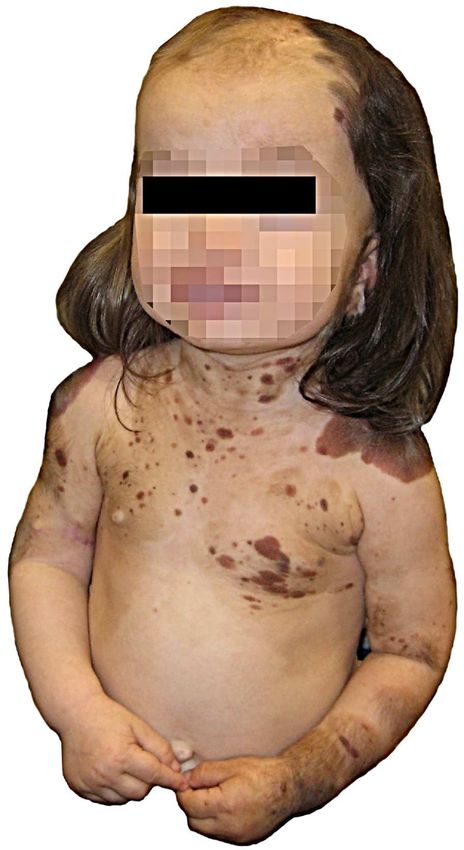
Shih et al. Acta Neuropathologica Communications 2014, 2:140                                                    Page 2 of 7
http://www.actaneurocomms.org/content/2/1/140

                                                                 on FLAIR. The periventricular cysts had increased in
                                                                 size and quantity. A new lesion in continuity with the
                                                                 choroid plexus was expanding the temporal horn of the
                                                                 right lateral ventricle. MR imaging of the brain at 21
                                                                 months age showed a new left occipital brain tumor that
                                                                 was T2 hyperintense and enhanced strongly following
                                                                 administration of gadolinium-DTPA. It grew from 1.6 ×
                                                                 1.5 × 1.2 cm to 4.0 × 3.6 × 3.2 cm 4 months later
                                                                 (Figure 2). A left occipital craniotomy was performed
                                                                 at 24.5 months age. The tumor was not in contact with
                                                                 the meninges. It had a single vascular pedicle. There
                                                                 was an easily identified plane between it and the adja-
                                                                 cent brain. The tumor was removed in one piece with-
                                                                 out complication. MR imaging of the brain 26 months
                                                                 after surgery (51 months age) showed no recurrence of
                                                                 the tumor; a vague region of non-enhancing T2 signal
                                                                 abnormality in the right medial occipital lobe had ex-
                                                                 panded slightly.

                                                                 Histopathological characterization
                                                                 The tumor was a 4.5 × 3.5 × 3 cm firm tan nodule
                                                                 with a smooth external surface and uniform cut surface
                                                                 (Figure 3A). The brain tumor and skin biopsies were
                                                                 fixed in 10% neutral buffered formalin, dehydrated and
                                                                 embedded in paraffin wax for sectioning (5 uM in thick-
                                                                 ness). All samples were stained with hematoxylin and
                                                                 eosin for histologic evaluation. Immunohistochemistry
                                                                 was performed using primary antibodies against vimentin
                                                                 (mouse monoclonal, V9, Dako), CD34 (mouse monoclo-
                                                                 nal, QBEND-10, Dako), alpha B crystallin (mouse mo-
                                                                 noclonal, G2JF, Novocastra), CD56 (mouse monoclonal,
                                                                 CD564, Dako), D240 (mouse monoclonal, D2-40, Dako),
                                                                 CD99 (mouse monoclonal, 12E71, Dako), Bcl2 (mouse
                                                                 monoclonal, 124, Dako), p53 (mouse monoclonal, D0-7,
 Figure 1 Photograph of the patient at 14 months age showing     Dako), Ki67 (mouse monoclonal, MIB-1, Dako), HLA-DR
 giant congenital melanocytic nevi on each shoulder and
                                                                 (mouse monoclonal, CR3/43, Dako), Factor 13a (mouse
 numerous smaller satellite nevi on the neck, scalp, arms, and
 upper trunk.                                                    monoclonal, EP3372, Cellmarque), GFAP (rabbit poly-
                                                                 clonal, Dako), S100 (rabbit polyclonal, Dako), Collagen
                                                                 IV (mouse monoclonal, C1V22, Dako), pan-cytokeratin
with signal characteristics the same as cerebrospinal            (mouse monoclonal, AE1/AE3, Dako), EMA (mouse mo-
fluid (CSF) were present around the atria of the lateral         noclonal, E29, Dako), CD57 (mouse monoclonal, TB01,
ventricles in the right cerebellopontine cistern along           Dako), HMB-45 (mouse monoclonal, HMB45, Dako),
with small cysts around the bodies of the lateral ventri-        MART-1/Melan-A (mouse monoclonal, A103, Dako), cal-
cles. The left cerebral hemisphere was larger than the           retinin (rabbit polyclonal, Cellmarque), neurofilament
right, and parietal white matter volume was abnormally           (mouse monoclonal, 2 F11, Dako), synaptophysin (mouse
small. The combination of giant congenital nevi along            monoclonal, 27G12, Novocastra), NeuN (mouse mono-
with cerebral melanin deposition led to a clinical diagno-       clonal, A60, Lifespan Biosciences). Antigen retrieval was
sis of NCMS [10]. A CT scan of the head performed at             performed in a Bull’s Eye Decloaking chamber (Biocare
4 months age during an episode of status epilepticus             Medical, Concord, CA) for 1 minute at 125°C utilizing a
showed no additional abnormalities. An abdominal so-             Dako pH9 retrieval solution. All antibodies were detected
nogram performed the following day showed no ab-                 using the Dako Envision system (Dako) and diamino-
normalities. Repeat MR imaging showed the presumed               benzidine precipitation solution. A sample of the tumor
melanocytic nodule had increased to 3 mm; it was hy-             was fixed in 2.5% buffered glutaraldehyde, post-fixed
perintense on T1, hypointense on T2, and hyperintense            in osmium tetroxide, dehydrated in graded ethanol, and
Shih et al. Acta Neuropathologica Communications 2014, 2:140 Page 3 of 7
http://www.actaneurocomms.org/content/2/1/140
Microscopic examination showed an indistinct inter-
face with the brain parenchyma. The tumor had a diffuse
pattern of stellate and elongated cells with delicate
processes in a loose myxoid background. There were no
pigmented cells. Numerous small, multinucleated cells
were evenly distributed within the lesion (Figure 3B).
Rarely the cells clustered around blood vessels. Very rare
mitotic figures were present and there was no necrosis
or endothelial hyperplasia. Focal areas of the extracellu-
lar material stained with Alcian blue (Figure 3C). Reticu-
lin staining was negligible. By immunohistochemistry,
the cells were positive for vimentin, CD34 (Figure 3D),
CD56, D240, Bcl2, CD99 (weak), and P53. A subpopula-
tion of cells was Factor 13a positive. An estimated 5-10%
of nuclei were Ki67 positive. Only perivascular cells
(likely astrocytes) were positive for glial fibrillary acidic
protein (GFAP), S100, and alpha B crystallin. Blood ves-
sel walls were positive for E cadherin and collagen IV.
There was no immunoreactivity for cytokeratin (AE1/3),
epithelial membrane antigen (EMA), CD57, HMB-45,
MART-1/Melan-A, calretinin, neurofilament, synaptophy-
sin, or NeuN. Scattered HLA-DR positive cells were likely
infiltrating microglia. Electron microscopic examination
showed fusiform cells with abundant rough endoplasmic
reticulum and prominent intermediate filaments but
no specific secretory organelles or obvious inter-
cellular junctions. The cells lacked a well-defined base-
ment membrane and were surrounded by a flocculent
extracellular material with rare clusters of striated collagen
bundles (Figure 3E, 3F). The diagnostic categorization
arrived at locally and supported by external consultation
was myxoid mesenchymal brain tumor of uncertain
growth potential.
DNA extraction and sequencing
Genomic DNA was extracted from formalin-fixed paraffin-
embedded samples of the intracranial tumor, an excised
cutaneous nevus lesion, and grossly unaffected skin. DNA
was also extracted from swabbed buccal cells of the
patient and both of her parents. DNA was quantitated
using the Qubit 2.0 fluorometer (Life Technology). Fo-
cused deep sequencing of 10 ng of genomic DNA ex-
Figure 2 Magnetic resonance image of the brain. At 14 months tracted from the above samples was performed using the
age (upper image, coronal, T2 weighted) an enlarged left temporal
Ion AmpliSeq Cancer panel (Life Technology). This in-
lobe and a tumor of the right choroid plexus (arrow) were apparent.
At 23 months age (lower image, horizontal, T1-weighted) a 4 cm cludes PCR primers covering 739 potential cancer-related
tumor is present in the left occipital lobe. Periventricular cysts are hotspot mutations in 46 genes including KRAS, NRAS,
located adjacent to the right occipital horn (arrow). BRAF, PIK3CA, and IDH1 [11]. This technology permits
the interrogation of genetic alterations including mu-
tations and insertions/deletions, even minor alleles in
embedded in epoxy resin. Semithin sections (0.5 μm) were complex samples, in suboptimal specimens including
stained with toluidine blue, and ultrathin sections were formalin-fixed paraffin-embedded tissues. Processing of all
contrasted with uranyl acetate and lead citrate then viewed samples was performed according to the manufacturer’s
with electron microscopy using a JEM 1010 transmission protocol. Construction and enrichment of the emulsion
electron microscope (JEOL Ltd., Tokyo, Japan). PCR library was performed using the Ion OneTouchShih et al. Acta Neuropathologica Communications 2014, 2:140 Page 4 of 7
http://www.actaneurocomms.org/content/2/1/140
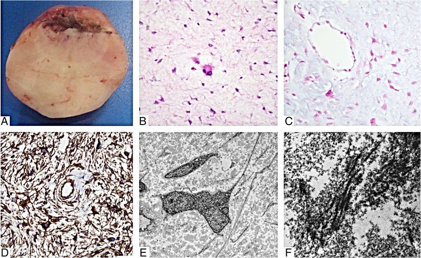
Figure 3 Macroscopic and microscopic appearance of the tumor. (A) The firm tumor was easily dissected from the surrounding brain tissue
and had a smooth tan-yellow glistening cut surface. (B) Microscopic examination revealed a paucicellular tumor consisting of predominantly
fusiform cells in a myxoid background. There is moderate nuclear pleomorphism with a multinucleated cell in the center (hematoxylin & eosin).
(C) The loose myxoid background stained positive for Alcian blue. (D) Immunohistochemical staining for CD34 is diffusely positive. (E) Electron
microscopic examination showed stellate cells with long delicate processes and occasional multinucleation. (F) The extracellular material is
abundant with clusters of collagen fibers. (Original magnifications: B-C, ×400; D, ×200; E, ×2000; F ×30000).
instrument. Sequencing was done on Ion 314 and Ion was found in the MET gene of the child’s tumor and
316 sequencing chips using the Ion Torrent Personal buccal swab as well as in the buccal swab of the father.
Genome Machine (Life Technology) following the manu-
facturer’s protocol. Data analysis including alignment to Discussion and conclusions
hg19 human reference genome, base calling, and identifi- Neurocutaneous melanosis (NCMS), first described by
cation of variants was done using the Ion Variant Caller Rokitansky in 1861 [12], is a rare congenital disorder
(version 2.2). Somatic variants from the brain tumor and consisting of multiple large melanocytic cutaneous nevi
the melanocytic nevus were identified after filtering out and melanocytic proliferations in the leptomeninges [13].
germline changes identified from the patient’s buccal Malformative lesions of the posterior fossa have rarely
swab. Allele frequency of a variant is calculated by dividing been described [14,15]. Recent genetic findings have clari-
the number of variant reads by total reads in the same fied the pathogenesis of NCMS and large/giant congenital
nucleotide position. All variants are covered by a mini-
mum of 500 reads. Table 1 Allelic frequency of nucleotide change in NRAS
The panel identified a missense mutation (chr1:1152 and BRAF from deep amplicon sequencing of pathology
58745; c.37G > C) in NRAS that results in a p. G13R specimens and buccal swabs
amino acid substitution. The allelic frequency of the nu- Gene NRAS BRAF
cleotide change in the brain tumor and the cutaneous Chromosome position (hg19) 1:115258745 7:140481411
nevus (58% and 33% respectively) is consistent with a Nucleotide change c.37G > C c.1397G > T
heterozygous mutation (Table 1). The same change was Amino acid change p. Gly13Arg p. Gly466Val
observed at an allelic frequency of 4.8% in the normal
Allelic frequencies (%)
skin and 2.7% in the buccal swab of the patient. Deep
sequencing of DNA extracted from the buccal swabs from Mesenchymal brain tumor 58.0 0.0
both parents did not reveal NRAS mutations. A BRAF Melanocytic nevus 33.0 32.0
somatic nucleotide change (chr7: 140481441; c.1397G > T) Normal skin 4.8 0.0
resulting in amino acid substitution p. G466V was de- Buccal swab 2.7 0.0
tected only in the nevus at an allelic frequency of 32%. A Mother buccal swab 0.0 0.0
single nucleotide polymorphism (SNP) resulting in a mis-
Father buccal swab 0.0 0.0
sense mutation (chr7: 116411990; c. 3209C > T; p.T1010I;)Shih et al. Acta Neuropathologica Communications 2014, 2:140 Page 5 of 7 http://www.actaneurocomms.org/content/2/1/140 melanocytic nevi [7,16]. Single postzygotic mutations of somatic mutations suggest cooperative involvement of NRAS codon 61 and associated mosaicism are responsible oncogenic pathways in the brain tumor and especially in for the majority of NCMS cases [5] and large/giant con- the skin lesions (BRAF p.G466V). Only a single sarcoma (a genital melanocytic nevi [17]. The same mutation is com- rhabdomyosarcoma) has been described with the NRAS p. mon in congenital melanocytic nevi [18]. The NRAS p. G13R mutation [35]. Somatic mosaics of NRAS mutations G13R somatic mosaicism in this patient is unusual and, to in almost identical protein regions (G12D, G12S, G13D) our knowledge, is the first instance reported in association have been described in relation to juvenile myelomonocytic with NCMS. The presence mosaicism in combination leukemia [36,37]. Note that neurofibromin is a major regu- with the absence of mutation in the parents suggests this lator of the NRAS pathway [38]. NF1 is not assessed in the mutation likely occurred after conception. This is consist- AmpliSeq Cancer Panel and we did not attempt direct se- ent with the mosaicism hypotheses for NCMS and other quencing. However, given that the documented abnormal- phakomatoses [19]. This child does not show any dys- ities are distal to neurofibromin signaling, a mutation in morphic features associated with germline mutations NF1 is not necessary to explain this child’s phenotype. in NRAS, which are usually similar to Noonan syndrome A germline SNP was found in the MET gene of this [20]. NRAS mutations are considered important in the child (inherited from her father who is not known to have genesis of melanoma. NRAS activates four major signaling any neoplastic disease). The MET gene encodes the recep- pathways including RAF-MEK-ERK, RalGDS, PI3K-AKT/ tor for hepatic growth factor/scatter factor (HGF/SF) and PDK1, and PLC/PKC [21]. Mutations affecting codons 12, appears to be involved in cell motility, proliferation, and 13 and 61, lead to constitutive activation of RAS GTPase invasiveness [39]. Mice that overexpress HGF/SF over- in the absence of growth factor signaling and ultimately stimulate the MET pathway and develop melanosis in the neoplastic growth. The specific NRAS G13R mutation central nervous system and patterned hyperpigmentation identified in this case has been rarely found in melanomas of the skin similar to NCMS [40]. These mice also develop of the skin [22-24] and esophagus [25] as well as in 1/27 fibrosarcoma and rhabdomyosarcoma [41]. MET has been patients with large congenital melanocytic nevi [26,27]. In detected by immunohistochemistry in optic canal nevus melanoma the most common NRAS (Q61R, Q61L, Q61K) cells from a child with NCMS [42]. The p.T1010I variant and BRAF (V600E, V600K) mutation sites and substitu- has been identified in thyroid carcinomas [33], neuroen- tions differ from those found in this patient [28]. docrine carcinoma (NEC) of lung [43], a pleomorphic In addition to NCMS, this child had a low-grade mes- xanthoastrocytoma case [11], and has been implicated as a enchymal brain tumor, which itself is very rare. Typically risk factor for familial colorectal cancer [44]. One early re- children with NCMS do not develop sarcomas, although port described this change in MET as capable of altering one rhabdomyosarcoma was described in a congenital signaling in NEC [45], however more recently this vari- giant nevus [29]. Children with NCMS are reported to ation was not shown to alter c-Met phosphorylation in develop choroid plexus papilloma and meningioma [5]. NEC [46], nor does it seem capable of transforming the Primary sarcomas of the brain represented only 0.36% of Ba/F3 pro-B lymphocyte cell line [47]. To summarize, the brain tumors in one very large series [30] and 0.7% of all significance of the MET SNP in this child is unclear. sarcomas in another series [31]. Most likely arise from Hemimegalencephaly has been associated with other the meninges or blood vessels; among them are rhabdo- neurocutaneous syndromes including epidermal nevus myosarcoma, fibrosarcoma, leiomyosarcoma, and angio- syndrome, proteus syndrome, hypomelanosis of Ito, and sarcoma. Primary non-meningeal myxoid mesenchymal neurofibromatosis-1 [48,49]. Recently, de novo somatic intracranial tumors are especially rare. Reported cases mutations with mosaicism in the PI3K-AKT3-mTOR include two low-grade fibromyxoid sarcomas [32,33] and pathway were shown to cause hemimegalencephaly [50]. one myxofibrosarcoma, the diagnosis of which was based NRAS is known to have direct interaction with PI3K on fluorescent in situ hybridization (FISH) analysis of the [21]. Given that this child had no mutation in PI3K, FUS/CREB3L2 translocation [34]. Both of these tumors the NRAS mosaicism is likely the explanation for the have some features similar to the reported child’s brain hemimegalencephaly. tumor. It is important to emphasize that the tumor had no morphologic or immunophenotypic features of me- Conclusion lanoma. The relatively low-grade appearance of the In summary, development of a primary intracerebral brain tumor presented herein is discrepant with its mesenchymal neoplasm in a child with NCMS and he- rapid growth. The rapid increase in size could be mimegalencephaly can likely be explained by specific explained by expansion of the myxoid extracellular NRAS mutant mosaicism possibly in combination with material rather than neoplastic cell proliferation. a MET germline variation, which together constitute a The heterozygous state of NRAS in the melanocytic ne- unique combination. This case highlights the importance vus and the brain tumor combined with additional novel of DNA analysis from multiple sites, as well as from
Shih et al. Acta Neuropathologica Communications 2014, 2:140                                                                                              Page 6 of 7
http://www.actaneurocomms.org/content/2/1/140

parents, in individuals with complex disease states. The                                 neurocutaneous melanocytosis with MEK162, a MEK-inhibitor.
presence of an NRAS somatic mosaic supports the hy-                                      Acta Neuropathol Commun 2014, 2:41.
                                                                                   7.    Etchevers HC: Hiding in plain sight: molecular genetics applied to giant
pothesized developmental pathogenesis of NCMS. Further                                   congenital melanocytic nevi. J Invest Dermatol 2014, 134:879–882.
exploration of the role of NRAS and MET in develop-                                8.    Bekiesinska-Figatowska M, Szczygielski O, Boczar M, Madzik J, Klepacka T,
ment of the neural crest derived pigment cells will be                                   Michalak E, Romaniuk-Doroszewska A, Uliasz M, Peczkowski P, Sawicka E:
                                                                                         Neurocutaneous melanosis in children with giant congenital melanocytic
of interest.                                                                             nevi. Clin Imaging 2014, 38:79–84.
                                                                                   9.    Ramaswamy V, Delaney H, Haque S, Marghoob A, Khakoo Y: Spectrum of
Consent                                                                                  central nervous system abnormalities in neurocutaneous melanocytosis.
                                                                                         Dev Med Child Neurol 2012, 54:563–568.
Parents provided explicit consent for genetic tests and                            10.   Scattolin MA, Lin J, Peruchi MM, Rocha AJ, Masruha MR, Vilanova LC:
use of photographs.                                                                      Neurocutaneous melanosis: follow-up and literature review. J Neuroradiol
                                                                                         2011, 38:313–318.
Abbreviations                                                                      11.   Yang MM, Singhal A, Rassekh SR, Yip S, Eydoux P, Dunham C: Possible
BRAF: B-Raf proto-oncogene, serine/threonine kinase; DNA: Deoxyribonucleic               differentiation of cerebral glioblastoma into pleomorphic
acid; FISH: Fluorescent in situ hybridization; MET: MET proto-oncogene,                  xanthoastrocytoma: an unusual case in an infant. J Neurosurg Pediatr
receptor tyrosine kinase; MR: Magnetic resonance; NCMS: Neurocutaneous                   2012, 9:517–523.
melanosis; NRAS: Neuroblastoma RAS viral (v-ras) oncogene homolog;                 12.   Rokitansky J: Ein ausgezeichneter Fall von Pigment-mal mit ausgebreiteter
PCR: Polymerase chain reaction.                                                          Pigmentierung der inneren Hin- und Ruchenmarkshaute. Allg Wien Med Z
                                                                                         1861, 6:113–116.
Competing interests                                                                13.   Fu YJ, Morota N, Nakagawa A, Takahashi H, Kakita A: Neurocutaneous
The authors declare that they have no competing interests.                               melanosis: surgical pathological features of an apparently
                                                                                         hamartomatous lesion in the amygdala. J Neurosurg Pediatr 2010, 6:82–86.
Authors’ contributions                                                             14.   De Cock J, Snauwaert J, Van Rompaey W, Morren MA, Demaerel P: A newborn
FS – Literature review and initial drafts of manuscript. SY – Genetic analysis           with neurocutaneous melanocytosis and Dandy-Walker malformation.
and drafting manuscript. PJM – Neurosurgical care of child, editing                      Pediatr Neurol 2014, 50:276–278.
manuscript. AEC – Clinical care of child, genetic explanation, editing             15.   Frieden IJ, Williams ML, Barkovich AJ: Giant congenital melanocytic nevi:
manuscript. MRD – Conceived project, final synthesis of manuscript.                      brain magnetic resonance findings in neurologically asymptomatic
All authors read and approved the final manuscript.                                      children. J Am Acad Dermatol 1994, 31:423–429.
                                                                                   16.   Gerami P, Paller AS: Making a mountain out of a molehill: NRAS, mosaicism,
Acknowledgements                                                                         and large congenital nevi. J Invest Dermatol 2013, 133:2127–2130.
Dr. Del Bigio holds the Canada Research Chair in Developmental                     17.   Charbel C, Fontaine RH, Malouf GG, Picard A, Kadlub N, El-Murr N, How-Kit
Neuropathology. We thank Dr. Peter Burger (Johns Hopkins University) for his             A, Su X, Coulomb-L’Hermine A, Tost J, Mourah S, Aractingi S, Guegan S:
consultation on the brain tumor. We thank Tiana Baskin, Sharon Allen and                 NRAS mutation is the sole recurrent somatic mutation in large
Susan Janeczko for their technical assistance. We are grateful to the family             congenital melanocytic nevi. J Invest Dermatol 2014, 134:1067–1074.
for their cooperation with these investigations and permission to publish the      18.   Bauer J, Curtin JA, Pinkel D, Bastian BC: Congenital melanocytic nevi
findings. SY would like to acknowledge the funding support of BrainCare BC.              frequently harbor NRAS mutations but no BRAF mutations. J Invest
                                                                                         Dermatol 2007, 127:179–182.
Author details                                                                     19.   Hamm H: Cutaneous mosaicism of lethal mutations. Am J Med Genet
1
 Diagnostic Services Manitoba, Winnipeg, MB, Canada. 2Department of                      1999, 85:342–345.
Pathology & Laboratory Medicine, University of British Columbia, Vancouver,        20.   Cirstea IC, Kutsche K, Dvorsky R, Gremer L, Carta C, Horn D, Roberts AE,
BC, Canada. 3Section of Neurosurgery, University of Manitoba, Winnipeg, MB,              Lepri F, Merbitz-Zahradnik T, Konig R, et al: A restricted spectrum of NRAS
Canada. 4Department of Biochemistry & Medical Genetics, University of                    mutations causes Noonan syndrome. Nat Genet 2010, 42:27–29.
Manitoba, Winnipeg, MB, Canada. 5Department of Pathology, University of            21.   Ross AL, Sanchez MI, Grichnik JM: Molecular nevogenesis. Dermatol Res
Manitoba and Diagnostic Services Manitoba, Room 401 Brodie Centre, R3E                   Pract 2011, 2011:463184.
3P5 Winnipeg, MB, Canada.                                                          22.   Ball NJ, Yohn JJ, Morelli JG, Norris DA, Golitz LE, Hoeffler JP: Ras mutations
                                                                                         in human melanoma: a marker of malignant progression. J Invest
Received: 30 July 2014 Accepted: 8 September 2014                                        Dermatol 1994, 102:285–290.
                                                                                   23.   van Elsas A, Zerp SF, van der Flier S, Kruse KM, Aarnoudse C, Hayward NK,
                                                                                         Ruiter DJ, Schrier PI: Relevance of ultraviolet-induced N-ras oncogene
References                                                                               point mutations in development of primary human cutaneous
1. Pavlidou E, Hagel C, Papavasilliou A, Giouroukos S, Panteliadis C:                    melanoma. Am J Pathol 1996, 149:883–893.
    Neurocutaneous melanosis: report of three cases and up-to-date review.         24.   Pavey S, Johansson P, Packer L, Taylor J, Stark M, Pollock PM, Walker GJ,
    J Child Neurol 2008, 23:1382–1391.                                                   Boyle GM, Harper U, Cozzi SJ, Hansen K, Yudt L, Schmidt C, Hersey P,
2. Makkar HS, Frieden IJ: Neurocutaneous melanosis. Semin Cutan Med Surg                 Ellem KA, O'Rourke MG, Parsons PG, Meltzer P, Ringner M, Hayward NK:
    2004, 23:138–144.                                                                    Microarray expression profiling in melanoma reveals a BRAF mutation
3. Kinsler VA, Anderson G, Latimer B, Natarajan D, Healy E, Moore GE, Sebire NJ:         signature. Oncogene 2004, 23:4060–4067.
    Immunohistochemical and ultrastructural features of congenital                 25.   Sekine S, Nakanishi Y, Ogawa R, Kouda S, Kanai Y: Esophageal melanomas
    melanocytic naevus cells support a stem-cell phenotype. Br J Dermatol                harbor frequent NRAS mutations unlike melanomas of other mucosal
    2013, 169:374–383.                                                                   sites. Virchows Arch 2009, 454:513–517.
4. Online Mendelian Inheritance in Man: Melanosis, Neurocutaneous; NCMS;           26.   Dessars B, De Raeve LE, Morandini R, Lefort A, El Housni H, Ghanem GE, Van
    MIM#249400. Baltimore, MD: Johns Hopkins University; 2014.                           den Eynde BJ, Ma W, Roseeuw D, Vassart G, Libert F, Heimann P: Genotypic
5. Kinsler VA, Thomas AC, Ishida M, Bulstrode NW, Loughlin S, Hing S, Chalker J,         and gene expression studies in congenital melanocytic nevi: insight into
    McKenzie K, Abu-Amero S, Slater O, Chanudet E, Palmer R, Morrogh D,                  initial steps of melanotumorigenesis. J Invest Dermatol 2009, 129:139–147.
    Stanier P, Healy E, Sebire NJ, Moore GE: Multiple congenital melanocytic       27.   Phadke PA, Rakheja D, Le LP, Selim MA, Kapur P, Davis A, Mihm MC Jr,
    nevi and neurocutaneous melanosis are caused by postzygotic                          Hoang MP: Proliferative nodules arising within congenital melanocytic
    mutations in codon 61 of NRAS. J Invest Dermatol 2013, 133:2229–2236.                nevi: a histologic, immunohistochemical, and molecular analyses of
6. Kusters-Vandevelde HV, Willemsen AE, Groenen PJ, Kusters B, Lammens M,                43 cases. Am J Surg Pathol 2011, 35:656–669.
    Wesseling P, Djafarihamedani M, Rijntjes J, Delye H, Willemsen MA, van         28.   Colombino M, Capone M, Lissia A, Cossu A, Rubino C, De Giorgi V, Massi D,
    Herpen CM, Blokx WA: Experimental treatment of NRAS-mutated                          Fonsatti E, Staibano S, Nappi O, Pagani E, Casula M, Manca A, Sini M, Franco R,
Shih et al. Acta Neuropathologica Communications 2014, 2:140 Page 7 of 7
http://www.actaneurocomms.org/content/2/1/140
Botti G, Caraco C, Mozzillo N, Ascierto PA, Palmieri G: BRAF/NRAS mutation 47. Tyner JW, Fletcher LB, Wang EQ, Yang WF, Rutenberg-Schoenberg ML,
frequencies among primary tumors and metastases in patients with Beadling C, Mori M, Heinrich MC, Deininger MW, Druker BJ, Loriaux MM:
melanoma. J Clin Oncol 2012, 30:2522–2529. MET receptor sequence variants R970C and T992I lack transforming
29. Zuniga S, Las Heras J, Benveniste S: Rhabdomyosarcoma arising in a capacity. Cancer Res 2010, 70:6233–6237.
congenital giant nevus associated with neurocutaneous melanosis in a 48. Flores-Sarnat L: Hemimegalencephaly: part 1. Genetic, clinical, and
neonate. J Pediatr Surg 1987, 22:1036–1038. imaging aspects. J Child Neurol 2002, 17:373–384.
30. Paulus W, Slowik F, Jellinger K: Primary intracranial sarcomas: 49. Flores-Sarnat L: Neurocutaneous melanocytosis. Handb Clin Neurol 2013,
histopathological features of 19 cases. Histopathology 1991, 18:395–402. 111:369–388.
31. Toro JR, Travis LB, Wu HJ, Zhu K, Fletcher CD, Devesa SS: Incidence 50. Lee JH, Huynh M, Silhavy JL, Kim S, Dixon-Salazar T, Heiberg A, Scott E,
patterns of soft tissue sarcomas, regardless of primary site, in the Bafna V, Hill KJ, Collazo A, Funari V, Russ C, Gabriel SB, Mathern GW, Gleeson
surveillance, epidemiology and end results program, 1978–2001: JG: De novo somatic mutations in components of the PI3K-AKT3-mTOR
An analysis of 26,758 cases. Int J Cancer 2006, 119:2922–2930. pathway cause hemimegalencephaly. Nat Genet 2012, 44:941–945.
32. Tun K, Ozen O, Kaptanoglu E, Gurcan O, Beskonakli E, Celasun B:
Primary intracranial low-grade fibromyxoid sarcoma (Evans tumor). doi:10.1186/s40478-014-0140-8
J Clin Neurosci 2008, 15:1298–1301. Cite this article as: Shih et al.: Oncogenic codon 13 NRAS mutation in a
33. Ricarte-Filho JC, Ryder M, Chitale DA, Rivera M, Heguy A, Ladanyi M, primary mesenchymal brain neoplasm and nevus of a child with
Janakiraman M, Solit D, Knauf JA, Tuttle RM, Ghossein RA, Fagin JA: neurocutaneous melanosis. Acta Neuropathologica Communications
Mutational profile of advanced primary and metastatic radioactive 2014 2:140.
iodine-refractory thyroid cancers reveals distinct pathogenetic roles for
BRAF, PIK3CA, and AKT1. Cancer Res 2009, 69:4885–4893.
34. Buccoliero AM, Castiglione F, Garbini F, Rossi Degl’Innocenti D, Moncini D,
Franchi A, Paglierani M, Simoni A, Baroni G, Daniele D, Sardi I, Giordano F,
Mussa F, Arico M, Genitori L, Taddei GL: Primary cerebral
myxofibrosarcoma: clinical, morphologic, immunohistochemical,
molecular, and ultrastructural study of an infrequent tumor in an
extraordinary localization. J Pediatr Hematol Oncol 2011, 33:e279–e283.
35. Stratton MR, Fisher C, Gusterson BA, Cooper CS: Detection of point mutations
in N-ras and K-ras genes of human embryonal rhabdomyosarcomas using
oligonucleotide probes and the polymerase chain reaction. Cancer Res 1989,
49:6324–6327.
36. Doisaki S, Muramatsu H, Shimada A, Takahashi Y, Mori-Ezaki M, Sato M,
Kawaguchi H, Kinoshita A, Sotomatsu M, Hayashi Y, Furukawa-Hibi Y,
Yamada K, Hoshino H, Kiyoi H, Yoshida N, Sakaguchi H, Narita A, Wang X,
Ismael O, Xu Y, Nishio N, Tanaka M, Hama A, Koike K, Kojima S: Somatic
mosaicism for oncogenic NRAS mutations in juvenile myelomonocytic
leukemia. Blood 2012, 120:1485–1488.
37. De Filippi P, Zecca M, Lisini D, Rosti V, Cagioni C, Carlo-Stella C, Radi O,
Veggiotti P, Mastronuzzi A, Acquaviva A, D'Ambrosio A, Locatelli F, Danesino C:
Germ-line mutation of the NRAS gene may be responsible for the
development of juvenile myelomonocytic leukaemia. Br J Haematol 2009,
147:706–709.
38. Harrisingh MC, Lloyd AC: Ras/Raf/ERK signalling and NF1. Cell Cycle 2004,
3:1255–1258.
39. Boccaccio C, Comoglio PM: Invasive growth: a MET-driven genetic
programme for cancer and stem cells. Nat Rev Cancer 2006, 6:637–645.
40. Takayama H, La Rochelle WJ, Anver M, Bockman DE, Merlino G: Scatter
factor/hepatocyte growth factor as a regulator of skeletal muscle and
neural crest development. Proc Natl Acad Sci U S A 1996, 93:5866–5871.
41. Takayama H, LaRochelle WJ, Sharp R, Otsuka T, Kriebel P, Anver M, Aaronson
SA, Merlino G: Diverse tumorigenesis associated with aberrant
development in mice overexpressing hepatocyte growth factor/scatter
factor. Proc Natl Acad Sci U S A 1997, 94:701–706.
42. Takayama H, Nagashima Y, Hara M, Takagi H, Mori M, Merlino G, Nakazato Y:
Immunohistochemical detection of the c-met proto-oncogene product
in the congenital melanocytic nevus of an infant with neurocutaneous
melanosis. J Am Acad Dermatol 2001, 44:538–540.
43. Tengs T, Lee JC, Paez JG, Zhao X, LaFramboise T, Giannoukos G, Thomas RK:
A transforming MET mutation discovered in non-small cell lung cancer Submit your next manuscript to BioMed Central
using microarray-based resequencing. Cancer Lett 2006, 239:227–233. and take full advantage of:
44. Neklason DW, Done MW, Sargent NR, Schwartz AG, Anton-Culver H, Griffin CA,
Ahnen DJ, Schildkraut JM, Tomlinson GE, Strong LC, Miller AR, Stopfer JE,
• Convenient online submission
Burt RW: Activating mutation in MET oncogene in familial colorectal cancer.
BMC Cancer 2011, 11:424. • Thorough peer review
45. Ma PC, Kijima T, Maulik G, Fox EA, Sattler M, Griffin JD, Johnson BE, Salgia R: • No space constraints or color figure charges
c-MET mutational analysis in small cell lung cancer: novel
juxtamembrane domain mutations regulating cytoskeletal functions. • Immediate publication on acceptance
Cancer Res 2003, 63:6272–6281. • Inclusion in PubMed, CAS, Scopus and Google Scholar
46. Voortman J, Harada T, Chang RP, Killian JK, Suuriniemi M, Smith WI, Meltzer PS,
• Research which is freely available for redistribution
Lucchi M, Wang Y, Giaccone G: Detection and therapeutic implications of
c-Met mutations in small cell lung cancer and neuroendocrine tumors.
Curr Pharm Des 2013, 19:833–840. Submit your manuscript at
www.biomedcentral.com/submitYou can also read