Multimethod investigation of the neurobiological basis of ADHD symptomatology in children aged 9-10: baseline data from the ABCD study - Nature
←
→
Page content transcription
If your browser does not render page correctly, please read the page content below
Owens et al. Translational Psychiatry (2021)11:64
                                    https://doi.org/10.1038/s41398-020-01192-8                                                                                                     Translational Psychiatry

                                     ARTICLE                                                                                                                                                    Open Access

                                    Multimethod investigation of the neurobiological
                                    basis of ADHD symptomatology in children aged
                                    9-10: baseline data from the ABCD study
                                    Max M. Owens 1, Nicholas Allgaier1, Sage Hahn1, DeKang Yuan1, Matthew Albaugh1, Shana Adise1, Bader Chaarani1,
                                    Joseph Ortigara1, Anthony Juliano 1, Alexandra Potter1 and Hugh Garavan1

                                      Abstract
                                      Attention deficit/hyperactivity disorder is associated with numerous neurocognitive deficits, including poor working
                                      memory and difficulty inhibiting undesirable behaviors that cause academic and behavioral problems in children. Prior
                                      work has attempted to determine how these differences are instantiated in the structure and function of the brain, but
                                      much of that work has been done in small samples, focused on older adolescents or adults, and used statistical
                                      approaches that were not robust to model overfitting. The current study used cross-validated elastic net regression to
                                      predict a continuous measure of ADHD symptomatology using brain morphometry and activation during tasks of
                                      working memory, inhibitory control, and reward processing, with separate models for each MRI measure. The best
                                      model using activation during the working memory task to predict ADHD symptomatology had an out-of-sample
                                      R2 = 2% and was robust to residualizing the effects of age, sex, race, parental income and education, handedness,
                                      pubertal status, and internalizing symptoms from ADHD symptomatology. This model used reduced activation in task
                  1234567890():,;
1234567890():,;
                  1234567890():,;
1234567890():,;

                                      positive regions and reduced deactivation in task negative regions to predict ADHD symptomatology. The best model
                                      with morphometry alone predicted ADHD symptomatology with an R2 = 1% but this effect dissipated when including
                                      covariates. The inhibitory control and reward tasks did not yield generalizable models. In summary, these analyses
                                      show, with a large and well-characterized sample, that the brain correlates of ADHD symptomatology are modest in
                                      effect size and captured best by brain morphometry and activation during a working memory task.

                                    Introduction                                                                                        working memory, and attention7–9. Differences in indivi-
                                      There is substantial evidence that individuals with                                               duals with ADHD have also been found in activation in
                                    Attention Deficit Hyperactivity Disorder (ADHD) have                                                 the ventral striatum, ventromedial prefrontal cortex
                                    below average working memory1–3 and difficulty with                                                  (VMPFC), and ACC during monetary reward tasks10.
                                    response inhibition4–6. These deficits may be explained by                                           Additionally, a recent review of morphometric differences
                                    reduced activation in brain regions, such as the dorso-                                             measured by structural magnetic resonance imaging
                                    lateral prefrontal cortex (DLPFC), anterior cingulate                                               (sMRI) in adolescents with ADHD reported abnormalities
                                    cortex (ACC), posterior parietal cortex (PPC), ven-                                                 in cortical thickness (CT) in the lateral prefrontal and
                                    trolateral prefrontal cortex (VLPFC), insula thalamus, and                                          parietal cortices, as well as gray matter volume of the
                                    striatum, which have been found to differ in individuals                                            thalamus as the most frequently identified effects11.
                                    with ADHD in meta-analyses of functional magnetic                                                   However, these reviews and meta-analyses included few
                                    resonance imaging (fMRI) tasks of inhibitory control,                                               studies with more than 50 participants and none over 100.
                                                                                                                                          Two recent large studies of adolescents (N > 1000) have
                                                                                                                                        aimed to characterize the neuroanatomical correlates of
                                    Correspondence: Max M. Owens (mowens1@uvm.edu)                                                      ADHD. These studies both investigated surface-based
                                    1
                                     Department of Psychiatry, University of Vermont, Burlington, VT 05401, USA

                                    © The Author(s) 2021, corrected publication 2021
                                                        Open Access This article is licensed under a Creative Commons Attribution 4.0 International License, which permits use, sharing, adaptation, distribution and reproduction
                                                        in any medium or format, as long as you give appropriate credit to the original author(s) and the source, provide a link to the Creative Commons license, and indicate if
                                    changes were made. The images or other third party material in this article are included in the article’s Creative Commons license, unless indicated otherwise in a credit line to the material. If
                                    material is not included in the article’s Creative Commons license and your intended use is not permitted by statutory regulation or exceeds the permitted use, you will need to obtain
                                    permission directly from the copyright holder. To view a copy of this license, visit http://creativecommons.org/licenses/by/4.0/.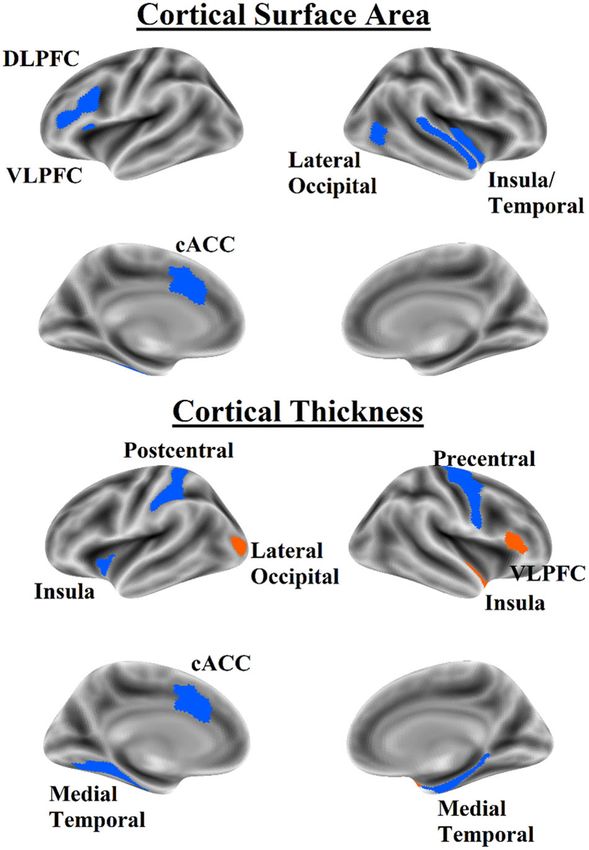
Owens et al. Translational Psychiatry (2021)11:64                                                               Page 2 of 11

morphometry in a non-clinical sample of adolescents               An additional weakness in the existing literature on
using the IMAGEN study dataset (https://imagen-europe.          the neurobiological basis of ADHD is a primary focus on
com/), which includes participants with a range of ADHD         categorical diagnoses of ADHD. There is strong
symptoms. They identified lower levels of CT in the              empirical evidence that child psychopathology is better
ventromedial prefrontal cortex12,13 and cortical surface        suited to dimensional measurement19. This is true spe-
area (CSA) in the DLPFC and ACC14, which were also              cifically for ADHD as well20. Sub-diagnostic ADHD
linked to performance on several neuropsychological             symptomatology has been linked to poorer educational
measures known to be disrupted in individuals with              outcomes21 and differences in cortical maturation in
ADHD. Additionally, a recent mega-analysis (N = 1713            children22,23. Continuous measures of ADHD sympto-
with ADHD, 1529 controls) found smaller gray matter             matology show stronger genetic correlations than
volume (GMV) in children with ADHD in several sub-              ADHD diagnoses24 suggesting that continuous mea-
cortical structures (nucleus accumbens, caudate, puta-          sures are investigating the same construct at a higher
men, amygdala, and hippocampus)15. However,                     resolution (as opposed to another phenomenon alto-
comparably powered studies have not been published for          gether). Furthermore, the use of a dimensional approach
task fMRI.                                                      is more consistent with the National Institute of Mental
   In response to concerns of mounting replication failures     Health’s Research Domain Criteria program25. Thus, the
in the social sciences, reducing overfitting and assessing       current study used a continuous measure of ADHD
generalizability of models have become critical. To achieve     symptomatology, the “attention problems” scale of the
this, methods such as regularized regression and cross-         child behavior checklist (CBCL)26.
validation are frequently used16. A recent large machine          Given the established relationship of working memory,
learning study (N = 2713) found only modest prediction          inhibitory control, and reward processing with ADHD, we
accuracies when distinguishing children with ADHD from          hypothesized that ADHD symptomatology would be
controls using sMRI with more rigorous cross-validation17.      predictable using each of the three fMRI tasks. We
It stands in contrast to prior smaller studies, which had       expected that the features used by these models would be
shown much more optimistic predictions using more               regions activated by the tasks in prior studies, such as the
questionable approaches (e.g., failure to tune regularization   DLPFC, VLPFC, and ACC for working memory and
hyperparameters, not validating tuned hyperparameters on        inhibition tasks and the striatum, VMPFC, and ACC for
an independent sample, and using accuracy as an outcome         the reward task. Based on prior work linking structural
for imbalanced groups). However, the majority of existing       MRI data to ADHD, we also hypothesized that structural
machine learning work on ADHD focuses on only one               MRI would predict ADHD symptomatology. We expected
imaging modality or task, making it difficult to compare         that the features used in these models would be the same
classification accuracies across ADHD studies. Additionally,     as those whose morphometry had been found to be
the use of non-linear modeling strategies makes the inter-      associated with ADHD in prior studies, such as the
pretation of model features in many machine learning            DLPFC, ACC, VMPFC, and striatum.
models difficult.
   Another approach to improving replicability has been         Materials and methods
the initiation of large-scale open-data projects. The cur-      Procedures
rent study used baseline data from the Adolescent Brain           The Adolescent Brain Cognitive Developmentsm Study
and Cognitive Development (ABCD; www.ABCDstudy.                 (ABCD) is an ongoing multisite, longitudinal neuroi-
org) study to predict current ADHD symptomatology               maging study following a cohort of 11,875 youths over
using elastic net regression models with brain activation       10 years. In this analysis, we use data from the baseline
during fMRI tasks of working memory, inhibitory control,        visits at which participants were 9–10 years old. All data
and the anticipation and receipt of monetary reward, as         used in the current study (i.e., fMRI, questionnaire,
well as brain morphometry measured by sMRI. Among               neuropsychological tasks) were collected at a single visit
machine learning approaches, elastic net regression is          or across two visits that occurred within 30 days of each
particularly effective at making predictions when the           other. The ABCD study® was approved by the institu-
predictors are highly intercorrelated, has been shown to        tional review board of the University of California,
be effective at making predictions using neuroimaging           San Diego (IRB# 160091). Additionally, the institutional
data18, and is one of the approaches in which model             review boards of each of the 21 data collection
features are most interpretable, making it ideal for            sites approved the study. Informed consent was
attempting to understand the specific brain regions most         obtained from all parents and informed assent was
critical to ADHD. The current analyses also used cross-         obtained from participants. Data can be accessed
validation with an external test set to ensure that effects     through registration with the ABCD study at https://
identified were replicable in an independent dataset.            nda.nih.gov/abcd.
Owens et al. Translational Psychiatry (2021)11:64                                                                            Page 3 of 11

Participants                                                               fMRI scans pass the image quality control performed by
  Demographics for the full sample are shown in Table 1                    the DAIRC (which was similar to the DAIRC Freesurfer
and the distribution of ADHD symptomatology is shown                       QC reported above27), or failing to meet additional quality
in Supplemental Fig. 1. While the ABCD Data Analysis,                      control criterion specific to this report. These additional
Informatics & Resource Center (DAIRC) creates several                      quality control steps included having: (1) hemispheric
indices of data quality, all exclusions were done by the                   mean beta-weights more than two standard deviations
research team of the current study starting from the total                 from the sample mean, (2) fewer than 200 degrees of
11,875 participants enrolled in the ABCD study. Partici-                   freedom over the two runs, (3) mean framewise displace-
pants for all MRI/fMRI modalities were excluded if they                    ment > 0.9 mm for both runs, (4) failed to meet task-
had incomplete data for the CBCL “Attention Problems”                      specific performance criteria (described in Casey et al.28).
scale, were missing sociodemographic covariates, or failed                 Because of a data processing error (https://github.com/
the FreeSurfer quality control assessments performed by                    ABCD-STUDY/fMRI-cleanup), participants were exclu-
the DAIRC. In the DAIRC Freesurfer quality control,                        ded who were collected on Philips scanners for all fMRI
trained technicians reviewed each subject’s cortical                       tasks. Additionally, for the SST only, a small group of
reconstruction, judging the severity of five problem types:                 participants were excluded because of a glitch in the SST
motion, intensity inhomogeneity, white matter under-                       task (when the stop signal delay is 50 msec, a response that
estimation, pial overestimation, and magnetic suscept-                     is faster than 50 msec is erroneously recorded as the
ibility artifact. Reconstructions were rated for each                      response for all subsequent Stop trials, see
problem as “none”, “mild”, “moderate”, or “severe”. A                      Garavan et al.29). This resulted in participant totals of 8596
subject was recommended for exclusion if any of the five                    for Structural MRI (sMRI), 5417 for the EN-Back task,
categories are rated as “severe” and these recommenda-                     5959 for the monetary incentive delay (MID) task, and
tions were summarized as an overall binary QC score (i.e.,                 5020 for Stop Signal Task (SST). Participant exclusion
pass or fail), which was used in the current manuscript27.                 steps are reported in Supplemental Table 1.
For each fMRI task, participants were excluded for having
any missing fMRI data on that task, having fewer than two                  Measures
                                                                           Child behavior checklist
Table 1 Descriptive statistics on samples for each MRI                       The “Attention Problems” empirically derived syn-
paradigm.                                                                  drome scale of the CBCL parent-report questionnaire was
                                                                           used to evaluate symptoms of ADHD symptomatology as
                    sMRI          EN-Back       SST          MID
                                                                           a continuous variable30. Despite its name, the CBCL
                    Mean    SD    Mean    SD    Mean    SD   Mean    SD    “Attention Problems” scale evaluates a broad constellation
                                                                           of ADHD symptomatology (attention, hyperactivity, and
Child age
                                                                           impulsivity) and has been shown to be an excellent pre-
  Months            119.18 7.46 119.61 7.53 119.53 7.54 119.42 7.52
                                                                           dictor of ADHD diagnosis derived from clinical inter-
                    Total   %     Total   %     Total   %    Total   %     view31,32. Additionally, the CBCL internalizing composite
                                                                           was used as a covariate to account for depression and
Child sex
                                                                           anxiety, which are frequently comorbid in children with
  Female            4179    49% 2681      49% 2524      50% 2973     50%
                                                                           ADHD. This composite is made up of the anxious/
Child race
  White             5917    69% 3878      72% 3596      72% 4174     70%
                                                                           depressed scale, the withdrawn/depressed scale, and the
  Black             1140    13% 592       11% 570       11% 712      12%   somatic symptoms scale.
  Asian              177     2% 111        2% 105        2% 118       2%
  Other             1362    16% 836       15% 749       15% 955      16%   Demographic Questionnaire
Child ethnicity                                                              A demographics questionnaire was administered to the
  Hispanic          1604    19%    997    18%    911    18% 1122     19%   participant’s parent/guardian to determine demographic
Household income                                                           information including sex, age, race, household income,
  $100 K           3819    44% 2516      46% 2330      46% 2709     45%
                                                                           Pubertal Development Scale
Parent education
                                                                             Pubertal status was assessed using the pubertal devel-
Owens et al. Translational Psychiatry (2021)11:64 Page 4 of 11
Edinburgh handedness inventory - short form “stop signal” has been presented and functions as an index
The Edinburgh Handedness Inventory – Short Form34 for inhibitory speed.
is a 4-item self-report scale that produces a handedness
score of “right”, “mixed”, or “left” by asking about pre- Magnetic resonance imaging acquisition and data
ferred hand used for writing, throwing, using a spoon, and processing
using a toothbrush. The measure has been shown to have Structural and functional MRI scans were acquired at
good reliability and to correlate highly with longer mea- sites across the United States using 26 different scanners
sures of handedness34. from two vendors (Siemens and General Electric); there
were also three sites using Philips scanners that were
Medications survey inventory excluded from analyses due to an error in processing prior
Parents completed a survey in which they listed the to their release. MRI sequences are reported in Supple-
names and dosages of all medications taken by the child. mental Methods and in prior work31. All sMRI and fMRI
From this we created a binary variable if participants were data were preprocessed by the DAIRC using pipelines that
taking one or more stimulant medications prescribed to have been detailed in prior work27. Briefly, sMRI data
treat ADHD (e.g., Adderall, Ritalin, amphetamine). For a were preprocessed using FreeSurfer version 5.327 to pro-
full list of medications included in this variable, see duce CT and CSA measures for each of the 74 Destrieux
Supplemental Table 2. atlas36 regions of interest in each hemisphere (148 regions
total) and GMV for nine subcortical regions in Free-
fMRI tasks Surfer’s ASEG parcellation in each hemisphere (18
The tasks used in the current study have been described regions total), plus the brainstem which was not split by
previously28,35 and are detailed in Supplemental Materials hemisphere. All structural MRI data were visually exam-
and a schematic of the tasks is shown in Supplemental Fig. ined by a trained ABCD technician, who rated them from
2. In short, the EN-Back task was a modified version of a zero to three on five dimensions: motion, intensity
traditional N-Back task in which participants viewed a homogeneity, white matter underestimation, pial over-
series of stimuli and for each responded if that stimulus estimation, and magnetic susceptibility artifact. From this
matched the one they saw N items ago (i.e., “N back”). The an overall score was generated recommending inclusion
current task version incorporated added elements of facial or exclusion27. All subjects recommended for exclusion
and emotional processing, though these were not a focus based on their Freesurfer data were excluded from all
of the current analysis. The task had two conditions: a 2- analyses in the current study, including fMRI analyses as
back as its active condition and a 0-back as the baseline the fMRI processing pipeline relies on the Freesurfer
condition. d’ (z(hit rate) − z(false alarm rate)) was used as cortical reconstruction.
the performance measure for the EN-back tasks. The MID fMRI data were preprocessed using a multi-program
task included both anticipation and receipt of reward and pipeline that yielded neural activation in these same
loss. In this task, participants viewed an incentive cue for cortical and subcortical regions for each fMRI contrast.
2 sec (anticipation) and then quickly respond to a target to The contrasts used for the SST were incorrect stop –
win or avoid losing money ($5.00 or $0.20). Participants correct go and correct stop – correct go; for the EN-Back
were then given feedback about their performance the only contrast was 2-Back vs. 0-Back; for the MID,
(receipt). The baseline used was “neutral” trials in which contrasts were reward anticipation – neutral anticipation
participants completed the same action but with no and positive reward outcome – negative reward outcome
money available to be won or lost. The current study (i.e., win – loss).
focused only on reward trials (i.e., trials in which parti-
cipants win money), as most prior research on ADHD Data analysis
symptomatology has focused on reward trials10. The SST Preliminary analyses
consisted of serial presentations of leftward and rightward To investigate the relationship between covariates and
facing arrows. Participants were instructed to indicate the ADHD symptomatology, we examined the association of
direction of the arrows using a two-button response box each covariate with ADHD symptomatology in a separate
(the “go” signal), except when the left or right arrow was mixed effect model for each covariate using R version
followed by an arrow pointing upward (the “stop” signal). 3.6.1. Code for all analyses is available at https://github.
Participants were also instructed to respond as “quickly com/owensmax/ADHD. Covariates were participants’
and accurately as possible”. Trials were then categorized age, sex, race, pubertal status, handedness, internalizing
based on the participant’s accuracy (“correct” and symptom score from the CBCL, parent’s highest educa-
“incorrect”). The performance variable used for the SST tion level, and family income. An additional analysis was
was stop signal reaction time (SSRT), which represents also added that also used stimulant medication status
the duration required to inhibit a “go” response after a (yes/no) as a covariate and so we examined stimulantOwens et al. Translational Psychiatry (2021)11:64 Page 5 of 11
medication status’ association with ADHD symptoma- internal cross-validation framework with 80% of the data
tology. Additionally, framewise displacement from the used for model training and 20% of the data used as an
relevant fMRI scan was used as a covariate and for internal validation set to assess the model’s performance
structural MRI the average of framewise displacement out-of-sample. Regularization hyperparameter tuning was
from the fMRI scans was used. In structural MRI analyses, conducted through a further nesting of a 20-fold cross-
total intracranial volume was also used as covariate. To validation within the 5-fold cross validation (see Supple-
account for the large numbers of siblings and multiple mental Methods for details on the cross-validation fra-
data collection sites, family ID (used to denote sibling mework). Prediction accuracy was measured in R2. For the
status) was modeled as a random effect nested inside a internal cross-validation, each R2 represents the accuracy
random effect of scanner in all mixed effects models1, as of predicting the internal validation set (the 5th fold)
has been recommended37 and is standard on the ABCD using the model built on the training set (folds 1–4); to
Data Exploration and Analysis Portal (DEAP). This nested confirm the model’s generalizability, another R2 was
approach was used since all siblings in each family were derived by predicting the external test set (i.e., 20% of total
collected at the same scanner site. In these analyses, the data) using the most successful of the 5 models built in
covariates were the independent variables and ADHD the 5-fold cross-validation. See Supplemental Methods for
symptomatology was the dependent variable. Addition- more details on the elastic net cross-validation scheme.
ally, to investigate if working memory and inhibitory
control were associated with ADHD symptomatology, we Primary analysis: confirmatory univariate analyses
examined the association of behavioral performance on To aid with the interpretation of elastic net regression
the EN-Back and Stop Signal tasks with attention symp- analyses, follow-up analyses were conducted testing the
toms. d′ (z(hit rate) − z(false alarm rate)) was used as the associations of ADHD symptomatology with each of the
performance measure for the EN-Back tasks and stop brain features from the best elastic net regression model
signal reaction time was used as the performance metric from each modality. This was done with a separate mixed
for SST. In these analyses, the performance metrics were effects model for each brain feature, which was used as a
used as the independent variables, along with all fixed fixed effect. Covariates were fixed effects, family and
effect covariates, and ADHD symptomatology was used as scanner were random effects, and ADHD symptomatol-
the dependent variable. ogy was the dependent variable. Since these analyses were
confirming relationships of coefficients found in elastic
net models, a threshold of p < 0.05 was used to indicate
Primary analyses: elastic net regression significance. Regions included in elastic net regression
Elastic net regression was used to build predictive models were only considered as neural correlates of
models for each of the imaging modalities (structural ADHD symptomatology if they were also associated in the
MRI, EN-Back, SST, and MID) using the glmnet package same direction in confirmatory univariate analyses.
in MATLAB R2018b. Separate models were built for each
MRI paradigm (i.e., each of three fMRI task + sMRI), with Secondary analysis: categorical analyses
all brain variables used as features (i.e., independent Because of the non-normal distribution of ADHD
variables) and ADHD symptomatology used as the target symptomatology (see Supplemental Fig. 1), we also com-
(i.e., dependent variable). Three versions of the analysis pleted a supplementary analysis to ensure that results
were run, one in which covariates were not accounted for, were not being driven by this distribution. For this ana-
one in which the ADHD symptomatology score was lysis we created a categorical variable of ADHD sympto-
residualized so that its shared variance with the covariates matology based on a tertile split of the variable (group 1:
was removed, and one in which covariates including no symptoms, 0 on CBCL; group 2: low symptoms, 1–3
medication status were residualized from ADHD on CBCL; and group 3: high symptoms, ≥3 on the CBCL).
symptomatology. The three ADHD symptomatology groups were roughly
Initially, data were divided in an 80%/20% split, with the equal in size (see Supplemental Fig. 3). Then we repeated
80% used as a training/validation set for model building the primary analyses using this categorical ADHD
and the 20% used as a final external test set. Then, elastic symptomatology variable as the dependent variable.
net regression model training was conducted in a 5-fold Additionally, to determine if continuous findings of
1
ADHD symptomatology would generalize to a clinical
Of note, when examined with a chi-square test, we found that the inclusion
of family ID had no impact on the fit of the random effects model (comparison diagnosis of ADHD, we also repeated the primary analysis
of models with and without family ID: λ2 = 0, p = 1). To ensure that its using ADHD diagnosis from the K-SADS as the depen-
inclusion was not adversely affecting our analyses, we also ran all analyses dent variable (K-SADS measure described in Supple-
using a simpler model that included only a random effect of scanner. Results
using this approach were identical (within rounding), both for the elastic net mental Methods). There were 727 participants with an
and mixed effect model statistics. ADHD diagnosis vs. 7745 controls with valid sMRI dataOwens et al. Translational Psychiatry (2021)11:64 Page 6 of 11
(N.B., 124 participants from the primary analyses did not Table 2 Associations of demographic covariates with
have K-SADS ADHD diagnosis data and were excluded attention deficit hyperactivity disorder symptoms.
from this analysis). See Supplemental Fig. 4 for visuali-
Phenotype (IV) B SE p R2
zation of distribution of K-SADS diagnosis.
Age −0.004 0.005 0.450 0.000
Results
Pubertal status −0.105 0.049 0.031 0.001
Preliminary mixed effect analyses
In the full sample, all demographic covariates but age Sex (male) 1.036 0.073Owens et al. Translational Psychiatry (2021)11:64 Page 7 of 11
Table 3 Prediction accuracy (R2) for elastic net regression
models.
Raw ADHD EN-back (%) Structural SST (%) MID (%)
symptomatology MRI (%)
Model 1 −0.1 0.7 −1.2 −0.2
Model 2 2.0 0.8 −0.9 −0.3
Model 3 1.3 1.1 0.5 0.0
Model 4 −0.2 0.7 −0.1 0.1
Model 5 1.0 0.4 0.0 −0.1
Mean 0.8 0.7 0.3 −0.1
External test 1.9 0.8 0.1 0.1
Covs residualized EN-back (%) Structural SST (%) MID (%)
ADHD MRI (%)
symptomatology
Model 1 −0.5 0.1 −0.6 0.0
Model 2 1.2 0.2 −0.9 −0.1
Model 3 1.4 −0.3 0.1 −0.1
Model 4 −0.6 0.0 −0.1 0.0
Model 5 1.0 −0.4 0.3 0.0
Mean 0.5 −0.1 −0.2 0.0
External test 1.0 0.0 −0.3 0.1
Covs + medication EN-back (%) Structural SST (%) MID (%)
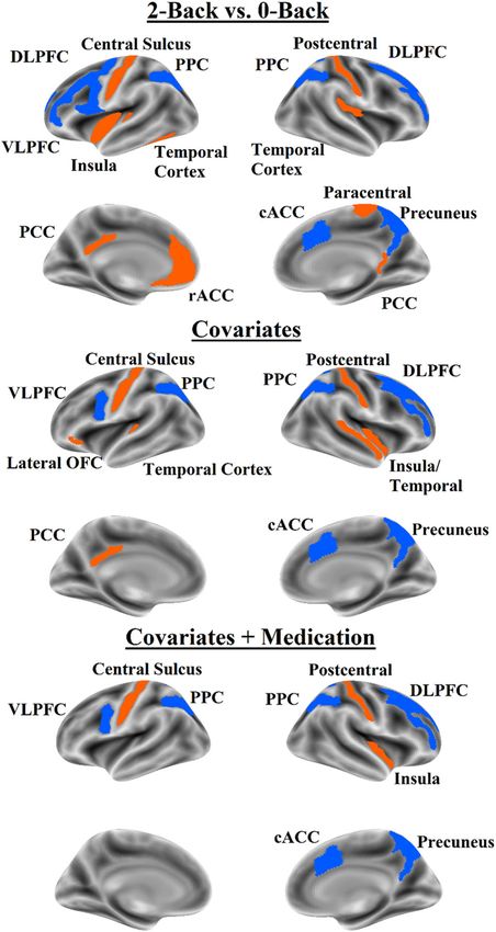
Fig. 1 Brain map for sMRI features that predicted ADHD
residualized ADHD MRI (%) Symptomatology in the elastic net regression and were
symptomatology associated with ADHD symptomatology in univariate mixed
effect models. Orange indicates positive coefficients; blue indicates
Model 1 −0.9 −0.3 −0.9 −0.2 negative coefficients. DLPFC dorsolateral prefrontal cortex, VLPFC
ventrolateral prefrontal cortex, cACC caudal anterior cingulate cortex.
Model 2 0.6 0.0 −0.6 −0.2
Model 3 1.0 −0.1 0.0 −0.1
Model 4 −0.9 −0.2 −0.1 0.0
Supplemental Table 6 (accounting for covariates includ-
Model 5 0.4 −0.2 −0.1 0.0 ing medication).
Mean 0.0 −0.2 −0.3 −0.1 The best model for SST predicted raw ADHD symp-
External test 0.6 0.1 −0.1 0.0
tomatology with R2 = 0.5% in the internal validation set
and with R2 = 0.1% in the external test set. The best
Models 1–5 indicate the R2 of the models predicting the internal validation set in model for SST predicting ADHD symptomatology with
the training phase. “External Validation” indicates the R2 of the best model from
the training phase being tested on the external test set. Full elaboration of the covariates residualized had an R2 = 0.1% on the internal
models can be found in Supplemental Tables. validation set and an R2 = −0.03% on the external test set;
SST stop signal task, MID monetary incentive delay task.
a
Indicates model used no features (i.e., predicted intercept for all cases). in this analysis, negative R2 values means the best models
were still worse than predicting the mean ADHD symp-
tomatology score for all cases. When medication was
considered the model also included less deactivation in added as a covariate, the best SST model had an R2 = 0%
the rostral ACC posterior cingulate cortex, lateral tem- on the internal validation and −0.1% on the external test
poral cortex, and paracentral lobule. Regions identified in set. The best model for the MID predicted ADHD
elastic net regression and confirmed in univariate analyses symptomatology with an R2 = 0.1% in the internal vali-
are shown in Fig. 2 and reported in Supplemental Table 4 dation set, although this was consistent with the external
(not accounting for covariates), Supplemental Table 5 test set (R2 = 0.1%). When covariates were residualized,
(accounting for covariates except medication), and the best MID model had an R2 = 0% on the internalOwens et al. Translational Psychiatry (2021)11:64 Page 8 of 11
Secondary analysis: categorical analyses
Results of the elastic net regression and mixed effects
modeling for categorical ADHD symptomatology using a
tertile split of the CBCL were quite similar to the primary
analyses of the study that used the CBCL ADHD symp-
tomatology measure continuously. In short, with the
categorical version of the CBCL the EN-Back predicted
categorical ADHD symptomatology beyond covariates,
with models using similar regions as those predicting
continuous ADHD symptomatology. sMRI predicted
categorical ADHD symptomatology but not beyond cov-
ariates. SST and MID did not predict categorical ADHD
symptomatology. In contrast, elastic net analyses were not
able to derive a model that robustly predicted ADHD
diagnosis from the K-SADS ADHD diagnosis using any of
the three task fMRI paradigms or sMRI. These results are
reported in Supplemental Results, Supplemental Tables
7–9, and Supplemental Figs. 5 and 6.
Discussion
The current study used elastic net regression with
nested cross-validation to build models for predicting
ADHD symptomatology out-of-sample using four sMRI
and fMRI paradigms. Results indicate that the fMRI EN-
Back working memory task was the most useful imaging
paradigm to predict ADHD symptomatology even when
accounting for numerous sociodemographic factors. Brain
morphometry was able to predict ADHD symptomatology
when sociodemographic factors were not accounted for,
but not when covariates were accounted for. Even without
accounting for sociodemographic factors, the best models
for SST and MID were not effective at predicting ADHD
symptomatology. Based on literature demonstrating
individuals with ADHD symptomatology show poorer
performance and different neural activation for working
memory38, inhibitory control9,11, and reward processing10,
we were surprised to see only working memory activation
Fig. 2 Brain map for fMRI features that predicted ADHD was able to predict ADHD symptomatology.
symptomatology in the elastic net regression and were
The features used by the best EN-Back models suggest
associated with ADHD symptomatology in univariate mixed
effect models. Orange indicates positive coefficients; blue indicates that as ADHD symptomatology increases, activation in
negative coefficients. Top panel = no covariate analysis, middle panel task positive regions (DLPFC, PPC, and caudal ACC)
= analysis with covariates except medication status; bottom panel = decreases and activation in task negative regions
all covariates including medication status. DLPFC dorsolateral (VMPFC, posterior cingulate cortex, lateral temporal
prefrontal cortex, VLPFC ventrolateral prefrontal cortex, cACC caudal
cortex, precentral, and postcentral gyri) increases. This
anterior cingulate cortex, rACC rostral anterior cingulate cortex, OFC
orbitofrontal cortex, PPC posterior parietal cortex, PCC posterior is highly consistent with the existing literature, which
cingulate cortex. has shown that during working memory tasks, indivi-
duals with ADHD show reduced activation in task
positive regions8,38,39 and increased activation in task
validation set and R2 = 0.1% on the external test set. negative regions40.
When medication was added as a covariate, the best MID The CSA features identified by the best sMRI model
model had an R2 = 0% on the internal validation set and overlapped considerably with those in the EN-Back
R2 = 0% on the external test set. Given the poor predictive models; ADHD symptomatology was predicted by less
ability of the SST and MID models, we did not investigate CSA in the DLPFC, caudal ACC, and lateral temporal
their features. cortex. These findings converge with the largest priorOwens et al. Translational Psychiatry (2021)11:64 Page 9 of 11 study of the association of ADHD symptomatology and individuals with ADHD. Future work might further CSA in adolescents, which also found that ADHD interrogate these tasks for relationships with ADHD symptomatology were linked to less CSA in the DLPFC symptomatology. For example, there are alternate and caudal ACC14. Regarding subcortical GMV, the approaches to analyzing these tasks (e.g., functional con- caudate was the only region in which GMV appeared in nectivity, computational modeling) that may prove better the best elastic net regression model and was confirmed for investigating how inhibitory control and reward pro- in univariate analysis to be associated with ADHD cessing differences in those with ADHD symptomatology symptomatology. This is consistent with the largest manifest in the brain. Alternately, it is also possible that examination of the correlates of ADHD for subcortical prior findings linking ADHD symptomatology to SST and GMV in youths, which also found lower caudate volume MID were spurious or that differences in the neural in children with ADHD15. In the current study, ADHD correlates of inhibitory control and reward processing are symptomatology was predicted by lower CT in the caudal limited to other age groups, as most prior studies exam- ACC, precentral and postcentral gyri, and medial tem- ined older adolescents and adults. poral lobe, and higher CT in the VLPFC and medial and lateral occipital cortex, partially overlapping with the Considerations regions observed in the EN-Back model. Given that CT A major consideration for the current study is the was not linked to ADHD symptomatology in the study by magnitude of the observed effect sizes. Even the best Bayard et al. and the literature on CT and ADHD model predicting ADHD symptomatology explained just symptomatology has not been consistent across small 2% of the variance out of sample – equivalent to a Pear- studies (see Lin and Roth11), we would describe the son’s correlation of 0.14. According to Cohen’s heuristic, a regional findings detailed here as preliminary. correlation of 0.1 is considered small44. However, it has While the relations of CSA and caudate GMV have been been noted that these guidelines understate the impor- reproduced across studies when covariates are not tance of associations between 0.1 and 0.2, as many rela- accounted for (or are minimally accounted for), the tionships in day-to-day life that most would consider question remains as to whether it is more appropriate to meaningful fall within this range45, such as the relation- correct for demographic factors that likely are related to ship of antihistamine use to allergy symptoms (Pearson’s both ADHD symptomatology and brain structure and r = 0.11), ibuprofen to headache severity (Pearson’s r = function and may play a causal role in their development. 0.14), combat exposure in the Vietnam war to likelihood In the current study, accounting for age, sex, race, of developing PTSD (Pearson’s r = 0.11), college grades to handedness, pubertal status, comorbid internalizing psy- job performance (Pearson’s r = 0.14), and the critical chopathology, parental income, and parental education ratings to a film’s box office success (Pearson’s r = 0.17)46. reduced the prediction accuracy of the elastic net As larger datasets become available, it is becoming more regression considerably (best model R2 = 0.2% on internal apparent that prior small studies showing “large” asso- validation set, 0.0% on external test set). Covariate use in ciations between brain and behavior were much too the structural MRI literature is varied41 and it creates optimistic47. It is also worth noting that small effect sizes considerable ambiguities in interpreting findings; in the found in the current study are consistent with other large present findings, it is unclear if the factors proposed as MRI studies on the neurobiological correlates of ADHD. covariates should be considered confounding third vari- For example, a recent ENIGMA ADHD study found small ables or meaningful and potentially causal factors. For effect sizes for differences in gray matter volume in example, it is very likely that growing up in impoverished individuals with ADHD (Cohen’s d ~ 0.1, equivalent to R2 circumstances affects the brain’s development and = 2%)15. Likewise, a recent IMAGEN study found small increases the risk of developing ADHD symptomatol- effect sizes for associations of cortical thickness to ADHD ogy42,43; consequently to remove variance in brain and symptoms in adolescents (f2 = 0.01, equivalent to R2 = ADHD symptomatology that is shared by socioeconomic 1%). The largest and most thorough machine learning status may serve to deflate the magnitude of the associa- study to examine ADHD in children (also using the tion between the two. While the field of cognitive neu- ENIGMA dataset) found that their best model using sMRI roscience works towards a consensus on how best to predicted ADHD with an AUC = 0.67 (equivalent to R2 = handle covariates, we opted to report results both with 9%)17. This is slightly larger than the effects in the current and without covariates. study but is consistent when considering that these stu- It is notable that neither the SST nor MID models were dies were comparing a clinical sample of individuals with effective at predicting ADHD symptomatology. However, an ADHD diagnosis and controls without ADHD symp- we do not think that the current results should be inter- tomatology. There have not been any comparable and preted as definitive evidence that there are no differences adequately powered studies with which to compare effect in brain activation during inhibitory or reward tasks in sizes to the current results for the EN-Back.
Owens et al. Translational Psychiatry (2021)11:64                                                                               Page 10 of 11

   Another consideration is that of in-scanner motion.          driver of differences in brain structure and function in
Children with ADHD symptomatology are known to be               individuals with ADHD symptomatology. This was
prone to fidgeting during MRI sessions and in the current        expected based on prior literature, which suggested that
study in-scanner motion explained 1–2% of the variance          medication effects would not represent a substantial
in ADHD symptomatology across fMRI scans. As a con-             confound to the current study. The largest previous study
sequence, we made several attempts to account for in-           on this topic to date found no differences in morphometry
scanner motion, such as using a stringent exclusion cri-        between children with ADHD on and off stimulant
teria for excess in-scanner motion (total framewise dis-        medication15. Several meta-analyses of task fMRI differ-
placement >0.9 mm and volume censoring leaving fewer            ences in children with ADHD have found similar patterns
than 200 TRs of usable data) and covarying for in-scanner       of activation for children on and off medication7,8.
motion in all analyses. While this provides protection
against the possibility that results are entirely a con-        Conclusion
sequence of movement-induced scanner artifact, it also            Using elastic net regression with nested cross-valida-
means that children with the most severe ADHD symp-             tion, the current study found compelling evidence that
tomatology were the most likely to be excluded, resulting       ADHD symptomatology is associated with less activation
in artificially truncated variance in ADHD symptomatol-          during engagement of working memory in task positive
ogy. This may contribute to the small effects seen in the       regions of the brain and more activation in task negative
current study and it is possible that effects may have been     regions, consistent with prior literature. The current study
somewhat larger if these exclusions were not necessary to       also confirmed prior work indicating that lower CSA and
guard against artifact. As such, technical innovations in       GMV of the caudate is associated with more ADHD
fMRI acquisition or processing that can reduce motion or        symptomatology, though this was not the case after
its impacts on data would be tremendously helpful in the        accounting for sociodemographic factors. Previously
study of the neurobiology of ADHD symptomatology.               identified associations of activation during behavioral
   It is also worth considering the measure used to assess      inhibition and reward processing were not confirmed in
ADHD symptomatology. The CBCL is among the best                 the current study.
validated parent-report measures of child ADHD symp-
                                                                Acknowledgements
tomatology48,49; it shows excellent concordance with            This work was funded by NIH/NIDA T32DA043593. Data used in the
clinical interviews done by mental health professionals and     preparation of this article were obtained from the Adolescent Brain Cognitive
convergent and discriminant validity with numerous other        Development (ABCD) Study (https://abcdstudy.org), held in the NIMH Data
                                                                Archive (NDA). This is a multisite, longitudinal study designed to recruit more
indicators of psychopathology. Increasingly, research sug-      than 10,000 children age 9–10 and follow them over 10 years into early
gests that ADHD is a dimensional construct and levels of        adulthood. The ABCD Study is supported by the National Institutes of Health
attention problems that do not meet the diagnostic              and additional federal partners under award numbers U01DA041022,
                                                                U01DA041028, U01DA041048, U01DA041089, U01DA041106, U01DA041117,
threshold for ADHD can have negative effects on indivi-         U01DA041120, U01DA041134, U01DA041148, U01DA041156, U01DA041174,
duals’ quality of life21. Given that the current study used a   U24DA041123, U24DA041147, U01DA041093, and U01DA041025. A full list of
non-clinical sample, we think measuring ADHD con-               supporters is available at https://abcdstudy.org/federal-partners.html. A listing
                                                                of participating sites and a complete listing of the study investigators can be
tinuously was the most appropriate approach. Notably,           found at https://abcdstudy.org/scientists/workgroups/. ABCD consortium
when we re-analyzed the data using ADHD diagnosis from          investigators designed and implemented the study and/or provided data but
the K-SADS as the target variable our elastic net approach      did not necessarily participate in analysis or writing of this report. This
                                                                manuscript reflects the views of the authors and may not reflect the opinions
was not able to build a model that could robustly predict       or views of the NIH or ABCD consortium investigators. The ABCD data
ADHD diagnosis. We suspect that this was a result of the        repository grows and changes over time. The ABCD data used in this report
loss of statistical power from artificially binarizing a con-    came from version 2.0.1.
tinuous phenomenon into imbalanced groups. However,
                                                                Conflict of interest
we do recognize this as a limitation of the study, as our       The authors declare that they have no conflict of interest.
results cannot be directly generalized to a formal ADHD
diagnosis, which (despite its flaws) does represent the
dominate approach in modern clinical psychiatry. It is also     Publisher’s note
                                                                Springer Nature remains neutral with regard to jurisdictional claims in
a limitation that our current results do not speak to spe-      published maps and institutional affiliations.
cific ADHD subtypes, but rather focus on ADHD symp-
tomatology in the aggregate.                                    Supplementary Information accompanies this paper at (https://doi.org/
                                                                10.1038/s41398-020-01192-8).
   One further consideration is the medication status of
the participants. Results were not affected substantially by    Received: 21 August 2020 Revised: 30 November 2020 Accepted: 4
including each participant’s medication status as a cov-        December 2020
ariate, suggesting that medication was not the primary
Owens et al. Translational Psychiatry (2021)11:64 Page 11 of 11
References 25. Insel, T. et al. Research Domain Criteria (RDoC): toward a new classification
1. Alderson, M. R., Kasper, L. J., Hudec, K. L. & Patros, C. H. G. Attention-deficit/ framework for research on mental disorders. Am. J. Psychiatry 167, 748–751
hyperactivity disorder (ADHD) and working memory in adults: a meta-analytic (2010).
review. Neuropsychology 27, 287–302 (2013). 26. Achenbach, T. Manual for the ASEBA School-Age Forms & Profiles An Integrated
2. Kasper, L. J., Alderson, R. M. & Hudec, K. L. Moderators of working memory System of Multi-informant Assessment (ASEBA, 2007).
deficits in children with attention-deficit/hyperactivity disorder (ADHD): A 27. Hagler, D. J. et al. Image processing and analysis methods for the Adolescent
meta-analytic review. Clin. Psychol. Rev. 32, 605–617 (2012). Brain Cognitive Development Study. Neuroimage 202, 116091 (2019).
3. Martinussen, R., Hayden, J., Hogg-Johnson, S. & Tannock, R. A meta-analysis of 28. Casey, B. J. et al. The Adolescent Brain Cognitive Development (ABCD) study:
working memory impairments in children with attention-deficit/hyperactivity Imaging acquisition across 21 sites. Dev. Cogn. Neurosci. 32, 43–54 (2018).
disorder. J. Am. Acad. Child Adolesc. Psychiatry 44, 377–384 (2005). 29. Garavan, H. et al. The ABCD stop signal data: response to Bissett et al. Preprint
4. Alderson, M. R., Rapport, M. D. & Kofler, M. J. Attention-deficit/hyperactivity at https://doi.org/10.1101/2020.07.27.223057 (2020).
disorder and behavioral inhibition: a meta-analytic review of the stop-signal 30. Achenbach, T. M. & Ruffle, T. M. The child behavior checklist and related forms
paradigm. J. Abnorm. Child Psychol. 35, 745–758 (2007). for assessing behavioral/emotional problems and competencies. Pediatr. Rev.
5. Lipszyc, J. & Schachar, R. Inhibitory control and psychopathology: a meta- 21, 265–271 (2000).
analysis of studies using the stop signal task. J. Int. Neuropsychol. Soc. 16, 31. Chen, W. J., Faraone, S. V., Biederman, J. & Tsuang, M. T. Diagnostic accuracy of
1064–1076 (2010). the child behavior checklist scales for attention-deficit hyperactivity disorder: a
6. Schachar, R. J., Tannock, R. & Logan, G. Inhibitory control, impulsiveness, and receiver-operating characteristic analysis. J. Consult. Clin. Psychol. 62,
attention deficit hyperactivity disorder. Clin. Psychol. Rev. 13, 721–739 (1993). 1017–1025 (1994).
7. Dickstein, S. G., Bannon, K., Xavier Castellanos, F. & Milham, M. P. The neural 32. Lampert, T. L., Polanczyk, G., Tramontina, S., Mardini, V. & Rohde, L. A. Diag-
correlates of attention deficit hyperactivity disorder: an ALE meta-analysis. J. nostic performance of the CBCL-attention problem scale as a screening
Child Psychol. Psychiatry 47, 1051–1062 (2006). measure in a sample of Brazilian children with ADHD. J. Atten. Disord. 8, 63–71
8. Cortese, S. et al. Toward systems neuroscience of ADHD: a meta-analysis of 55 (2004).
fMRI sudies. Am. J. Psychiatry 169, 1038–1055 (2012). 33. Petersen, A. C., Crockett, L., Richards, M. & Boxer, A. A self-report measure of
9. Hart, H., Radua, J., Nakao, T., Mataix-Cols, D. & Rubia, K. Meta-analysis of pubertal status: reliability, validity, and initial norms. J. Youth Adolesc. 17,
functional magnetic resonance imaging studies of inhibition and attention in 117–133 (1988).
attention-deficit/hyperactivity disorder: exploring task-specific, stimulant 34. Veale, J. F. Edinburgh handedness inventory - short form: a revised version
medication, and age effects. JAMA Psychiatry 70, 185 (2013). based on confirmatory factor analysis. Laterality 19, 164–177 (2014).
10. Plichta, M. M. & Scheres, A. Ventral-striatal responsiveness during reward 35. Chaarani, B. et al. Brain function in the pre-adolescent brain: results from the
anticipation in ADHD and its relation to trait impulsivity in the healthy ABCD study. Nat. Neurosci. (in press).
population: A meta-analytic review of the fMRI literature. Neurosci. Biobehav. 36. Destrieux, C., Fischl, B., Dale, A. & Halgren, E. Automatic parcellation of human
Rev. 38, 125–134 (2014). cortical gyri and sulci using standard anatomical nomenclature. Neuroimage
11. Lin, G. & Roth, R. M. The status of structural and functional MRI in adoles- 53, 1–15 (2010).
cents with attention-deficit/hyperactivity disorder. Psychol. Inj. Law 10, 37. Heeringa, S. & Berglund, P. A guide for population-based analysis of the
209–222 (2017). adolescent brain cognitive development (ABCD) study baseline data. Preprint
12. Albaugh, M. D. & Potter, A. S. The ventromedial prefrontal cortex: a putative at https://doi.org/10.1101/2020.02.10.942011 (2020).
locus for trait inattention. Neuropsychopharmacology 44, 226–227 (2019). 38. Farruggia, M. C., Laird, A. R. & Mattfeld, A. T. Common default mode network
13. Albaugh, M. D. et al. Inattention and reaction time variability are linked to dysfunction across psychopathologies: a neuroimaging meta-analysis of the
ventromedial prefrontal volume in adolescents. Biol. Psychiatry 82, 660–668 n-back working memory paradigm. bioRxiv https://doi.org/10.1101/
(2017). 2020.01.30.927210 (2020).
14. Bayard, F. et al. Distinct brain structure and behavior related to ADHD and 39. Christakou, A. et al. Disorder-specific functional abnormalities during sustained
conduct disorder traits. Mol. Psychiatry 25, 3020–3033 (2018). attention in youth with Attention Deficit Hyperactivity Disorder (ADHD) and
15. Hoogman, M. et al. Subcortical brain volume differences in participants with with Autism. Mol. Psychiatry 18, 236–244 (2013).
attention deficit hyperactivity disorder in children and adults: a cross-sectional 40. Metin, B. et al. Dysfunctional modulation of default mode network activity in
mega-analysis. Lancet Psychiatry 4, 310–319 (2017). attention-deficit/hyperactivity disorder. J. Abnorm. Psychol. 124, 208–214
16. Yarkoni, T. & Westfall, J. Choosing prediction over explanation in psychology: (2015).
lessons from machine learning. Perspect. Psychol. Sci. 12, 1100–1122 (2017). 41. Hyatt, C. S. et al. The quandary of covarying: a brief review and empirical
17. Zhang-james, A. Y. et al. Machine learning classification of attention-deficit/ examination of covariate use in structural neuroimaging studies on psycho-
hyperactivity disorder using structural MRI data. Preprint at https://doi.org/ logical variables. Neuroimage 205, 116225 (2019).
10.1101/546671 (2020). 42. Russell, A. E., Ford, T., Williams, R. & Russell, G. The Association Between
18. Jollans, L. et al. Quantifying performance of machine learning methods for Socioeconomic Disadvantage and Attention Deficit/Hyperactivity Disorder
neuroimaging data. Neuroimage 199, 351–365 (2019). (ADHD): a systematic review. Child Psychiatry Hum. Dev. 47, 440–458 (2016).
19. Hudziak, J. J., Achenbach, T. M., Althoff, R. R. & Pine, D. S. A dimensional 43. Cheung, C. H. M. et al. Childhood predictors of adolescent and young adult
approach to development psychopathology. Int. J. Methods Psychiatr. Res. 16, outcome in ADHD. J. Psychiatr. Res. 62, 92–100 (2015).
S16–S23 (2007). 44. Cohen, J. Statistical Power Analysis For The Behavioral Sciences. 2nd edn.
20. Willcutt, E. G. et al. Validity of DSM-IV attention–deficit/hyperactivity disorder (Lawrence Erlbaum Associates, 1988).
symptom dimensions and subtypes. J. Abnorm. Child Psychol. 121, 991–1010 45. Funder, D. C. & Ozer, D. J. Evaluating effect size in psychological research:
(2012). sense and nonsense. Adv. Methods Pract. Psychol. Sci. 2, 156–168 (2019).
21. Loe, I. M. & Feldman, H. M. Academic and educational outcomes of children 46. Meyer, G. J. et al. Psychological testing and psychological assessment: a review
with ADHD. J. Pediatr. Psychol. 32, 643–654 (2007). of evidence and issues. Am. Psychol. 56, 128–165 (2001).
22. Shaw, P. et al. Cortical development in typically developing children with 47. Button, K. S. et al. Power failure: why small sample size undermines the
symptoms of hyperactivity and impulsivity: support for a dimensional view of reliability of neuroscience. Nat. Rev. Neurosci. 14, 365–376 (2013).
attention deficit hyperactivity disorder. Am. J. Psychiatry 168, 143–151 (2011). 48. Raiker, J. S. et al. Accuracy of Achenbach Scales in the screening of attention-
23. Ducharme, S. et al. Decreased regional cortical thickness and thinning rate are deficit/hyperactivity disorder in a community mental health clinic. J. Am. Acad.
associated with inattention symptoms in healthy children. J. Am. Acad. Child Child Adolesc. Psychiatry 56, 401–409 (2017).
Adolesc. Psychiatry 51, 18–27.e2 (2012). 49. Hudziak, J. J., Copeland, W., Stanger, C. & Wadsworth, M. Screening for
24. Derks, E. M. et al. Genetic and environmental influences on the relation DSM-IV externalizing disorders with the Child Behavior Checklist: a
between attention problems and attention deficit hyperactivity disorder. receiver-operating characteristic analysis. J. Child Psychol. Psychiatry 45,
Behav. Genet. 38, 11–23 (2008). 1299–1307 (2004).You can also read