Double core hole valence-to-core x-ray emission spectroscopy: A theoretical exploration using time-dependent density functional theory
←
→
Page content transcription
If your browser does not render page correctly, please read the page content below
Double core hole valence-to-core x-ray
emission spectroscopy: A theoretical
exploration using time-dependent density
functional theory
Cite as: J. Chem. Phys. 151, 144114 (2019); https://doi.org/10.1063/1.5111141
Submitted: 23 May 2019 . Accepted: 22 September 2019 . Published Online: 11 October 2019

Yu Zhang      , Uwe Bergmann          , Robert Schoenlein, Munira Khalil   , and Niranjan Govind

J. Chem. Phys. 151, 144114 (2019); https://doi.org/10.1063/1.5111141                               151, 144114
The Journal
                                                                                                 ARTICLE           scitation.org/journal/jcp
 of Chemical Physics

    Double core hole valence-to-core x-ray emission
    spectroscopy: A theoretical exploration using
    time-dependent density functional theory
    Cite as: J. Chem. Phys. 151, 144114 (2019); doi: 10.1063/1.5111141
    Submitted: 23 May 2019 • Accepted: 22 September 2019 •
    Published Online: 11 October 2019

    Yu Zhang,1,a)           Uwe Bergmann,2,b)               Robert Schoenlein,3 Munira Khalil,4       and Niranjan Govind5,c)

    AFFILIATIONS
    1
        Stanford PULSE Institute, SLAC National Accelerator Laboratory, Menlo Park, California 94025, USA
    2
        LCLS and Stanford PULSE Institute, SLAC National Accelerator Laboratory, Menlo Park, California 94025, USA
    3
        SLAC National Accelerator Laboratory, Menlo Park, California 94025, USA
    4
        Department of Chemistry, University of Washington, Seattle, Washington 98195, USA
    5
        Environmental Molecular Sciences Laboratory, Pacific Northwest National Laboratory, Richland, Washington 99352, USA

    Note: This paper is part of the JCP Special Collection on Ultrafast Spectroscopy and Diffraction from XUV to X-ray.
    a)
       Electronic mail: yzhang15@slac.stanford.edu
    b)
       Electronic mail: bergmann@slac.stanford.edu
    c)
       Electronic mail: niri.govind@pnnl.gov

    ABSTRACT
    With the help of newly developed X-ray free-electron laser (XFEL) sources, creating double core holes (DCHs) simultaneously at the same
    or different atomic sites in a molecule has now become possible. DCH X-ray emission is a new form of X-ray nonlinear spectroscopy
    that can be studied with a XFEL. Here, we computationally explore the metal K-edge valence-to-core (VtC) X-ray emission spectroscopy
    (XES) of metal/metal and metal/ligand DCH states in a series of transition metal complexes with time-dependent density functional theory.
    The simulated DCH VtC-XES signals are compared with conventional single core hole (SCH) XES signals. The energy shifts and intensity
    changes of the DCH emission lines with respect to the corresponding SCH-XES features are fingerprints of the coupling between the second
    core hole and the occupied orbitals around the DCHs that contain important chemical bonding information of the complex. The differ-
    ence between delocalized/localized core hole models on DCH VtC-XES is also briefly discussed. We theoretically demonstrate that DCH
    XES provides subtle information on the local electronic structure around metal centers in transition metal complexes beyond conventional
    linear XES. Our predicted changes from calculations between SCH-XES and DCH-XES features should be detectable with modern XFEL
    sources.
    https://doi.org/10.1063/1.5111141., s

    I. INTRODUCTION                                                            have been several theoretical DCH studies and related spectroscopic
                                                                               signals with various methods including many-body Green’s func-
         One of the striking effects of intense X-ray-matter interaction       tion,2–4 multiple configuration self-consistent field (MCSCF),5–13
    is the creation of multiple core holes. Theoretical insight into this      density functional theory (DFT),8,9,11,14–18 time-dependent density
    phenomenon was provided long before any realistic experiments on           functional theory (TDDFT),19 Møller–Plesset perturbation theory,16
    double core hole (DCH) states were performed. More than three              and the Z + 1 approximation,20 respectively.
    decades ago, Cederbaum et al. studied DCHs in small molecules                   DCH spectroscopy was originally studied with synchrotron
    theoretically and predicted that the electron binding energies asso-       radiation,21–27 where the absorption of one photon is accompanied
    ciated with DCHs at different atomic sites could sensitively probe         by the ejection of two core electrons. In this process, the
    the chemical environment of the ionized atoms.1 Since then, there          correlation between the two ejected core electrons plays an

J. Chem. Phys. 151, 144114 (2019); doi: 10.1063/1.5111141                                                                                151, 144114-1
The Journal
                                                                                                   ARTICLE            scitation.org/journal/jcp
 of Chemical Physics

    important role. While multiphoton processes with synchrotron radi-          facility.43 In previous DCH photoelectron spectroscopy experi-
    ation are unlikely, they become possible with more intense X-ray            ments,29 XFEL pulses with a duration of ∼10 fs and an intensity over
    free-electron laser (XFEL) pulses. Thanks to the rapid development          1016 W/cm2 were used. Since hard X-ray core electron photoion-
    of XFEL sources, two-photon photoelectron spectroscopy or dou-              ization cross sections are generally one order of magnitude smaller
    ble core hole (DCH) spectroscopy has been shown to be a pow-                than soft X-ray core electron photoionization cross sections,44 we
    erful tool to probe the chemical environment of specific atomic             believe that even shorter and more intense XFEL pulses are needed
    sites in molecules.11,15,28–31 Recently, DCH states have been cre-          for metal-metal DCH spectroscopy experiments. The experiments
    ated using XFEL pulses at the Linac Coherent Light Source (LCLS)            of metal/ligand DCH-XES simulated in this study are still not cur-
    facility in neon,32 nitrogen gas,29,33,34 N2 O, CO2 and CO,29 and           rently available because of the difficulty of combining hard and soft
    the aminophenol molecule.35 Differences between the photoelectron           XFEL pulses, but this technique could be possible with the planned
    spectral data taken with focused and unfocused laser beams were             Tender X-ray Instrument (TXI) in the under-constructing LCLS-II
    studied in order to extract the DCH contribution to the signal.29           facility. Moreover, for metal/metal DCH-XES, the ultrashort lifetime
    It is believed that two-site DCH (ts-DCH) spectroscopy provides             of DCH states not only requires extremely short and intense XFEL
    a more sensitive probe for the local chemical environment of the            pulses but also broadens the emission lines, which makes the obser-
    excited atoms than does single core hole (SCH) spectroscopy.29,30           vation of the fine features in experiment very challenging. This line
    Mukamel et al. theoretically studied X-ray four-wave mixing spec-           broadening issue could be remedied by using the recently developed
    troscopy involving DCHs.12,19,20 The corresponding experiments              stimulated XES technique45–47 with which specific emission lines
    require well-controlled intense X-ray pulses that are not currently         could be selectively enhanced and narrowed.
    available. In this study, we focus on ts-DCH spectroscopy because
    it is more sensitive for chemical analysis than single-site (ss) DCH        II. COMPUTATIONAL DETAILS
    spectroscopy. Previous DCH spectroscopy experiments measured
    photoelectrons28,29,36–38 and Auger electrons,24,33,34,37,39,40 which are         All calculations were performed at the DFT and TDDFT
    not suitable for solution samples common in chemistry. We pro-              [within the Tamm-Dancoff approximation (TDA)48 ] levels of
    pose the use of intense XFEL pulses to create DCHs at different sites       theory with the NWChem quantum chemistry package.49 No sym-
    in the system and to study the corresponding X-ray emissions. We            metry restrictions have been applied. The PBE050,51 hybrid func-
    envision that DCH-X-ray emission spectroscopy (XES) signals can             tional was used for all calculations. All geometries were opti-
    provide information beyond what single core hole X-ray emission             mized at the PBE0/Def2-TZVP52 level of theory. The X-ray
    spectroscopy (SCH-XES) signals can tell us. Even though this spec-          emission calculations have been performed using the FCH (full core
    troscopy has not been realized in transition metal complexes, we            hole)/TDDFT approach described in our previous publication.53
    hope our theoretical work in this manuscript can serve as a guide for       Here, this approach has been extended to explore double core hole
    future experiments. Our findings suggest how the additional core            states. In our SCH and DCH signal simulations, both α and β ion-
    hole affects the local electronic structure of both core holes and          ization channels and all of their possible combinations were con-
    allows one to probe the electronic coupling between the two atomic          sidered with equal weights. As an illustration, a sample NWChem
    sites. In other words, the additional core hole offers more “control        input file with notes is provided in the supplementary material. For
    knobs” to detect different occupied orbitals of the system and further      the binuclear complexes, all calculations have been performed in
    complement SCH-XES.                                                         the gas phase, while for the Mn mononuclear complexes with avail-
          In this paper, we study ts-DCH spectroscopy from a theoreti-          able VtC-XES experimental results, all calculations were performed
    cal standpoint using TDDFT. We calculate the VtC-XES41,42 signals           in the CH2 Cl2 solution phase (ε = 9.08), which is described by the
    resulting from DCHs of a series of mono- and binuclear transition           conductorlike screening model (COSMO).54 For the TDDFT calcu-
    metal model complexes. VtC-XES signals carry more chemical infor-           lations of the core hole states, the Sapporo-TZP-201255 /Def2-TZVP
    mation of the system than Kα and Kβ mainline emissions because              basis sets have been used for the metal and nonmetal atoms, respec-
    they directly probe the valence orbitals. For the binuclear complexes       tively. In the localized N1s core hole calculations, in order to fix a
    with metal-metal direct bonds, the VtC-XES signals of the metal 1s          1s core hole at an individual N atom, we use an all-electron basis
    DCHs at different sites were studied. Mononuclear complexes with            set representation only on one N atom and use effective core poten-
    different Mn oxidation states (II, III) have been used to investigate       tials (ECPs) and the corresponding basis sets to describe the other
    the emissions of the metal-1s/ligand-1s DCH states, from which the          N atoms. Specifically in this study, the Def2-TZVP basis set has
    information on the chemical bonds between the metal center and the          been used for the N atom with a 1s core hole, and the Stevens-
    coordinating atoms are revealed.                                            Basch-Krauss-Jasien-Cundari ECP56 together with the correspond-
          Here, we have focused on only two-site DCH XES signals and            ing polarized basis set57 has been used for the other N atoms without
    not single-site DCH-XES, which could also be interesting. For deep          1s core holes. In the SCH and DCH calculations of the studied bin-
    ss-DCHs (e.g., 1s/1s single-site DCHs), the overall effect of the sec-      uclear complexes, transitions to core holes at both metal atoms are
    ond core hole on the XES spectrum is mainly a constant shift of             included.
    all emission lines and thus less interesting. However, 1s/2p single-
    site DCH-XES is more informative because the 2p core hole unlike            III. RESULTS AND DISCUSSION
    the spherical 1s core hole couples with the 3d and other valence
    orbital in different ways. In collaboration with Fuller et al., recently,   A. Binuclear transition metal complexes
    we observed Fe1s/Fe2p single-site DCH Kα XES in Fe systems                      We have studied the VtC-XES signals of metal 1s/metal 1s
    at the SPring-8 Angstrom Compact free electron LAser (SACLA)                ts-DCH states of binuclear transition metal complexes with strong

J. Chem. Phys. 151, 144114 (2019); doi: 10.1063/1.5111141                                                                                   151, 144114-2
The Journal
ARTICLE scitation.org/journal/jcp
of Chemical Physics
metal-metal bonds. Polynuclear transition metal complexes with One can clearly see that the hole on the right Fe atom induces
direct metal-metal bonds58 have attracted the attention of chemists a significant electron density redistribution (blue means hole and
for a long time, not only for their high bond orders (>3) but also for red means particle density). There are also some p-type electron
their important applications in making metal-organic frameworks, density changes on the Cp ring and O atom of the CO ligand,
molecular conductors, photosensitizers, and catalysts.59 Results which suggests that emission involving orbitals with similar char-
from theoretical calculations on binuclear metal complexes with acter could be significantly affected by the second core hole. For
metal-metal bonds have been reviewed recently.60 Here, we chose Fe-cplx, both the SCH and DCH spectra have a shoulder feature
one Fe complex Cp2 Fe(μ-CO)2 (Fe-cplx, Cp = cyclopentadienyl) above 7123 eV [labeled S1 in Fig. 1(a) and D1 in panel (b)], but the
and one Co complex (C4 H6 )2 Co2 (μ-CO) (Co-cplx) as candidate sys- DCH-XES peak is red-shifted by ∼0.5 eV and much weaker (relative
tems. The structures of the two complexes can be found in Figs. 1 to the strongest peak) compared with the corresponding SCH-XES
and 2. The optimized Fe–Fe and Co–Co bond lengths are 2.130 and feature.
2.110 Å, respectively. These bond lengths are close to the previously Molecular orbital (MO) analysis of the representative tran-
reported theoretical values (2.120 Å for Fe–Fe and 2.142 Å for Co– sitions of the two peaks shows that the largest contribution of
Co).61,62 Bond valence analysis shows that Fe-cplx has a formal bond each comes from the Fe–Fe d π orbitals (see Table S1 in the
order of 361 and Co-cplx has a formal bond order of 4.62 supplementary material for the plots of the MOs discussed for Fe-
In general, compared with SCH states, the second core hole in cplx). However, for the SCH-XES transition, the dominant MOs
ts-DCH states have two types of effects on XES: (1) emission energy are at the Fe atoms, while for the DCH-XES transition, MOs on
shifts due to the additional attractive potential of the second core the Cp ring are also involved because of the second core hole.
hole; (2) emission intensity changes due to the additional perturba- The involvement of the Cp ring orbitals may explain the reduced
tions on the molecular orbital (MO) shapes caused by the second intensity of the corresponding transition in the DCH-XES spec-
core hole. An electron in a localized orbital near the second deep 1s trum. For the strongest SCH-XES features around 7120.4 eV (S2
core hole will feel a very strong attraction, resulting in a blue shift of and D2 in Fig. 1), the DCH-XES peak D2 is blue-shifted by ∼0.7 eV
the corresponding emission energies. For other orbitals not localized compared to S2. Local Fe d orbitals and Fe–Fe d π orbitals con-
near the second core hole, the attraction and screening of the core tribute significantly to such transitions, and the influence of the
hole can have an overall effect on the orbital shape. On the other second core hole on these transitions is manifest in the energy
hand, if the two core holes are uncorrelated or independent, both shift.
emission energy and intensity changes will be negligible and should The DCH-XES spectrum has relatively stronger shoulder fea-
result in a spectrum almost identical to the corresponding SCH-XES tures between 7118 and 7120 eV [D3 and D4 on the high platform in
spectrum. Comparing the emission energies and intensities (Figs. 1 Fig. 1(b)], respectively. These transitions involve local Fe d orbitals
and 2) allows one to shed light on the strength of the interaction and Fe–Fe d σ bonding orbitals, which are also heavily affected by the
second core hole. These emission lines contain information about
between the two core holes.
the Fe–Fe direct bonding, but cannot be clearly resolved in the SCH-
XES spectrum as it is suppressed by the strongest peak S2 nearby.
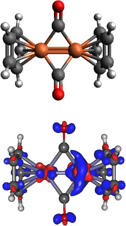
1. Fe-cplx The S3 peak mainly represents transitions from orbitals on the CO
In Fig. 1(b), we show the calculated electron density difference ligands, while the D5 peak has many transition components from the
between the DCH and the SCH states of Fe-cplx (ρDCH − ρSCH ). Cp ring C p orbitals, which again is the effect of the second core hole.
FIG. 1. Calculated SCH and DCH
Fe1s VtC-XES signals of Cp2 Fe(μ-CO)2
(Fe-cplx, Cp = cyclopentadienyl). All cal-
culated spectra have not been shifted
and have been Lorentzian broadened by
1.3 eV. SCH and DCH spectra are scaled
differently for the convenience of plot-
ting. Stick heights in different panels are
not calibrated. (a) Calculated SCH Fe1s
VtC-XES signals. Important features are
labeled S1–7. The molecular structure is
also shown. Color code: brown, Fe; red,
O; dark gray, C; light gray, H. (b) Cal-
culated two-site Fe1s/Fe1s DCH Fe1s
VtC-XES signals. Important features are
labeled D1–9. The calculated electron
density difference between the DCH and
the SCH state is also shown (ρDCH
− ρSCH , surface isovalue = 0.005). Red
and blue denote positive and negative
values, respectively.
J. Chem. Phys. 151, 144114 (2019); doi: 10.1063/1.5111141 151, 144114-3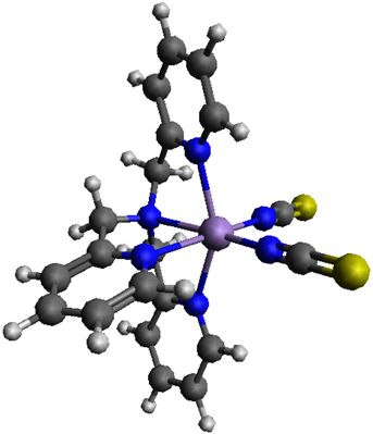
The Journal
                                                                                                 ARTICLE            scitation.org/journal/jcp
 of Chemical Physics

                                                                                                                 FIG. 2. Calculated SCH and DCH Fe1s
                                                                                                                 VtC-XES signals of (C4 H6 )2 Co2 (μ-CO)
                                                                                                                 (Co-cplx). All calculated spectra have not
                                                                                                                 been shifted and have been Lorentzian
                                                                                                                 broadened by 1.4 eV. SCH and DCH
                                                                                                                 spectra are scaled differently for the
                                                                                                                 convenience of plotting. Stick heights
                                                                                                                 in different panels are not calibrated.
                                                                                                                 (a) Calculated SCH Co1s VtC-XES sig-
                                                                                                                 nals. Important features are labeled S1–
                                                                                                                 8. The molecular structure is also shown.
                                                                                                                 Color code: pink, Co; red, O; dark gray,
                                                                                                                 C; light gray, H. (b) Calculated two-
                                                                                                                 site Co1s/Co1s DCH Co1s VtC-XES sig-
                                                                                                                 nals. Important features are labeled D1–
                                                                                                                 9. The calculated electron density differ-
                                                                                                                 ence between the DCH and the SCH
                                                                                                                 state is also shown (ρDCH − ρSCH , sur-
                                                                                                                 face isovalue = 0.006). Red and blue
                                                                                                                 denote positive and negative values,
                                                                                                                 respectively.

    Compared to S3, D3 is blue-shifted by ∼0.5 eV. All the other              transitions have significant contributions from both CO p π orbitals
    peaks (S4–7 and D6–9) have dominant transition contributions              and σ bonding orbitals on the C4 H6 ligands. Both S6 and D7 mainly
    from the orbitals on the CO and Cp ligands, which are not heav-           represent emissions from C and O 2s orbitals on the CO ligand. D7
    ily affected by the second core hole, and thus similar in both            is blue-shifted by about 0.4 eV compared to S6. Going further to
    the SCH-XES and DCH-XES spectra. From the analysis above,                 the lower energy range, D8 is shifted by ∼0.7 eV compared to S7
    we see that the second Fe1s core hole energy shifts and inten-            and D9 is shifted by ∼0.6 eV compared to S8. All of these can be
    sity changes in the higher energy Kβ2,5 region are affected to            considered as transitions from the C 2s orbitals from the C4 H6 lig-
    a greater extent compared with the lower energy Kβ′′ features.            ands. Unlike the Fe-cplx, for the Co-cplx, it seems that both Kβ2,5
    This is probably because that the Kβ2,5 features involve MOs with         and Kβ′′ emission lines are shifted in the DCH-XES spectrum com-
    more metal d orbital character than those involved by the Kβ′′            pared to the corresponding SCH-XES. As Kβ′′ emissions mainly
    features.                                                                 come from ligand orbitals, the shifting of these lines tells us in Co-
                                                                              cplx the metal-ligand interaction is stronger than that in Fe-cplx, and
    2. Co-cplx                                                                DCH-XES may contain coordination chemical information beyond
                                                                              SCH-XES.
         In Fig. 2(b), we also show the calculated electron density differ-
    ence between the DCH and the SCH state of Co-cplx (ρDCH − ρSCH ).
    Similar to the case of Fe-cplx, one can see that the hole on the right    B. Mn mononuclear complexes
    Co atom induces electron density changes around it and some p-type              In Sec. III A, we studied the case of the VtC-XES of two-
    electron density deficiency on the C4 H6 ligand and O atom of the         site metal 1s/metal 1s DCH states, which, in principle, can be
    CO ligand. Peak S1 is much stronger (relative to the strongest peak)      created by a single intense hard X-ray pulse with enough bril-
    than D1 (see Fig. 2 for labeling). An inspection of the contributing      liance. Here, we propose creating metal 1s/ligand 1s DCH states
    MOs involved in the transitions (see Table S2 in the supplementary        in transition metal complexes, which may be achieved by com-
    material) suggests that S1 involves localized Co d orbitals bonded        bining hard and soft X-ray pulses. Using typical Mn mononu-
    with p orbitals on the C4 H6 ligand only around the SCH, while            clear complexes as candidate systems, we investigate the VtC-XES
    peak D1 involves localized Co d orbitals of both Co atoms, which          of Mn1s/ligand 1s DCH states. Compared with metal core holes,
    reduces its emission intensity. The strongest DCH-XES peak D2 is          core holes on ligands might have greater impact on the valence
    blue-shifted by about 0.8 eV compared to the strongest SCH-XES            orbitals around the metal center, and therefore, the corresponding
    peak S2. This shift is even larger than that of Fe-coplx (∼0.6 eV) and    VtC-XES spectra could be more informative about the coordination
    should be detectable at the current levels of instrumentation. Both       bonds.
    transitions involve d orbitals at two Co sites interacting (bonding or          The calculated Mn1s SCH and Mn1s/N1s and Mn1s/Cl1s DCH
    antibonding) with the C p orbitals on the C4 H6 ligand. D3–5 form a       VtC-XES signals of a representative high-spin Mn(II) mononu-
    rising shoulder beside the strongest peak D2. Similar to the case of      clear complex [Mn(II)(terpy)Cl2 ] (Mn_II-cplx, terpy = 2,2′ ; 6′ ,2′′ -
    Fe-cplx, D4 contains the information of direct Co–Co bonding and          terpyridine, sextet) at the Mn K-edge are presented in Fig. 3. We
    the corresponding peak cannot be resolved in the SCH-XES spec-            note that in all the Mn1s/Cl1s DCH calculations, the Cl1s core hole
    trum. D6 is blue-shifted by around 0.5 eV compared to S5. Their           is localized on one of the Cl atoms, while for the Mn1s/N1s DCH

J. Chem. Phys. 151, 144114 (2019); doi: 10.1063/1.5111141                                                                                      151, 144114-4
The Journal
ARTICLE scitation.org/journal/jcp
of Chemical Physics
D2 in panel (b) has the same character as S1 (see Table S3), but
is red-shifted by ∼0.5 eV. This is the effect of the N1s core hole.
However, the N1s core hole has almost no effect on D3 since it
is essentially the same as S2. The Mn1s/Cl1s spectrum [panel (c)
in Fig. 3] is quite different from the other two spectra in the fig-
ure. The strongest peak D4 is of the same character as S1, but is
blue-shifted by about 0.8 eV. The small shoulder D5 is in the same
energy range of S1 but of totally different character: it mainly rep-
resents transitions from the Mn–Cl coordination bonding orbitals
(the Cl atom has not a core hole). D6 at ∼6532.8 eV is similar to
S2 in character, but is blue-shifted by ∼0.8 eV. The relatively strong
peak D7 at ∼6531.1 eV represents transitions from 3p orbitals of the
Cl with the 1s core hole, which is very different from D3 and S2 in
character.
For comparison, we study another high-spin Mn mononuclear
complex [Mn(III)(terpy)Cl3 ] (Mn_III-cplx, quintet). The calculated
Mn1s SCH and Mn1s/N1s, Mn1s/Cl1s DCH VtC-XES signals at the
Mn K-edge are shown in Fig. 4. As indicated in panels (a) and (b),
the Mn1s/N1s DCH VtC-XES signals are very different from the
Mn1s SCH counterpart. The strongest peak D1 is blue-shifted by
about 2.5 eV compared to S1! A MO analysis reveals that the strong
transitions in the broad peak S1 mainly involve MOs on the two
side pyridine rings, while strong transitions in D1 and D2 have sig-
nificant contributions from the Cl 3p orbitals and the MOs on the
middle pyridine ring (see Table S4 in the supplementary material).
This D1 peak resembles the D1 peak in Fig. 3 in MO character. The
huge shift of D1 indicates that the N1s core hole drastically changes
the local electronic structure around the Mn metal center and there
is much stronger strong hybridization between the N 2p and Mn
3d orbitals in Mn_III-cplx compared to Mn_III-cplx. Peak D4 is
similar to S2, and both represent transitions from Cl 3s orbitals.
D3 features with energies more than 2 eV higher than those of S2
also represent transitions from Cl 3s orbitals, but it is not seen in
FIG. 3. Calculated SCH and DCH Mn1s VtC-XES signals of [Mn(II)(terpy)Cl2 ] the SCH spectrum and should be considered as the effect of the
(Mn_II-cplx). All calculated spectra have been red-shifted by 14.07 eV to be N1s core hole. Because there are two chemically nonequivalent Cl
compared with the conventional SCH VtC-XES experiment63 and have been atoms in this complex, we chose to put the 1s core hole at one of
Lorentzian broadened by 1.2 eV. SCH and DCH spectra are scaled differently the Cl atoms perpendicular to the terpy plane, as labeled with an
for the convenience of plotting. Stick heights in different panels are not calibrated. asterisk symbol in panel (b) of Fig. 4. The Mn1s/Cl1s DCH spec-
(a) Experimental and calculated SCH Mn1s VtC-XES signals. Important features trum has a very broad shoulder on the lower energy side of the
are labeled S1 and S2. (b) Calculated Mn1s/N1s DCH Mn1s VtC-XES signals.
Important features are labeled D1–3. The molecular structure of Mn_II-cplx is also strongest peak D5, lacking characteristic features. The strong tran-
shown. (c) Calculated Mn1s/Cl1s DCH Mn1s VtC-XES signals. Important features sitions above 6534 eV mainly involve Cl 3p orbitals, Mn–Cl and
are labeled D4–7. Mn–N coordination bonding orbitals. The weak peak D7 is similar
to D4 and S2. D6 resembles D3 in MO character but is red-shifted
by ∼0.8 eV because its transition orbital is on the Cl atom with a core
hole.
calculations, the N1s core hole is delocalized to all N atoms. The From this analysis, we see that metal-ligand DCH VtC-XES can
issue of localized/delocalized core hole in DCH calculations will be have shifted or new features compared to conventional SCH VtC-
addressed in Sec. III C. We focus on the important features labeled XES. These shifted or new features potentially contain additional
in Fig. 3. S1 around 6535.4 eV is the strongest peak in the con- chemical information of the coordination bonds between the metal
ventional SCH VtC-XES spectrum, of which the major contributing center and ligands.
occupied MO has significant components as the Mn–N coordina-
tion bond and sigma bonds on the pyridine rings (see Table S3 in
the supplementary material). S2 at 6532.0 eV also represents transi- C. Localized and delocalized core hole models
tions from orbitals on the pyridine rings. For the Mn1s/N1s DCH When studying core holes on multiple chemically equivalent
spectrum, the shoulder peak D1 around 6536.0–6535.5 eV denotes atoms in a molecule, a question arises whether the core hole should
transitions from the Mn–Cl bonds and Cl 3p orbitals. This peak can- be considered as localized on one atom or delocalized to all equiva-
not be resolved in the SCH spectrum. It is interesting that the N1s lent atoms. Chemically equivalent atoms are symmetric and a local-
core hole favors the transition from the Cl atoms. The strongest peak ized core hole on one of them breaks the symmetry, leading to the
J. Chem. Phys. 151, 144114 (2019); doi: 10.1063/1.5111141 151, 144114-5The Journal
ARTICLE scitation.org/journal/jcp
of Chemical Physics
both important.66 This can be used to explain why the localized core
hole model works very well for calculating core ionization poten-
tials with independent particle theories such as Hartree-Fock. Since
these two seminal studies, more evidence favoring localized models
in determining core hole properties has emerged.67–71 The symme-
try breaking issue can be remedied by employing high-level electron
correlation methods such as MCSCF methods.72 A specific double
excitation configuration describing a core-core excitation coupled
to a valence-valence excitation under the symmetry restriction helps
one to reduce the symmetry breaking relaxation error.73 In exper-
iment, both localized and delocalized core holes can be selectively
detected.74,75
A complete comparison of localized/delocalized core hole mod-
els in DCH spectroscopy goes beyond the scope of this paper. Here,
we present only a special case of localized/delocalized ligand core
hole models in metal 1s/ligand 1s DCH VtC-XES. In our calcula-
tions reported in Sec. III B, although no symmetry was enforced,
we found that for the 1s core holes on light atoms such as N,
self-consistent field (SCF) calculations often converge to a delocal-
ized core hole state in which the core hole is almost equally dis-
tributed over all N atoms in the molecular complex. We note that
in our complexes the N atoms are not chemically equivalent and
this is an example of hole delocalization without strict symmetry.
However, for deeper Cl1s and metal 1s core holes, we did not see
core hole delocalization in the SCF calculations without symme-
try constraints. In order to steer the SCF calculation to our tar-
get localized core hole state, we must freeze all the N1s electrons
but one. Our strategy is to apply pseudopotentials to represent all
N 1s electrons but the target one. See Sec. II for computational
details.
The calculated Mn1s/N1s DCH Mn1s VtC-XES signals of
[Mn(II)(tpa)(NCS)2 ] (Mn_tpa-cplx, sextet) using the delocalized and
localized core hole models are shown in Fig. 5. We chose this com-
plex for our study because it has only N atoms in its coordination
FIG. 4. Calculated SCH and DCH Mn1s VtC-XES signals of
sphere. We note that we did not impose any symmetry in our cal-
[Mn(III)(terpy)Cl3 ](Mn_III-cplx). All calculated spectra have been red-shifted culations, so all the N atoms [including N6 and N7 in panel (b)] are
by 12.04 eV to be compared with the conventional SCH VtC-XES experiment63 not equivalent in our optimized geometry. As we described above,
and have been Lorentzian broadened by 1.2 eV. SCH and DCH spectra are the signals in panels (a) and (b) of Fig. 5 were calculated with all-
scaled differently for the convenience of plotting. Stick heights in different panels electron basis sets and the signals in panels (c)–(h) of Fig. 5 were
are not calibrated. (a) Experimental and calculated SCH Mn1s VtC-XES signals. calculated with pseudopotential basis sets, and a direct quantita-
Important features are labeled S1 and S2. (b) Calculated Mn1s/N1s DCH Mn1s tive comparison on the signals from the delocalized/localized core
VtC-XES signals. Important features are labeled D1–4. The molecular structure of
Mn_III-cplx is also shown. The asterisk symbol on the Cl atom denotes the Cl1s hole models may be misleading. Therefore, we focus on the spec-
core hole site. (c) Calculated Mn1s/Cl1s DCH Mn1s VtC-XES signals. Important tral profiles in this section. From Figs. 5(a) and 5(b), we can see
features are labeled D5–7. despite the intensity difference between the shoulder peaks A and
A′ , the Mn1s/delocalized N1s DCH VtC-XES signal is very simi-
lar to the conventional Mn1s SCH VtC-XES signal. However, the
spectral profiles of Mn1s/localized N1s DCH VtC-XES signals [pan-
Löwdin’s symmetry lemma of Hartree-Fock theory.64 This core hole els (c)–(h)] are different from those in panels (a) and (b). This is
localization and symmetry breaking issue has been raised and dis- understandable since a 1s core hole localized to individual N atoms
cussed in theoretical and experimental studies for decades. Bagus would induce a very different electron density redistribution from
and Schaeffer discovered that in diatomic molecules the error of that caused by a more spherical delocalized N1s core hole. We
core ionization potentials from Hartree-Fock calculations could be also notice that in the localized core hole model different N1s core
greatly reduced if a localized core hole model, rather than a delocal- holes lead to different DCH VtC-XES signals, which may be used
ized core hole model, is used.65 Cederbaum and Domcke pointed to probe the physical occurrence of localized DCH ionizations. An
out that in a decomposition of the core ionization potential, the easy inspection of the curves in panels (c)–(h) groups the N atoms
relaxation energy contribution is much larger than the correlation into 3 categories: (d) and (f) both have a flat shoulder in the low
energy contribution if a localized core hole model is used, while in energy range; (e), (g), and (h) all have mainly two broad strong
a delocalized core hole model, relaxation and correlation effects are peaks; and (c) stands on its own because it has 3 major features. This
J. Chem. Phys. 151, 144114 (2019); doi: 10.1063/1.5111141 151, 144114-6The Journal
ARTICLE scitation.org/journal/jcp
of Chemical Physics
FIG. 5. Calculated Mn1s/N1s
DCH Mn1s VtC-XES signals of
[Mn(II)(tpa)(NCS)2 ] [Mn_tpa-cplx, tpa
= Tris(2-pyridylmethyl)amine] from
the delocalized and localized core
hole models. All calculated spectra
have been red-shifted by 14.55 eV to
be compared with experiment63 and
have been Lorentzian broadened by
1.2 eV. SCH and DCH spectra are
scaled differently for the convenience of
plotting. Stick heights in different panels
are not calibrated. (a) Experimental
and calculated SCH Mn1s VtC-XES
signals. (b) Calculated Mn1s/N1s DCH
Mn1s VtC-XES signals with N1s core
hole delocalized to all N atoms. The
molecular structure and N atom labeling
is also shown. Color code: purple, Mn;
blue, N; yellow, S; dark gray, C; light
gray, H. [(c)–(h)] Calculated Mn1s/N1s
DCH Mn1s VtC-XES signals with the
N1s core hole localized at the specific
N atoms as labeled on the molecular
structure in panel (b). The dotted curve
in panel (h) represents the average of all
the spectra in panels (c)–(h).
3-group classification is chemically intuitive: N3 and N5 [see panel research tool in transition metal complex chemistry and ultrafast
(b) for labeling] belong to the –NCS group; N4, N6, N7 are pyri- science studies.
dine nitrogens and N2 is the only amine nitrogen atom. Without Generally multiple ionization can happen either at the same
experimental support, one has difficulty to judge which core hole atom or at different atoms. It is difficult to selectively ionize two
model gives more reasonable DCH VtC-XES signals, but they dif- specific atoms if there are multiple atoms of the same element in
fer qualitatively in the spectral profile: in all curves calculated with a molecule. Except for the case of adjacent two-site DCHs, which
the delocalized core hole model and the conventional SCH VtC- is the main topic of this study, double core holes can be created
XES experiment, the higher energy peak is stronger than the lower either at the same atomic site or at different atomic sites which are
energy peak (C0 > B0 , C > B, . . .), while for the curves calculated not necessarily adjacent. XES signals from different types of DCHs
with the localized core hole model, there are more cases of the lower can be selectively detected. This is because two-site DCH emission
energy peak stronger than the higher energy peak; thus, the aver- lines are significantly different from single-site DCH emission lines
age spectrum has a stronger lower energy peak [see the dotted curve in energy (the two types of lines could be tens or even hundreds
in panel (h), suppose all N atoms have equal chances for ionization]. of electronvolts apart, depending on how deep the core holes are).
Checking the relative intensities of the major peaks in the higher and In addition, the energy shifts and intensity changes of DCH XES
lower energy ranges in the experimental DCH XES spectra would signals compared to their corresponding SCH XES signals disap-
give an easy test of both the delocalized and localized core hole pear if the created two core holes are not close; thus, those non-
models. neighboring DCHs produce almost identical XES spectra to those of
SCHs and are eliminated as the SCH signal background.29 In other
words, the single-site DCH signal is outside the energy window cal-
IV. CONCLUSIONS AND BRIEF OUTLOOK culated and/or detected, and only those adjacent DCHs contribute
In this paper, we theoretically explored ts-DCH metal 1s/metal to the studied DCH XES signals. In summary, DCHs might be cre-
1s and metal 1s/ligand 1s DCH VtC-XES of representative transi- ated at different sites in systems but one can selectively detect the
tion metal model complexes. DCH VtC-XES is a new form of X-ray XES signals from specific types of DCHs (single-site or adjacent
nonlinear spectroscopy enabled by the rapid development of XFELs. two-site).
Our simulations show that through the perturbation introduced by Finally, VtC-XES is only the starting point of theoretical
a second core hole near the studied core hole, DCH VtC-XES can DCH spectroscopy. Fast and reliable relativistic quantum chem-
go beyond the conventional SCH VtC-XES techniques and provide istry methods describing 2p core holes with spin-orbit coupling and
further information on the local electronic structure of the core holes real-time simulations on ultrafast core hole dynamics are needed
and especially the interaction between the two atoms with core holes. for a comprehensive understanding of other DCH spectroscopy
In the near future, DCH VtC-XES has the potential to become a new techniques.
J. Chem. Phys. 151, 144114 (2019); doi: 10.1063/1.5111141 151, 144114-7The Journal
ARTICLE scitation.org/journal/jcp
of Chemical Physics
SUPPLEMENTARY MATERIAL “KL double core hole pre-edge states of HCl,” Phys. Chem. Chem. Phys. 20,
2724–2730 (2018).
See the supplementary material for the plots of the molecular 14
O. Takahashi, K. Yamasaki, S. ichi Nagaoka, and K. Ueda, “Molecular double
orbitals with dominant contributions to the representative emis- core–hole electron spectroscopy of large molecules for probing molecular size: A
sion transitions of the different features discussed in the main text series of bridged trihalosilyl–trimethylsilyl molecules,” Chem. Phys. Lett. 518, 44–
and an example NWChem input file with notes for DCH VtC-XES 48 (2011).
15
calculations. K. Ueda and O. Takahashi, “Extracting chemical information of free molecules
from K-shell double core-hole spectroscopy,” J. Electron Spectrosc. Relat.
Phenom. 185, 301–311 (2012).
16
T. D. Thomas, “Single and double core-hole ionization energies in molecules,”
ACKNOWLEDGMENTS J. Phys. Chem. A 116, 3856–3865 (2012).
17
This material is based on the work supported by the U.S. O. Takahashi and K. Ueda, “Molecular double core-hole electron spectroscopy
Department of Energy, Office of Science, Office of Basic Energy Sci- for probing chemical bonds: C60 and chain molecules revisited,” Chem. Phys. 440,
64–68 (2014).
ences under the Contract Nos. DE-AC02-76SF00515 (Y.Z., U.B., 18
O. Takahashi, “Theoretical double-core-hole spectroscopy of cytosine
and R.S.) including the Laboratory Directed Research and Devel- tautomers,” J. Electron Spectrosc. Relat. Phenom. 223, 72–78 (2018).
opment funding (Y.Z. and U.B.), and KC030105172685 (N.G.) and 19
Y. Zhang, D. Healion, J. D. Biggs, and S. Mukamel, “Double-core excitations
DE-SC0019277 (M.K.). This research was performed using EMSL, a in formamide can be probed by X-ray double-quantum-coherence spectroscopy,”
DOE Office of Science User Facility sponsored by the Office of Bio- J. Chem. Phys. 138, 144301 (2013).
20
logical and Environmental Research and located at PNNL. PNNL is I. V. Schweigert and S. Mukamel, “Coherent ultrafast core-hole correlation
operated by Battelle Memorial Institute for the United States Depart- spectroscopy: X-ray analogues of multidimensional NMR,” Phys. Rev. Lett. 99,
163001 (2007).
ment of Energy under DOE Contract No. DE-AC05-76RL1830. This 21
P. Linusson, O. Takahashi, K. Ueda, J. H. D. Eland, and R. Feifel, “Structure sen-
research also benefited from resources provided by the National sitivity of double inner-shell holes in sulfur-containing molecules,” Phys. Rev. A
Energy Research Scientific Computing Center (NERSC), a DOE 83, 022506 (2011).
Office of Science User Facility supported by the Office of Science 22
P. Lablanquie, T. P. Grozdanov, M. Žitnik, S. Carniato, P. Selles, L. Andric,
of the U.S. Department of Energy under Contract No. DE-AC02- J. Palaudoux, F. Penent, H. Iwayama, E. Shigemasa, Y. Hikosaka, K. Soejima,
05CH11231. M. Nakano, I. H. Suzuki, and K. Ito, “Evidence of single-photon two-site core
double ionization of C2 H2 molecules,” Phys. Rev. Lett. 107, 193004 (2011).
23
P. Lablanquie, F. Penent, J. Palaudoux, L. Andric, P. Selles, S. Carniato, K. Bučar,
REFERENCES M. Žitnik, M. Huttula, J. H. D. Eland, E. Shigemasa, K. Soejima, Y. Hikosaka,
1
L. S. Cederbaum, F. Tarantelli, A. Sgamellotti, and J. Schirmer, “On double I. H. Suzuki, M. Nakano, and K. Ito, “Properties of hollow molecules probed by
vacancies in the core,” J. Chem. Phys. 85, 6513–6523 (1986). single-photon double ionization,” Phys. Rev. Lett. 106, 063003 (2011).
24
2
E. M. L. Ohrendorf, L. S. Cederbaum, and F. Tarantelli, “Double vacancies in the F. Penent, L. Andric, J. Palaudoux, and P. Lablanquie, “Auger decay of molec-
cores of silane and tetrafluorosilane,” Phys. Rev. A 44, 205–217 (1991). ular double core-hole and its satellite states: Comparison of experiment and
3 calculation,” J. Chem. Phys. 137, 224306 (2012).
R. Santra, N. V. Kryzhevoi, and L. S. Cederbaum, “X-ray two-photon photo- 25
M. Nakano, F. Penent, M. Tashiro, T. P. Grozdanov, M. Žitnik, S. Carniato,
electron spectroscopy: A theoretical study of inner-shell spectra of the organic
P. Selles, L. Andric, P. Lablanquie, J. Palaudoux, E. Shigemasa, H. Iwayama,
para-aminophenol molecule,” Phys. Rev. Lett. 103, 013002 (2009).
4 Y. Hikosaka, K. Soejima, I. H. Suzuki, N. Kouchi, and K. Ito, “Single photon K −2
N. V. Kryzhevoi, R. Santra, and L. S. Cederbaum, “Inner-shell single and dou-
and K −1 K −1 double core ionization in C2 H2n (n = 1–3), CO, and N2 as a potential
ble ionization potentials of aminophenol isomers,” J. Chem. Phys. 135, 084302
new tool for chemical analysis,” Phys. Rev. Lett. 110, 163001 (2013).
(2011). 26
5 S. Carniato, P. Selles, L. Andric, J. Palaudoux, F. Penent, M. Žitnik, K. Bučar,
H. Ågren and H. J. A. Jensen, “Relaxation and correlation contributions to
M. Nakano, Y. Hikosaka, K. Ito, and P. Lablanquie, “Single photon simultane-
molecular double core ionization energies,” Chem. Phys. 172, 45–57 (1993).
6
ous K-shell ionization and K-shell excitation. I. Theoretical model applied to
M. Tashiro, M. Ehara, H. Fukuzawa, K. Ueda, C. Buth, N. V. Kryzhevoi, and the interpretation of experimental results on H2 O,” J. Chem. Phys. 142, 014307
L. S. Cederbaum, “Molecular double core hole electron spectroscopy for chemical (2015).
analysis,” J. Chem. Phys. 132, 184302 (2010). 27
7
F. Penent, M. Nakano, M. Tashiro, T. Grozdanov, M. Žitnik, K. Bučar, S.
M. Tashiro, M. Ehara, and K. Ueda, “Double core–hole electron spectroscopy for Carniato, P. Selles, L. Andric, P. Lablanquie, J. Palaudoux, E. Shigemasa,
open-shell molecules: Theoretical perspective,” Chem. Phys. Lett. 496, 217–222 H. Iwayama, Y. Hikosaka, K. Soejima, I. Suzuki, N. Berrah, A. Wuosmaa,
(2010). T. Kaneyasu, and K. Ito, “Double core hole spectroscopy with synchrotron radi-
8
O. Takahashi, M. Tashiro, M. Ehara, K. Yamasaki, and K. Ueda, “Theoretical ation,” J. Electron Spectrosc. Relat. Phenom. 204, 303–312 (2015), special issue
spectroscopy on K −2 , K −1 L−1 , and L−2 double core hole states of SiX4 (X = H, F, on Gas Phase Spectroscopic and Dynamical Studies at Free-Electron Lasers and
Cl, and CH3 ) molecules,” Chem. Phys. 384, 28–35 (2011). Other Short Wavelength Sources.
9
O. Takahashi, M. Tashiro, M. Ehara, K. Yamasaki, and K. Ueda, “Theoretical 28
N. Berrah, L. Fang, B. Murphy, T. Osipov, K. Ueda, E. Kukk, R. Feifel, P.
molecular double-core-hole spectroscopy of nucleobases,” J. Phys. Chem. A 115, van der Meulen, P. Salen, H. T. Schmidt, R. D. Thomas, M. Larsson, R. Richter,
12070–12082 (2011). K. C. Prince, J. D. Bozek, C. Bostedt, S.-i. Wada, M. N. Piancastelli, M. Tashiro,
10
M. Tashiro, K. Ueda, and M. Ehara, “Double core–hole correlation satellite and M. Ehara, “Double-core-hole spectroscopy for chemical analysis with an
spectra of N2 and CO molecules,” Chem. Phys. Lett. 521, 45–51 (2012). intense X-ray femtosecond laser,” Proc. Natl. Acad. Sci. U. S. A. 108, 16912–16915
11
O. Takahashi, N. V. Kryzhevoi, and K. Ueda, “Probing chemical environment (2011).
with molecular double core-hole electron spectroscopy,” J. Electron Spectrosc. 29
P. Salén, P. van der Meulen, H. T. Schmidt, R. D. Thomas, M. Larsson, R. Feifel,
Relat. Phenom. 204, 290–302 (2015). M. N. Piancastelli, L. Fang, B. Murphy, T. Osipov, N. Berrah, E. Kukk, K. Ueda,
12
W. Hua, K. Bennett, Y. Zhang, Y. Luo, and S. Mukamel, “Study of double J. D. Bozek, C. Bostedt, S. Wada, R. Richter, V. Feyer, and K. C. Prince, “Experi-
core hole excitations in molecules by X-ray double-quantum-coherence signals: mental verification of the chemical sensitivity of two-site double core-hole states
A multi-configuration simulation,” Chem. Sci. 7, 5922–5933 (2016). formed by an x-ray free-electron laser,” Phys. Rev. Lett. 108, 153003 (2012).
13 30
D. Koulentianos, R. Püttner, G. Goldsztejn, T. Marchenko, O. Travnikova, M. N. Piancastelli, “K-shell double core-hole spectroscopy in molecules,” Eur.
L. Journel, R. Guillemin, D. Céolin, M. N. Piancastelli, M. Simon, and R. Feifel, Phys. J. Spec. Top. 222, 2035–2055 (2013).
J. Chem. Phys. 151, 144114 (2019); doi: 10.1063/1.5111141 151, 144114-8The Journal
ARTICLE scitation.org/journal/jcp
of Chemical Physics
31
N. Berrah and L. Fang, “Chemical analysis: Double core-hole spectroscopy with X-ray laser at 1.46 nanometres pumped by an X-ray free-electron laser,” Nature
free-electron lasers,” J. Electron Spectrosc. Relat. Phenom. 204, 284–289 (2015), 481, 488–491 (2012).
special issue on Gas Phase Spectroscopic and Dynamical Studies at Free-Electron 46
H. Yoneda, Y. Inubushi, K. Nagamine, Y. Michine, H. Ohashi, H. Yumoto,
Lasers and Other Short Wavelength Sources. K. Yamauchi, H. Mimura, H. Kitamura, T. Katayama, T. Ishikawa, and
32
L. Young, E. P. Kanter, B. Krässig, Y. Li, A. M. March, S. T. Pratt, R. Santra, M. Yabashi, “Atomic inner-shell laser at 1.5-ångström wavelength pumped by an
S. H. Southworth, N. Rohringer, L. F. DiMauro, G. Doumy, C. A. Roedig, X-ray free-electron laser,” Nature 524, 446–449 (2015).
N. Berrah, L. Fang, M. Hoener, P. H. Bucksbaum, J. P. Cryan, S. Ghimire, J. M. 47
T. Kroll, C. Weninger, R. Alonso-Mori, D. Sokaras, D. Zhu, L. Mercadier,
Glownia, D. A. Reis, J. D. Bozek, C. Bostedt, and M. Messerschmidt, “Femtosec- V. P. Majety, A. Marinelli, A. Lutman, M. W. Guetg, F.-J. Decker, S. Boutet,
ond electronic response of atoms to ultra-intense X-rays,” Nature 466, 56–61 A. Aquila, J. Koglin, J. Koralek, D. P. DePonte, J. Kern, F. D. Fuller, E. Pastor,
(2010). T. Fransson, Y. Zhang, J. Yano, V. K. Yachandra, N. Rohringer, and U. Bergmann,
33
J. P. Cryan, J. M. Glownia, J. Andreasson, A. Belkacem, N. Berrah, C. I. Blaga, “Stimulated x-ray emission spectroscopy in transition metal complexes,” Phys.
C. Bostedt, J. Bozek, C. Buth, L. F. DiMauro, L. Fang, O. Gessner, M. Guehr, Rev. Lett. 120, 133203 (2018).
J. Hajdu, M. P. Hertlein, M. Hoener, O. Kornilov, J. P. Marangos, A. M. March, 48
S. Hirata and M. Head-Gordon, “Time-dependent density functional theory
B. K. McFarland, H. Merdji, V. S. Petrović, C. Raman, D. Ray, D. Reis, F. Tarantelli, within the Tamm–Dancoff approximation,” Chem. Phys. Lett. 314, 291–299
M. Trigo, J. L. White, W. White, L. Young, P. H. Bucksbaum, and R. N. Coffee, (1999).
“Auger electron angular distribution of double core-hole states in the molecular 49
M. Valiev, E. Bylaska, N. Govind, K. Kowalski, T. Straatsma, H. V. Dam,
reference frame,” Phys. Rev. Lett. 105, 083004 (2010). D. Wang, J. Nieplocha, E. Apra, T. Windus, and W. de Jong, “NWChem:
34
L. Fang, M. Hoener, O. Gessner, F. Tarantelli, S. T. Pratt, O. Kornilov, C. Buth, A comprehensive and scalable open-source solution for large scale molecular
M. Gühr, E. P. Kanter, C. Bostedt, J. D. Bozek, P. H. Bucksbaum, M. Chen, R. simulations,” Comput. Phys. Commun. 181, 1477–1489 (2010).
Coffee, J. Cryan, M. Glownia, E. Kukk, S. R. Leone, and N. Berrah, “Double core- 50
J. P. Perdew, M. Ernzerhof, and K. Burke, “Rationale for mixing exact
hole production in N2 : Beating the Auger clock,” Phys. Rev. Lett. 105, 083005 exchange with density functional approximations,” J. Chem. Phys. 105, 9982–9985
(2010). (1996).
35
V. Zhaunerchyk, M. Kamińska, M. Mucke, R. J. Squibb, J. H. D. Eland, M. N. 51
C. Adamo and V. Barone, “Toward reliable density functional methods with-
Piancastelli, L. J. Frasinski, J. Grilj, M. Koch, B. K. McFarland, E. Sistrunk, out adjustable parameters: The PBE0 model,” J. Chem. Phys. 110, 6158–6170
M. Gühr, R. N. Coffee, C. Bostedt, J. D. Bozek, P. Salén, P. van der Meulen, (1999).
P. Linusson, R. D. Thomas, M. Larsson, L. Foucar, J. Ullrich, K. Motomura, 52
F. Weigend and R. Ahlrichs, “Balanced basis sets of split valence, triple zeta
S. Mondal, K. Ueda, R. Richter, K. C. Prince, O. Takahashi, T. Osipov, L. Fang,
valence and quadruple zeta valence quality for H to Rn: Design and assessment
B. F. Murphy, N. Berrah, and R. Feifel, “Disentangling formation of multiple-core
of accuracy,” Phys. Chem. Chem. Phys. 7, 3297 (2005).
holes in aminophenol molecules exposed to bright X-FEL radiation,” J. Phys. B: 53
At., Mol. Opt. Phys. 48, 244003 (2015). Y. Zhang, S. Mukamel, M. Khalil, and N. Govind, “Simulating valence-to-
36 core x-ray emission spectroscopy of transition metal complexes with time-
M. Larsson, P. Salén, P. van der Meulen, H. T. Schmidt, R. D. Thomas, R. Feifel,
dependent density functional theory,” J. Chem. Theory Comput. 11, 5804–5809
M. N. Piancastelli, L. Fang, B. F. Murphy, T. Osipov, N. Berrah, E. Kukk, K. Ueda,
(2015).
J. D. Bozek, C. Bostedt, S. Wada, R. Richter, V. Feyer, and K. C. Prince, “Double 54
core-hole formation in small molecules at the LCLS free electron laser,” J. Phys. B: A. Klamt and G. Schüürmann, “COSMO: A new approach to dielectric screen-
At., Mol. Opt. Phys. 46, 164030 (2013). ing in solvents with explicit expressions for the screening energy and its gradient,”
37 J. Chem. Soc., Perkin Trans. 2 1993, 799–805.
M. Mucke, V. Zhaunerchyk, L. J. Frasinski, R. J. Squibb, M. Siano, J. H. D. Eland, 55
P. Linusson, P. Salén, P. van der Meulen, R. D. Thomas, M. Larsson, L. Foucar, T. Noro, M. Sekiya, and T. Koga, “Segmented contracted basis sets for atoms H
J. Ullrich, K. Motomura, S. Mondal, K. Ueda, T. Osipov, L. Fang, B. F. Murphy, through Xe: Sapporo-(DK)-nZP sets (n = D, T, Q),” Theor. Chem. Acc. 131, 1124
N. Berrah, C. Bostedt, J. D. Bozek, S. Schorb, M. Messerschmidt, J. M. Glownia, (2012).
56
J. P. Cryan, R. N. Coffee, O. Takahashi, S. Wada, M. N. Piancastelli, R. Richter, W. J. Stevens, H. Basch, and M. Krauss, “Compact effective potentials and effi-
K. C. Prince, and R. Feifel, “Covariance mapping of two-photon double core hole cient shared-exponent basis sets for the first- and second-row atoms,” J. Chem.
states in C2 H2 and C2 H6 produced by an x-ray free electron laser,” New J. Phys. Phys. 81, 6026–6033 (1984).
57
17, 073002 (2015). N. P. Labello, A. M. Ferreira, and H. A. Kurtz, “Correlated, relativistic, and basis
38
D. Koulentianos, S. Carniato, R. Püttner, G. Goldsztejn, T. Marchenko, set limit molecular polarizability calculations to evaluate an augmented effective
O. Travnikova, L. Journel, R. Guillemin, D. Céolin, M. L. M. Rocco, M. N. core potential basis set,” Int. J. Quantum Chem. 106, 3140–3148 (2006).
58
Piancastelli, R. Feifel, and M. Simon, “Double-core-hole states in CH3 CN: Pre- Multiple Bonds between Metal Atoms, 3rd ed., edited by F. A. Cotton,
edge structures and chemical-shift contributions,” J. Chem. Phys. 149, 134313 C. A. Murillo, and R. A. Walton (Springer-Verlag, New York, 2005).
59
(2018). J. F. Berry and C. C. Lu, “Metal–metal bonds: From fundamentals to applica-
39
J. H. D. Eland, M. Tashiro, P. Linusson, M. Ehara, K. Ueda, and R. Feifel, “Dou- tions,” Inorg. Chem. 56, 7577–7581 (2017).
60
ble core hole creation and subsequent Auger decay in NH3 and CH4 molecules,” R. H. D. Lyngdoh, H. F. Schaefer, and R. B. King, “Metal–metal (MM) bond
Phys. Rev. Lett. 105, 213005 (2010). distances and bond orders in binuclear metal complexes of the first row transition
40
G. Goldsztejn, T. Marchenko, R. Püttner, L. Journel, R. Guillemin, S. Carniato, metals titanium through zinc,” Chem. Rev. 118, 11626–11706 (2018).
P. Selles, O. Travnikova, D. Céolin, A. Lago, R. Feifel, P. Lablanquie, 61
H. Wang, Y. Xie, R. B. King, and H. F. Schaefer, “Unsaturation in binuclear
M. Piancastelli, F. Penent, and M. Simon, “Double-core-hole states in neon: Life- cyclopentadienyliron carbonyls,” Inorg. Chem. 45, 3384–3392 (2006).
time, post-collision interaction, and spectral assignment,” Phys. Rev. Lett. 117, 62
Q. Fan, H. Feng, W. Sun, Y. Zeng, Y. Xie, and R. B. King, “Open chains ver-
133001 (2016). sus closed rings: Comparison of binuclear butadiene cobalt carbonyls with cyclic
41
E. Gallo and P. Glatzel, “Valence to core x-ray emission spectroscopy,” Adv. hydrocarbon analogs,” Inorg. Chim. Acta 388, 22–32 (2012).
Mater. 26, 7730–7746 (2014). 63
M. Beckwith, M. Roemelt, M.-N. Collomb, C. DuBoc, T.-C. Weng,
42
M. Bauer, “HERFD-XAS and valence-to-core-XES: New tools to push the limits U. Bergmann, P. Glatzel, F. Neese, and S. DeBeer, “Manganese Kβ X-ray emis-
in research with hard X-rays?,” Phys. Chem. Chem. Phys. 16, 13827–13837 (2014). sion spectroscopy as a probe of metal-ligand interactions,” Inorg. Chem. 50, 8397
43
F. Fuller et al. (unpublished). (2013).
44 64
J. H. Scofield, “Theoretical photoionization cross sections from 1 to 1500 kev,” P.-O. Löwdin, “Wave and reaction operators in the quantum theory of many-
Technical Report No. UCRL-51326, 1973. particle systems,” Rev. Mod. Phys. 35, 702–707 (1963).
45 65
N. Rohringer, D. Ryan, R. A. London, M. Purvis, F. Albert, J. Dunn, J. D. Bozek, P. S. Bagus and H. F. Schaefer, “Localized and delocalized 1s hole states of the
C. Bostedt, A. Graf, R. Hill, S. P. Hau-Riege, and J. J. Rocca, “Atomic inner-shell O+2 molecular ion,” J. Chem. Phys. 56, 224–226 (1972).
J. Chem. Phys. 151, 144114 (2019); doi: 10.1063/1.5111141 151, 144114-9The Journal
ARTICLE scitation.org/journal/jcp
of Chemical Physics
66 72
L. S. Cederbaum and W. Domcke, “Localized and delocalized core holes and G. B. Bacskay, G. Bryant, and N. S. Hush, “Hole localization and broken sym-
their interrelation,” J. Chem. Phys. 66, 5084–5086 (1977). metry: A theoretical study of core electron ionization in the Li2 molecule,” Int. J.
67
D. Dill, S. Wallace, J. Siegel, and J. L. Dehmer, “Molecular-photoelectron angu- Quantum Chem. 31, 471–487 (1987).
73
lar distributions as a probe of dynamic symmetry breaking,” Phys. Rev. Lett. 41, V. Carravetta and H. Ågren, “Symmetry breaking and hole localization in
1230–1233 (1978). multiple core electron ionization,” J. Phys. Chem. A 117, 6798–6802 (2013).
68 74
J. Müller, H. Ågren, and O. Goscinski, “Role of localization in the prediction of M. S. Schoffler, J. Titze, N. Petridis, T. Jahnke, K. Cole, L. P. H. Schmidt, A.
core ESCA lineshapes of N2 , O2 and NO,” Chem. Phys. 38, 349–359 (1979). Czasch, D. Akoury, O. Jagutzki, J. B. Williams, N. A. Cherepkov, S. K. Semenov,
69
H. Ågren and J. Nordgren, “Ab initio Hartree-Fock calculations of molecular C. W. McCurdy, T. N. Rescigno, C. L. Cocke, T. Osipov, S. Lee, M. H. Prior, A.
X-ray intensities. Validity of one-center approximations,” Theor. Chim. Acta 58, Belkacem, A. L. Landers, H. Schmidt-Bocking, T. Weber, and R. Dorner, “Ultrafast
111–119 (1981). probing of core hole localization in N2 ,” Science 320, 920–923 (2008).
70 75
R. Arneberg, J. Müller, and R. Manne, “Configuration interaction calculations R. Guillemin, P. Decleva, M. Stener, C. Bomme, T. Marin, L. Journel,
of satellite structure in photoelectron spectra of H2 O,” Chem. Phys. 64, 249–258 T. Marchenko, R. K. Kushawaha, K. Jänkälä, N. Trcera, K. P. Bowen, D. W.
(1982). Lindle, M. N. Piancastelli, and M. Simon, “Selecting core-hole localization or
71
D. P. Chong, “Localized and delocalized 1s core-holes in DFT calculations,” delocalization in CS2 by photofragmentation dynamics,” Nat. Commun. 6, 6166
J. Electron Spectrosc. Relat. Phenom. 159, 94–96 (2007). (2015).
J. Chem. Phys. 151, 144114 (2019); doi: 10.1063/1.5111141 151, 144114-10You can also read