Influence of Implant Surfaces on Osseointegration
←
→
Page content transcription
If your browser does not render page correctly, please read the page content below
Braz Dent J (2010) 21(6): 471-481                   Implant surfaces in osseointegration                                    ISSN 0103-6440
                                                                                                                                       471
Invited Review Article

       Influence of Implant Surfaces on Osseointegration
                                                 Arthur Belém NOVAES Jr.1
                                             Sérgio Luis Scombatti de SOUZA1
                                            Raquel Rezende Martins de BARROS1
                                            Karina Kimiko Yamashina PEREIRA1
                                                      Giovanna IEZZI2
                                                    Adriano Piattelli2

                       1Department  of Oral and Maxillofacial Surgery and Traumatology and Periodontology,
                          Ribeirão Preto Dental School, University of São Paulo, Ribeirão Preto, SP, Brazil
                                     2Dental School, University of Chieti-Pescara, Chieti, Italy

The biological fixation between the dental implant surfaces and jaw bones should be considered a prerequisite for the long-term success
of implant-supported prostheses. In this context, the implant surface modifications gained an important and decisive place in implant
research over the last years. As the most investigated topic in, it aided the development of enhanced dental treatment modalities and
the expansion of dental implant use. Nowadays, a large number of implant types with a great variety of surface properties and other
features are commercially available and have to be treated with caution. Although surface modifications have been shown to enhance
osseointegration at early implantation times, for example, the clinician should look for research evidence before selecting a dental
implant for a specific use. This paper reviews the literature on dental implant surfaces by assessing in vitro and in vivo studies to
show the current perspective of implant development. The review comprises quantitative and qualitative results on the analysis of
bone-implant interface using micro and nano implant surface topographies. Furthermore, the perspective of incorporating biomimetic
molecules (e.g.: peptides and bone morphogenetic proteins) to the implant surface and their effects on bone formation and remodeling
around implants are discussed.

Key Words: dental implants, topography, surface modifications, biomimetic coating, osseointegration.

INTRODUCTION                                                             the mechanism of osseointegration with faster and
                                                                         stronger bone formation, to confer better stability during
       Osseointegration is seen as the close contact                     the healing process, thus allowing more rapid loading
between bone and implant (1), and the interest on surface                of the implant (7,8).
engineering has to be understood as an important and                             Some of the objectives for the development of
natural trend. The bone response, which means rate,                      implant surface modifications are to improve the clinical
quantity and quality, are related to implant surface                     performance in areas with poor quantity or quality of
properties. For example, the composition and charges                     bone, to accelerate the bone healing and thereby allowing
are critical for protein adsorption and cell attachment                  immediate or early loading protocols and also stimulating
(2). Hydrophilic surfaces seem to favor the interactions                 bone growth in order to permit implant placement in sites
with biological fluids and cells when compared to the                    that lack sufficient residual alveolar ridge, thus providing
hydrophobic ones (3,4), and hydrophilicity is affected                   them a jumping gap ability, for example.
by the surface chemical composition.                                             Implant morphology influences bone metabolism:
       Various techniques of surface treatments have                     rougher surfaces stimulates differentiation, growth and
been studied and applied to improved biological                          attachment of bone cells, and increases mineralization;
surface properties, which favors the mechanism of                        furthermore, the degree of roughness is important.
osseointegration (5,6). This strategy aims at promoting                  Implants may have “smooth” (machined) or rough

Correspondence: Prof. Dr. Arthur B. Novaes Jr., Departamento de Cirurgia, Traumatologia Buco-Maxilo-Facial e Periodontia, Faculdade de
Odontologia de Ribeirão Preto, Universidade de São Paulo, Avenida do Café, S/N, 14040-904 Ribeirão Preto, SP, Brasil. Tel: +55-16-3602-3979.
Fax: +55-16-3602-4788. e-mail: novaesjr@forp.usp.br

                                                                                                                      Braz Dent J 21(6) 2010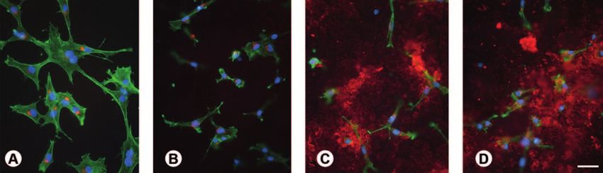
472                                                A.B. Novaes Jr. et al.

surfaces. The main methods that are reported in                   rough ones (21). However, osteoblasts present higher rate
the literature to create implant roughness are acid               of differentiation and matrix mineralization and higher
etching, sandblasting, titanium plasma spraying and               production of growth factors in the presence of rough
hydroxyapatite (HA) coating. A current tendency is the            substrates (22). Also, bone matrix proteins, alkaline
manufacturing of implants with micro and submicro                 phosphatase and osteocalcin, important indicators of
(nano) topography. Furthermore, the biofunctionalization          osteogenic differentiation and bone tissue formation,
of implants surfaces, by adding different substances to           have been shown to express at higher levels on rougher
improve its biological characteristics, has also been             titanium surfaces (23).
recently investigated (8-10).                                             The literature has shown that the surface
        However, the large number of commercially                 topography of titanium can be modified by different
available implant types, varying in surface properties            treatments, in order to obtain a surface with specific
and other features (11) have to be treated with caution.          properties, which have direct influence on the process
Considering that different methods for implant surface            of osseointegration (24,25). It has been suggested that
engineering may lead to different and unique surface              surface roughness in 1-2 μm range are beneficial for
properties that might affect the host-to-implant response,        biomechanical anchorage of dental implants (26).
it seems to be reasonable to test new implant surfaces                    Methods for altering surface texture can be
as new biomaterials (12). The evaluation should ideally           classified as either techniques that add particles on the
follow a hierarchical approach, where in vitro testing            biomaterial, creating a surface with bumps (additive
followed by in vivo animal studies evolves to clinical            mechanisms), and techniques that remove material
trials in humans (13).                                            from the surface, creating pits or pores (subtraction
        This paper reviews the literature on dental implant       mechanisms). Examples of additive processes are: HA
surfaces by assessing in vitro and in vivo studies to show        and calcium phosphate (CaP) coatings, titanium plasma-
the current perspective of implant development.                   sprayed and ion deposition. Examples of subtraction
                                                                  processes are: electro- or mechanical polishing, grit-
IN VITRO STUDIES                                                  blasting, acid-etching, grit-blasting followed by acid-
                                                                  etching and oxidation (27).
        Cell culture models are routinely used to study the               Numerous reports demonstrate that the surface
response of osteoblastic cells in contact with different          roughness of titanium implants affects the rate of
substrates for implantation in bone tissue. Cell cultures         osseointegration through the speed and amount of
focused on the morphological aspect, growth capacity              bone tissue formed at the interface. Comparison of the
and the state of differentiation of the cells on materials        behavior of different cell types on materials shows that
with various chemical, composition and topography (14).           they are influenced by surface roughness (28).
        The literature shows that the biochemistry and                    Roughness gradients of osteoblastic cells increase
topography of biomaterials’ surfaces play a key role              proliferation in close correlation with increasing surface
on success or failure upon placement in a biological              roughness. It was observed doubling of the rate of
environment (15). Wettability, texture, chemical                  osteoblast proliferation on titanium blasted with TiO2
composition and surface topography are properties of the          particles compared to smooth surfaces (29,30). Similar
biomaterials that directly influence their interaction with       results are reported using discs blasted with SiO2 particles
cells (16-18). The interactions of cells and extracellular        (31), and Al2O3 particles (29).
matrix affect directly the cellular processes of adhesion,                Subcultures of rat osteogenic cells grown on
proliferation and differentiation (19). Thus, the surface         grit-blasted and acid-etched surfaces demonstrated
properties of biomaterials are essential to the response          significant formation of bone-like nodules (32). MG63
of cells at biomaterial interface, affecting the growth           osteoblast-like cells (human osteosarcoma cell line)
and quality of newly formed bone tissue (16,17,20).               cultured on rough titanium surfaces exhibited increased
        In the search for new methods, much attention             adhesion and phenotype differentiation, and higher levels
has been focused on topographical characteristics,                of growth factors compared to smooth ones (4).
especially changes in surface roughness. In vitro studies                 There is agreement that the microtopography
have shown that osteoblastic cells attach, spread and             creates an environment favorable for cells and cell-
proliferate more rapidly on smooth surfaces than on               extracellular matrix interactions (33) and increases

Braz Dent J 21(6) 2010
Implant surfaces in osseointegration 473
production of growth factors (34). The microtopography A recent report demonstrated that titanium surfaces
provides increased cell differentiation of osteogenic with nanotopographies obtained from H2SO4/H2O2
cells, resulting in high activity of alkaline phosphatase mixture can promote osteoblast proliferation and inhibit
and osteocalcin synthesis (35). fibroblast growth (40). It was found that titanium surface
Recent studies have shown that the association of modifications on the nanoscale alters the adhesion,
different topographies may be beneficial. Indeed, using spreading, and growth of osteoblastic cells. Furthermore
osteogenic cell culture models, synergistic effects of the extracellular accumulation of osteopontin and
substrates with micron- and submicron-scale resulting bone sialoprotein, two major bone matrix proteins,
in bone tissue formation have been reported (30,32). increased significantly on the titanium substrate with
Titanium surfaces with microtopography and additional nanotopography, indicating that cellular differentiation
submicrotopography have been shown to promote early is accelerated, and the proteins are adsorbed more
development of mineralized matrix, which was observed efficiently on the nanostrutured substrates (41).
occasionally on the surfaces with microtopography and At the present moment, a huge number of
was absent on machined surfaces (36). experimental investigations have clearly demonstrated
The cellular behavior is also influenced by surfaces that the bone response is influenced by the implant
with nanomorphology. The complex interactions cell- surface topography. Furthermore, more recent data,
matrix-substrate and cell signaling events occur at the have suggested that titanium surface modifications
nanoscale (37,38). Different signaling pathways regulate with bioactive molecules enhance and/or accelerates the
adhesion, migration, differentiation and gene expression process of osteoblastic differentiation. As the molecules
in osteoblasts cells (39). Thus, it has been shown that are integrated into the structure of the implant, they are
different nanotopography influence protein adsorption, released gradually, acting as a slow release system of
cell adhesion, cell proliferation and synthesis, and osteogenic agents at the implantation site (42) (Fig. 1).
secretion of extracellular matrix molecules in vitro (37). Among all engineering-based implant surface
Nanoporous surfaces topography tend to favor modifications, the CaP coating have received significant
the proliferation and differentiation processes, acting attention (43). The interest of using this material is
directly on the selective adhesion of osteoblastic cells because of its chemical similarity to natural bone, and
on the surface, which can accelerate the healing process the fact that coatings can be applied along the implant
around implants (27,37). surfaces by different industrial processing methods.
In the same way as microstrutureted surfaces, Biomimetic CaP coatings improve the osteoconductivity
nanotopographies can be created by techniques such as of implants and show promising as slow delivery systems
anodization and oxidation. The production of substrates for growth factors and other bioactive molecules (44).
with nanoporous surfaces appears to strongly influence Other examples of biochemical modifications of
the host response at both cellular and tissue levels (12). biomaterial surfaces are found in the literature, such as
Figure 1. Epifluorescence of osteogenic cell cultures grown on machined (A), microstructured (B), nanostrutured (C), and synthetic
peptide coating (D) surfaces at day 3. Red indicates OPN labeling, green reveals actin cytoskeleton, and blue shows cell nuclei.
Extracellular OPN labeling is abundant and prominent in cultures grown on nanostrutured and synthetic peptide coating surfaces
(C,D). Scale bar: A-D=50 μm.
Braz Dent J 21(6) 2010474 A.B. Novaes Jr. et al.
the use of protein-like collagen, bone morphogenetic spreading of cells and speeding cell adhesion length
proteins and peptides and/or protein domain (27). (48). An in vitro study using bone marrow cells also
The biological effects that surfaces have on cell showed that surface coating with collagen type I showed
attachment are mainly mediated by integrins that bind high ALP activity, collagen synthesis, and mineralized
to sequences/domains arginine-glycine-aspartate (Arg- matrix formation (49).
Gly-Asp or RGD) of proteins (45). These Arg-Gly-Asp or A recent study comparing the development of
RGD are expressed in several bone extracellular matrix the osteoblastic phenotype of human alveolar bone-
proteins. Titanium surfaces modified with peptides and/ derived cells showed that collagen type I-coated titanium
or protein domains with RGD seem to facilitate the surface favors cell growth during the proliferative
mechanisms of adhesion and cell signaling via signal phase and osteoblastic differentiation, as demonstrated
transduction, which have shown positive effects on the by changes in mRNA expression profile during the
differentiation of osteoblasts (46). matrix mineralization phase. This suggests that surface
Since then, researchers have studied coatings modification may affect bone formation (50).
based on peptides containing a sequence of amino acids Some authors have used chemical modification,
to promote cell adhesion to the biomaterial (47). A new such as addition of fluoride to implant surface, to
strategy in the use of bioactive molecules involves the improve the biocompatibility of titanium and promote
addition of extracellular matrix proteins such as collagen. osteogenesis. This process is based on the formation of
Coating titanium surface with collagen enhanced the fluorapatite from interaction of fluoride and HA present
Figure 2. Histologic images evidencing a high level of bone-implant contact achieved with different improvements on implant surfaces.
A= Sandblasted and acid-etched; B= Nanostructured; C= Anodized; D= Biofunctionalized.
Braz Dent J 21(6) 2010Implant surfaces in osseointegration 475
in bone tissue, followed by promotion of osteoblast Finally, the biomechanical tests (torque, push-out, pull-
proliferation and stimulation of alkaline phosphatase out) usually measure the amount of force that a torque
activity. Currently, there are implants treated with needs to fail the bone-implant interface surrounding
fluoride as a biomimetic agent, commercially available different implant surfaces.
for clinical use (51). Considering the several factors that influence
Despite the interesting results in vitro it seems the osseointegration, the evaluation of the largest
that more research is still needed to enhance our possible number of host/implant response parameters
understanding of how this surface modifications actually is desirable for better understanding the bone healing
promotes fast osseointegration. around different implant surfaces (Fig. 2), clarifying
their indications of use and supporting their immediate/
IN VIVO ANIMAL STUDIES early loading. For descriptive purposes, the physical
and chemical surface properties will be separated in
In general, cell culture studies evaluate cell different categories.
morphology, adhesion, migration, proliferation and
differentiation on implant surfaces. However, outcomes Topographic Surface Modifications
on the initial biological behavior of new biomaterials
obtained in vitro cannot be fully correlated to in vivo Machined implant surfaces represent the starting
performance. Cell cultures cannot reproduce the dynamic point of implant surface design. They were used for
environment that involves the in vivo bone/implant decades according to the classical protocols in which
interaction, and their results can only be confirmed in several months were required for osseointegration (52).
animal models and subsequently in clinical trials (13). It has been demonstrated that the modification
The most frequently used animal models for dental on the topographic pattern of surface increases not only
bone-implant interface studies are rabbits and dogs. The the bone-implant contact, but also the biomechanical
rabbit model has some disadvantages when compared interaction of that interface at early implantation periods
to larger animals, such as the size when a number of (1). Rough surfaces have found widespread use in oral
control and experimental implants are recommended per implantology and replaced implants with machined
animal. Additionally the bone structure of the tibia and surfaces to a great extent in clinical applications (46).
femur of rabbits (e.g.; the amount of trabecular bone), Various methods have been developed in order to
are significantly different when compared to human. create a rough surface and improve the osseointegration
Otherwise the canine intraoral environment provides of titanium dental implants. These methods use titanium
a bone microstructure with a trabecular/cortical ratio plasma-spraying, blasting with ceramic particles, acid-
similar to that found in the human mandible, in addition etching and anodization.
to similar saliva and microflora.
Irrespective to the different animal models (rat, Titanium plasma-spraying
rabbits, sheep, dogs, pigs or nonhuman primates) or
surgical sites, valuable information can be retrieved from This method consists in injecting titanium
properly designed animal studies. Static and dynamic powders into a plasma torch at high temperature. The
histomorphometric parameters plus biomechanical titanium particles are projected onto the surface of
testing are recommended as measurable indicators of the the implants where they condense and fuse together,
host/implant response where different surface designs resulting in a titanium plasma sprayed (TPS) coating with
are compared. Bone-to-implant contact (BIC), which an average roughness of around 7 μm. This procedure
that is the most often evaluated parameter in in vivo increases substantially the surface area of the implants.
studies, together with bone density and amount and type Al-Nawas et al. (53) evaluated different types
of cellular content, are examples of static parameters. of macro and microstructure implant surfaces in a dog
Differently, mineral apposition rate and fluorescence model. After a healing period of 8 weeks and a loading
analysis temporally evaluates bone modeling/remodeling period of 3 months, machined surfaces were compared
processes. As dynamic measurements, they may provide to the TPS counterparts, used as a rough control, and
valuable information about the healing around different also with blasted/acid-etched surface. The evaluation
implant surfaces, but these parameters are rarely used. of the BIC areas revealed the benefit of rough surfaces
Braz Dent J 21(6) 2010476 A.B. Novaes Jr. et al.
relative to machined ones. However, the intra-individual of silica (sand), alumina, titanium oxide or CaP for
difference between the TPS and the blasted/acid-etched example. The most commonly used acid-etching agents
counterparts showed no significant difference. are hydrofluoric, nitric, sulfuric acids and combinations.
In a different animal model, Klokkevold et al. An example of this group of surfaces was investigated by
(54) compared the torque resistance to remove screw- Sammons et al. (55), evidencing a Ra value of 2.75 μm
shaped titanium implants having a dual acid-etched and irregular micropores with approximately 3-5 μm in
surface (DAE) with implants having either a machined diameter and 2-3 μm in depth. Even smaller micropores
surface, or a TPS surface that exhibited a significantly are located within these micropores.
more complex surface topography. After implantation, Novaes et al. (56) compared grit-blasted/acid-
the groups of 6 rabbits were sacrificed following etched implants to titanium plasma spray implants
1-, 2- and 3-month healing periods. Implants were immediately installed into periodontally infected sites.
removed by reverse torque rotation with a digital torque- The histomorphometric analyses showed percentages
measuring device. Three implants with machined surface of bone to implant contact of 52.7% and 42.7% for
preparation failed to achieve endosseous integration. All grit-blasted/acid-etched implants and titanium plasma
other implants were anchored by bone. Mean torque spray implants, respectively. The bone density analysis
values for machined, DAE and TPS implants at 1, 2 revealed percentages of 66.6% and 58.8% in the adjacent
and 3 months were 6.00 ± 0.64 N/cm, 9.07 ± 0.67 N/cm areas of grit-blasted/acid-etched implants and titanium
and 6.73 ± 0.95 N/cm; 21.86 ± 1.37 N/cm, 27.63 ± 3.41 plasma spray implants, respectively. These differences
N/cm and 27.40 ± 3.89 N/cm; and 27.48 ± 1.61 N/cm, between the groups were not statistically significant,
44.28 ± 4.53 N/cm and 59.23 ± 3.88 N/cm, respectively. but indicated a slightly better performance of the grit-
Clearly, the stability of DAE implants at the earliest time blasted/acid-etched surfaces when compared to the
point was comparable to that of TPS implants, while that titanium plasma spray surface, even in a challenging
of the machined implants was an order of magnitude healing situation.
lower. The TPS implants increased resistance to reverse Recently, it was seen that changing the
torque removal over the 3-month period. These results sterilization and storage method of an original sand-
indicate that dual acid etching of titanium enhances early blasted/acid-etched surface is another possible way to
endosseous integration to a level that is comparable to modify implant surface. In other words, the new surface
that achieved by the topographically more complex TPS is rinsed under nitrogen protection to prevent exposure
surfaces. Furthermore, this study confirmed an enhanced to air and then stored in a sealed tube containing an
bone anchorage to rough surface implants as compared isotonic saline solution (4). This treatment obtains the
to machined implants. hydroxylation of titanium oxides without changing the
TPS processing is one of the methods that further surface topography, this procedure improved the surface
increase the surface roughness profile and consequently wettability of the new surface when compared to the
the surface area. Such characteristics recommend its original one in a statistically significant level (57).
use in regions with low bone density (12). However, According to animal experiments, both
it has to be considered that the increase in surface area biomechanical and histomorphometric evaluations
that represents an effective increase in osseointegration showed better results for the modified surface compared
area provides spaces greater than 50 μm that facilitates to the original sand-blasted/acid-etched surface at early
the migration of pathogens when the implant surface is periods. In the miniature pig model, Buser et al. (58)
exposed to the oral fluids. investigated the interfacial stiffness values, which were
calculated from the torque-rotation curve, and found on
Blasting with ceramic particles/acid-etching average 9-14% higher values for the modified surface
compared to the original sand-blasted/ acid-etched
Surface acid-etching and grit-blasting/acid- surface. This difference was statistically significant
etching are very diffuse methods to obtain rough implant and they concluded that the modified provided better
surfaces. A great part of the commercially available grit- bone anchorage than the original surface. Moreover,
blasted implant surfaces are subsequently acid-etched. Schwarz et al. (59) evaluated the bone regeneration in
Generally, the grit-blasting procedure is dehiscence-type defects with these implants in beagle
performed by propulsion of particles of different sizes dogs. After 2 weeks of healing, the modified group
Braz Dent J 21(6) 2010Implant surfaces in osseointegration 477
achieved 74% of BIC, while the original group achieved widely used commercial CaP implant surface coating.
56%, and this difference was statistically significant. CaP ceramics are considered to have bioactive properties,
However, after 12 weeks of healing, BIC was 84% for which involves the strong interaction between materials
the modified group and 76% for the original group, and surrounding bone by means of a chemical bonding
without statistically significant and this difference. Thus, (2). Substrates containing CaP coating is expected to
it could be concluded that the modified implant surface render a faster biological fixation between implant
promoted enhanced bone apposition during the early and bone tissue when compared to those without CaP
stages of bone formation. In accordance to this, Bornstein coatings (65-67). However, the thick and non-uniform
et al. (60) comparing the same implant surfaces in a dog coating performed by the plasma-spraying method,
model, showed significantly higher percentage of bone in which HA ceramic particles are injected into a
in contact with the modified surface when compared plasma torch at high temperature and projected on
to the original sand-blasted/acid-etched surface after 2 to the surface of the titanium, have also been related
weeks, but no statistically significant difference after to some disadvantages. The possible delamination
4 weeks of implantation. These results suggest that a of the coating from the implant surface is generally
chemical modification on a microstructured implant highlighted, making possible the clinical failure of the
surface may interfere in the biomechanical and bone implant (68). Additionally, the transmucosal zone of
apposition properties at early phases after implantation. plasma sprayed HA implants represents a challenge
(43) in terms of periimplantitis infection. Based on
Electrochemical anodization these reasons, the clinical use of the plasma sprayed HA
implants decreased, but the osteoconductive property of
Another method that has been shown to increase this bioactive ceramic coating remains as a factor that
surface microtexture and change surface chemistry may contribute to additional bone attachment in areas
is electrochemical anodization. The combination of of poor quality or quantity of bone.
potentiostatic or galvanostatic anodization of titanium in As a new trend, the changes of surface roughness
strong acids at high current density or potential, results in at the nanoscale level seem to strongly influence the
thickening of the titanium oxide layer. Anodized surfaces host response at both cellular and tissue levels. In this
interfere positively in bone response with higher values context, it should be mentioned that some strategies
for biomechanical and histomorphometric tests when have been developed to improve the plasma sprayed
compared to machined surfaces (61,62). HA coating process. Thus methods such as sol-gel
Burgos et al. (63) selected a commercially implant deposition, electrophoretic deposition and discrete
surface manufactured by anodic oxidation to compare crystalline deposition were developed in order to obtain
to turned surfaces in a rabbit model. BIC values were significant thinner coating thicknesses when compared
20% (after 7 days), 23% (after 14 days), and 46% (after to the plasma sprayed HA technique.
28 days) around the oxidized surfaces and 15% (after It is already available for clinical use the result
7 days), 11% (after 14 days), and 26% (after 28 days) of a CaP nanoparticle modification of a minimally
around the machined surfaces. It was concluded that the rough titanium implant surface. It has been created by
moderately rough oxidized surfaces follows a different the combination of the sol-gel and discrete crystalline
pattern of osseointegration. deposition (DCD) of CaP. Mendes et al. (69) suggested
Differently, Huang et al. (64) evaluated the that the nano-feature size of the tightly adherent adsorbed
oxidized implant surfaces installed in the posterior CaP/DCD crystal is of 20-100 nm.
maxilla of monkeys. After 16 weeks, the mean BIC In the rat model, Mendes et al. (70) have shown
was of 74%. The authors suggested that this oxidized significantly greater average disruption forces with
surface detains a considerable osteoconductive potential DCD samples when compared to dual acid-etched
promoting a high level of implant osseointegration in samples. The authors concluded that an increase in
type IV bone in the posterior maxilla. the complexity of the surface topography can render a
bone-bonding ability. Recently, the same group, again
CaP coatings in the rat model (71), demonstrated significant increase
in osteoconduction as a function of the enhanced surface
Up to now, plasma-spraying remains as the most nanotopography obtained by the CAP nanocrystals in
Braz Dent J 21(6) 2010478 A.B. Novaes Jr. et al.
the rat model. difference was found between collagen coating and RGD
In summary, highly roughened implants, such as coatings. After 3 months, BIC was significantly higher in
TPS or grit-blasted implants, have been shown to favor all implants with organic coating than in implants with
mechanical anchorage and primary fixation to bone, machined surfaces. Periimplant BVD was significantly
while topographies in the nanometer level focus on the increased in all coated implants in comparison to
enhancement of the host response by means of promoting machined surfaces. The authors concluded that organic
protein adsorption and osteoblastic cell adhesion during coating of machined screw implant surfaces providing
the early stages of healing in the periimplant region. binding sites for integrin receptors can enhance bone
implant contact and periimplant bone formation. In
Biomimetic Surface Modifications addition, Germanier et al. (74) compared RGD peptide
polymer modified implant surfaces with sandblasted and
Biomolecules coated onto titanium surface acid-etched implant surfaces placed in the maxillae of
miniature pigs, and confirmed that the functionalization
A common theme in the engineering of cell and may promote enhanced bone apposition during the early
tissue behavior on device surfaces is to modify the stages of bone regeneration.
material to selectively interact with a specific cell type However,it should be mentioned that the success
through biomolecular recognition events. Typically, of such functionalization seems to be strongly dependent
peptides containing the cell-binding domains found on type, delivery and concentration of the coating. For
in the extracellular matrix proteins are immobilized example, some studies showed confusing results when
on the material to promote cell adhesion via ligand- evaluating implant surfaces modified by BMP coatings.
receptor interaction (72,73). Integrins are an example BMPs are a class of growth factors that promote
of cell adhesion receptors that bind to specific amino bone formation, but they also stimulate the action of
acid sequences, such as the RGD that is found in type I osteoclasts. It seems that the dose of the drug is critical
collagen, fibronectin, osteopontin and bone sialoprotein. for the final result.
Apart from cell attachment, extracellular matrix may Liu et al. (75) evaluated the effects of BMP-2
also act on cellular migration and proliferation events. and its mode of delivery on the osteoconductivity of
The concept of functionalizing the implant dental implants with either a naked titanium surface
surfaces with native or synthetic molecules based on or a calcium-phosphate-coated one in the maxillae
peptides, proteins and growth factors emerged from the of miniature pigs. After 3 weeks, the volume of bone
hypothesis that the ability of imitating the environment deposited within the osteoconductive space (periimplant)
of bone, which is composed of an organic matrix (mainly was highest for coated and uncoated implants bearing
collagenous proteins) and inorganic CaP, could enhance no BMP-2, while the lowest value was achieved with
the implant surface performance, encouraging the initial coated implants bearing only adsorbed BMP-2. It was
biologic response. concluded that the osteoconductivity of functionalized
Animal studies support the in vivo osteoconductive implant surfaces depends on the mode of BMP-2
potential of the RGD peptide sequence as a potential delivery, being drastically impaired when BMP-2 was
method of functionalizing titanium implant surfaces. In present as a superficially adsorbed depot upon CaP
the dog model, Schliephake et al. (46) compared implants coated or uncoated surfaces.
with machined titanium surface, coated with collagen In a different dog model, Wikesjö et al. (76)
I, coated with collagen I and low RGD concentrations studied the ability of recombinant human BMP-2
(100 μmol/mL), and coated with collagen I and high (rhBMP-2) coated onto a titanium porous oxide
RGD concentrations (1000 μmol/mL). The BIC and implant surface to stimulate local bone formation,
volume density of the newly formed periimplant bone including osseointegration and vertical augmentation
(BVD) was assessed histomorphometrically after 1 of the alveolar ridge. Thus critical-size, 5 mm, supra-
and 3 months. After 1 month, BIC was significantly alveolar, periimplant defects were created and implants
enhanced only in the group of implants coated with the coated with rhBMP-2 at 0.75 or 1.5 or 3.0 mg/mL or
higher concentration of RGD peptides. Volume density uncoated control were installed and compared. The
of the newly formed periimplant bone was significantly histologic evaluation showed newly formed bone with
higher in all implants with organic coating. No significant characteristics of the adjoining resident type II bone
Braz Dent J 21(6) 2010Implant surfaces in osseointegration 479
including cortex formation for sites receiving implants contexto, as modificações nas superfícies de implante ganharam
coated with rhBMP-2 at 0.75 or 1.5 mg/mL. Sites um lugar importante e decisivo na pesquisa em Implantodontia
nos últimos anos. Sendo o tópico mais estudado, colaboraram
receiving implants coated with rhBMP-2 at 3.0 mg/mL para o melhoramento de modalidades de tratamento dental, assim
exhibited more immature trabecular bone formation, como para a expansão de uso dos implantes dentais. Hoje, um
seroma formation and periimplant bone remodelling grande número de diferentes implantes com uma grande variedade
resulting in undesirable implant displacement. Control de propriedades de superfícies, entre outras características, está
comercialmente disponível e isto deve ser tratado com cuidado.
implants exhibited minimal, if any, bone formation. In Apesar das modificações nas superfícies terem melhorado
summary, rhBMP-2 coating onto titanium porous oxide a osseointegração em tempos precoces de implantação, por
implant surfaces induced clinically relevant local bone exemplo, o clínico deve procurar evidências científicas antes de
formation including vertical augmentation of the alveolar selecionar um implante dental para uso específico. Este artigo
fará uma revisão da literatura sobre superfícies de implantes
ridge and osseointegration, but higher concentrations/ osseointegráveis, analisando estudos in vitro e in vivo, a fim de
doses were associated with negative effects. mostrar uma perspectiva atual do desenvolvimento dos implantes.
Finally, non-BMP growth factors have also been Esta abordagem englobará os resultados obtidos com micro e nano
tested as potential agents to improve the osseointegration topografias, em termos quantitativos e qualitativos, avaliando
a interface osso-implante. Além disso, discutirá também as
parameters. Park et al. (77) evaluated the osseointegration perspectivas da incorporação de substâncias biomiméticas (como
of anodized titanium implants coated with fibroblast peptídeos e proteínas morfogenéticas) à superfície dos implantes
growth factor-fibronectin (FGF-FN) fusion protein that e seus efeitos na modulação da neoformação óssea periimplantar.
were placed in rabbit tibiae. The removal torque values
as well as the percentages of BIC of the test group were REFERENCES
better than those found for the implants that were not
biofunctionalized. 1. Albrektsson T, Wennerberg A. Oral implant surfaces: Part
1- review focusing on topographic and chemical properties of
different surfaces and in vivo responses to them. Int J Prosthodont
CONCLUDING REMARKS 2004;17:536-543.
2. Junker R, Dimakis A, Thoneick M, Jansen JA. Effects of implant
surface coatings and composition on bone integration: a systematic
There are a huge number of types of implant review. Clin Oral Implants Res 2009;20:185-206.
surfaces in the market, from different implant 3. Buser D, Broggini N, Wieland M, Schenk RK, Denzer AJ, Cochran
manufacturers, all of them claiming to have better clinical DL, et al.. Enhanced bone apposition to a chemically modified
SLA titanium surface. J Dent Res 2004;83:529-533.
results. It is important that the clinician selects for use in 4. Zhao G, Schwartz Z, Wieland M, Rupp F, Geis-Gerstorfer J,
their patients the surfaces that have shown good results Cochran DL, et al.. High surface energy enhances cell response
in the scientific literature. to titanium substrate microstructure. J Biomed Mater Res A
2005;74:49-58.
The majority of currently available in vitro and in
5. Wong M, Eulenberger J, Schenk R, Hunziker E. Effect of surface
vivo studies seem to indicate that implant surfaces with topology on the osseointegration of implant materials in trabecular
micro and submicro (nano) topography bring forward bone. J Biomed Mater Res 1995;29:1567-1575.
benefits to the process of interaction between bone cells 6. Wennerberg A, Albrektsson T. On implant surfaces: a review of
current knowledge and opinions. Int J Oral Maxillofac Implants
and implant surfaces, accelerating and increasing the 2010;25:63-74.
quality of BIC. 7. Wennerberg A, Albrektsson T. Effects of titanium surface
Finally, based on the state of the art of implant topography on bone integration: a systematic review. Clin Oral
Implants Res 2009;20:172-184.
development, it is possible to predict that, within some 8. Beutner R, Michael J, Schwenzer B, Scharnweber D. Biological
time, implant surfaces coated with substances with nano-functionalization of titanium-based biomaterial surfaces: a
biomimetic capacity will be available for clinical use. flexible toolbox. J R Soc Interface 2010;7:S93-S105.
9. Barros RR, Novaes AB Jr, Papalexiou V, Souza SL, Taba M Jr,
This process of biofuncionalization of implant aims at Palioto DB, et al.. Effect of biofunctionalized implant surface on
modulating new bone formation around implants, and osseointegration: a histomorphometric study in dogs. Braz Dent J
it is the next step in implant development. 2009;20:91-98.
10. Lutz R, Srour S, Nonhoff J, Weisel T, Damien CJ, Schlegel KA.
Biofunctionalization of titanium implants with a biomimetic active
RESUMO peptide (P-15) promotes early osseointegration. Clin Oral Implants
Res 2010;21:726-734.
A fixação biológica entre as superfícies de implante e os ossos 11. Binon PP. Implants and components: entering the new millennium.
maxilares deve ser considerada como um pré-requisito para o Int J Oral Maxillofac Implants 2000;15:76-94.
sucesso em longo prazo de próteses implanto-suportadas. Neste 12. Coelho PG, Granjeiro JM, Romanos GE, Suzuki M, Silva NR,
Cardaropoli G, et al.. Basic research methods and current trends
Braz Dent J 21(6) 2010480 A.B. Novaes Jr. et al.
of dental implant surfaces. J Biomed Mater Res B Appl Biomater interaction of topographic features in the production of bone-
2009;88:579-596. like nodules on Ti surfaces by rat osteoblasts. Biomaterials
13. Lemons JE. Biomaterials, biomechanics, tissue healing, 2005;26:1119-1130.
and immediate-function dental implants. J Oral Implantol 33. Matsuzaka K, Walboomers XF, Yoshinari M, Inoue T, Jansen JA.
2004;30:318-324. The attachment and growth behavior of osteoblast-like cells on
14. Anselme K. Osteoblast adhesio on biomaterials. Biomaterials microtextured surfaces. Biomaterials 2003;24:2711-2719.
2000;21:667-681. 34. Boyan BD, Lossdorfer S, Wang L, Zhao G, Lohmann CH, Cochran
15. Kasemo B. Biological surface science. Surf Sci 2002;500:656-677. DL, et al.. Osteoblasts generate an osteogenic microenvironment
16. Deligianni DD, Katsala N, Ladas S, Sotiropoulou D, Amedee J, when grown on surfaces with rough microtopographies. Eur Cell
Missirlis YF. Effect of surface roughness of the titanium alloy Ti- Mater 2003;6:22-27.
6Al-4V on human broadcast marrow cell response and on protein 35. Schwartz Z, Lohmann CH, Oefinger J, Bonewald LF, Dean
adsorption. Biomaterials 2001;22:1241-1251. DD, Boyan BD. Implant surface characteristics modulate
17. Lamers E, Walboomers XF, Domanski M, te Riet J, van Delft FC, differentiation behavior of cells in the osteoblastic lineage. Adv
Luttge R, et al.. The influence of nanoscale grooved substrates Dent Res 1999;13:38-48.
on osteoblast behavior and extracellular matrix deposition. 36. Schwartz Fo HO, Novaes AB Jr, de Castro LMS, Rosa AL, de
Biomaterials 2010;31:3307-3316. Oliveira PT. In vitro osteogenesis on a microstructured titanium
18. Mendonça G, Mendonça DB, Aragão FJ, Cooper LF. The surface with additional submicron-scale topography. Clin Oral
combination of micron and nanotopography by H(2)SO(4)/H(2) Implants Res 2007;18:333-344.
O(2) treatment and its effects on osteoblast-specific gene 37. De Oliveira PT, Nanci A. Nanotexturing of titanium-based surfaces
expression of hMSCs. J Biomed Mater Res A 2010;94:169-179. upregulates expression of bone sialoprotein and osteopontin by
19. Lincks J, Boyan BD, Blanchard CR, Lohmann CH, Liu Y, Cochran cultured osteogenic cells. Biomaterials 2004;25:403-413.
DL, et al.. Response of MG63 osteoblast-like cells to titanium and 38. De Oliveira PT, Zalzal SF, Beloti MM, Rosa AL, Nanci A.
titanium alloy is dependent on surface roughness and composition. Enhancement of in vitro osteogenesis on titanium by chemically
Biomaterials 1998;19:2219-2232. produced nanotopography. J Biomed Mater Res A 2007;80:554-
20. von der Mark K, Park J, Bauer S, Schmuki P. Nanoscale 564.
engineering of biomimetic surfaces: cues from the extracellular 39. Schneider GB, Zaharias R, Stanford C. Osteoblast integrin
matrix. Cell Tissue Res 2010;339:131-153. adhesion and signaling regulate mineralization. J Dent Res
21. Anselme K, Bigerelle M. Topography effects of pure titanium 2001;80:1540-1544.
substrates on human osteoblast long-term adhesion. Acta Biomater 40. Variola F, Yi JH, Richert L, Wuest JD, Rosei F, Nanci A. Tailoring
2005;1:211-222. the surface properties of Ti6Al4V by controlled chemical
22. Cooper LF, Masuda T, Yliheikkila PK, Felton DA. Generalizations oxidation. Biomaterials 2008;29:1285-1298.
regarding the process and phenomenon of osseointegration. Part 41. Vetrone F, Variola F, Tambasco de Oliveira P, Zalzal SF, Yi JH,
II. In vitro studies. Int J Oral Maxillofac Impl 1998;13:163-174. Sam J, et al.. Nanoscale oxidative patterning of metallic surfaces
23. Davies JE. Mechanisms of endosseous integration. Int J to modulate cell activity and fate. Nano Lett 2009;9:659-665.
Prosthodont 1998;11:391-401. 42. Liu Y, de Groot K, Hunziker EB. Osteoinductive implants: the
24. Lim YJ, Oshida Y, Andres CJ, Barco MT. Surface characterizations mise-en-scène for drug-bearing biomimetic coatings. Ann Biomed
of variously treated titanium materials. Int J Oral Maxillof Imp Eng 2004;32:398-406.
2001;16:333-342. 43. Yang Y, Kim KH, Ong JL. A review on calcium phosphate coatings
25. Dohan Ehrenfest DM, Coelho PG, Kang BS, Sul YT, Albrektsson produced using a sputtering process-an alternative to plasma
T. Classification of osseointegrated implant surfaces: materials, spraying. Biomaterials 2005;26:327-337.
chemistry and topography. Trends Biotechnol 2010;28:198-206. 44. Barrère F, Layrolle P, van Blitterswijk CA, de Groot K. Biomimetic
26. Wennerberg A, Albrektsson T. Suggested guidelines for the calcium phosphate coatings on Ti6AI4V: a crystal growth study of
topographic evaluation of implant surfaces. Int J Oral Maxillofac octacalcium phosphate and inhibition by Mg2+ and HCO3-. Bone
Implants 2000;15:331-344. 1999;25:107S-111S.
27. Le Guéhennec L, Soueidan A, Layrolle P, Amouriq Y. Surface 45. Tosatti S, Schwartz Z, Campbell C, Cochran DL, Vandevondele
treatments of titanium dental implants for rapid osseointegration. S, Hubbel JA, et al.. RGD-containing peptide GCRGYGRGDSPG
Dent Mater 2007;23:844-854. reduces enhancement of osteoblast differentiation by poly(L-
28. Healy KE, Thomas CH, Rezania A, Kim JE, McKeown PJ, Lom lysine)-graft-poly(ethylene glycol)-coated titanium surfaces. J
B, et al.. Kinetics of bone cell organization and mineralization Biomed Mater Res A 2004;68:458-472.
on materials with patterned surface chemistry. Biomaterials 46. Schliephake H, Scharnweber D, Dard M, Sewing A, Aref A,
1996;17:195-208. Roessler S. Functionalization of dental implant surfaces using
29. Rosa AL, Beloti MM. Effect of cpTi surface roughness on human adhesion molecules. J Biomed Mater Res B Appl Biomater
bone marrow cell attachment, proliferation, and differentiation. 2005;73:88-96.
Braz Dent J 2003;14:16-21. 47. Puleo DA, Nanci A. Understanding and controlling the bone-
30. Zinger O, Anselme K, Denzer A, Habersetzer P, Wieland M, implant interface. Biomaterials 1999;20:2311-2321.
Jeanfils J, et al.. Time-dependent morphology and adhesion of 48. Geissler U, Hempel U, Wolf C, Scharnweber D, Worch H, Wenzel
osteoblastic cells on titanium model surfaces featuring scale- KW. Collagen type I coating of Ti6Al4V promotes adhesion of
resolved topography. Biomaterials 2004;25:2695-2711. osteoblasts. J Biomed Mater Res 2000;51:752-760.
31. Mustafa K, Wennerberg A, Wroblewski J, Hultenby K, Lopez BS, 49. Mizuno M, Imai T, Fujisawa R, Tani H, Kuboki Y. Bone
Arvidson K. Determining optimal surface roughness of TiO(2) sialoprotein (BSP) is a crucial factor for the expression of
blasted titanium implant material for attachment, proliferation and osteoblastic phenotypes of bone marrow cells cultured on type I
differentiation of cells derived from human mandibular alveolar collagen matrix. Calcif Tissue Int 2000;66:388-396.
bone. Clin Oral Implants Res 2001;12:515-525. 50. De Assis AF, Beloti MM, Crippa GE, de Oliveira PT, Morra M,
32. Wieland M, Textor M, Chehroudi B, Brunette DM. Synergistic Rosa AL. Development of the osteoblastic phenotype in human
Braz Dent J 21(6) 2010Implant surfaces in osseointegration 481
alveolar bone-derived cells grown on a collagen type I-coated 64. Huang YH, Xiropaidis AV, Sorensen RG, Albandar JM, Hall J,
titanium surface. Clin Oral Implants Res 2009;20:240-246. Wikesjö UME. Bone formation of titanium porous oxide (TiUnite)
51. Cooper LF, Zhou Y, Takebe J, Guo J, Abron A, Holmén A, et al.. oral implants in type IV bone. Clin Oral Impl Res 2005;16:105-
Fluoride modification effects on osteoblast behavior and bone 111.
formation at TiO2 grit-blasted c.p. titanium endosseous implants. 65. Barrère F, van der Valk CM, Meijer G, Dalmeijer RA, de Groot K,
Biomaterials 2006;27:926-936. Layrolle P. Osteointegration of biomimetic apatite coating applied
52. Albrektsson T, Sennerby L. State of the art in oral implants. J Clin onto dense and porous metal implants in femurs of goats. J Biomed
Periodontol 1991;18:474-481. Mater Res 2003;67:655-665.
53. Al-Nawas B, Groetz KA, Goetz H, Duschner H, Wagner W. 66. Quaranta A, Iezzi G, Scarano A, Coelho PG, Vozza I, Marincola
Comparative histomorphometry and resonance frequency analysis M, et al.. A histomorphometric study of nanothickness and plasma-
of implants with moderately rough surfaces in a loaded animal sprayed calcium-phosphorous-coated implant surfaces in rabbit
model. Clin Oral Implants Res 2008;19:1-8. bone. J Periodontol 2010;81:556-561.
54. Klokkevold PR, Johnson P, Dadgostari S, Caputo A, Davies JE, 67. Yang GL, He FM, Hu JA, Wang XX, Zhao SF. Biomechanical
Nishimura RD. Early endosseous integration enhanced by dual comparison of biomimetically and electrochemically deposited
acid etching of titanium: a torque removal study in the rabbit. Clin hydroxyapatite-coated porous titanium implants. J Oral Maxillofac
Oral Implants Res 2001;12:350-357. Surg 2010;68:420-427.
55. Sammons RL, Lumbikanonda N, Gross M, Cantzler P. Comparison 68. Lee J, Rouhfar L, Beirne O. Survival of hydroxypatite-coated
of osteoblast spreading on microstructured dental implant surfaces implants: a meta-analytic review. J Oral Maxillofac Surg
and cell behavior in an explants model of osseointegration. A 2000;58:1372-1379.
scanning electron microscopy study. Clin Oral Implants Res 69. Mendes VC, Moineddin R, Davies JE. Discrete calcium phosphate
2005;16:657-666. nanocrystalline deposition enhances osteoconduction on titanium-
56. Novaes AB Jr, Papalexiou V, Grisi MF, Souza SS, Taba M based implant surfaces. J Biomed Mater Res A 2009;90:577-585.
Jr, Kajiwara JK. Influence of implant microstructure on the 70. Mendes VC, Moineddin R, Davies JE. The effect of discrete
osseointegration of immediate implants placed in periodontally calcium phosphate nanocrystals on bone-bonding to titanium
infected sites. A histomorphometric study in dogs. Clin Oral surfaces. Biomaterials 2007;28:4748-4755.
Implants Res 2004;15:34-43. 71. Mendes VC, Moineddin R, Davies JE. Discrete calcium phosphate
57. Rupp F, Scheideler L, Olshanska N, de Wild M, Wieland M, Geis- nanocrystalline deposition enhances osteoconduction on titanium-
Gerstorfer J. Enhancing surface free energy and hydrophilicity based implant surfaces. J Biomed Mater Res A 2009;90:577-585.
through chemical modification of microstructure titanium implant 72. Rezania A, Healy KE. Biomimetic peptide surfaces that regulate
surfaces. J Biomed Mater Res A 2006;76:323-334. adhesion, spreading, cytoskeletal organization, and mineralization
58. Buser D, Nydegger T, Oxland T, Cochran DL, Schenk RK, Hirt of the matrix deposited by osteoblast-like cells. Biotechnol Prog
HP, et al.. Interface shear strength of titanium implants with a 1999;15:19-32.
sandblasted and acid-etched surface: a biomechanical study in the 73. Rezania A, Thomas CH, Branger AB, Waters CM, Healy KE. The
maxilla of miniature pigs. J Biomed Mater Res 1999;45:75-83. detachment strength and morphology of bone cells contacting
59. Schwarz F, Herten M, Sager M, Wieland M, Dard M, Becker materials modified with a peptide sequence found within bone
J. Bone regeneration in dehiscence-type defects at chemically sialoprotein. J Biomed Mater Res 1997;37:9-19.
modified (SLAactive®) and conventional SLA titanium implants: 74. Germanier Y, Tosatti S, Broggini N, Textor M, Buser D. Enhanced
a pilot study in dogs. J Clin Periodontol 2007;34:78-86. bone apposition around biofunctionalized sandblasted and acid-
60. Bornstein MM, Valderrama P, Jones AA, Wilson TG, Seibl R, etched titanium implant surfaces. A histomorphometric study in
Cochran DL. Bone apposition around two different sandblasted miniature pigs. Clin Oral Impl Res 2006;17:251-257.
and acid-etched titanium implant surfaces: a histomorphometric 75. Liu Y, Enggist L, Kuffer AF, Buser D, Hunziker EB. The influence
study in canine mandibles. Clin Oral Impl Res 2008;19:233-241. of BMP-2 and its mode of delivery on the osteoconductivity
61. Sul YT, Johansson CB, Jeong Y, Wennerberg A, Albrektsson T. of implant surfaces during the early phase of osseointegration.
Resonance frequency and remova l torque analysis of implants Biomaterials 2007;28:2677-2686.
with turned and anodized surface oxides. Clin Oral Implants Res 76. Wikesjö UM, Qahash M, Polimeni G, Susin C, Shanaman RH,
2002;13:252-259. Rohrer MD, et al.. Alveolar ridge augmentation using implants
62. Rocci A, Martignoni M, Gottlow J. Immediate loading of coated with recombinant human bone morphogenetic protein-2:
Branemark System TiUnite and machined-surface implants in the histologic observations. J Clin Periodontol 2008;35:1001-1010.
posterior mandible: a randomized open-ended clinical trial. Clin 77. Park JM, Koak JY, Jang JH, Han CH, Kim SK, Heo SJ.
Implant Dent Relat Res 2003;5:557-563. Osseointegration of anodized titanium implants coated with
63. Burgos PM, Rasmusson L, Meirelles L, Senerby L. Early bone fibroblast growth factor-fibronectin (FGF-FN) fusion protein. Int
tissue responses to turned and oxidized implants in a rabbit tibia. J Oral Maxillofac Implants 2006;21:859-866.
Clin Impl Dent Relat Res 2008;10:181-190.
Accepted November 16, 2010
Braz Dent J 21(6) 2010You can also read