A subset of liver resident natural killer cells is expanded in hepatitis C infected patients with better liver function - Nature
←
→
Page content transcription
If your browser does not render page correctly, please read the page content below
www.nature.com/scientificreports
OPEN A subset of liver resident natural
killer cells is expanded in hepatitis
C‑infected patients with better liver
function
Erin H. Doyle1, Costica Aloman2, Ahmed El‑Shamy1, Francis Eng1, Adeeb Rahman3,
Arielle L. Klepper1, Brandy Haydel4, Sander S. Florman4, M. Isabel Fiel5, Thomas Schiano4 &
Andrea D. Branch1*
Viral hepatitis leads to immune-mediated liver injury. The rate of disease progression varies between
individuals. We aimed to phenotype immune cells associated with preservation of normal liver
function during hepatitis C virus (HCV) infection. Clinical data and specimens were obtained from
19 HCV-infected patients undergoing liver transplantation. Liver and peripheral blood mononuclear
cells were isolated and eight subsets of innate immune cells were delineated by multiparameter
flow cytometry. Cytokine assays and microarrays were performed. Intrahepatic CD56Bright/CD16-
natural killer (NK) cells comprised the only subset correlating with better liver function, i.e., lower
bilirubin (p = 0.0002) and lower model for end stage of liver disease scores (p = 0.03). The signature
of liver NK cells from HCV-infected patients included genes expressed by NK cells in normal liver
and by decidual NK cells. Portal vein blood had a higher concentration of interleukin (IL)-10 than
peripheral blood (p = 0.03). LMCs were less responsive to toll-like receptor (TLR) stimulation than
PBMCs, with fewer pro-inflammatory gene-expression pathways up-regulated after in vitro exposure
to lipopolysaccharide and a TLR-7/8 agonist. Hepatic CD56Bright/CD16- NK cells may be critical for
maintaining liver homeostasis. Portal vein IL-10 may prime inhibitory pathways, attenuating TLR
signaling and reducing responsiveness to pro-inflammatory stimuli.
Abbreviations
ALT Alanine aminotransferase
CD Cluster differentiation
GGT Gamma-glutamyl transferase
HBV Hepatitis B virus
HCV Hepatitis C virus
IFN Interferon
IL Interleukin
INR International normalized ratio
IQR Interquartile range
LMC Liver mononuclear cell
LPS Lipopolysaccharide
MELD Model of end-stage liver disease
NK Natural killer
PAMPS Pathogen associated molecular patterns
PBMC Peripheral blood mononuclear cell
TLR Toll-like receptor
TNFα Tumor necrosis factor alpha
1
Division of Liver Diseases, Icahn School of Medicine at Mount Sinai School, 1425 Madison Ave., Icahn 11‑23, New
York, NY 10029, USA. 2Rush University Medical Center, Chicago, IL, USA. 3Human Immune Monitoring Core, Icahn
School of Medicine at Mount Sinai, New York, NY, USA. 4Recanati Miller Transplantation Institute, The Mount Sinai
Hospital, New York, NY, USA. 5Department of Pathology, The Mount Sinai Hospital, New York, NY, USA. *email:
Andrea.Branch@mssm.edu
Scientific Reports | (2021) 11:1551 | https://doi.org/10.1038/s41598-020-80819-8 1
Vol.:(0123456789)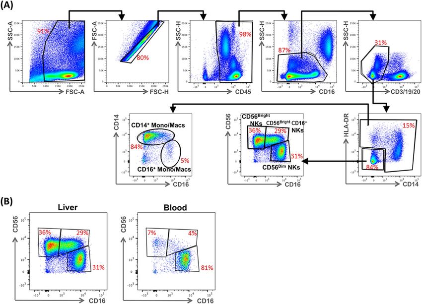
www.nature.com/scientificreports/
The global burden of advanced liver disease is increasing rapidly, far out stripping increases in cardiovascular
and chronic pulmonary d iseases1. It is therefore a priority to increase knowledge of the hepatic cells, including
the immune cell subsets, that preserve and restore liver function. Liver immune cells have distinctive functional
properties. They cleanse portal vein blood of lipopolysaccharide (LPS) and other microbial toxins without under-
going pro-inflammatory responses2 and create a tolerogenic microenvironment that protects liver grafts from
chronic rejection3. Single cell RNA sequencing and mass cytometry are revealing the heterogeneity of human
liver immune c ells4,5 and highlighting the need to integrate data about cell populations with clinical data so that
translational research can focus on the key subtypes.
Approximately 1.34 million people die each year from hepatitis B virus (HBV) and hepatitis C virus (HCV)
infection6. Direct acting antiviral drugs allow most HCV infections to be cured and reduce HBV disease; how-
ever, these viruses remain major public health threats. Patients cured of HCV continue to have an elevated risk
of liver-related morbidity and mortality. Regardless of etiology, the extent of liver damage varies from person to
person as a consequence of genetic, environmental, and immunological factors. Although liver damage is often
immune-mediated7; immunosuppression can accelerate pathogenesis, as seen in HCV-infected patients with
inborn immunodeficiencies8, HIV infection9, or on immunosuppressive d rugs10, implying that, in the absence
of immunosuppression, cytotoxic immune cells are held in check by other immune cell subsets. A promising
strategy for improving outcomes in liver disease patients is to identify the immune cell subsets that modulate
immunopathology, promote repair, and maintain homeostasis.
In this study, we analyzed innate immune cell populations in liver and peripheral blood of 19 HCV-infected
adults undergoing liver transplantation, seeking subsets expanded in patients with relatively well-preserved liver
function. Model for end stage liver disease (MELD) scores ranged from 7 to 42 in the study group. Uniquely,
liver CD56Bright/CD16- natural killer (NK) cells were expanded in patients with lower MELD scores and lower
serum bilirubin, consistent with evidence that the abundance of intrahepatic NK cells correlates inversely with
serum transaminase values and ISHAK fibrosis scores in HCV-infected p atients11,12. Gene expression profiling
showed a relationship between liver NK cells and decidual NK cells; these cells promote placental formation
largely by secreting growth factors, rather than by killing neighboring cells13. If liver CD56Bright/CD16- NK cells
act through similar mechanisms, greater knowledge of their secretome and cell to cell interactions could lead to
interventions that increase their activity and thereby improve liver function.
Results
NK cells dominate the intrahepatic innate immune cell population and transcriptome. We
began our search for an immunoprotective cell subset by surveying liver and peripheral blood mononuclear cells
from 19 HCV-infected patients undergoing liver transplantation, using the flow cytometry gating strategy in
Fig. 1. The median age of the patients was 62 years [interquartile range (IQR), 59–65)]; 80% were male (Table 1).
Innate immune cells ( CD45+ CD3- CD19- CD20-) were a similar percentage of the C D45+ mononuclear cells in
liver and peripheral blood: 46% in LMCs and 43% in PBMCs (Fig. 2A). Our multiparameter flow panel deline-
ated three subsets of natural killer (NK) cells, two subsets of monocytes/macrophages, and three subsets of den-
dritic cells. The distributions of innate immune cells in liver and blood are presented in Fig. 2B–F.
NK cells were a higher percentage of the C D45+ innate immune mononuclear cells in liver than in blood.
Bright -
CD56 /CD16 NK cells were an average of 23% in liver, but only 2% in blood, a highly-significant tenfold
difference (p < 0.0001; Fig. 2B,C) that is consistent with published d ata14. The liver contained two additional NK
subpopulations: CD56 Bright
/CD16 NK cells, which were almost exclusively localized to liver (Fig. 2D; p < 0.0001)
+
and CD56Dim/CD16+ NK cells, which had a similar abundance in liver and blood (Fig. 2E). CD14+ mono-
cytes/macrophages were 50% of the innate immune mononuclear cells in blood, but only 21% in liver (Fig. 2F;
p < 0.0001). CD16+ monocytes/macrophages were also enriched about two-fold in the blood (Fig. 2B; p = 0.02).
Dendritic cells were a similar percentage of liver and blood innate immune cells; the compartmentalization of
individual dendritic cell subsets (plasmacytoid, classical, and cross-presenting) was reported b efore4.
After comparing the two compartments based on the distribution of cell subsets, we compared them at the
transcriptomic level. In this investigation of microarray data, we used gene set enrichment analysis (GSEA) and
the blood transcriptome modules (BTM)15 to compare gene expression of freshly-isolated ex vivo cells from the
liver and the blood. Three of the top five gene sets expressed at higher levels in LMCs were related to NK cells;
the other two were related to T cells (Supplementary Table 1). Because three of the top five gene sets expressed
at a higher level in LMCs were related to NK cells, we conclude that NK cells are more important in the hepatic
microenvironment than in blood. Upregulated genes included multiple killer cell immunoglobulin-like receptor
(KIR) genes16, and EOMES and MYBL117, two transcription factors expressed in immature NK cells18. Additional
upregulated genes encode granzymes A and K 19, two proteases that are critical for NK function.
The signatures of freshly isolated (ex vivo) LMCs and PBMCs were also compared to the single-cell RNA
sequencing (scRNAseq) signature of liver NK cells from healthy liver organ donors analyzed by Aizarani et al.5
(Fig. 3A). The transcriptome of LMCs from the HCV-infected livers were significantly more similar to the sig-
nature of liver NK cells of organ donors (who do not have any underlying liver disease) than was the transcrip-
tome of PBMCs from the HCV-infected patients (FDR = 0.003, Fig. 3A). The leading edge genes shared between
LMCs of HCV-infected patients and uninfected liver NK cells included EOMES and CD69, a marker of tissue
residency14. These data indicate that NK cells in the LMCs of HCV-infected patients retain key features of liver
resident NK cells in uninfected patients; this important finding aligns with data of Cosgrove and c olleagues12 who
found, “the liver resident NK immunophenotype appeared unperturbed in the context of chronic HCV infection”.
Hepatic immune cells have a blunted response to pro‑inflammatory stimuli compared to
blood. Pathogen associated molecular patterns (PAMPs) activate antimicrobial defenses and alter immune
Scientific Reports | (2021) 11:1551 | https://doi.org/10.1038/s41598-020-80819-8 2
Vol:.(1234567890)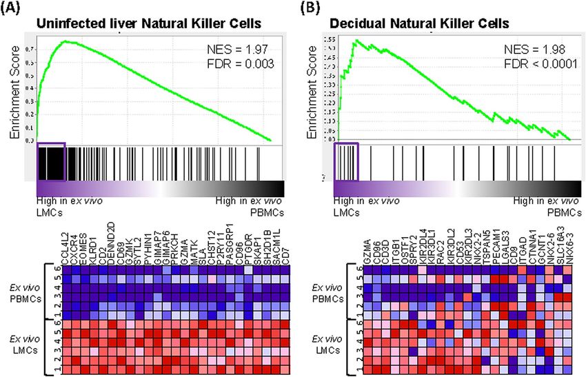
www.nature.com/scientificreports/
Figure 1. Identification of hepatic innate immune cell populations by multiparameter flow cytometry, the
gating strategy. (A) Hepatic mononuclear cells from a representative liver were stained with a nine-color
antibody panel: CD45, CD3, CD19, CD20, HLA-DR, CD14, CD16, CD123, BDCA1, BDCA3, and CD56.
Innate immune mononuclear cells were selected based on viability (live/dead), unicellularity (singlets), CD45
expression (CD45+), intracellular complexity (non-granulocytes/granulocytes), and lack of expression of lineage
markers (CD3-, CD19-, CD20-). (B) Difference in expression of C
D16+ NK cells in liver compared to blood for a
representative patient.
cell function. Compared to PBMCs, LMCs of HCV-infected patients are exposed to higher concentrations of
HCV-related PAMPS and disease-associated molecular patterns (DAMPs) released from infected and dam-
aged cells. Despite chronic exposure to these alarmins, the LMCs of the HCV-infected patients were far less
responsive to LPS or the toll-like receptor (TLR)-7/8 agonist, R848, than PBMCs, as shown by the analysis of
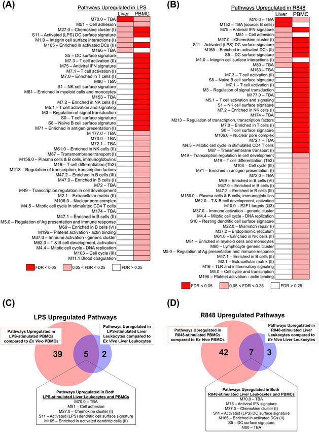
up-regulated gene expression pathways in Fig. 4. The LMC’s blunted response to LPS and to TLR 7/8 stimulation
demonstrates that the liver’s well-known tolerogenic properties persist during HCV infection.
In this set of experiments, the pathways up-regulated by incubation in LPS (or R848) were first determined
for the cells from each compartment (using FDR < 0.25 as the threshhold) and then the two sets of upregulated
pathways were compared to each other. LPS induced 44 pathways in PBMCs, but only seven in LMCs (Fig. 4A,C).
Similar to LPS, R848 up-regulated 49 pathways in PBMCs and only 10 pathways in LMCs (Fig. 4B,D). The leading
edge genes of the pathways up-regulated in both LMCs and PMBCs are listed in Supplementary Table 2 (LPS-
stimulation) and Supplementary Table 3 (R848-stimulation).
IL22 was among the differentially expressed genes in cells exposed to LPS. It was more strongly up-regulated
in LMCs than in PBMCs, p = 0.0002, FDR = 0.025. IL-22 stimulates repair processes in other t issues20 and, thus,
our findings could indicate that LMCs are programmed to activate tissue repair pathways in response to pro-
inflammatory stimulation20. In the liver, IL-22′s primary role is in tissue repair rather than inflammation21.
Mice expressing IL22 under an albumin promoter do not develop liver inflammation but are resistant to ConA
T cell-mediated hepatitis. After partial hepatectomy, liver regeneration in these animals is accelerated through
enhanced hepatocyte survival and proliferation22. Using single cell mass cytometry, we attempted to confirm
that liver NK cells produce IL-22, as has been shown for other NK populations, but the antibody signal was too
weak to identify IL-22-positive cells.
To gain further insights into the effects of alarmins and to relate them to NK abundance in individual liver
specimens, LMCs and PBMCs were incubated for 4 h in media, LPS or R848 and concentrations of the monokine,
IL-12, and the cytokine, IFNγ, which are important for NK terminal differentiation and effector function, were
Scientific Reports | (2021) 11:1551 | https://doi.org/10.1038/s41598-020-80819-8 3
Vol.:(0123456789)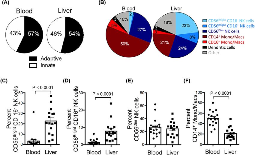
www.nature.com/scientificreports/
Clinical characteristics, median (IQR), N (%) Patients, N = 19
Age (years) 62 (59–65)
Male 15 (79%)
Hispanic 4 (21%)
Race
Asian 1 (5%)
Black 3 (16%)
Caucasian 13 (68%)
Mixed 1 (5%)
Other 1 (5%)
Reason for liver transplantation
Hepatocellular carcinoma 13 (68%)
End-stage liver disease 6 (32%)
CMV IgG Ab
Negative 5 (26%)
Positive 14 (74%)
18 (13–32)
Natural MELD
Range: 7–42
Total bilirubin (mg/dL) 2.9 (1.55–6.85)
Normal: 0.1–1.2 Range: 0.5–31
GGT (U/L) 48 (30–83)
Normal: 9–48 Range: 22–239
INR 1.55 (1.3–2.5)
Normal: ≤ 1.1 Range: 1.1–4.2
Creatinine (mg/dL) 1.13 (0.96–2.47)
Normal: 0.6–1.2 Range: 0.69–9.7
Albumin (g/dL) 3.3 (2.8–3.9)
Normal: 3.4–5.4 Range: 2.4–4.6
Platelets (× 103/μL) 75 (57–95)
Normal: 150–450 Range: 21–146
White blood cell count (× 103/μL) 4.65 (3.43–7.18)
Normal: 4.5–11 Range: 2.5–9.4
Table 1. Characteristics of the study population. IQR interquartile range, INR international normalized ratio,
MELD model for end-stage liver disease, GGTgamma-glutamyl transpeptidase.
measured in cell culture supernatants. The amount of IL-12 secreted by LMCs following R848 stimulation cor-
related with the prevalence of intrahepatic C D56Bright/CD16- NK cells, as expected based on the well-known
relationship between IL-12 and NK cells (Fig. 5A,C, p = 0.007). IFNγ produced by LMCs in response to TLR7/8
stimulation correlated with the abundance of CD56Bright/CD16- NK cells (Fig. 5B,D, p = 0.03), suggesting that
these cells contribute significantly to intrahepatic IFNγ production. LMCs incubated in media (with no TLR
stimulation) secreted more of this anti-fibrotic cytokine than PBMCs (Fig. 5B), which may reflect the higher
concentration of HCV RNA in liver tissue. These results suggest that liver resident NK cells may restrain liver
fibrogenesis through the constitutive expression of IFNγ23,24.
CD56Bright CD16‑ NK cells are uniquely correlated with indicators of better liver function. We
next examined the relationship between the prevalence of each of the sixteen innate immune cell populations
(eight in LMCs and eight in PBMCs) and seven indicators of chronic liver disease severity. This analysis was
possible because our study group included patients with a spectrum of liver disease severity: MELD scores
ranged from 7 to 42, total bilirubin concentrations ranged from 0.5 to 31 mg/dL, and platelets ranged from 21
to 146 × 103/μL (Table 1). The abundance of only one subset—hepatic CD56Bright/CD16- NK cells—correlated
with better liver function, as indicated by lower total serum bilirubin values (R2 = 0.65, p = 0.0002, Fig. 6A) and
lower MELD scores (R2 = 0.28, p = 0.03, Fig. 6B). This association was highly specific for hepatic C D56Bright/
CD16- NK cells. No other subset of NK cells and none of the subsets of other innate immune cells had a sta-
tistically significant relationship with lower bilirubin and/or lower MELD scores. Bilirubin concentration was
not correlated with blood C D56Bright/CD16- NK cells (Fig. 6C, p = 0.99) or hepatic C
D56Bright/CD16+ NK cells
(Fig. 6E, p = 0.21), and MELD scores were not correlated with blood CD56Bright/CD16- NK cells (Fig. 6D, p = 0.85)
or hepatic CD56Bright/CD16+ NK cells (Fig. 6F, p = 0.72).
In contrast to the hepatic CD56Bright/CD16- NK cells, greater abundance of hepatic CD16+/CD14- monocytes/
macrophages correlated with higher INR values (R2 = 0.42, p = 0.007, Supplementary Fig. 1A) and higher MELD
scores (R2 = 0.35, p = 0.016, Supplementary Fig. 1B). Opposite, greater abundance of hepatic CD14+ monocytes/
macrophages correlated with higher total bilirubin values ( R2 = 0.27, p = 0.04, Supplementary Fig. 1C), consistent
with published data showing that liver monocytes/macrophages promote liver i nflammation25. Overall, hepatic
CD56 Bright/CD16- NK cells emerged as the subset most likely to have a protective effect.
Single sample gene set enrichment analysis (ssGSEA) was used to investigate the impact of CD56Bright/CD16-
NK cells on the gene expression signature of surrounding tissue, with the expectation that the signature will be
dominated by hepatocytes and the other major types of liver cells (endothelial cells, stellate cells, biliary epithelial
Scientific Reports | (2021) 11:1551 | https://doi.org/10.1038/s41598-020-80819-8 4
Vol:.(1234567890)www.nature.com/scientificreports/
Figure 2. Comparison of the abundance of innate immune cells in the liver and in peripheral blood. (A) Pie
charts depicting the composition of innate and adaptive immune cells from peripheral blood (left) and liver
(right). (B) Pie charts depicting the composition of C
D45+ lineage negative ( CD3-, CD19-, CD20-) innate
immune populations of the peripheral blood (left) and liver (right). (C) The percentage of C D56Bright CD16- NK
cells of total hematopoietic ( CD45+), lineage negative (CD3-, CD19-, CD20-) cells from liver and peripheral
blood mononuclear cells. (D) The percentage of CD56Bright CD16+ NK cells. (E) The percentage of CD56Dim
CD16+ NK cells. (F) The percentage of C D14+ monocytes/macrophages. Horizontal bars depict the mean ± SEM.
N = 16 liver leukocytes, n = 18 PBMCs by unpaired t test.
cells. The ssGSEA enrichment score denotes the degree to which the genes in a particular pathway are upregulated
in an individual s pecimen26. As was done b efore13, non-tumor tissue was used if a patients had hepatocellular
carcinoma. ssGSEA showed that the abundance of CD56Bright/CD16- NK cells correlated positively with two
pathways associated with normal liver function (Supplementary Table 4): “Metabolism of porphyrins” (heme
biosynthesis and breakdown are important hepatocyte functions) and “Circadian rhythm” (normal liver func-
tions follow a pronounced circadian rhythm). Pathways with negative correlations included “Platelet adhesion to
exposed collagen”, “Signaling by PDGF”, “Apoptotic executive phase” and “Apoptotic cleavage of cellular proteins”.
Collectively, the ssGSEA analysis suggests that C D56Bright/CD16- NK cells may promote liver homeostatic func-
tions in surrounding cells. Consistent with the negative correlation with apoptotic pathways, a separate analysis
showed that the abundance of intrahepatic C D56Bright/CD16- NK cells did not significantly correlate with serum
levels of HCV RNA (R = 0.19, p = 0.09), as might be expected if their beneficial effects are mediated by pathways
2
that do not involve killing HCV-infected hepatocytes..
Killer functions are inhibited in liver resident CD56Bright CD16‑ NK cells. The positive association
between hepatic CD56Bright/CD16- NK cells and liver homeostatic pathways suggested that they may have func-
tional similarities to decidual NK cells, a key population that produces angiogenic factors and promotes vascular
remodeling during the early phases of placental formation27. To test this, we compared a signature of decidual
NK cells from Koopman et al.28 to the gene expression profiles of ex vivo LMCs and PBMCs. The LMCs were
much more similar to the decidual NK cells than were the PBMCs (FDR < 0.0001, Fig. 3B). Several of the lead-
ing edge genes encode proteins that inhibit NK functions, including KIR2DL4, KIR3DL1, CD53, KIR2DL316,
and SPRY229, suggesting that liver resident CD56Bright/CD16- NK cells are primed to resist activation of killing
pathways.
Because the portal vein supplies the majority of blood to the liver, cytokines in portal vein blood may influence
the programming of the hepatic immune response. Importantly, the concentration of IL-10, an immunosup-
pressive factor, was significantly higher in portal vein plasma than in peripheral blood plasma (Fig. 7A), while
concentrations of the other cytokines (CXCL10, IFNα, IFNγ, IL-6, IL-12, TNFα) were similar in the two com-
partments (Fig. 7B–F). The high concentration of IL-10 in portal vein blood could prime LMCs for a blunted
and tolerogenic response to pro-inflammatory TLR ligands.
Scientific Reports | (2021) 11:1551 | https://doi.org/10.1038/s41598-020-80819-8 5
Vol.:(0123456789)www.nature.com/scientificreports/
Figure 3. Liver mononuclear cells from H
CV+ individuals are more similar to natural killer cells from healthy
decidua and healthy liver. (A) GSEA of gene sets from Aizarani et al.5 related to “Uninfected Liver Natural
Killer Cells” comparing ex vivo LMCs to ex vivo PBMCs from HCV-infected patients. (B) GSEA of gene sets
from Koopman et al.28 related to “Decidual Natural Killer Cells” comparing ex vivo LMCs to ex vivo PBMCs.
Pathways with a false discovery rate (FDR) below 0.25 were considered significant. Genes contributing to
pathway enrichment (leading edge genes) are boxed and presented in the heatmaps.
Discussion
This study revealed a strong association between the abundance of human liver C D56Bright/CD16- NK cells and
measures liver function, such as lower total bilirubin concentration in serum, lower MELD scores, and expres-
sion of molecular pathways associated with normal liver functions. The abundance of liver C D56Bright/CD16- NK
cells accounted for 65% of the variance in total bilirubin in serum. In contrast to the NK cells, the abundance
of hepatic monocytes/macrophages was associated with increased liver injury and dysfunction, consistent with
published data implicating these cells in liver damage. Monocyte recruitment and expansions in liver accelerate
fibrogenesis in rat and mouse models30; and the number of CD14- and CD16-expressing hepatic cells is positively
correlated with scar tissue and liver damage25. While hepatic monocytes/macrophages cause pro-inflammatory
liver damage, hepatic C D56Bright/CD16- NK emerged from our data as the subset of innate immune cells most
likely to have a protective effect. Cosgrove and colleagues reached a similar conclusion. They also found a strong
inverse relationship between serum bilirubin and intrahepatic CD56Bright NKG2D+ NK cells (r = − 0.861) and
proposed that NK cells attenuate liver fibrogenic pathways in chronic HCV i nfection12.
Our findings cconfirm previous data showing that CD56Bright NK cells are highly enriched in liver compared
to blood12,14,31–35 and they suggest several mechanisms hepatic C D56Bright/CD16- NK cells might use to promote
liver homeostasis and repair. The main cytokines NK cells are known to produce are IFNγ, TNFα, and IL-1036.
Of these, our data support a role for IFNγ. Without TLR stimulation, liver leukocytes produced more IFNγ than
PBMCs, suggesting that the liver cells are primed for spontaneous secretion. The amount of IFNγ produced by
LMCs in response to TLR7/8 stimulation correlated with the abundance of hepatic C D56Bright/CD16- NK cells,
indicating that these cells may be a major source of hepatic IFNγ. In other settings, IFNγ has antifibrotic effects
and is part of regulatory circuits that down-modulate pathways of tissue injury37. IFNγ from liver NK cells may
contribute to analogous processes in liver.
In addition to a possible antifibrotic effect of IFNγ, the positive association between hepatic CD56Bright/CD16-
NK cells and liver homeostatic gene expression pathways suggested that these cells may have functional similari-
ties to decidual NK cells, a key population that produces angiogenic factors and promotes vascular remodeling
during the early phases of placental formation27. The signature of decidual NK cells from Koopman et al.28 had
several genes in common with that of liver NK cells from our patients. A number of the shared leading edge
genes encode proteins that inhibit NK functions, including KIR2DL4, KIR3DL1, CD53, KIR2DL3, and SPRY2,
Scientific Reports | (2021) 11:1551 | https://doi.org/10.1038/s41598-020-80819-8 6
Vol:.(1234567890)www.nature.com/scientificreports/
Figure 4. Fewer gene pathways are upregulated in stimulated LMCs than matched PBMCs. (A) Differential modulation of
molecular pathways from the blood transcriptome modules (BTM) of ex vivo LMCs versus LPS-stimulated LMCs was compared to
the modulation of molecular pathways from the same BTM pathways of ex vivo PBMCs versus LPS-stimulated PBMCs, analyzed
by GSEA. (B) Differential modulation of molecular pathways from the BTMs of ex vivo LMCs versus R848-stimulated LMCs was
compared to the modulation of molecular pathways from the same BTM pathways of ex vivo PBMCs versus R848-stimulated PBMCs,
analyzed by GSEA. Pathways that were had a false discovery rate (FDR) of less than 0.05 are in red, between 0.05 and 0.25 FDR in
pink, and greater than 0.25 FDR in white. (C) Shared BTM pathways upregulated with a FDR < 0.25 in both LPS-stimulated LMCs
versus ex vivo LMCs and LPS-stimulated PBMCs versus ex vivo PBMCs. Leading edge genes driving these pathways are listed in
Supplementary Table 2. (D) Shared BTM pathways upregulated with a FDR < 0.25 in both R848-stimulated LMCs versus ex vivo LMCs
and R848-stimulated PBMCs versus ex vivo PBMCs. Leading edge genes driving these pathways are listed in Supplementary Table 3.
Scientific Reports | (2021) 11:1551 | https://doi.org/10.1038/s41598-020-80819-8 7
Vol.:(0123456789)www.nature.com/scientificreports/
Figure 5. IFNγ secretion of TLR-stimulated LMCs is correlated with hepatic C D56Bright CD16- NK cells. LMCs
and PBMCs were stimulated with LPS, R848 or media alone prior to analysis for cytokine production. Total
pg/mL for (A) IL-12 and (B) IFNγ. Horizontal bars depict the mean ± SD. N = 16. Comparisons were made
using paired t-tests. (C) IL-12 secreted from LMCs stimulated with R848 correlated with the percent hepatic
CD56Bright CD16- NK cells. (D) IFNγ secreted from LMCs stimulated with R848 correlated with the percent
hepatic CD56Bright CD16- NK cells. N = 16, calculated using the Pearson’s correlation coefficient.
indicating that the liver resident C D56Bright/CD16- NK cells may be primed to resist activation of killing pathways.
Bright
CD56 /CD16 NK cells express low levels of perforin and they have low c ytotoxicity36. Taken together with
-
published data showing that liver NK abundance is inversely related to serum markers of hepatocyte damage,
ALT and AST11, our results indicate that hepatic CD56Bright/CD16- NK cells preserve liver function through
mechanisms independent of cell killing. Interestingly, decidual NK cells produce IFNγ constituitively, suggesting
that this cytokine may contribute to an essential function of the resident NK cells in these two compartments.
After LPS stimulation, LMCs up-regulated expression of IL22. Because IL-22 is associated with liver regenera-
tion in other s ettings20, this up-regualtion could indicate that the LMCs are primed to up-regulate tissue repair
pathways in response to pro-inflammatory s timulation20. Further investigation is needed to determine whether
liver NK cells secrete this cytokine. Additional mechanisms liver NK cells might use to promote liver function
could include an enhancement of hepatocyte survival and proliferation21, stimulation of liver repair38, down-
modulation of pro-inflammatory responses, and killing hepatic stellate c ells39.
Portal vein blood could play a role in the programming of liver NK cells for tissue repair. The liver receives
most of its blood directly from the gut vasculature through the portal vein, and thus hepatic cells are exposed
to high levels of food antigens and bacterial PAMPS, including LPS. The liver must clear these non-self antigens
without becoming inflamed, while also maintaining antimicrobial defenses. The liver immune system helps
Scientific Reports | (2021) 11:1551 | https://doi.org/10.1038/s41598-020-80819-8 8
Vol:.(1234567890)www.nature.com/scientificreports/
Figure 6. Correlation between the abundance of liver resident CD56Bright CD16- NK and better clinical status.
The correlation between the abundance of hepatic CD56Bright CD16- NK cells and serum total bilirubin (A) and
the natural MELD score calculated for the day of transplantation (B). The correlation between the abundance of
peripheral blood CD56Bright CD16- NK cells and serum total bilirubin (C) and the natural MELD score (D). The
correlation between the abundance of hepatic C D56Bright CD16+ NK cells and serum total bilirubin (E) and the
natural MELD score (F). N = 16, calculated using the Pearson’s correlation coefficient.
achieve the balance necessary for homeostasis. The tolerogenic nature of the liver innate immune system is due to
lower levels of TLRs40, cytokine-mediate suppression41, liver microenvironment reprograming42, and enrichment
of tolerogenic innate immune p opulations43. In this study of HCV-infected patients, we found that liver leuko-
cytes were far less responsive to TLR stimuli than PBMCs, demonstrating that the liver’s hypo-responsiveness
to pro-inflammatory stimuli continues during HCV infection. The high concentration of IL-10 in portal vein
blood may help establish and maintain this hypo-responsiveness. IL-10 is important for maintaining N KG2A+
NK cells in a hypo-responsive state that promotes the induction of T regulatory c ells44. It is thus plausible that
IL-10 in portal vein blood modulates liver NK cells and down-regulates their cytototoxic activities.
The study has several strengths. It is a prospective investigation using specimens that were processed in an
expedited manner to preserve cell viability. The sample size of 19 is robust for a study of this depth. Multipa-
rameter flow cytometry allowed eight subsets of hepatic innate immune cells to be delineated and compared to
PBMCs. Finally, the flow cytometry data were integrated with clinical, molecular, and secretomic analyses. The
study also has limitations. All the patients had HCV infection; however, Cosgrove and colleagues compared NK
cells from patients with and without HCV infection and found that “the profile of intrahepatic NK cells was
remarkably unaltered in chronic HCV infection” 12, and we found an association between the gene expression
profile of LMCs and liver NK cells of patients with no underlying liver d isease5. Additionally, our experiments
did not prove that there is a causal relationship between the abundance of liver NK cells and better liver function.
Scientific Reports | (2021) 11:1551 | https://doi.org/10.1038/s41598-020-80819-8 9
Vol.:(0123456789)www.nature.com/scientificreports/
Figure 7. Portal vein blood has a higher concentration of IL-10 than peripheral blood. Portal vein plasma and
peripheral plasma was analyzed for concentrations of (A) IL-10, (B) CXCL10, (C) IFNγ, (D) IL-12, (E) IL-6,
and (F) TNFα. Horizontal bars depict the mean ± SD. N = 16 peripheral plasma, n = 13 portal vein plasma by
unpaired t test.
Our results allow other interpretations: C D56Bright/CD16- NK cells could be a non-functional precursor popula-
tion, for example. Despite these limitations, our study achieved its primary objective: Delineating eight innate
immune cell subsets in human liver and blood and identifying the one that is most strongly associated with
maintenance of normal liver homeostatic functions.
In summary, our findings suggest that hepatic CD56Bright/CD16- NK cells may be a source of immunoprotec-
tive factors in the liver. These cells may counter the inflammatory effects of macrophages, which would otherwise
cause excessive liver damage, and they may share functions with decidual NK cells. The hyporesponsiveness of
liver innate immune cells to TLR4 or 7/8 stimulation is maintained in patients with a chronic viral infection,
despite inflammation and activation of IFN-stimulated antiviral defense p athways12,45.
Methods
Study design. This is a prospective study of 19 HCV-positive adults who underwent liver transplantation at
the Mount Sinai Medical Center between November 2013 and August 2014. Informed consent was obtained in
writing from patients before transplantation. The study protocol was approved by the Icahn School of Medicine
at Mount Sinai’s institutional review board and adhered to all its guidelines. Medical records were reviewed for
clinical/demographic data.
Specimen collection. Blood for research and for clinical testing was collected prior to surgery and liver
explant tissue and portal vein blood were obtained at the time of surgery. After the recipient hepatic veins and
arteries were clamped, portal vein blood was collected and used to prepare portal vein plasma. Liver explants
were accessed by liver pathologists in an expedited manner to allow cell preparation to begin as quickly as pos-
sible, optimizing cell viability. Liver tissue arrived at the cell isolation laboratory within a median of 45 min of
surgery. Tissue was processed, as described before4. Mononuclear cells were isolated using a percoll gradient.
Scientific Reports | (2021) 11:1551 | https://doi.org/10.1038/s41598-020-80819-8 10
Vol:.(1234567890)www.nature.com/scientificreports/
Flow cytometry. Nine-color multiparameter flow cytometry was used to delineate cell subsets using
the antibodies reported previously4 for CD45, CD3, CD19, CD20, HLA-DR, CD14, CD16, CD123, BDCA1,
BDCA3, and CD56, using the gating strategy in Fig. 1. NK cells were defined as CD45+ CD3- CD19- CD20-
HLA-DR- CD14- and classified in 3 subsets based on CD56 and CD16 expression: CD56Bright/CD16-, CD56Bright/
CD16+, and C D56Dim/CD16+. Subpopulations of monocytes/macrophages were defined by the following mark-
ers: CD45+ CD3- CD19- CD20- HLA-DR+ CD14+/- CD16+/-.
Cell stimulation in vitro, proteomics, and RNA extraction. Liver mononuclear cells (LMCs)
and peripheral blood mononuclear cells (PBMCs) were stimulated with two toll-like receptor (TLR) agonists
[200 ng/mL lipopolysaccharide (LPS) or 1 μg/mL resiquimod (R848), a TLR7/8 agonist] or incubated in media
for four hours at 37 °C. Supernatants were collected for secretomic analysis. Luminex multiplex cytokine assays
(Millipore) quantified interferon (IFN) gamma-induced protein 10 (IP10 aka CXCL10), interleukin 6 (IL-6),
IL-10, IL-12p70, IFNγ and tumor necrosis factor (TNF)α. Cells were collected in Trizol (Life Technologies) and
RNA was purified using RNeasy mini kits (Qiagen), according to manufacturer’s i nstructions4.
Microarray analysis. Profiling data from Illumina Human-HT-Expression Beadchips were normalized
using GenomeStudio’s quantile method. GenePattern was used for gene set enrichment analysis (GSEA) and
single sample GSEA (ssGSEA) of immune pathways46 using blood transcriptomic modules (BTMs)15, KEGG
pathways47, and Reactome pathways48. A false discovery rate (FDR) below 0.25 was considered statistically sig-
nificant based on the Broad Institute’s guidelines49 Comparative Marker Selection (GenePattern) was used to
determine the genes that were most differentially expressed. LMCs/PBMCs: To obtain sufficient RNA, LMC
samples of matched pairs of patients were pooled. Matching was based on age, gender, HCV genotype, baseline
HCV RNA, natural MELD score, and HCC (yes/no). PBMCs were pooled similarly. Whole liver: Whole liver
efore4.
microarray data of 11 of the 19 patients consented for this analysis was used, as b
Statistical analysis. GraphPad Prism was used for statistical analysis. T-tests, both paired and unpaired,
were performed. Linear regression was used for clinical correlations.
Received: 11 September 2020; Accepted: 23 November 2020
References
1. Asrani, S.K. et al. Increasing health care burden of chronic liver disease compared with other chronic diseases, 2004–2013. Gas-
troenterology 155, 719–729 e714 (2018).
2. Crispe, I. N. Hepatic T cells and liver tolerance. Nat. Rev. Immunol. 3, 51–62 (2003).
3. Sanchez-Fueyo, A. & Strom, T. B. Immunologic basis of graft rejection and tolerance following transplantation of liver or other
solid organs. Gastroenterology 140, 51–64 (2011).
4. Doyle, E. H. et al. Individual liver plasmacytoid dendritic cells are capable of producing IFNalpha and multiple additional cytokines
during chronic HCV infection. PLoS Pathog. 15, e1007935 (2019).
5. Aizarani, N. et al. A human liver cell atlas reveals heterogeneity and epithelial progenitors. Nature 572, 199–204 (2019).
6. Organization, W.H. Global Health Estimates 2016: Disease burden by Cause, Age, Sex, by Country and by Region, 2000–2016. (2018).
7. Lohse, A. W., Weiler-Normann, C. & Tiegs, G. Immune-mediated liver injury. J. Hepatol. 52, 136–144 (2010).
8. Zignego, A. L., Giannini, C., Gragnani, L., Piluso, A. & Fognani, E. Hepatitis C virus infection in the immunocompromised host:
A complex scenario with variable clinical impact. J. Transl. Med. 10, 158 (2012).
9. Hernandez, M. D. & Sherman, K. E. HIV/hepatitis C coinfection natural history and disease progression. Curr. Opin. HIV AIDS
6, 478–482 (2011).
10. Neuberger, J. Treatment of hepatitis C virus infection in the allograft. Liver Transpl. 9, S101-108 (2003).
11. Varchetta, S. et al. Impaired intrahepatic natural killer cell cytotoxic function in chronic hepatitis C virus infection. Hepatology
56, 841–849 (2012).
12. Cosgrove, C. et al. Chronic HCV infection affects the NK cell phenotype in the blood more than in the liver. PLoS ONE 9, e105950
(2014).
13. Hoshida, Y. et al. Prognostic gene expression signature for patients with hepatitis C-related early-stage cirrhosis. Gastroenterology
144, 1024–1030 (2013).
14. Hudspeth, K. et al. Human liver-resident CD56(bright)/CD16(neg) NK cells are retained within hepatic sinusoids via the engage-
ment of CCR5 and CXCR6 pathways. J. Autoimmun. 66, 40–50 (2016).
15. Li, S. et al. Molecular signatures of antibody responses derived from a systems biology study of five human vaccines. Nat. Immunol.
15, 195–204 (2014).
16. Pende, D. et al. Killer Ig-like receptors (KIRs): Their role in NK cell modulation and developments leading to their clinical exploita-
tion. Front. Immunol. 10, 1179 (2019).
17. Allan, D. S. J. et al. Transcriptome analysis reveals similarities between human blood CD3(-) CD56(bright) cells and mouse
CD127(+) innate lymphoid cells. Sci. Rep. 7, 3501 (2017).
18. Harmon, C. et al. Tissue-resident Eomes(hi) T-bet(lo) CD56(bright) NK cells with reduced proinflammatory potential are enriched
in the adult human liver. Eur. J. Immunol. 46, 2111–2120 (2016).
19. Pardo, J., Balkow, S., Anel, A. & Simon, M. M. Granzymes are essential for natural killer cell-mediated and perf-facilitated tumor
control. Eur. J. Immunol. 32, 2881–2887 (2002).
20. Khawar, M. B., Azam, F., Sheikh, N. & Abdul Mujeeb, K. How does interleukin-22 mediate liver regeneration and prevent injury
and fibrosis?. J. Immunol. Res. 2016, 2148129 (2016).
21. Radaeva, S., Sun, R., Pan, H. N., Hong, F. & Gao, B. Interleukin 22 (IL-22) plays a protective role in T cell-mediated murine hepatitis:
IL-22 is a survival factor for hepatocytes via STAT3 activation. Hepatology 39, 1332–1342 (2004).
22. Park, O. et al. In vivo consequences of liver-specific interleukin-22 expression in mice: Implications for human liver disease pro-
gression. Hepatology 54, 252–261 (2011).
Scientific Reports | (2021) 11:1551 | https://doi.org/10.1038/s41598-020-80819-8 11
Vol.:(0123456789)www.nature.com/scientificreports/
23. Jeong, W. I., Park, O. & Gao, B. Abrogation of the antifibrotic effects of natural killer cells/interferon-gamma contributes to alcohol
acceleration of liver fibrosis. Gastroenterology 134, 248–258 (2008).
24. Horras, C. J., Lamb, C. L. & Mitchell, K. A. Regulation of hepatocyte fate by interferon-γ. Cytokine Growth Factor Rev. 22, 35–43
(2011).
25. Tan-Garcia, A. et al. Intrahepatic CD206+ macrophages contribute to inflammation in advanced viral-related liver disease. J.
Hepatol. (2017).
26. Barbie, D. A. et al. Systematic RNA interference reveals that oncogenic KRAS-driven cancers require TBK1. Nature 462, 108–112
(2009).
27. Tabiasco, J. et al. Human decidual NK cells: Unique phenotype and functional properties—A review. Placenta 27(Suppl A), S34-39
(2006).
28. Koopman, L. A. et al. Human decidual natural killer cells are a unique NK cell subset with immunomodulatory potential. J. Exp.
Med. 198, 1201–1212 (2003).
29. Hanafusa, H., Torii, S., Yasunaga, T. & Nishida, E. Sprouty1 and Sprouty2 provide a control mechanism for the Ras/MAPK signal-
ling pathway. Nat. Cell Biol. 4, 850–858 (2002).
30. Karlmark, K. R. et al. Hepatic recruitment of the inflammatory Gr1+ monocyte subset upon liver injury promotes hepatic fibrosis.
Hepatology 50, 261–274 (2009).
31. Caligiuri, M. A. Human natural killer cells. Blood 112, 461–469 (2008).
32. Mozer-Lisewska, I. et al. Detection and significance of cytotoxic cell subsets in biopsies of HCV-infected human livers. Arch.
Immunol. Ther. Exp. 62, 153–160 (2014).
33. Doherty, D. G. & O’Farrelly, C. Innate and adaptive lymphoid cells in the human liver. Immunol. Rev. 174, 5–20 (2000).
34. Doherty, D. G. et al. The human liver contains multiple populations of NK cells, T cells, and CD3+CD56+ natural T cells with
distinct cytotoxic activities and Th1, Th2, and Th0 cytokine secretion patterns. J. Immunol. 163, 2314–2321 (1999).
35. Stegmann, K. A. et al. CXCR6 marks a novel subset of T-bet(lo)Eomes(hi) natural killer cells residing in human liver. Sci. Rep. 6,
26157 (2016).
36. Vivier, E., Tomasello, E., Baratin, M., Walzer, T. & Ugolini, S. Functions of natural killer cells. Nat. Immunol. 9, 503–510 (2008).
37. Wang, H. & Yang, Y. G. The complex and central role of interferon-gamma in graft-versus-host disease and graft-versus-tumor
activity. Immunol. Rev. 258, 30–44 (2014).
38. Feng, D. et al. Interleukin-22 promotes proliferation of liver stem/progenitor cells in mice and patients with chronic hepatitis B
virus infection. Gastroenterology 143, 188–198 e187 (2012).
39. Melhem, A. et al. Anti-fibrotic activity of NK cells in experimental liver injury through killing of activated HSC. J. Hepatol. 45,
60–71 (2006).
40. De Creus, A. et al. Low TLR4 expression by liver dendritic cells correlates with reduced capacity to activate allogeneic T cells in
response to endotoxin. J. Immunol. 174, 2037–2045 (2005).
41. Chen, Y. et al. Distinct response of liver myeloid dendritic cells to endotoxin is mediated by IL-27. J. Hepatol. 51, 510–519 (2009).
42. Xia, S. et al. Hepatic microenvironment programs hematopoietic progenitor differentiation into regulatory dendritic cells, main-
taining liver tolerance. Blood 112, 3175–3185 (2008).
43. Pillarisetty, V. G., Shah, A. B., Miller, G., Bleier, J. I. & DeMatteo, R. P. Liver dendritic cells are less immunogenic than spleen
dendritic cells because of differences in subtype composition. J. Immunol. 172, 1009–1017 (2004).
44. Lassen, M. G., Lukens, J. R., Dolina, J. S., Brown, M. G. & Hahn, Y. S. Intrahepatic IL-10 maintains NKG2A+Ly49- liver NK cells
in a functionally hyporesponsive state. J. Immunol. 184, 2693–2701 (2010).
45. Bohne, F. et al. HCV-induced immune responses influence the development of operational tolerance after liver transplantation in
humans. Sci. Transl. Med. 6, 242ra281 (2014).
46. Reich, M. et al. GenePattern 2.0. Nat. Genet. 38, 500–501 (2006).
47. Kanehisa, M., Furumichi, M., Tanabe, M., Sato, Y. & Morishima, K. KEGG: New perspectives on genomes, pathways, diseases and
drugs. Nucleic Acids Res. 45, D353–D361 (2017).
48. Fabregat, A. et al. Reactome pathway analysis: A high-performance in-memory approach. BMC Bioinform. 18, 142 (2017).
49. Broad Institute. 3.2 Why does GSEA use a false discovery rate (FDR) of 0.25 rather than the more classic 0.05? http://softwa re.broad
instit ute.org/cancer /softwa re/gsea/wiki/index. php/FAQ#Why_does_GSEA_use_a_false_ discov ery_rate_.28FDR. 29_of_0.25_rathe
r_than_the_more_classic_0.05.3F. Accessed 3 Oct 2020 (2020).
Acknowledgements
This research was supported by Merck Pharmaceuticals, as well as by NIOSH, NIAAA, NIAID, NIDA and
NIDDK. We thank Amir Horowitz and the Icahn School of Medicine’s Human Immune Monitoring Core who
provided insight and expertise that greatly assisted the research.
Author contributions
E.H.D.: design and perform experiments, analyze data, manuscript draft. C.A.: design experiments, analyze data,
manuscript revision. A.E.: design and perform experiments, analyze data. F.E.: design experiments, analyze data.
A.R.: design experiments, analyze data. A.L.K.: design experiments, analyze data. B.H.: design experiments,
analyze data. S.S.F.: design experiments, analyze data. M.I.F.: design experiments, analyze data. T.S.: design
experiments, analyze data. A.D.B.: design and perform experiments, analyze data, manuscript draft and revisions.
Funding
This research was funded in part by a research grant from Merck Pharmaceuticals to TS (https://www.msdgr
ants.com), the CDC’s National Institute for Occupational Safety and Health (CDC/NIOSH01148) to ADB, the
National Institute on Drug Abuse (DA031095) to ADB, the Prevent Cancer Foundation to ADB, the National
Institute of Diabetes and Digestive and Kidney Diseases (DK088954) to CA and the National Institute on Alcohol
Abuse and Alcoholism (AA024762) to CA. EHD was supported by the NIH’s Virus-Host Interactions training
grant (5T32AI007647-17). AR was supported by the NIH GI training grant.
Competing interests
The authors declare no competing interests.
Additional information
Supplementary Information The online version contains supplementary material available at https://doi.
org/10.1038/s41598-020-80819-8.
Scientific Reports | (2021) 11:1551 | https://doi.org/10.1038/s41598-020-80819-8 12
Vol:.(1234567890)www.nature.com/scientificreports/
Correspondence and requests for materials should be addressed to A.D.B.
Reprints and permissions information is available at www.nature.com/reprints.
Publisher’s note Springer Nature remains neutral with regard to jurisdictional claims in published maps and
institutional affiliations.
Open Access This article is licensed under a Creative Commons Attribution 4.0 International
License, which permits use, sharing, adaptation, distribution and reproduction in any medium or
format, as long as you give appropriate credit to the original author(s) and the source, provide a link to the
Creative Commons licence, and indicate if changes were made. The images or other third party material in this
article are included in the article’s Creative Commons licence, unless indicated otherwise in a credit line to the
material. If material is not included in the article’s Creative Commons licence and your intended use is not
permitted by statutory regulation or exceeds the permitted use, you will need to obtain permission directly from
the copyright holder. To view a copy of this licence, visit http://creativecommons.org/licenses/by/4.0/.
© The Author(s) 2021
Scientific Reports | (2021) 11:1551 | https://doi.org/10.1038/s41598-020-80819-8 13
Vol.:(0123456789)You can also read