Original Article HNSC exosome-derived MIAT improves cognitive disorders in rats with vascular dementia via the miR-34b-5p/CALB1 axis
←
→
Page content transcription
If your browser does not render page correctly, please read the page content below
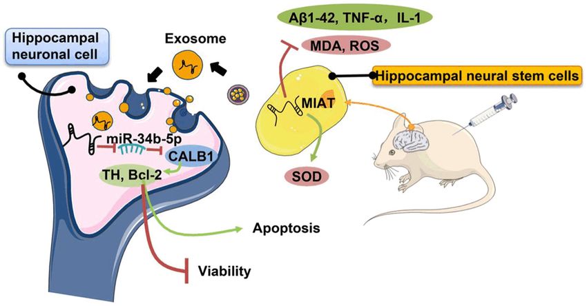
Am J Transl Res 2021;13(9):10075-10093
www.ajtr.org /ISSN:1943-8141/AJTR0134015

Original Article
HNSC exosome-derived MIAT improves cognitive
disorders in rats with vascular dementia
via the miR-34b-5p/CALB1 axis
Dan Qi1*, Xiaojun Hou2*, Chunfeng Jin4, Xi Chen3, Chengli Pan2, Hongjuan Fu2, Leifeng Song1, Jujun Xue2

Departments of 1Neurology, 2Gerontological Neurology, 3Experimental Diagnosis, Heilongjiang Provincial Hospital,
Harbin, Heilongjiang Province, China; 4Department of Neurology, Harbin Second Hospital, Harbin, Heilongjiang
Province, China. *Equal contributors and co-first authors.
Received April 7, 2021; Accepted June 24, 2021; Epub September 15, 2021; Published September 30, 2021

Abstract: Objective: To explore the molecular mechanism by which hippocampal neural stem cell (HNSC) exosome
(exo)-derived MIAT improves cognitive disorders in rats with vascular dementia (VD). Methods: Rat hippocampal
tissues were collected, and HNSCs and hippocampal neuronal cells (HNCs) were isolated, purified, and identified.
Then the exosomes (exo) of the HNSCs were extracted and identified. A VD rat model was constructed. HE staining
was used to evaluate the hippocampal pathology in each group. The expressions of the RNAs in the HNSCs were
intervened, and the cells were then grouped. ELISA was used to measure the of TNF-α, IL-1, and Aβ1-42 expression
levels. The kits were used to determine the oxidative stress factor levels. The targeting relationships among MIAT,
miR-34b-5p, and CALB1 were measured using dual-luciferase assays. The MIAT expressions in exo were measured
using qRT-PCR. The proliferation and apoptosis of the HNCs were determined using CCK-8 and Annexin V-FITC/PI
staining, respectively. The CALB1, TH, and Bcl-2 protein expressions were determined using Western blot. The Morris
water maze test was used for the spatial learning and memory testing. Results: The hippocampal tissues in the
model group were clearly damaged, but the pathological characteristics were significantly improved in the exo group.
The exo group also showed an increased SOD level, decreased MDA and ROS levels, and down-regulated TNF-α, IL-1,
and Aβ1-42 expressions (all P
HNSC exo-derived MIAT improves vascular dementia

in the treatments [4]. Exo is one of these vesi-    Materials and methods
cles. It has now been discovered that exo can
interact with cells through secretions and the      Bioinformatics methods
delivery of proteins and nucleotides. Exo can
be used as diagnostic markers for many dis-         We used the Gene Expression Omnibus data-
eases, but its specific mechanism remains to        base (https://www.ncbi.nlm.nih.gov/geo/) to
be determined [5-7].                                retrieve the microarray data of the VD-related
                                                    expression profile by using “Vascular demen-
Studies have proved that long non-coding RNAs       tia” as the keyword, then GSE122063 was
(LncRNAs) are involved in a variety of diseases,    selected for the subsequent analysis. GSE-
such as cancer, and metabolic, neurological,        122063 is the dataset of altered gene expres-
and skin diseases [8-11]. Gao et al. demon-         sions in the aortas of 32-week ApoE-/- mice
strated that knocking down LncRNA SNHG1             and wild-type mice. Meanwhile, the aorta
could reduce amyloid-induced neuronal dam-          expression profiles in ApoE-/- mice and wild-
age in Alzheimer’s dementia [12]. Zhuang et al.     type mice were selected from GSE72248 to
found decreased levels of LncRNA MALAT1 in          analyze the differentially expressed genes.
the cerebrospinal fluid and plasma in patients      The Affy package from the R programming lan-
with Alzheimer’s dementia [13]. The exo-            guage was used for the background correc-
derived LncRNA myocardial infraction associa-       tions and normalization of the microarray data
tion transcript (MIAT) is a gene located at         (http://www.bioconductor.org/packages/relea-
chr22:27,042,392-27072441 (GRCh37/hg19).            se/bioc/html/affy.html). The limma package
Moreover, a study showed that the loss of           (http://master.bioconductor.org/packages/
MIAT can lead to cerebral microvascular dis-        release/bioc/html/limma.html) was used to
eases, neurodegeneration, neuron loss, and          standardize the microarray data and screen
Alzheimer’s dementia, suggesting that MIAT          the differentially expressed genes (DEGs). The
                                                    differential gene screening conditions were
may be a key factor in neurovascular diseases
                                                    FDR1.5. The
[14]. The absence of MIAT may also promote
                                                    pheatmap package (https://cran.r-project.org/
the development of VD. Therefore, this study
                                                    web/packages/pheatmap/index.html) was us-
aimed to explore the biological effects of MIAT
                                                    ed to draw a heat map of the DEGs. “Calculate
on the progression of VD.
                                                    and draw custom Venn diagrams” (http://bioin-
LncRNAs affect physiological and pathological       formatics.psb.ugent.be/webtools/Venn/) was
processes mainly through the competitive            used to compare and analyze the different sets
adsorption of microRNA to further regulate          of data as well as to draw the Venn diagrams.
mRNAs, the mechanism of which has been              The candidate miRNAs of the target genes
studied in detail for many diseases [15]. In this   were searched using miRDB (http://mirdb.org),
study, we also further explored the down-           miRWalk (http://mirwalk.umm.uni-heidelberg.
                                                    de), and RNA22 (http://www.rna-society.org/
stream molecular pathways of MIAT and found
                                                    raid/index.html). Then DisgeNET (http://www.
that MIAT can regulate miR-34b-5p/CALB1.
                                                    disgenet.org/?1) was used to search for VD
One study showed that treatment with miR-34
                                                    associated risk genes in the disease-related
inhibitors can improve memory deficits in aged
                                                    gene database. The interactions between the
rats [16]. As a member of the superfamily of
                                                    target gene and the protein were searched in
calcium-binding proteins, CALB1 widely pres-
                                                    the STRING database (https://string-db.org/).
ents in neurons. A study showed that the posi-
                                                    The visualization was implemented using the
tive neurons of CALB1 are relatively reduced in
                                                    Cytoscape_v3.6.0 software.
Parkinson’s disease [17]. However, the role of
MIAT in regulating the miR-34b-5p/CALB1 axis        Isolation and identification of the HNSCs and
in VD remains to be investigated. Therefore,        HNCs
this study aimed to clarify the effect of HNSC
exo-derived MIAT on improving cognitive disor-      This study was approved by the Medical
ders by regulating the miR-34b-5p/CALB1 axis        Research Ethics Committee of Heilongjiang
in VD rats, in order to provide a new biomarker     Provincial Hospital, and the animal experiments
for the prevention and treatment of VD.             were carried out in strict accordance with the

10076                                                   Am J Transl Res 2021;13(9):10075-10093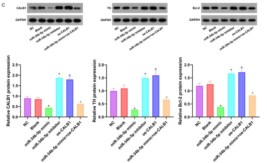
HNSC exo-derived MIAT improves vascular dementia

regulations of the Heilongjiang Provincial           and 4°C. Then the sample was mixed with PBS
Hospital Committee on the Care and Use of            containing a protease inhibitor and centrifuged
Laboratory Animals, and we tried our best to         for 1 h to obtain the exo-containing particles.
alleviate the suffering of the animals. A total of   The particles were resuspended in PBS con-
50 SD rats used in this experiment were pro-         taining protease inhibitors, identified and ana-
vided by the Experimental Animal Center of           lyzed for the morphology of exo with a transmis-
Heilongjiang Provincial Hospital. The rats were      sion electron microscope.
sacrificed using intraperitoneal injections of
3% sodium pentobarbital (0.15 mL/100 g,              Construction of the animal model and the in
Shanghai Sixin, China), then disinfected with        vivo transfection
75% alcohol on the skin to harvest the brain
                                                     The VD rat model was established using bilat-
tissues and the hippocampus using cranioto-
                                                     eral common carotid artery occlusion [20]. A
mies. Part of each hippocampus was stored in
                                                     total of 50 SD rats weighing 200-230 g were
liquid nitrogen for later use, and each remain-
                                                     used as the experimental animals, regardless
ing part was used to separate the HNSCs and
                                                     of their sex. The rats were anesthetized using
HNCs. The meninges and blood vessels were
                                                     intraperitoneal injections of 250 mg/kg tribro-
removed in pre-cooled DMEM/F12 medium
                                                     moethanol, then placed supine on a constant
(Thermo Fisher, USA) and rinsed carefully. Then
                                                     temperature pad, shaved, had their skin disin-
each hippocampus was cut into fragments of
                                                     fected, and a 3 cm incision was cut along the
about 1 mm3. The HNSCs were separated us-
                                                     midline of the neck to expose the left and right
ing trypsin digestion. Trypsin (0.25%, Thermo        common carotid arteries, which were ligated
Fisher, USA) was added for digestion to prepare      with 3-0 surgical sutures, then the incisions
the cell suspension which was filtered with a        were sutured [21]. We applied the same opera-
200-mesh filter [18]. The HNCs were separated        tion in the sham group but without blocking
using trypsin-free mechanical pipetting. The         the arteries. The survival rate of the model-
hippocampus tissues were placed in a centri-         operated rats was 73.5%, so 36 rats were
fuge tube, mixed with 4 mL of cell inoculum,         used for the follow-up experiments. The experi-
pipetted with 1 mL and 200 μL pipettors. The         mental animals were divided into the sham
pipetting was stopped when the clumpy tis-           group (n=9, with a tail vein injection of PBS
sues disappeared. The cells separated using          equivalent to exo), the model group (n=9, with
the above two methods were centrifuged at            a tail vein injection of PBS equivalent to exo),
1000 r/min for 3 min, then the supernatant           the model + exo group (n=9, with a tail vein
was removed, and a DMEF/F12 medium (con-             injection of 100 μg at 3*108 particles/μL in
taining 2% B27, 20 μg/L of epidermal growth          the modeled rats), and the oe-MIAT-exo group
factor and 20 μg/L of basic fibroblast growth        (n=9, with a tail vein injection of exo with over-
factors) was added. Thereafter, the cells were       expressed MIAT in the model rats) [22]. The
cultivated in a 37°C, 5% CO2 incubator, then         sequence of the oe-MIAT was 5’-GACTTT-
examined for the HNSC markers PE-CD133               ATAGCGATCGAGCTCGATT-3’. The rats were
(ab252128, ABCAM, UK), APC-Nestin (ab187-            anesthetized using pentobarbital sodium at a
846, ABCAM, UK), HNCs marker PE-CD24                 concentration of 40 mg/kg, and then the rats
(ab25494, ABCAM, UK, 0.2 µg for 106 cells),          were sacrificed by having their necks broken.
and FITC-Tyrosine hydroxylase (TH, 10009396-         Their brain tissues were collected for the sub-
1, Cayman, USA) using flow cytometry [19].           sequent experiments. This study was conduct-
                                                     ed in accordance with the regulations for the
Exo collection and identification in the HNSCs       management of experimental animals and was
                                                     approved by the Animal Ethics Committee of
The exo was extracted using ultracentrifuga-         our hospital (Approval No. (2021) 031).
tion. The HNSCs were cultured in a serum-free
DMEF/F12 medium. After 24 hours, 225 mL of           HE staining
the HNSC conditioned medium was collected
and centrifuged at 2000× g for 10 min at 4°C.        The brain tissues of the rats in each group were
The supernatant was centrifugated for another        collected, routinely fixed, and embedded in
10 min at 1000× g and 4°C, and another 3 h in        paraffin. The embedded tissues were cut into 3
a 60 Ti centrifuge (Beckman) at 126,000× g           μm slices and prepared as paraffin sections.

10077                                                    Am J Transl Res 2021;13(9):10075-10093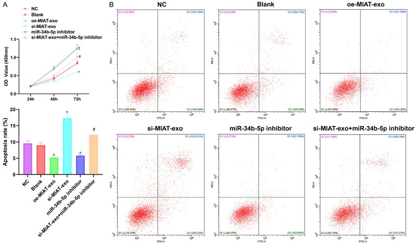
HNSC exo-derived MIAT improves vascular dementia

The sections were dewaxed using xylene, dehy-       Western blot
drated using decreasing concentrations of eth-
anol, rinsed with distilled water for 2 min, and    The stored hippocampal tissues were ground
stained with a hematoxylin solution (Shanghai       and then mixed with a RIPA lysate (Beijing
Gefan Biotechnology, China) for 10 min. The         Soleibao, China) to extract the protein. The pro-
slices were immersed in tap water for 10 min,       tein concentrations were determined using a
washed with distilled water, dehydrated with        BCA protein quantification kit (Shanghai
95% ethanol, and then stained with an eosin         Yisheng, China). Sodium dodecyl sulfate-poly-
solution (Shanghai Gefan Biotechnology, China)      acrylamide gel electrophoresis was used to
for 1 min. After being washed again with 70%        separate the total proteins at 45°C, and the
ethanol, the slices were observed to determine      separated proteins were then transferred to
the staining results under a microscope. Five       polyvinylidene fluoride membranes (Sigma-
fields of view were randomly selected to count      Aldrich, USA). The membranes were blocked in
the number of necrotic pyramidal nerve cells        5% skim milk and mixed with the primary anti-
within 1 mm2 [23].                                  bodies CALB1 (1:3000, ab108404, Abcam,
                                                    UK), TH (1:1000, ab137869, Abcam, UK), Bcl-2
ELISA                                               (1:1000, Ab32124, Abcam, UK), and GAPDH
                                                    (1:1000, ab181602, Abcam, UK) to incubate
In this experiment, ELISA was used to measure       overnight at 4°C. The membranes were then
the tumor necrosis factor-α (TNF-α), interleu-      washed with PBST, and we added horseradish
kin-1 (IL-1), and amyloid (Aβ1-42) secretion lev-   peroxidase-labeled secondary antibody IgG
els in the rat serum. The blood samples from        (1:5000, ab190492, Abcam, UK) and incubate
the abdominal aortae were collected, placed at      the membranes at 37°C for 2 h. Thereafter,
4°C for 30 min, then centrifugated at 4000 r        an enhanced chemiluminescence reagent
for 10 min to obtain the serum samples. The         (Shanghai Yisheng, China) was used to visual-
measurements were conducted using TNF-α             ize the protein signals. Image J software was
detection kits (LE-B0126), IL-1 detection kits,     used to analyze the gray values (the ratio of the
and Aβ1-42 detection kits from Hefei Laier,         gray values of the target proteins to the internal
China. The experimental procedures were car-        reference proteins).
ried out in strict accordance with the kits’
instructions. A microplate reader (Thermo           FISH
Fisher, USA) was used to measure the optical
density (OD) at a wavelength of 450 nm. A           FISH test kits (1002-30, Guangzhou Boxiny,
curve of the OD values was plotted, and the         China) were used to measure the MIAT localiza-
concentration values were calculated.               tions and expressions. The cells were routinely
                                                    fixed and embedded. Then the pre-hybridiza-
Measurement of the oxidative stress levels          tion reactions, the hybridization reactions, and
                                                    the staining of the cell nuclei with the fluoro-
The oxidative stress levels were measured in        chromes DAPI were performed according to the
the rat serum which was obtained using the          kit instructions. The staining results were
same method we used with the ELISA. The             observed under a fluorescence microscope.
superoxide dismutase (SOD), malondialdehyde
(MDA), and reactive oxygen species (ROS) were       Dual-luciferase reporter assays
used as evaluation indicators. The content of
SOD was measured using WST-8 according to           The online biological prediction tools predicted
the instructions of the total SOD activity detec-   the downstream target miR-34b-5p for MIAT
tion kit (Shanghai Beyotime, China), and the        and the downstream target gene CALB1 for
OD was measured at 450 nm. The MDA was              miR-34b-5p. A dual-luciferase reporter assay
measured using colorimetry according to the         was designed according to the complementary
instructions of the MDA detection kit (Shanghai     base-pairing to verify the targeting relation-
Beyotime, China), and the OD was determined         ships. The 3’UTR end of MIAT/CALB1 was
with a microplate reader (Thermo Fisher, US) at     inserted into the pGL3-basic vector to con-
532 nm. The ROS content was determined              struct wild-type and mutant luciferase reporter
using fluorescent probe DCFH-DA according to        plasmids. The reporter plasmids were then co-
the instructions of the ROS detection kit           transfected with miR-34b-5p NC and miR-34b-
(Shanghai Beyotime, China).                         5p mimics, and the transfection steps were car-

10078                                                   Am J Transl Res 2021;13(9):10075-10093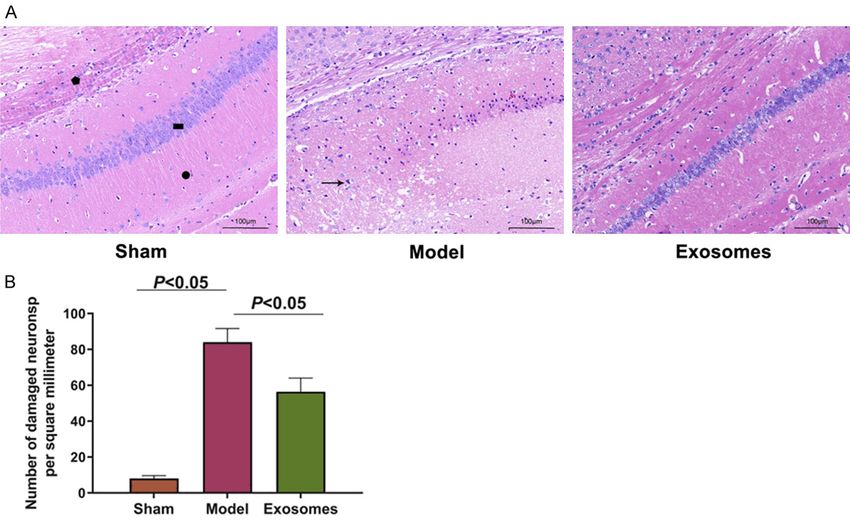
HNSC exo-derived MIAT improves vascular dementia

Table 1. Primer sequence of qRT-PCR                                           was intervened in the HNCs. The
               Primer sequence (5’-3’)                                        cells were then divided into the fol-
 Gene                                                                         lowing groups, the blank group
               Forward primer                    Reverse primer
                                                                              (HNCs + empty-vector-exo), the NC
 MIAT          GGACGTTCACAACCACACTG TCCCACTTTGGCATTCTAGG
                                                                              group (HNCs + NC-vector-exo), the
 miR-34b-5p GGGTAGGCAGTGTCATTAGC AACAACCAACACAACCCAAC
                                                                              oe-MIAT-exo group (HNCs + oe-
 CALB1         CTCCGACGGCAATGGGTAC GGTGTTAAGTCCAAGCCTGCC                      MIAT-exo), the si-MIAT-exo group
 GAPDH         CACCCACTCCTCCACCTTTG CCACCACCCTGTTGCTGTAG                      (HNCs + si-MIAT-exo), the miR-
 U6            GCCCTCTGTGCTACTTACTC GCTGGTTGTGGGTTACTCTC                      34b-5p inhibitor group (HNCs +
 Note: MIAT: myocardial infraction association transcript.                    miR-34b-5p inhibitor), the miR-
                                                                              34b-5p mimic group (HNCs + miR-
                                                                              34b-5p mimic), the miR-34b-5p
 Table 2. Sequence of vectors                                                 inhibitor + oe-MIAT-exo group
 NC                             5’-TCTATGCGAGCAGCGCGTC-3’                     (HNCs + miR-34b-5p inhibitor +
 oe-MIAT                        5’-GACTTTATAGCGATCGAGCTCGATT-3’               oe-MIAT-exo), the oe-CALB1 group
 si-MIAT                        5’-AACTGCTCGAATATCGCAGC-3’                    (HNCs + oe-CALB1), and the miR-
 miR-34b-5p inhibitor           5’-TAGCGAGCTCGCGAGCGAGCCT-3’                  34b-5p mimic + oe-CALB1 group
 miR-34b-5p mimic               5’-GGCTATTAGCGCTTCGGCTTAT-3’                  (HNCs + miR-34b-5p mimic + oe-
 oe-CALB1                       5’-CATATCGAGGCACTTCTCGTC-3’                   CALB1). The miR-34b-5p mimic
 Note: MIAT: myocardial infraction association transcript.                    and the miR-34b-5p inhibitor were
                                                                              purchased from Thermo Fisher,
                                                                              US. The recombinant plasmids
ried out in accordance with the requirements of                 were designed by Shanghai Gema Gene, China.
Lipofectamine 3000 (Thermo Fisher, USA). The                    Lipofectamine 3000 (Thermo Fisher, USA) was
medium was refreshed after 6 hours. The cells                   used for the plasmid transfection, and the
were washed with PBS after 48 hours and test-                   operations were carried out in accordance with
ed for luciferase activity according to the gene                the kit instructions. The transfection sequenc-
detection system of the dual-luciferase report-                 es in this study are shown in Table 2.
er assay (Shanghai, China). The Renilla lucifer-
ase activity was used as an internal reference.                 CCK-8

qRT-PCR                                                       The transfected HNCs were spread onto a
                                                              96-well plate for incubation. To each well, 10 μL
Total RNA was extracted from the HNCs and                     of LCCK8 solution (Beijing Soleibao, China) was
exo using TRlzol reagent (Thermo Fisher, USA),                added at 24 h, 48 h, and 72 h. The plate was
and the purity of the RNA was measured using                  placed in a 5% CO2 incubator at 37°C for 4 h.
a spectrophotometer (Mettler-Toledo, Switzer-
                                                              Then, the OD value at 450 nm was measured
land). Then cDNA was synthesized using
                                                              using a microplate reader (Thermo Fisher, USA).
reverse transcription kits (Shanghai Yisheng,
China). Real-time PCR was conducted using                     Annexin V-FITC/PI double labeling
2× HieffCanace PCR Master Mix (Shanghai
Yisheng, China). GAPDH was the internal refer-                Annexin V-FITC/PI cell apoptosis detection kits
ence for MIAT and CALB1, and U6 was the                       (Shanghai Qianchen, China) were used for this
internal reference for miR-34b-5p. The relative               experiment. The cells were centrifuged at 300
expression levels were calculated using the                   g for 5 min at 4°C, washed with pre-cooled
2-ΔΔCt formula. The primers were designed and                 PBS, centrifuged again at 300 g for 5 min at
synthesized by Shanghai Gema Gene, China.
                                                              4°C, collected and resuspended in a 1× binding
The sequences are shown in Table 1.
                                                              buffer. Then, the cells were mixed with 5 μL of
Collecting exo from the silenced and overex-                  Annexin V-FITC and 10 μL of PI staining solu-
pressed genes                                                 tion, mixed and incubated in the dark for 15
                                                              min. 1× binding buffer was then added, and the
The exo were collected after the HNSCs were                   cells were placed on ice, and examined for
transfected with the plasmids of the silenced or              apoptosis with the use of a flow cytometer
overexpressed genes. The expression of RNAs                   (Beckman Coulter, USA).

10079                                                              Am J Transl Res 2021;13(9):10075-10093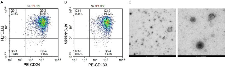
HNSC exo-derived MIAT improves vascular dementia

Water maze test                                       were double positive for CD133 and Nestin
                                                      (98.22%). See Figure 1B. The results indicated
The Morris water maze test was used to evalu-         the high purity of the separated HNCs and
ate the rats’ cognitive function [21]. The experi-    HNSCs. The separated exo were observed in a
ment was performed in a quiet and spacious            transmission electron microscope and found to
room with a water maze basin (31 cm in depth,         have elliptical shapes with bright outer edges,
94 cm in diameter) in the center of the room,         and they were mostly around 100-120 nm in
and a FLOOR lamp with a red safety bulb in a          size. See Figure 1C.
corner of the room. A camera was fixed on the
ceiling above the water maze basin and con-           The exo treatment reduced the hippocampal
nected to a computer equipped with an auto-           pathological features
matic tracking system. A heater was used to
keep the temperature of the water at around           The HNSCs-derived exo were injected into the
23°C.                                                 brains of the VD rats, and the pathological con-
                                                      ditions in area CA1 of the hippocampi were
A circular platform was put in one of the quad-       observed using HE staining. It was found that
rants, 15 cm from the edge. Then the safety           the molecular layer (pentagonal), the pyramidal
light was turned on for testing. The rat was put      cell layer (rectangular), and the polymorphic
in the water maze and observed to determine           layer (circular) showed clear boundaries and
the time it took to find the platform within 90       complete nuclei in the sham group. In the
s. If a rat could find the platform within 90 s, it   model group, the molecular layer, the pyramidal
was allowed to stay on the platform for 10 s          cell layer, and the polymorphic cell layer of the
and then moved out of the water maze. If a rat        hippocampal tissues were disordered. The cell
failed to find the platform within 90 s, it was       nuclei of the pyramidal layers were pykeptic,
manually guided to the platform and also              and the cells were dehydrated and necrotic
stayed on the platform for 10 s. The experi-          (see the arrow) (P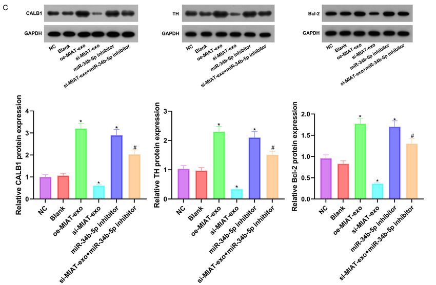
HNSC exo-derived MIAT improves vascular dementia

Figure 1. Identification of the HNCs, HNSCs and exo. A: The identification of the HNCs using PE-CD24 and FITC-TH (double positive); B: The identification of the
HNSCs sing PE-CD133 and APC-Nestin (double positive); C: The identification of exo (10000×, 40000×). N=3, and the experiment was repeated 3 times. HNSCs:
hippocampal neural stem cells; HNCs: hippocampal neuronal cells; exo: exosome.

10081                                                                                                            Am J Transl Res 2021;13(9):10075-10093
HNSC exo-derived MIAT improves vascular dementia
Figure 2. The pathological characteristics of the hippocam-
pus (HE staining). A: The pathological results of the HE stain-
ing (100×); B: The number of necrotic pyramidal neurons per
square millimeter. Pentagon: molecular layer; rectangle: py-
ramidal cell layer; circle: polymorphic cell layer; arrow: dehy-
dration and pyknosis of pyramidal cell nucleus. N=9, and the
experiment was repeated 3 times.
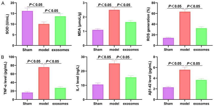
Figure 3. Exo improved the oxidative stress and secretion levels of the inflammatory factors in the VD rats. A: Com-
parison of SOD, MDA, and ROS in each group; B: Comparison of TNF-α, IL-1 and Aβ1-42 in each group. N=9, and
the experiment was repeated 3 times. Exo: exosome; VD: vascular dementia; SOD: superoxide dismutase; MDA:
malondialdehyde; ROS: reactive oxygen species; TNF-α: tumor necrosis factor-α; IL-1: interleukin-1; Aβ1-42: amyloid.
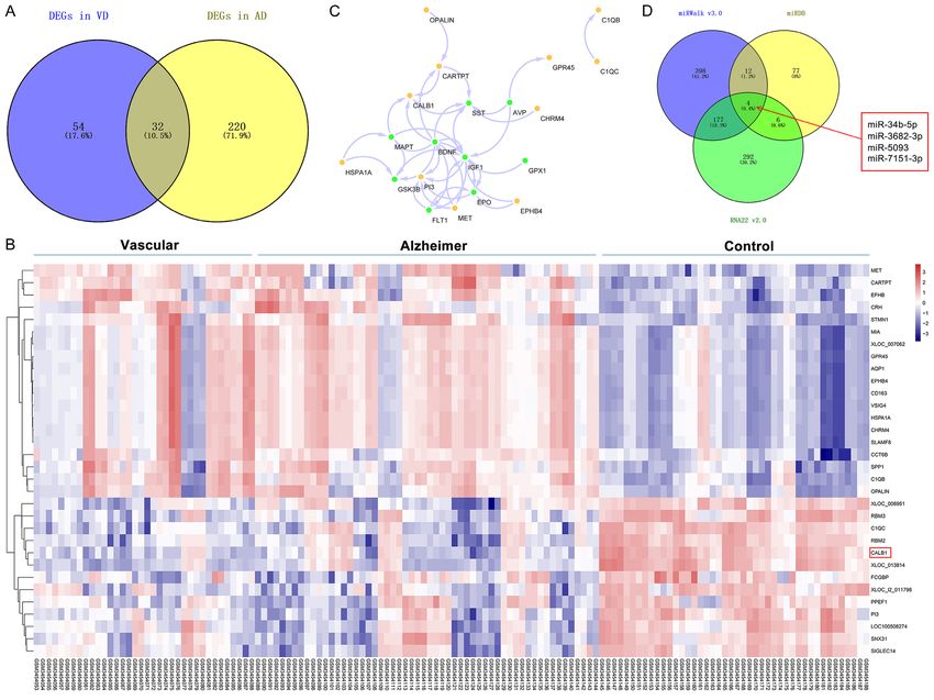
pared to the control) of Alzheimer’s dementia expression profile of the intersected genes was
and VD in GSE122063 (|LogFC|>1.5, FDR< plotted to explore the protein interaction of the
0.05). The mutual phenotypes of the two were VD-associated risk genes. See Figure 4B and
analyzed to determine the differential genes 4C. Among the above-mentioned intersected
that exist in both Alzheimer’s dementia and VD. genes, CALB1, a member of the superfamily of
See Figure 4A. A heat map representing the calcium-binding proteins, was down-regulated
10082 Am J Transl Res 2021;13(9):10075-10093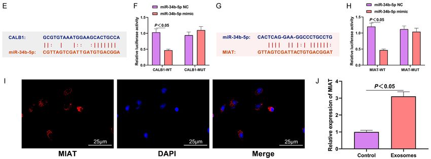
HNSC exo-derived MIAT improves vascular dementia

10083                                                 Am J Transl Res 2021;13(9):10075-10093
HNSC exo-derived MIAT improves vascular dementia

Figure 4. MIAT regulated the expression of CALB1 by adsorbing the miR-34b-5p. A: The intersection of the differential genes between Alzheimer’s disease and VD;
B: Heat map of the expression profiles of the differential genes (horizontal axis: sample number; vertical axis: gene symbol); the red in the upper right corner indi-
cates up-regulation, and the blue indicates down-regulation; C: The interaction network between the intersected genes and the VD risk gene set; the yellow circles
represent the intersected genes; the green circles represent the VD risk genes; the arrows indicate the regulatory relationship; D: The miRNAs targeting CALB1 pre-
dicted by miRDB, miRWalk and RNA22; E: Complementary base-pairing of CALB1 and miR-34b-5p; F: Verification of the targeting relationship between CALB1 and
miR-34b-5p; G: Complementary base-pairing of MIAT and miR-34b-5p; H: Verification of the targeting relationship between CALB1 and miR-34b-5p; I: Subcellular
localization of MIAT in neural stem cells (400×); J: Expression of MIAT in exo. N=6, and the experiment was repeated 3 times. VD: vascular dementia; exo: exosome;
MIAT: myocardial infraction association transcript.

10084                                                                                                                Am J Transl Res 2021;13(9):10075-10093
HNSC exo-derived MIAT improves vascular dementia

in Alzheimer’s disease and VD compared with        or silencing MIAT in the HNSCs, the exo were
the controls, and CALB1 interacted more close-     collected and co-cultured with HNCs transfect-
ly with the proteins of the VD associated genes    ed with a miR-34b-5p inhibitor. Compared with
MAPT, BDNF, and SST (Figure 4C). One study         the NC group, the oe-MIAT-exo group and the
found that the superfamily of calcium-binding      miR-34b-5p inhibitor group showed increased
proteins is involved in the development of the     cell viability, up-regulated protein expressions
hippocampus, and the low expressions of its        of CALB1, TH, and Bcl-2, as well as reduced
members are related to neurodegenerative dis-      apoptosis, while the si-MIAT-exo group showed
eases such as Alzheimer’s disease [24].            the opposite results (all P
HNSC exo-derived MIAT improves vascular dementia

10086                                                 Am J Transl Res 2021;13(9):10075-10093
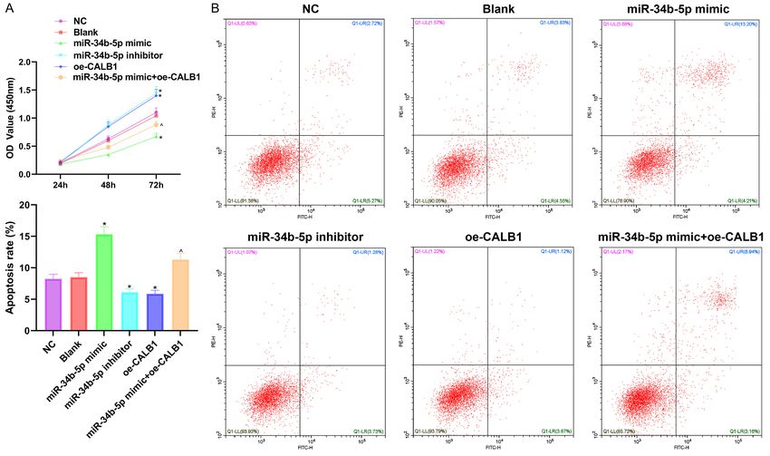
HNSC exo-derived MIAT improves vascular dementia

Figure 5. The overexpression of MIAT can protect HNCs. A: Measuring the cell viability using CCK8; B: Measuring the apoptosis using Annexin V-FITC/PI double label-
ing; C: Measuring the protein expressions of CALB1, TH and Bcl-2 using Western blot. Compared with the NC group, *P
HNSC exo-derived MIAT improves vascular dementia

10088                                                 Am J Transl Res 2021;13(9):10075-10093
HNSC exo-derived MIAT improves vascular dementia

Figure 6. The overexpression of miR-34b-5p can inhibit the proliferation of HNCs and induce apoptosis, and the effects cab be partially reversed by CALB1. A: Mea-
suring the cell viability using CCK8; B: Measuring the apoptosis using Annexin V-FITC/PI double labeling; C: Measuring the protein expressions of CALB1, TH, and
Bcl-2 using Western blot. Compared with the NC group, *P
HNSC exo-derived MIAT improves vascular dementia

Figure 7. The effects of MIAT and exo on the VD rats measured with water maze tests. A: Comparison of the incu-
bation periods; B: Comparison of the number of times crossing the platform; C: The mRNA levels of MIAT in the
hippocampi. Compared with the Sham group, &P
HNSC exo-derived MIAT improves vascular dementia

Discussion                                           sion of miR-34b also plays a role in Alzheimer’s
                                                     disease and VD [25]. In our study, it was proved
VD is a type of dementia second only to              that the overexpression of MIAT has a protec-
Alzheimer’s disease. People with cerebrovascu-       tive effect on neurological function, but this
lar risks are likely to develop VD which threat-     effect can be partially resisted by miR-34b-5p.
ens the quality of life of the elderly, especially
among people over 65 years old [26]. LncRNAs         CALB1, a member of the superfamily of calci-
regulate many diseases, including VD [27]. A         um-binding proteins, is a downstream target
previous study showed that the down-regula-          gene of miR-34b-5p. One study found that
tion of MIAT is associated with the pathogene-       CHI3L2 might be involved in the progression of
sis of Alzheimer’s disease [14]. In this study, we   Alzheimer’s disease, but the expressions of
have confirmed that MIAT exists in exo. We           CALB1 and CHI3L2 were negatively correlated,
established a VD rat model using bilateral com-      and the expression of CALB1 was correlated
mon carotid artery occlusion. After treatment        with age in patients with Alzheimer’s disease
with exo, the pathological characteristics of the    [33]. Studies have also shown that calcium
rats were significantly improved. A previous         binding proteins are involved in important
study showed that the combination of recep-          developmental processes of the striatum, the
tors for advanced glycation end products and         hippocampal cortex, and the cerebellar cortex
Aβ1-42 can mediate the inflammatory res-             [34, 35]. Our research found that miR-34b-5p
ponse, increase the expression of the pro-           can inhibit the growth of neuronal cells, but this
inflammatory factors (such as TNF-α), and            effect is partially reversed by CALB1.
accelerate the degeneration of nerve tissues
                                                     This study also has some limitations. The sam-
[28]. This study also showed elevated expres-
                                                     ple size was limited. Our In vivo experiments
sions of these factors in the VD rats, and the
                                                     only verified the role of the exo-derived MIAT in
secretions of these factors were controlled
                                                     improving VD through behavioral studies but
using exo treatment. Oxidative stress refers to
                                                     did not measure the apoptosis or the oxidative
excessive oxidation in the body which causes
                                                     stress levels. We will conduct future studies
inflammatory stress, produces a large number
                                                     with improvements in the above aspects.
of oxidative intermediates, and leads to various
diseases and aging [29]. Our research also           In summary, our experiment proves that exo
confirmed that exo treatment can improve the         can improve the pathological damage caused
oxidative stress levels in VD rats. In addition,     by VD, and exo treatment is achieved by MIAT
our in vivo experiments further proved that          regulating the miR-34b-5p/CALB1 axis, which
the overexpression of exo-derived MIAT can           suggests that MIAT has a protective effect on
improve the memory and learning abilities of         nerve function. Our study fills the vacancy of
VD rats. Therefore, we believe that exo-derived      the effects of MIAT on the biological study of VD
MIAT is likely to be a new target for the treat-     and provides a reference for the targeted ther-
ment of VD.                                          apy of VD.
In order to prove the mechanism of exo treat-        Acknowledgements
ment for VD, we used online bioinformatics pre-
diction tools to conduct research. The Gene          This work was supported by the Heilongjiang
Expression Omnibus database combined with            Province Natural Joint Guidance Project (LH-
miRDB, miRWalk and RNA22 predicted that              2021H064).
MIAT can regulate miR-34b-5p and CALB1 in
                                                     Disclosure of conflict of interest
VD. The interaction of this molecular pathway
was then verified using a dual-luciferase report-    None.
er assay. A type of miRNA, miR-34b-5p can be
infiltrated by LncRNA, which inhibits the expres-    Address correspondence to: Jujun Xue, Depart-
sion of miRNA to weaken its function. This           ment of Gerontological Neurology, Heilongjiang
mechanism has been confirmed in various              Provincial Hospital, No. 82 Zhongshan Road,
studies [30, 31]. The up-regulation of miR-34b       Xiangfang District, Harbin 150001, Heilongjiang
is found in Parkinson’s disease and in multiple      Province, China. Tel: +86-0451-87131560; Fax:
system atrophy [32]. The differential expres-        +86-0451-88025770; E-mail: xxuejjun@163.com

10091                                                    Am J Transl Res 2021;13(9):10075-10093
HNSC exo-derived MIAT improves vascular dementia
References [12] Gao Y, Zhang N, Lv C, Li N, Li X and Li W. ln-
cRNA SNHG1 knockdown alleviates amyloid-β-
[1] Zhou L, Yang R and Wu F. Efficacy and safe- induced neuronal injury by regulating ZNF217
ty of butylphthalide as adjunctive therapy for via sponging miR-361-3p in Alzheimer’s dis-
vascular dementia: a protocol for systematic ease. J Alzheimers Dis 2020; 77: 85-98.
review and meta-analysis. Medicine (Balti- [13] Zhuang J, Cai P, Chen Z, Yang Q, Chen X, Wang
more) 2020; 99: e23236. X and Zhuang X. Long noncoding RNA MALAT1
[2] Li J, Shang Y, Wang L, Zhao B, Sun C, Li J, Liu S, and its target microRNA-125b are potential
Li C, Tang M, Meng FL and Zheng P. Genome biomarkers for Alzheimer’s disease manage-
integrity and neurogenesis of postnatal hippo- ment via interactions with FOXQ1, PTGS2 and
campal neural stem/progenitor cells require CDK5. Am J Transl Res 2020; 12: 5940-5954.
a unique regulator filia. Sci Adv 2020; 6: [14] Jiang Q, Shan K, Qun-Wang X, Zhou RM, Yang
eaba0682. H, Liu C, Li YJ, Yao J, Li XM, Shen Y, Cheng H,
[3] Amin LE and Montaser M. Comparative evalu- Yuan J, Zhang YY and Yan B. Long non-coding
ation of pulpal repair after direct pulp capping RNA-MIAT promotes neurovascular remodeling
using stem cell therapy and biodentine: an in the eye and brain. Oncotarget 2016; 7:
animal study. Aust Endod J 2021; 47: 11-19. 49688-49698.
[4] Li L, Wang YK, Yu XB, Bao YM, An LJ, Wei XW, [15] Wang Y, Fu J, Yang L and Liang Z. Long non-
Yu WT, Liu BY, Li JL, Yang JH, Xia Y, Liu G, Cao F, coding RNA SNHG20 promotes colorectal can-
Zhang XZ and Zhao DW. Bone marrow mesen- cer cell proliferation, migration and invasion
chymal stem cell-derived exosomes promote via miR-495/STAT3 axis. Mol Med Rep 2021;
plasminogen activator inhibitor 1 expression in 23: 31.
vascular cells in the local microenvironment [16] Hernandez-Rapp J, Rainone S and Hébert SS.
during rabbit osteonecrosis of the femoral MicroRNAs underlying memory deficits in neu-
head. Stem Cell Res Ther 2020; 11: 480. rodegenerative disorders. Prog Neuropsycho-
pharmacol Biol Psychiatry 2017; 73: 79-86.
[5] Domenis R, Marino M, Cifù A, Scardino G, Cur-
[17] Mizuta I, Tsunoda T, Satake W, Nakabayashi Y,
cio F and Fabris M. Circulating exosomes ex-
Watanabe M, Takeda A, Hasegawa K, Nakashi-
press α4β7 integrin and compete with CD4+ T
ma K, Yamamoto M, Hattori N, Murata M and
cells for the binding to Vedolizumab. PLoS One
Toda T. Calbindin 1, fibroblast growth factor
2020; 15: e0242342.
20, and alpha-synuclein in sporadic Parkin-
[6] Li Y, Yin ZR, Fan JS, Zhang SY and Yang WB.
son’s disease. Hum Genet 2008; 124: 89-94.
The roles of exosomal miRNAs and lncRNAs in
[18] Tian JY, Chen WW, Cui J, Wang H, Chao C, Lu ZY
lung diseases. Signal Transduct Target Ther
and Bi YY. Effect of lycium bararum polysac-
2019; 4: 47.
charides on methylmercury-induced abnormal
[7] Vlachakis D, Mitsis Τ, Nicolaides N, Efthimiad-
differentiation of hippocampal stem cells. Exp
ou A, Giannakakis A, Bacopoulou F and Chrou- Ther Med 2016; 12: 683-689.
sos GP. Functions, pathophysiology and cur- [19] Monaco S, Baur K, Hellwig A, Hölzl-Wenig G,
rent insights of exosomal endocrinology Mandl C and Ciccolini F. A flow cytometry-
(review). Mol Med Rep 2021; 23: 26. based approach for the isolation and charac-
[8] Liu H, Zhang L, Ding X and Sui X. LINC00861 terization of neural stem cell primary cilia.
inhibits the progression of cervical cancer cells Front Cell Neurosci 2018; 12: 519.
by functioning as a ceRNA for miR-513b-5p [20] Venkat P, Chopp M and Chen J. Models and
and regulating the PTEN/AKT/mTOR signaling mechanisms of vascular dementia. Exp Neurol
pathway. Mol Med Rep 2021; 23: 24. 2015; 272: 97-108.
[9] Zhai HY, Yan RH, Yang TZ, Zhou ZH, Gao L and [21] Kim Y and Kim YJ. Effect of obesity on cogni-
Li J. LncRNA BCAR4 up-regulates EGFR and tive impairment in vascular dementia rat mod-
thus promotes human thyrocyte proliferation. el via BDNF-ERK-CREB pathway. Biol Res Nurs
Neoplasma 2019; 66: 180105N12. 2021; 23: 248-257.
[10] Zhang Y, Xia QM and Lin J. LncRNA H19 attenu- [22] Gao BY, Zhou ST, Sun CC, Cheng DD, Zhang Y,
ates apoptosis in MPTP-induced Parkinson’s Li XT, Zhang L, Zhao J, Xu DS and Bai YL. Brain
disease through regulating miR-585-3p/PI- endothelial cell-derived exosomes induce neu-
K3R3. Neurochem Res 2020; 45: 1700-1710. roplasticity in rats with ischemia/reperfusion
[11] Li HT, Yang C, Zhang J, Zhong W, Zhu L and injury. ACS Chem Neurosci 2020; 11: 2201-
Chen YF. Identification of potential key mRNAs 2213.
and LncRNAs for psoriasis by bioinformatic [23] Liu DH, Agbo E, Zhang SH and Zhu JL. Anticon-
analysis using weighted gene co-expression vulsant and neuroprotective effects of paeonol
network analysis. Mol Genet Genomics 2020; in epileptic rats. Neurochem Res 2019; 44:
295: 741-749. 2556-2565.
10092 Am J Transl Res 2021;13(9):10075-10093HNSC exo-derived MIAT improves vascular dementia
[24] Emmanuele V, Garcia-Cazorla A, Huang HB, [30] Chen WJ, Zhai LL, Liu HM, Li YT, Zhang Q, Xu
Coku J, Dorado B, Cortes EP, Engelstad K, De DD and Fan WY. Downregulation of lncRNA
Vivo DC, Dimauro S, Bonilla E and Tanji K. De- ZFAS1 inhibits the hallmarks of thyroid carci-
creased hippocampal expression of calbindin noma via the regulation of miR-302-3p on cy-
D28K and cognitive impairment in MELAS. J clin D1. Mol Med Rep 2021; 23: 2.
Neurol Sci 2012; 317: 29-34. [31] Li XQ, Mo JX, Li J and Chen YL. lncRNA CASC2
[25] Barbagallo C, Mostile G, Baglieri G, Giunta F, inhibits lipopolysaccharide-induced acute lung
Luca A, Raciti L, Zappia M, Purrello M, Ragusa injury via miR-27b/TAB2 axis. Mol Med Rep
M and Nicoletti A. Specific signatures of serum 2020; 22: 5181-5190.
miRNAs as potential biomarkers to discrimi- [32] Vallelunga A, Ragusa M, Di Mauro S, Iannitti T,
nate clinically similar neurodegenerative and Pilleri M, Biundo R, Weis L, Di Pietro C, De Iuliis
vascular-related diseases. Cell Mol Neurobiol A, Nicoletti A, Zappia M, Purrello M and Anto-
2020; 40: 531-546. nini A. Identification of circulating microRNAs
[26] Perera G, Rijnbeek PR, Alexander M, Ansell D, for the differential diagnosis of Parkinson’s
Avillach P, Duarte-Salles T, Gordon MF, Lapi F, disease and multiple system atrophy. Front
Mayer MA, Pasqua A, Pedersen L, van Der Lei Cell Neurosci 2014; 8: 156.
J, Visser PJ and Stewart R. Vascular and meta- [33] Sanfilippo C, Castrogiovanni P, Imbesi R and Di
bolic risk factor differences prior to dementia Rosa M. CHI3L2 expression levels are corre-
diagnosis: a multidatabase case-control study lated with AIF1, PECAM1, and CALB1 in the
using European electronic health records. BMJ brains of Alzheimer’s disease patients. J Mol
Open 2020; 10: e038753. Neurosci 2020; 70: 1598-1610.
[27] Nuthikattu S, Milenkovic D, Rutledge J and [34] Bernácer J, Prensa L and Giménez-Amaya JM.
Villablanca A. The western diet regulates hip- Distribution of GABAergic interneurons and do-
pocampal microvascular gene expression: an paminergic cells in the functional territories of
integrated genomic analyses in female mice. the human striatum. PLoS One 2012; 7:
Sci Rep 2019; 9: 19058. e30504.
[28] C RC, Lukose B and Rani P. G82S RAGE poly- [35] Flace P, Lorusso L, Laiso G, Rizzi A, Cagiano R,
morphism influences amyloid-RAGE interac- Nico B, Ribatti D, Ambrosi G and Benagiano V.
tions relevant in Alzheimer’s disease patholo- Calbindin-D28K immunoreactivity in the hu-
gy. PLoS One 2020; 15: e0225487. man cerebellar cortex. Anat Rec (Hoboken)
[29] Gamage R, Wagnon I, Rossetti I, Childs R, Nie- 2014; 297: 1306-1315.
dermayer G, Chesworth R and Gyengesi E.
Cholinergic modulation of glial function during
aging and chronic neuroinflammation. Front
Cell Neurosci 2020; 14: 577912.
10093 Am J Transl Res 2021;13(9):10075-10093You can also read