Clinical and Histological Effects of the Intrathecal Administration of Methylprednisolone in Dogs
←
→
Page content transcription
If your browser does not render page correctly, please read the page content below
Pain Physician 2010; 13:493-501 • ISSN 1533-3159

           Animal Trial

                 Clinical and Histological Effects of the
                 Intrathecal Administration of
                 Methylprednisolone in Dogs
                       Rodrigo Moreira Lima, MD1, Lais H. Navarro, MD, PhD2, Jeffrey M. Carness2,
                       Guilherme A. Barros , MD, PhD1, Mariangela EA Marques, MD, PhD1,
                       Daneshvari Solanki, MD2, and Eliana M. Ganem, MD, PhD1

    From: 1São Paulo State University     Background: Methylprednisolone is one of the most commonly used steroids for
  Botucatu, Sao Paulo; 2University of     management of chronic back pain via epidural injection. Its inadvertent injection into the
 Texas Medical Branch, Galveston, TX      intrathecal space is associated with complications such as adhesive arachnoiditis.
       Dr. Lima is with São Paulo State
                                          Objective: The present study aimed to assess the clinical and histological changes associated
   University, Botucatu, Sao Paulo; Dr.
      Navarro, and Dr. Solanki are with   with the injection of methylprednisolone into the intrathecal space of dogs.
    the Department of Anesthesiology,
   University of Texas Medical Branch,    Study Design: A randomized, double blind, controlled animal trial.
     Galveston, TX; Carness is a Fourth
    year medical student, University of   Methods: After approval by the animal research ethics committee, 14 dogs were studied
 Texas Medical Branch, Galveston, TX;
                                          in a randomized double blind controlled trial. They were assigned to one of 2 groups:
Dr. Marques is with the Department of
Pathology, São Paulo State University;    Group I received 1 mL of 0.9% normal saline; Group II received 1 mL (1.15mg/kg) of
Dr. Barros and Dr. Ganem are with the     methylprednisolone into the intrathecal space. Animals were clinically evaluated for 21 days,
   Department of Anesthesiology, São      and then sacrificed. The lumbar and sacral portions of their spinal cords were removed for
 Paulo State University, Botucatu, Sao    histological examination.
                                 Paulo.
                                          Results: In Group I, there were no clinical or histological changes. All animals in Group
            Address correspondence:
                    Rodrigo Lima, MD      II showed no clinical changes but all exhibited histological changes in the spinal cord. The
       Department of Anesthesiology       main histological changes consisted of meningeal thickening and lymphocytic infiltrates in
   University of Texas Medical Branch     the blood vessels. In 3 animals, adhesion of pia, arachnoid, and dura matter was noted and
          Galveston TX – 77555-1102       the nerve roots were surrounded by fibrosis. In one animal, necrosis of the spinal cord was
         E-mail: rmelima@gmail.com        evident.
Disclaimer: Study supported by Brazil
  Federal Agency for the Support and      Limitations: The limitations of the present study include: small sample of animals (n=14),
    Evaluation of Graduate Education      relative short clinical follow-up (21 days), and use of a commercially available drug solution,
                              (CAPES).    which is not preservative free.
            Conflict of interest: None.
                                          Conclusion: The present study demonstrated that the intrathecal administration of
    Manuscript received: 04/16/2010
                                          commercially available methylprednisolone was responsible for causing histological changes
        Revised manuscript received:
                        06/007/2010       in the spinal cord and meninges of the animals studied.
Accepted for publication: 06/14/2010
                                          Key words: Methylprednisolone; intrathecal injection; steroids; adhesive arachnoiditis; low
                Free full manuscript:     back pain; epidural injection; spinal cord.
       www.painphysicianjournal.com
                                          Pain Physician 2010; 13:493-501

C       hronic low back pain related to radicular
        compression is the most common spinal pain
        treated with epidural corticosteroids (1-22).
Steroids have enjoyed a relatively safe profile (23-
26). However, debate continues about their use in the
                                                                         epidural space (1-22,27-35). The use of epidural steroids
                                                                         in the treatment of back pain was first reported in
                                                                         1952 and the technique has been widely practiced
                                                                         since that time (36). Even though reports indicated a
                                                                         favorable response to this therapy, debate continues

                                               www.painphysicianjournal.com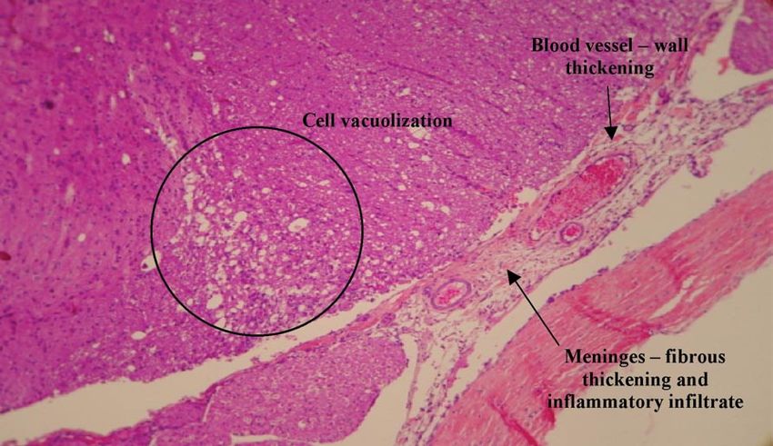
Pain Physician: September/October 2010; 13:493-501

not only about effectiveness but also safety. Double           tal groups according to the type of solution to be ad-
blind studies also have shown that steroid treatment           ministered into the intrathecal space. Seven dogs were
is beneficial both in short term as well as long term          allocated to each group. Group I was defined as the
evaluations (2-5,15-22,37,38).                                 control group, in which saline 0.9% was injected. Group
     Administration of corticosteroids in the epidural         II received methylprednisolone (1.15mg/kg). The dose
space utilized the anti-inflammatory action relatively         of the solution was calculated based on the dose epidu-
close to the nerve roots that are irritated. They then, pre-   rally employed to treat radicular pain in a 70 kg adult
sumably have less systemic effects. However, there are         patient (80 mg methylprednisolone acetate). Both so-
reports in the literature of Cushing like symptoms and         lutions had the same 1 mL volume. All the syringes in
other systemic side effects from epidural steroids (28,39-     our study were covered with sterile opaque white tape
42). Epidural injection also have their own potential com-     to keep the investigator blinded to the solution being
plications such as local discomfort, infection, inadvertent    injected, as methylprednisolone is a milky solution com-
dural puncture, post-dural puncture headache, epidural         pared to saline, which is a clear solution.
hematoma, nerve injury, coma, and, in rare cases, death
(1-7,41-44). Some of these complications can be due to         Spinal Puncture Protocol
vasospasm, direct vascular trauma, or embolus from par-             The dogs were fasted 12 hours before the proce-
ticulate steroids (44-46). Inaccurate placement of epidu-      dure, with water ad libitum. All animals were anesthe-
ral needles into the vein occurs in 1.9-11.2% of lumbar        tized with an intravenous administration of etomidate
epidural injections (44,47,48), while intrathecal injection    (2 mg/kg) and fentanyl (0.005 mg/kg). A 10 cm area
occurs in 5%-6% of epidural steroid injections when the        around the site of the spinal puncture at the L6-7 inter-
needle insertion is done under fluoroscopy (49-51).            vertebral space level was washed with water and soap,
     The use of intrathecal steroids has been associ-          followed by hair removal and skin cleansing with sa-
ated with severe nervous system complications such as          line. Finally, the naked skin was disinfected with a 2%
adhesive arachnoiditis (AA). It is more likely to occur        chlorexidine gluconate solution and sterile fields were
when methylprednisolone is used (52,53). Animal ex-            appropriately positioned.
perimentation is the first step to establish the safety             The subarachnoid puncture was performed through
of intrathecal methylprednisolone injection (54). To           the midline, approximately 450 to the skin, with a 22-
our knowledge there are no reports in the literature of        gauge Quincke needle. Any difficulties during the pro-
untoward clinical and histological effects of intrathecal      cedure and any changes in the color of the cerebrospi-
methylprednisolone injection in the animal model. The          nal fluids (CSF) were recorded. When a traumatic spinal
objective of this study was to determine the clinical and      puncture was identified, as defined by the presence of
histological effects of injecting methylprednisolone           hemorrhagic CSF or the need for more than one punc-
into the intrathecal space of dogs.                            ture, the animal was excluded from the study. Once
                                                               the needle was properly placed and clear CSF could be
Methods                                                        identified, 1 mL of the randomized solution was injected
                                                               for approximately 10 seconds through 1mL disposable
Animal Model                                                   syringes.
     After approval by the animal research ethics com-
mittee, 14 male adult mongrel dogs were obtained from          Solution Specification
the Experimental Animal Center at the Sao Paulo State                Preparations of methylprednisolone (Depo-medrol,
University at Botucatu campus. The animals weighed             Pfizer, Puurs, Belgium) contained methylprednisolone
from 7 to 14 kg, and the length of their vertebral col-        80mg/ml, polyethylene glycol (Macrogol 4000), myristil-
umn ranged from 54 to 68 cm. All animals were kept             gama-picolinine chloride, and saline. The pH of the final
under clinical observation in individual cells and were        product remains within the USP specified rate, between
assessed before spinal puncture and had normal neuro-          3.5-7.0. The saline (Baxter Healthcare Corp., Sao Paulo, Bra-
logical function. All tests were performed in accordance       zil) administered to the control group had a pH of 5.0.
with Ethical Guidelines in Conscious Animals (55).
                                                               Evaluation and outcomes
Experimental Groups                                                The animals were evaluated after the recovery
      The dogs were randomized to one of 2 experimen-          from the anesthesia and were followed daily during

494                                                                                            www.painphysicianjournal.com
Effects of Intrathecal Methylprednisolone in Dogs
the next 21 days by the same researcher every day. Each ware (GraphPod Software, La Jolla, CA) was used to
animal was evaluated regarding the following second- perform the statistical analysis.
ary outcomes: motor deficit, anal sphincter tonus, and
painful sensibility. The primary aim of the study was to
Results
evaluate the histological changes in the spinal cord and There were no significant differences between the
meninges of the dogs 21 days after subarachnoid ad- studied groups with regard to the weight (p = 0.18)
ministration of the methylprednisolone. or spinal length (p = 0.57). No animals were excluded
The presence of pain was defined by the follow- due to multiple or hemorrhagic punctures. None of the
ing: hind limb withdrawal, vocalization, and facial ex- animals demonstrated any clinical change, such as in-
pression. All 3 secondary outcomes (motor deficit, anal creased sensitivity to pain, decreased mobility, or anal
sphincter relaxation, and nociception) were classified sphincter relaxation.
dichotomously into absent or present. If the slightest There were no histological changes in the spinal
deficit in any of these dimensions was observed at clini- cord of the animals from Group I (Fig. 1). However, all
cal assessment, the animal would be classified as posi- seven animals in Group II presented with spinal injury
tive for the deficit according to the dichotomous clas- (p < 0.001). (Table 1; Figs. 2, 3, and 4). The main histo-
sification used. Nociception was assessed by reaction to logical changes observed in Group II were areas with
painful pressure and thermal stimuli. Motor deficit was meningeal thickening and lymphocytic infiltration in
determined by the inability to walk, jump, and sustain the blood vessel. Further histological examination also
the tail in an upward position. Anal sphincter relaxation revealed that in 3 animals there was adherence among
was ascertained through visual inspection. Pressure no- the pia, arachnoid, and dura matter and the nerve
ciceptive stimuli were elicited by the bilateral pinch of a roots were seen completely encircled by fibrous tissue.
skin fold over sacral, lumbar and thoracic dermatomes, One animal also demonstrated necrosis of the dorsal
and interdigital membranes of hind limbs. Thermal spinal cord.
painful stimuli were provided by thermoalgometer set
at 50oC for 10 seconds, touching the interdigital mem-
Discussion
branes of the hind limbs. Dogs were selected for this study because other
After 21 days, the animals were anesthetized with toxicity studies have been successfully performed in
intravenous sodium pentobarbital and then killed by this species. The anatomy of the dog also provides easy
electroshock. The lumbar and sacral segments of the access to the intervertebral space, and so facilitates
spinal cord with the surrounding meninges were quickly lumbar puncture and intrathecal injection (56,57). No
removed in less than 3 minutes to minimize the risk of animal had to be excluded from the present study due
ischemia and apoptosis of those tissues. The specimens to difficult or traumatic spinal puncture.
were fixed in formalin 10% solution for 7 days, and then The neurotoxic and inflammatory mediator phos-
histological sections were prepared from about 10 cm pholipase A2 (PLA2), which is normally contained in
above the level of the spinal puncture to the end of the the nucleus pulposus, is released after an annular in-
cauda equina. The histological sections were stained by jury (58). The PLA2 triggers the arachdonic acid cascade
hematoxilin-eosin and Masson trichome technique and that leads to localized inflammation mediated by pros-
examined by optical microscopy. Researchers who were taglandins and leukotrines. Corticosteroids have pow-
blinded to the solutions administered to each of the ani- erful anti-inflammatory effects because they inhibit
mals performed all clinical and histological evaluations. prostaglandin synthesis, block PLA2 activity, and stabi-
lize inflammatory cell membranes. Injection of steroid
Statistical analysis in the epidural space should result in a higher concen-
To evaluate the effectiveness of the randomization tration of the steroid in the epidural space compared
procedure and the comparability of the 2 groups, Stu- to oral or parenteral administration. Epidural injection
dent t-test was performed for the animals’ weight and is the only method of drug delivery that does not rely
the length of their vertebral columns. One-sided Fisher’s on blood flow and thus is deemed to be beneficial in
exact test was selected to compare the frequencies of compressive disc herniation in which blood flow to the
the findings on the outcomes between the methylpred- region might be impaired (3-6).
nisolone and the control groups. P values less than 0.05 Methyprednisolone is one of the commonly used
were considered statistically significant. Prism 4 soft- steroids in the treatment of lumbosciatic pain de-
www.painphysicianjournal.com 495Pain Physician: September/October 2010; 13:493-501
Fig. 1. Group I (saline). Segment of nerve and lumbar nervous tissues, meninges and blood vessels showing no abnormalities
(H&E 100X).
Table 1. Findings observed in Group II (methylprednisolone) during the review of histological sections.
Meninges Blood vessels Spinal cord
Histological Trabeculae formation
changes Fibrous Inflammatory Wall Inflammatory
(adherence among pia, Necrosis
thickening infiltration thickening infiltration
arachnoid, and dura matter)
Animal 1 present present absent present present absent
Animal 2 present present present present present absent
Animal 3 present present absent present present absent
Animal 4 present present present present present present
Animal 5 present present present present present absent
Animal 6 present present absent present present absent
Animal 7 present present absent present present absent
Animals in Group I (saline) have not presented with any change in the histological sections.
spite its association with adhesive arachnoiditis (AA) static state (25,27,39,44,54,60-62). It can cause bladder
(26,27,40). The present study confirms the potential for and bowel dysfunction, loss of sensation, and motor
spinal damage due to the intrathecal administration of weakness (53). In the present study, the animals had no
methylprednisolone. Arachnoiditis is a severe progres- clinical changes during the study period, though histo-
sive disorder characterized by an inflammatory process logical damages were observed in animals of Group II.
leading to fibrosis of the arachnoid and subarachnoid Different researchers, using different animal models,
space (59). The course of this disease can be irregular have reported similar findings, suggesting that clinical
with 1.8 to 33% of patients presenting a progressive findings do not always correlate with the degree of the
state and 50% to 59% of patients suffering with a histological changes (57,63,64). AA has an insidious on-
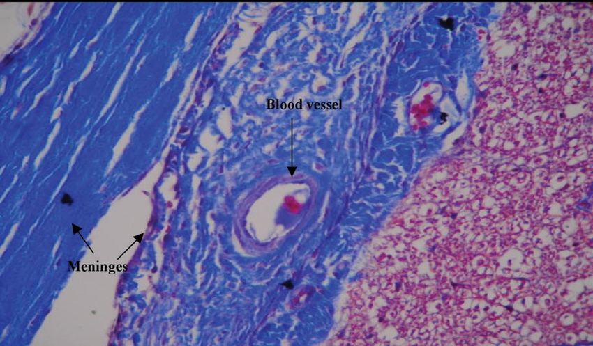
496 www.painphysicianjournal.comEffects of Intrathecal Methylprednisolone in Dogs Fig. 2. Group II (methylprednisolone). Segment of lumbar nervous tissues and meninges, showing thickness of the meninges and of the meningeal blood vessels (arrows). Nerve tissue necrosis with cell vacuolization is highlighted in the figure (H&E 100X). Fig. 3. Group II (methylprednisolone). Segment of lumbar nervous tissue and meninges, showing adherence among pia, arach- noid, and dura matter (in detail: early stage of adhesive arachinoidits) (H&E 100X). www.painphysicianjournal.com 497
Pain Physician: September/October 2010; 13:493-501

 Fig. 4. Group II (methylprednisolone). Segment of nerve and lumbar nervous tissues with fibrous tissue reaction in the meninges
 and meningeal blood vessels (Masson’s trichrome 400X).

set and the neurologic symptoms and signs are likely               South Wales, Australia) had published a warning rec-
to occur after an extensive period. This could be the              ommending that methylprednisolone should no longer
reason why the animals in the present study did not                be used for epidural steroid injections (66).” Ten years
manifest any clinical signs during the 21 days of the              later, the use of depo-steroids in the epidural space was
study. The lumbar puncture technique should not be                 recognized as safe in the review published by the Aus-
responsible for the lesions observed in the animals in             tralian Pain Society, but the authors do not recommend
Group II, since animals in Group I showed no signs of              the use of methylprednisolone (67).
histological abnormality.                                               Polyethylene glycol (PEG) is used as an excipient for
     Methylprednisolone, as a depo-steroid with a long             depo-steroid preparation. It is added to the commer-
half-life, will have prolonged contact with the neural             cially available methylprednisolone solutions and other
tissue and will allow for extensive distribution onto              steroids in order to increase their solubility. PEG might
the spinal cord surface. The meningeal thickening and              be responsible for the reports associated with AA re-
associated vascular inflammation found in the pres-                sulting from such methylprednisolone injections in the
ent study might be an indicator of the onset of AA.                intrathecal space (26,40,49,52). Methylprednisolone
Goldstein (65), in 1970, performed a clinical study with           and triamcinolone contain 3% PEG in their commercial-
intrathecal methylprednisolone injection in 38 patients            ly available solutions. Concerns have been raised about
with multiple sclerosis. One of these patients present-            the potential neurotoxic effects of 3% PEG injected in
ed with clinical AA. Since the time of this publication,           the human intrathecal space. Experiments with rabbit
physicians have been concerned about the risk of AA                nerve preparation has revealed no significant neuroly-
following intrathecal methylprednisolone (49). Based               sis or slowed conduction velocities with solutions con-
on the many AA cases reported (29,30,52,58,59,63-65),              taining 40% PEG. Histopathological studies were not
the Australian Physicians Medical Defense Union (New               done to corroborate these findings. Currently, there is

498                                                                                                  www.painphysicianjournal.com
Effects of Intrathecal Methylprednisolone in Dogs
no data on human toxicity with PEG (61). On the other edema and necrosis, while none of the controls with
hand, Selby (68) observed immediate demyelization non-particulate drug were affected (70). The occlusion
when PEG was injected into the peripheral nerves, op- of small arterioles should not be ignored as a reason-
tic nerve, nerve roots and spinal cord of rats and rab- able explanation for the spinal cord damages found in
bits. The drug preparation that we used in this study the present study.
contained 3% PEG and that might have contributed to It is clear that the present study has some limita-
the histological changes found in the spinal cord. Expo- tions. First, the small sample of animals can limit the
sure of the nerves of a rat’s paw to solutions containing applicability of the results. Second, we performed the
PEG for a period longer than one hour resulted in per- clinical follow-up for a short period (21 days). The
manent lesions and neuronal degeneration. Remark- present 21-day time course of the study might not be
ably, such lesions occurred with the administration of adequate to detect the complete extent of the spinal
methylprednisolone with concentration of PEG found damage caused by the methylprednisolone, since AA
in commercial preparations, which was supposed not to has an insidious onset and can take longer to produce
cause neurotoxicity (54,69). In our study we have dem- clinical symptoms. Third, the drug used in the present
onstrated that the dose of methylprednisolone we had study was not preservative free. Consequently, we can
used has the ability to produce histological changes in not assure that the spinal injuries observed were due
the meninges and the spinal cord of dogs. Most antioxi- to the methylprednisolone by itself or due to the other
dants, preservatives, and excipients are safe for human components of the presentation such as PEG. We de-
use. However, a word of caution seems appropriate for cided to use the same preparation that is used clinically
such additives when considered for epidural or intra- to treat spinal pain (i.e., with preservatives) because we
thecal injections (61). believe it has a higher clinical relevance. However, fur-
Another important complication related to the ther studies including intrathecally PEG solution injec-
epidural injection of particulate corticosteroid, such as tion might help to elucidate the role of the preservative
methylprednisolone, is brain and/or spinal cord infarct. in the development of histological changes observed in
Several hypotheses have been suggested, but the exact the present study.
mechanism is not yet known. The leading hypothesis is
that the inadvertent intra-arterial injection of particu-
Conclusion
late corticosteroid creates an embolus, causing a distal In this study we were able to show that intrathe-
infarct (31-34). Derby et al (35) showed in their study cal injection of commercially available methylpredniso-
that methylprednisolone particles were smaller than lone is capable of producing histological changes in the
red blood cells, but the particles were densely packed spinal cord and meninges of dogs. Further studies are
and few aggregations were observed. The risk of the necessary to elucidate the mechanism of the meningeal
densely packed particles suggests that they are capa- toxicity observed and to evaluate the clinical long-term
ble of forming an embolus that could occlude a small outcome in dogs and in different animal models.
arteriole. A study performed by Okubadejo et al (70),
comparing intravascular injection of particulate and
Acknowledgments
non particulate steroids in pigs (methylprednisolone vs. The authors wish to thank the editorial board of
dexamethasone vs. prednisolone), demonstrated that Pain Physician for review and criticism in improving the
all animals injected with methylprednisolone had neu- manuscript.
rologic deficit and histological changes consistent with
References
1. Staal JB, de Bie RA, de Vet HCW, Hil- 2009; 12:233-251. of the effectiveness of cervical epidur-
debrandt J, Nelemans P. Injection ther- 3. Conn A, Buenaventura R, Datta S, Abdi als in the management of chronic neck
apy for subacute and chronic low back S, Diwan S. Systematic review of caudal pain. Pain Physician 2009; 12:137-157.
pain: An updated Cochrane review. epidural injections in the management 5. Parr AT, Diwan S, Abdi S. Lumbar inter-
Spine (Phila PA 1976) 2009; 34:49-59. of chronic low back pain. Pain Physician laminar epidural injections in manag-
2. Buenaventura RM, Datta S, Abdi S, 2009; 12:109-135. ing chronic low back and lower extrem-
Smith HS. Systematic review of ther- 4. Benyamin RM, Singh V, Parr AT, Conn ity pain: A systematic review. Pain Phy-
apeutic lumbar transforaminal epidu- A, Diwan S, Abdi S. Systematic review sician 2009; 12:163-188.
ral steroid injections. Pain Physician
www.painphysicianjournal.com 499Pain Physician: September/October 2010; 13:493-501
6. Manchikanti L, Boswell MV, Singh V, pain: Part 1. Discogenic pain without ed Kingdom, and the USA. Pain Digest
Benyamin RM, Fellows B, Abdi S, Bue- disc herniation or radiculitis. Pain Phy- 1999; 9:226-234.
naventura RM, Conn A, Datta S, Derby sician 2008; 11:785-800. 25. Hooten WM, Mizerak A, Carns PE, Hunt-
R, Falco FJE, Erhart S, Diwan S, Hayek 16. Manchikanti L, Singh V, Cash KA, Pam- oon MA. Discitis after lumbar epidural
SM, Helm S, Parr AT, Schultz DM, Smith pati V, Damron KS, Boswell MV. Prelim- corticoid injection: A case report and
HS, Wolfer LR, Hirsch JA. Comprehen- inary results of a randomized, equiva- analysis of the case report literature.
sive evidence-based guidelines for in- lence trial of fluoroscopic caudal epidu- Pain Med 2006; 7:46-51.
terventional techniques in the manage- ral injections in managing chronic low 26. Raj PP. Epidural steroid injections. Pain
ment of chronic spinal pain. Pain Physi- back pain: Part 2. Disc herniation and Digest 1990; 9:235-240.
cian 2009; 12:699-802. radiculitis. Pain Physician 2008; 11:801- 27. Hartrick CT. Epidural steroid injection –
7. Manchikanti L, Boswell MV, Datta S, Fel- 815. How should the indications for use be
lows B, Abdi S, Singh V, Benyamin RM, 17. Manchikanti L, Singh V, Cash KA, Pam- derived: Systematic review or basic sci-
Falco FJE, Helm S, Hayek S, Smith HS. pati V, Datta S. Preliminary results of ence? Pain Practice 2009; 9:165-166.
Comprehensive review of therapeutic randomized, equivalence trial of fluo-
interventions in managing chronic spi- 28. Nelson DA, Landau WM. Intraspinal
roscopic caudal epidural injections in
nal pain. Pain Physician 2009; 12:E123- steroids: History, efficacy, accidentally,
managing chronic low back pain: Part 3.
E198. and controversy with review of United
Post surgery syndrome. Pain Physician
States Food and Drug Administration
8. Manchikanti L, Singh V, Derby R, Schul- 2008; 11:817-831. reports. J Neurol Neurosurg Psychiatry
tz DM, Benyamin RM, Prager JP, Hirsch 18. Manchikanti L, Cash KA, McManus CD, 2001; 70:433-443.
JA. Reassessment of evidence synthe- Pampati V, Abdi S. Preliminary results of
sis of occupational medicine practice 29. Nelson DA, Vates TS, Jr., Thomas RB, Jr.
a randomized, equivalence trial of flu-
guidelines for interventional pain man- Complications from intrathecal steroid
oroscopic caudal epidural injections in
agement. Pain Physician 2008; 11:393- therapy in patients with multiple scle-
managing chronic low back pain: Part
482. rosis. Acta Neurol Scand 1973; 49:176-
4. Spinal stenosis. Pain Physician 2008; 188.
9. Chou R, Huffman L. Evaluation and 11:833-848.
Management of Low Back Pain: Evi- 30. Nelson DA. Dangers from methylpred-
19. Manchikanti L, Cash KA, McManus CD,
dence Review. American Pain Society, nisolone acetate therapy by intraspinal
Pampati V, Benyamin R. Preliminary re-
Glenview, IL, 2009. injection. Arch Neurol 1988; 45:804-
sults of a randomized, double-blind, 806.
10. Manchikanti L, Datta S, Gupta S, Mung- controlled trial of fluoroscopic lumbar
lani R, Bryce DA, Ward SP, Benyamin interlaminar epidural injections in man- 31. Rathmell JP, Aprill C, Bogduk N. Cervi-
RM, Sharma ML, Helm II S, Fellows B, aging chronic lumbar discogenic pain cal transforaminal injection of steroids.
Hirsch JA. A critical review of the Ameri- without disc herniation or radiculitis. Anesthesiology 2004; 100:1595-1600.
can Pain Society clinical practice guide- Pain Physician 2010; 13:E279-E292. 32. Rathmell JP, Benzon HT. Transforam-
lines for interventional techniques: 20. Manchikanti L, Singh V, Falco FJE, Cash inal injection of steroids: Should we
Part 2. Therapeutic interventions. Pain KA, Pampati V. Evaluation of the effec- continue? Reg Anesth Pain Med 2004;
Physician 2010; 13:E215-E264. tiveness of lumbar interlaminar epidu- 29:397-399.
11. Manchikanti L, Singh V, Pampati V, ral injections in managing chronic pain 33. Scanlon GC, Moeller-Bertram T, Ro-
Smith HS, Hirsch JA. Analysis of growth of lumbar disc herniation or radiculitis: manoswsky SM, Wallace MS. Cervical
of interventional techniques in manag- A randomized, double-blind, controlled transforaminal epidural steroid injec-
ing chronic pain in the Medicare popu- trial. Pain Physician 2010; 13:343-355. tions: More danderous than we think?
lation: A 10-year evaluation from 1997 21. Manchikanti L, Cash KA, Pampati V, Spine (Phila Pa 1976) 2007; 32:1249-
to 2006. Pain Physician 2009; 12:9-34. Wargo BW, Malla Y. Cervical epidural 1256.
12. Manchikanti L, Boswell MV, Giordano J. injections in chronic discogenic neck 34. Tiso Rl, Cutler T, Catania JA, Whalen
Re: Friedly J, Chan L, Deyo R. Increases pain without disc herniation or radiculi- K. Adverse central nervous system
in lumbosacral injections in the Medi- tis: Preliminary results of a randomized, sequelae after selective tranforami-
care population: 1994 to 2001. Spine double-blind, controlled trial. Pain Phy- nal block: The role of corticosteroids.
(Phila PA 1976) 2007; 32:1754-1760. sician 2010; 13:E265-E278. Spine J 2004; 4:468-474.
Spine (Phila PA 1976) 2007; 32:3092. 22. Manchikanti L, Cash KA, Pampati V, War- 35. Derby R, Lee SH, Date ES, Lee JH, Lee
13. Deyo RA, Mirza SK, Turner JA, Martin BI. go BW, Malla Y. The effectiveness of flu- CH. Size aggregation of corticosteroids
Overtreating chronic back pain: Time to oroscopic cervical interlaminar epidural used for epidural injections. Pain Med
back off? J Am Board Fam Med 2009; injections in managing chronic cervical 2008; 9:227-234.
22:62-68. disc herniation and radiculitis: Prelim- 36. Robecchi A, Capra R. [Hydrocortisone
14. Friedly J, Chan L, Deyo R. Increases in inary results of a randomized, double- (compound F); first clinical experi-
lumbosacral injections in the Medicare blind, controlled trial. Pain Physician ments in the field of rheumatology]. Mi-
population: 1994 to 2001. Spine (Phila 2010; 13:223-236. nerva Med 1952; 43:1259-1263.
Pa 1976) 2007; 32:1754-1760. 23. Abram SE. Perceived dangers from in- 37. Riew KD, Park JB, Cho YS, Gilula L, Pa-
15. Manchikanti L, Cash KA, McManus CD, traspinal steroid injections. Arch Neurol tel A, Lenke LG, Bridwell KH. Nerve root
Pampati V, Smith HS. Preliminary re- 1989; 46:719-721. blocks in the treatment of lumbar ra-
sults of a randomized, equivalence tri- 24. Bogduk N. Current guidelines in the dicular pain: A minimum five-year fol-
al of fluoroscopic caudal epidural in- use of epidural steroids: Reports from low-up. J Bone Joint Surg Am 2006;
jections in managing chronic low back Australia, Belgium, Norway, the Unit- 88:1722-1725.
500 www.painphysicianjournal.comEffects of Intrathecal Methylprednisolone in Dogs
38. Riew KD, Yin Y, Gilula L, Bridwell KH, 49. Nelson DA. Intraspinal therapy using haemorrhage. Br J Neurosurgery 2008;
Lenke LG, Lauryssen C, Goette K. The methylprednisolone acetate. Twen- 22:578-579.
effect of nerve-root injection on the ty-three years of clinical controversy. 60. Guyer DW, Wiltse LL, Eskay ML, Guyer
need for operative treatment of lum- Spine (Phila Pa 1976) 1993; 18:278- BH. The long-range prognosis of arach-
bar radicular pain. J Bone Joint Surg Am 286. noiditis. Spine (Phila Pa 1976) 1989;
2000; 82:1589-1593. 50. Botwin KP, Gruber RD, Bouchlas CG, 14:1332-1341.
39. Kay J, Raff H. Epidural triamcinolone Torres-Ramos FM, Hanna A, Rittenberg 61. Hodgson PS, Neal JM, Pollock JE, Liu
causes prolonged and severe depres- J, Thomas SA. Complications of fluoro- SS. The neurotoxicity of drugs given in-
sion of the pituitary-adrenal axis. Anes- scopically guided caudal epidural in- trathecally. Anesth Analg 1999; 88:797-
thesiology 1991; 75:A694. jections. Am J Phys Med Rehabil 2001; 809.
40. McLain R. Point of view: The pathologic 80:416-424. 62. Koc Z, Ozcakir S, Sivrioglu K, Gurbet A,
effects of intrathecal betamethasone. 51. Botwin KP, Castellanos R, Rao S, Han- Kucukoglu S. Effectiveness of physical
Spine (Phila Pa 1976) 1997; 22:1562. na AF, Torres-Ramos FM, Gruber RD, therapy and epidural steroid injections
41. Abram SE, O’Connor TC. Complications Bouchlas CG, Fuoco GS. Complications in lumbar spinal stenosis. Spine (Phila
associated with epidural steroid injec- of fluoroscopically guided interlami- Pa 1976) 2009; 34:985-989.
tions. Reg Anesth 1996; 21:149-162. nar cervical epidural injections. Arch 63. Ready LB, Plumer MH, Haschke RH,
Phys Med Rehabil 2003; 84:627-633. Austin E, Sumi SM. Neurotoxicity of in-
42. Manchikanti L. Role of neuraxial ste-
roids in interventional pain manage- 52. Corrigan AB, Carr G, Tugwell S. Intra- trathecal local anesthetic in rabbits.
ment. Pain Physician 2002; 5:182-199. spinal corticosteroid injections. Med J Anesthesiology 1985; 63:364-370.
Aust 1982; 1:224-225. 64. Rosen MA, Baysinger CL, Shnider SM,
43. Fitzgibbon DR, Posner KL, Domino KB,
Caplan RA, Lee LA, Cheney FW. Chron- 53. Rice I, Wee MYK, Thompson K. Ob- Dailey PA, Norton M, Curtis JD, Collins
ic pain management: American Soci- stetric epidurals and chronic adhe- M, Davis RL. Evaluation of neurotoxic-
ety of Anesthesiologists Closed Claims sive arachnoiditis. Br J Anaesth 2004; ity after subarachnoid injection of large
Projects. Anesthesiology 2004; 100:98- 92:109-120. volumes of local anesthetic solutions.
105. 54. Johansson A, Hao J, Sjolound B. Lo- Anesth Analg 1983; 62:802-808.
44. Kim DW, Han KR, Kim C, Chae YJ. Intra- cal corticosteroid application blocks 65. Goldstein NP, McGuckin WF, McKen-
vascular flow patterns in transforami- transmission in normal nociceptive C- zie BF, Mattox VR. Experimental intra-
nal epidural injections: A comparative fibers. Acta Anesthesiol Scand 1990; thecal administration of methylpred-
study of the cervical and lumbar seg- 34:335-338. nisolone acetate in multiple sclerosis.
ments. Anesth Analg 2009; 109:233- 55. Zimmermann M. Ethical guidelines for Trans Am Neurol Assoc 1970; 95:243-
239. investigations of experimental pain in 244.
45. Glaser SE, Falco FJE. Paraplegia follow- conscious animals. Pain 1983; 16:109- 66. Gibb D. Spinal injection of corticoste-
ing a thoracolumbar transforaminal 110. roids. Med J Aust 1981; 2:302-303.
epidural steroid injection. Pain Physi- 56. Fukushima FB, Barros GAM, Marques 67. Gronow DW, Mendelson G. Epidural in-
cian 2005; 8:309-314. MEA, Vidal EIO, Ganem EM. The neu- jection of depot corticosteroids. Aus-
46. Glaser SE, Shah RV. Root cause analy- roaxial effects of intraspinal amitrip- tralian Pain Society Limited. Med J Aust
sis of paraplegia following transforam- tyline at low concentrations. Anesth 1992; 157:417-420.
inal epidural steroid injections: The Analg 2009; 109:965-971. 68. Selby R. To the editor. Neurosurgery
‘unsafe’ triangle. Pain Physician 2010; 57. Ganem EM, Vianna PT, Marques M, 1983; 12:591.
13:237-244. Castiglia YMM, Vane LA. Neurotox- 69. Abram SE. Need for precise diagnosis
47. Manchikanti L, Cash KA, Pampati V, icity of subarachnoid hyperbaric bu- prior to epidural steroids: Clinical con-
Damron KS, McManus CD. Evalua- pivacaine in dogs. Reg Anesth 1996; cepts and commentary. Anesthesiolo-
tion of lumbar transforaminal epidu- 21:234-238. gy 2000; 93:566-567.
ral injections with needle placement 58. Tachihara H, Sekiguchi M, Kikuchi SI, 70. Okubadejo GO, Talcott MR, Schmidt RE,
and contrast flow patterns: A prospec- Konno SI. Do corticosteroids produce Sharma A, Patel AA, Mackey RB, Guari-
tive, descriptive report. Pain Physician additional benefit in nerve root infiltra- no AH, Moran CJ, Riew KD. Perils of in-
2004; 7:217-223. tion for lumbar disc herniation? Spine travascular methylprednisolone injec-
48. Manchikanti L, Cash KA, Pampati V, (Phila Pa 1976) 2008; 33:743-747. tion into the vertebral artery. J Bone
McManus CD, Damron KS. Evaluation 59. Ginanneschi F, Palma L, Rossi A. Arach- Joint Surg Am 2008; 90:1932-1938.
of fluoroscopically guided caudal epi- noid cyst and arachnoiditis follow-
dural injections. Pain Physician 2004; ing idiopathic spinal subarachnoid
7:81-92.
www.painphysicianjournal.com 501You can also read